Immobilization of chondroitin sulfate to lipid membranes and its interactions with ECM proteins Noomi Altgärde a , Jana Becher b , Stephanie Möller b , Franz E. Weber c , Matthias Schnabelrauch b , Sofia Svedhem a,⇑ a Department of Applied Physics, Chalmers University of Technology, 412 96 Göteborg, Sweden b Biomaterials Department, INNOVENT e.V., Pruessingstrasse 27 B, 07745 Jena, Germany c Division of Cranio-Maxillofacial and Oral Surgery, Oral Biotechnology & Bioengineering, University Hospital, 8091 Zurich, Switzerland article info Article history: Received 18 May 2012 Accepted 24 July 2012 Available online 7 August 2012 Keywords: Supported lipid bilayer Chondroitin sulfate Bone morphogenetic protein-2 (BMP-2) Quartz Crystal Microbalance with Dissipation monitoring (QCM-D) abstract Glycosaminoglycans (GAGs) in the extracellular matrix (ECM) have multiple functions in tissues including providing support, mediating cell division and differentiation, and taking part in important interactions with proteins, e.g. growth factors. Studying GAG related interactions is inherently difficult and requires suit- able interaction platforms. We show two strategies to covalently couple the GAG chondroitin sulfate (CS) to supported lipid bilayers (SLBs), either by (a) activating carboxy-functionalized phospholipids in the lipid bilayer, followed by the addition of hydrazide-functionalized CS, or by (b) activating naturally occurring carboxyl groups on CS prior to addition to an amino-functionalized SLB. Bilayer formation and subsequent immobilization was followed in real-time using the Quartz Crystal Microbalance with Dissipation monitor- ing, a technique that provides unique information when studying highly hydrated molecular films. The two strategies yielded thin CS films (in the nanometer range) with similar viscoelastic properties. Fluidity of the lipid bilayer was retained when CS was coupled. The application of the CS interaction platform was exemplified for type I collagen and the bone inducing growth factor bone morphogenetic protein-2 (BMP-2). The addition of collagen to immoblized CS resulted in soft layers whereas layers formed by addition of BMP-2 were denser, independent on the immobilization strategy used. Ó 2012 Elsevier Inc. All rights reserved. 1. Introduction Research concerning supports for tissue-engineering and cell culture has progressed tremendously over the past decades [1], paralleled by an increased understanding of the role of the extra- cellular matrix (ECM) for cell behavior. Even so, much is still to be discovered about the supramolecular assemblies constituting the ECM and about the interactions between glycosaminoglycans (GAGs) and proteins. GAGs are long, unbranched polysaccharide chains that can be found both in the ECM and at the cell membrane, often bound to a protein core forming proteoglycans (e.g. the proteoglycan aggrecan depicted in Scheme 1(I)) [2]. Independent of their origin, GAGs are highly compatible with var- ious biological functions such as stabilizing growth factors, mediat- ing cellular processes [3] and regulating atherogenesis [4]. For example, chondroitin sulfate (CS) has recently gained increased interest because of its role in bone and cartilage tissue and its interactions with growth factors and proteins like type I collagen, the most abundant protein in the ECM [5–7]. Growth factors, for example bone morphogenetic protein 2 (BMP-2), are frequently used in research, primarily with the aim of stimulating bone growth and regeneration [8–11]. The short half-life of growth fac- tors, and their tendency to aggregate in solution aggravates the usage but combining positively charged BMP-2 with negatively charged GAGs [12,13], such as CS [14], has proven to stabilize them with retained functionality. GAGs are not encoded in the DNA but result from posttranslational modifications leading to e.g. different sulfation states [2,15]. Their changeable structural properties make them challenging to study and derivatives with well-defined chemical and physical properties are therefore necessary. 0021-9797/$ - see front matter Ó 2012 Elsevier Inc. All rights reserved. http://dx.doi.org/10.1016/j.jcis.2012.07.063 Abbreviations: GAG, glycosaminoglycan; ECM, extracellular matrix; CS, chon- droitin sulfate; QCM-D, Quartz Crystal Microbalance with Dissipation monitoring; SLB, supported lipid bilayer; BMP-2, bone morphogenetic protein-2; EDC, N-(3-dimethylaminopropyl)-N 0 -ethylcarbodiimide hydrochloride; NHS, N- hydroxysuccinimide; POPC, 1-palmitoyl-2-oleoyl-sn-glycero-3-phosphocholine; DOPE-COOH, 1,2-dioleoyl-sn-glycero-3-phosphoethanolamine-N-(lauroyl); DOPE- NH 2 , 1,2-dioleoyl-sn-glycero-3-phosphoethanolamine-N-(lauroylamine); NBD-PC, 1-palmitoyl-2-{12-[(7-nitro-2-1,3-benzoxadiazol-4-yl)amino]dodecanoyl}-sn-gly- cero-3-phosphocholine; DS hydr , degree of hydrazide substitution; SAM, self-assem- bled monolayer. ⇑ Corresponding author. E-mail addresses: [email protected](N. Altgärde), jb@innovent- jena.de (J. Becher), [email protected](S. Möller), [email protected](F.E. Weber), [email protected](M. Schnabelrauch), sofi[email protected](S. Svedhem). Journal of Colloid and Interface Science 390 (2013) 258–266 Contents lists available at SciVerse ScienceDirect Journal of Colloid and Interface Science www.elsevier.com/locate/jcis
Transcript
Journal of Colloid and Interface Science 390 (2013) 258–266
Contents lists available at SciVerse ScienceDirect
Journal of Colloid and Interface Science
www.elsevier .com/locate / jc is
Immobilization of chondroitin sulfate to lipid membranes and its interactionswith ECM proteins
Noomi Altgärde a, Jana Becher b, Stephanie Möller b, Franz E. Weber c, Matthias Schnabelrauch b,Sofia Svedhem a,⇑a Department of Applied Physics, Chalmers University of Technology, 412 96 Göteborg, Swedenb Biomaterials Department, INNOVENT e.V., Pruessingstrasse 27 B, 07745 Jena, Germanyc Division of Cranio-Maxillofacial and Oral Surgery, Oral Biotechnology & Bioengineering, University Hospital, 8091 Zurich, Switzerland
a r t i c l e i n f o
Article history:Received 18 May 2012Accepted 24 July 2012Available online 7 August 2012
Glycosaminoglycans (GAGs) in the extracellular matrix (ECM) have multiple functions in tissues includingproviding support, mediating cell division and differentiation, and taking part in important interactionswith proteins, e.g. growth factors. Studying GAG related interactions is inherently difficult and requires suit-able interaction platforms. We show two strategies to covalently couple the GAG chondroitin sulfate (CS) tosupported lipid bilayers (SLBs), either by (a) activating carboxy-functionalized phospholipids in the lipidbilayer, followed by the addition of hydrazide-functionalized CS, or by (b) activating naturally occurringcarboxyl groups on CS prior to addition to an amino-functionalized SLB. Bilayer formation and subsequentimmobilization was followed in real-time using the Quartz Crystal Microbalance with Dissipation monitor-ing, a technique that provides unique information when studying highly hydrated molecular films. The twostrategies yielded thin CS films (in the nanometer range) with similar viscoelastic properties. Fluidity of thelipid bilayer was retained when CS was coupled. The application of the CS interaction platform wasexemplified for type I collagen and the bone inducing growth factor bone morphogenetic protein-2(BMP-2). The addition of collagen to immoblized CS resulted in soft layers whereas layers formed byaddition of BMP-2 were denser, independent on the immobilization strategy used.
� 2012 Elsevier Inc. All rights reserved.
1. Introduction
Research concerning supports for tissue-engineering and cellculture has progressed tremendously over the past decades [1],paralleled by an increased understanding of the role of the extra-cellular matrix (ECM) for cell behavior. Even so, much is still tobe discovered about the supramolecular assemblies constitutingthe ECM and about the interactions between glycosaminoglycans
(GAGs) and proteins. GAGs are long, unbranched polysaccharidechains that can be found both in the ECM and at the cellmembrane, often bound to a protein core forming proteoglycans(e.g. the proteoglycan aggrecan depicted in Scheme 1(I)) [2].Independent of their origin, GAGs are highly compatible with var-ious biological functions such as stabilizing growth factors, mediat-ing cellular processes [3] and regulating atherogenesis [4]. Forexample, chondroitin sulfate (CS) has recently gained increasedinterest because of its role in bone and cartilage tissue and itsinteractions with growth factors and proteins like type I collagen,the most abundant protein in the ECM [5–7]. Growth factors, forexample bone morphogenetic protein 2 (BMP-2), are frequentlyused in research, primarily with the aim of stimulating bonegrowth and regeneration [8–11]. The short half-life of growth fac-tors, and their tendency to aggregate in solution aggravates theusage but combining positively charged BMP-2 with negativelycharged GAGs [12,13], such as CS [14], has proven to stabilize themwith retained functionality. GAGs are not encoded in the DNA butresult from posttranslational modifications leading to e.g. differentsulfation states [2,15]. Their changeable structural properties makethem challenging to study and derivatives with well-definedchemical and physical properties are therefore necessary.
Scheme 1. (I) Example of CS as part of the proteoglycans aggrecan in the ECM, andsyndecan in the cell membrane. CS: chondroitin sulfate, KS: keratan sulfate, HS:heparan sulfate, Hya: hyaluronic acid/hyaluronan, LP: link protein. (II) CS coupled toa supported lipid bilayer.
N. Altgärde et al. / Journal of Colloid and Interface Science 390 (2013) 258–266 259
Systematic screening of GAG related interactions requiresappropriate immobilization of the GAG using a general strategy.Several immobilization techniques have been presented in the lit-erature, e.g. immobilizing biotinylated GAGs to (strept)avidinmodified surfaces [16,17], coupling of thiolated carbohydrates toa maleimide presenting surface or vice versa [18,19] or direct cou-pling of the GAG via the reducing end to a hydrazide presentingsurface [20]. When the GAG is chemically modified, changes are in-duced in the primary structure and care must be taken as to not
Chart 1. Lipids used: (a) POPC, (b) DOPE-COOH, (c) DO
influence the binding characteristics to other biomolecules. Bind-ing characteristics can also depend on the supramolecular struc-ture of the GAG. Supported lipid bilayers (SLBs) are often used tomimic cell membranes and have proven to be a good platformwhen investigating various molecular interactions since non-spe-cific interactions are often minimized [21,22]. An important benefitof the lipid bilayer is the possibility to control and vary the lipidcomposition. Aspects of the lipid membrane to consider includethe (dynamic) distribution of lipids between the two leaflets andthe lateral diffusion of lipids. SLBs with immobilized peptides havebeen used as cell culture support [23,24], and attaching GAGs alongwith other molecules from the ECM would further increase theusefullness of this model system. There are some examples inthe literature of attaching GAGs to SLBs via streptavidin [22,25],but to our knowledge covalent attachment has not been shown.With well-designed GAG derivatives, high degree of control ofthe molecular configuration can be obtained as well as stable linksto the surface, which are advantageous for regeneration of thesensing element.
Here, we present two strategies to covalently couple CS to SLBs(Scheme 1(II)). The platform is applied by studying interactions ofCS with two different compounds; collagen type I and recombinanthuman BMP-2. The main surface sensitive technique used is theQuartz Crystal Microbalance with Dissipation monitoring (QCM-D). This technique is advancing as a tool in GAG research whereespecially the possibility to study biomolecular structures thatare associated with large amount of solvent is important [26].
2. Materials and methods
2.1. Chemicals
Unless otherwise stated, chemicals were bought from Sigma Al-drich. Amine coupling kit (EDC, NHS, ethanolamine) was purchasedfrom GE Healthcare Bioscience AB. 1-palmitoyl-2-oleoyl-sn-glyce-ro-3-phosphocholine (POPC), 1,2-dioleoyl-sn-glycero-3-phospho-ethanolamine-N-(lauroyl) (DOPE-COOH), 1,2-dioleoyl-sn-glycero-3-phosphoethanolamine-N-(lauroylamine) (DOPE-NH2) and1-palmitoyl-2-{12-[(7-nitro-2-1,3-benzoxadiazol-4-yl)amino]-dodecanoyl}-sn-glycero-3-phosphocholine (NBD-PC, fluorescentlylabeled at the fatty acid) were from Avanti Polar Lipids (Alabaster,AL) (Chart 1). Type I collagen (MW = �200 kDa) was purchased fromSigma Aldrich. Water was deionized (resistivity >18.2 MX/cm) andfiltered using a Milli-Q system (Millipore, Billerica, MA). Symmetricdisulfides of 7–8 ethylene glycol units with terminal hydroxyl(SS-OEG-OH, MW: 771 Da) or carboxyl groups (SS-OEG-COOH,
PE-NH2. Protonated as when at physiological pH.
260 N. Altgärde et al. / Journal of Colloid and Interface Science 390 (2013) 258–266
MW: 915.1 Da) were purchased from Polypure, Norway. Chondroi-tin sulfate (CS, 20 kDa, mixture of 70% chondroitin-4-sulfate(CS-A), and 30% chondroitin-6-sulfate (CS-C)) was purchased fromKraeber (Ellerbek, Germany). Other chemicals and solvents usedfor GAG synthesis were bought in analytical reagent grade fromFluka (Buchs, Switzerland) and Roth (Karlsruhe, Germany), andused without further purification. PBS buffer was prepared fromtablets or powder (0.01 mol/L phosphate, 0.0027 mol/L potassiumchloride, 0.137 mol/L sodium chloride, pH 7.4) and MES buffer100 mmol/L was prepared and pH adjusted to 4.7. Acetate buffer10 mmol/L was prepared from sodium acetate, in some caseswith 10 mmol/L calcium chloride or 140 mmol/L NaCl added, andpH adjusted to 5. Buffers were filtered and degassed before use.
2.2. Molecular weight determination of chondroitin sulfate (CS)
Gel permeation chromatography (GPC) analysis was performedusing the following systems: Jasco PU 980 pump, PostnovaAnalytics PN 3000 (15�) laser light scattering detector, JascoRID-1531 refraction detector, and Suprema-Gel 10 lm–100 Å,10 lm–1000 Å and 20 lm–30,000 Å columns. The eluent was PBSbuffer and the flow rate was 0.5 and 0.8 mL/min. The molecularweight of the commercial starting material, CS, was 19,763 g/moland the polydispersity index (size distribution, D (RI)) was 1.56.
2.3. Synthesis of hydrazide-functionalized chondroitin sulfate (h-CS)
Hydrazide-functionalized CS (h-CS) with different degrees ofhydrazide substitution was prepared as described [27]. Briefly,1 g (2.1 mmol) CS was dissolved in 250 mL water and 1.0, 0.5, or0.05 mol equivalents of adipic dihydrazide were added to preparehydrazide-containing CS with different degrees of substitution.The pH of the solution was adjusted to 4.75 by adding 1 N HCl,followed by the addition of 0.3, 0.15, or 0.05 mol equivalents ofEDC, respectively. The solution was stirred at room temperaturefor 6 h during which the pH was kept at 4.75. The pH was then in-creased to neutral conditions by adding 1 N NaOH, and the solutionwas dialyzed against water for 3–5 days. Subsequent lyophilizationand drying under high vacuum at 40 �C for 8 h lead to the desiredproduct. The average degree of hydrazide substituents per disac-charide repeating unit (DShydr.) was estimated by integration ofthe 1H-NMR signals at 3.68 ppm (invariable between CS andh-CS) and 2.27 ppm (invariable between adipic dihydrazide andh-CS). IR spectra were obtained on a FT-IR-Spectrometer FTS 175(BIO RAD, Krefeld, Germany) applying the KBr technique. IR (KBr)for h-CS: 2973, 2933, 2885, 1572, 1555, 1419, 1374, 1255, 1157,926, 886, 855 cm�1. NMR spectra were recorded in D2O on a BrukerAdvance 300 MHz spectrometer. 1H-NMR (d, D2O, 323 K): 4.75–4.45 (m, H-1, H-10), 4.22–4.18 (m, H-6, H-60, H-4), 4.03–3.80 (m,H-2, H-3, H-5), 3.68–3.59 (m, H-30 H-40, H-50), 3.38 (m, 1H, H-2),2.40 (m, 2H, CH2-hydrazide), 2.27 (m, 2H, hydrazide CH2), 2.04(m, 3H, CH3), 1.67 (m, 4H, CH2-hydrazide) ppm. 13C-NMR (d, D2O,323 K): 175.16 (C@O), 174.56 (C@O), 104.38 (C-1), 103.83 (C-1),101.63 (C-10), 101.06 (C-10), 80.63 (C-4), 80.19 (C-30), 76.88 (C-5),76.65 (C-5), 75.75 (C-40), 75.14 (C-50), 74.79 (C-50), 73.94 (C-3),73.81 (C-3), 72.71–72.50 (C-2), 61.21 (C-60), 51.74 (C-20), 51.05(C-20), 33.54 (NACH2), 33.33 (NACH2A), 24.68 (ACH2A24.56,(ACH2A22.75 (NACH3) ppm.
2.4. Preparation of bone morphogenetic protein 2, BMP-2
Osteoinductive human recombinant BMP-2 dimers were pro-duced in Escherichia coli followed by a refolding and dimerizationstep as previously described [28]. The final product had a molecu-lar weight of 26.08 kDa and a pI of 8.09, deduced from the aminoacid sequence. The bioactivity was tested in vivo as described
[28]. The purity of the sample also made it suitable for clinicaltrials [29].
2.5. Cleaning of QCM-D sensors
Silica-coated AT-cut quartz crystals (Q-sense, Göteborg,Sweden) were immersed in 10 mmol/L SDS for 5 min, rinsed inwater, dried in nitrogen and cleaned in a UV ozone system for30 min. Gold-coated AT-cut quartz crystals were cleaned in a5:1:1 solution of H2O:NH3:H2O2 at 80 �C for 10 min, rinsed inwater and dried in nitrogen.
2.6. Preparation of SAMs on QCM-D sensors
Gold coated crystals were incubated in a 0.5 mmol/L disulfidesolution of either 95% SS-OEG-OH+5% SS-OEG-COOH or 100%SS-OEG-COOH for >12 h, creating self-assembled monolayers withdifferent degrees of carboxyl groups (SAM-COOH). To removeloosely bound material, the crystals were ultrasonicated in ethanolfor 3 min and dried in nitrogen before mounting in the QCM-D flowchamber.
2.7. QCM-D measurements and modeling
Reactions taking place on the sensor surfaces were monitored inreal-time by QCM-D using an E4 instrument (Q-sense, Göteborg,Sweden). The technique is described elsewhere [30]. Briefly, it isbased on a quartz crystal disk (i.e. the QCM-D sensor) that oscil-lates at its resonance frequency when an alternating potential isapplied. When mass adsorbs on the surface, changes in resonancefrequency (Df) are measured. The driving potential is continuouslyswitched on and off and in this way the damping of the oscillatorymotion, the energy dissipation (DD), is also measured. The changein mass (Dm) caused by adsorption is related to Df through theSauerbrey equation [31],
Dm ¼ �CDfn
ð1Þ
where C is the mass sensitivity constant (17.7 ng/cm2 Hz) and n isthe overtone number. This equation is only valid for rigid adlayers,displaying low dissipation.
2.8. Lipid liposome preparation
Lipids (Chart 1) dissolved in chloroform were mixed in differentratios in a round-bottom flask and a stream of nitrogen wasapplied in order to form a thin lipid film on the wall of the flask.Solvent residues were removed under vacuum, 90 kPa, for >1 h.The lipid film was hydrated in PBS buffer by repeated vortexingto a final concentration of 1 mg/mL for solutions containing 55%of DOPE-NH2 and 5 mg/mL for all other solutions. Liposomes wereprepared by extruding the solution 11 times each through a200 nm (for 55% DOPE-NH2), a 100 nm and a 30 nm membrane.When using NBD-PC lipids, liposome preparation was made inexclusion of light.
2.9. Formation of SLBs
Cleaned crystals were immediately placed in the QCM-D flowchamber and a stable baseline was acquired in PBS buffer. Beforeuse, the lipid solution was diluted to 0.1 mg/mL and added to thechamber at 22 �C and a flow rate of 100 lL/min. The SLB formationcompleted in approx. 10 min depending on lipid composition, afterwhich rinsing with PBS buffer was performed.
N. Altgärde et al. / Journal of Colloid and Interface Science 390 (2013) 258–266 261
2.10. Covalent immobilization of hydrazide-functionalized chondroitinsulfate (h-CS) to SLBs and SAMs
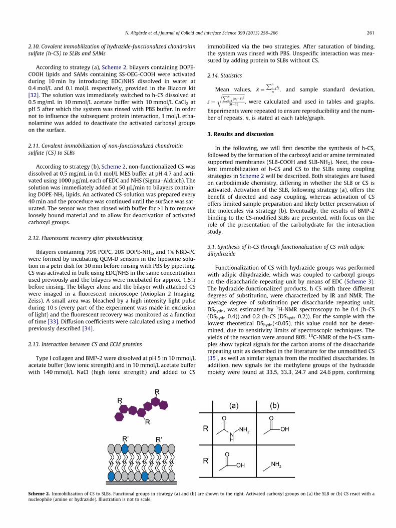
According to strategy (a), Scheme 2, bilayers containing DOPE-COOH lipids and SAMs containing SS-OEG-COOH were activatedduring 10 min by introducing EDC/NHS dissolved in water at0.4 mol/L and 0.1 mol/L respectively, provided in the Biacore kit[32]. The solution was immediately switched to h-CS dissolved at0.5 mg/mL in 10 mmol/L acetate buffer with 10 mmol/L CaCl2 atpH 5 after which the system was rinsed with PBS buffer. In ordernot to influence the subsequent protein interaction, 1 mol/L etha-nolamine was added to deactivate the activated carboxyl groupson the surface.
2.11. Covalent immobilization of non-functionalized chondroitinsulfate (CS) to SLBs
According to strategy (b), Scheme 2, non-functionalized CS wasdissolved at 0.5 mg/mL in 0.1 mol/L MES buffer at pH 4.7 and acti-vated using 1000 lg/mL each of EDC and NHS (Sigma–Aldrich). Thesolution was immediately added at 50 lL/min to bilayers contain-ing DOPE-NH2 lipids. An activated CS-solution was prepared every40 min and the procedure was continued until the surface was sat-urated. The sensor was then rinsed with buffer for >1 h to removeloosely bound material and to allow for deactivation of activatedcarboxyl groups.
2.12. Fluorescent recovery after photobleaching
Bilayers containing 79% POPC, 20% DOPE-NH2, and 1% NBD-PCwere formed by incubating QCM-D sensors in the liposome solu-tion in a petri dish for 30 min before rinsing with PBS by pipetting.CS was activated in bulk using EDC/NHS in the same concentrationused previously and the bilayers were incubated for approx. 1.5 hbefore rinsing. The bilayer alone and the bilayer with attached CSwere imaged in a fluorescent microscope (Axioplan 2 Imaging,Zeiss). A small area was bleached by a high intensity light pulseduring 10 s (every part of the experiment was made in exclusionof light) and the fluorescent recovery was monitored as a functionof time [33]. Diffusion coefficients were calculated using a methodpreviously described [34].
2.13. Interaction between CS and ECM proteins
Type I collagen and BMP-2 were dissolved at pH 5 in 10 mmol/Lacetate buffer (low ionic strength) and in 10 mmol/L acetate bufferwith 140 mmol/L NaCl (high ionic strength) and added to CS
Scheme 2. Immobilization of CS to SLBs. Functional groups in strategy (a) and (b) are snucleophile (amine or hydrazide). Illustration is not to scale.
immobilized via the two strategies. After saturation of binding,the system was rinsed with PBS. Unspecific interaction was mea-sured by adding protein to SLBs without CS.
2.14. Statistics
Mean values, �x ¼Pn
i¼1xi
n , and sample standard deviation,
s ¼ffiffiffiffiffiffiffiffiffiffiffiffiffiffiffiffiffiffiffiffiPn
i¼1ðxi��xÞ2
ðn�1Þ
r, were calculated and used in tables and graphs.
Experiments were repeated to ensure reproducibility and the num-ber of repeats, n, is stated at each table/graph.
3. Results and discussion
In the following, we will first describe the synthesis of h-CS,followed by the formation of the carboxyl acid or amine terminatedsupported membranes (SLB-COOH and SLB-NH2). Next, the cova-lent immobilization of h-CS and CS to the SLBs using couplingstrategies in Scheme 2 will be described. Both strategies are basedon carbodiimide chemistry, differing in whether the SLB or CS isactivated. Activation of the SLB, following strategy (a), offers thebenefit of directed and easy coupling, whereas activation of CSoffers limited sample preparation and likely better preservation ofthe molecules via strategy (b). Eventually, the results of BMP-2binding to the CS-modified SLBs are presented, with focus on therole of the presentation of the carbohydrate for the interactionstudy.
3.1. Synthesis of h-CS through functionalization of CS with adipicdihydrazide
Functionalization of CS with hydrazide groups was performedwith adipic dihydrazide, which was coupled to carboxyl groupson the disaccharide repeating unit by means of EDC (Scheme 3).The hydrazide-functionalized products, h-CS with three differentdegrees of substitution, were characterized by IR and NMR. Theaverage degree of substitution per disaccharide repeating unit,DShydr., was estimated by 1H-NMR spectroscopy to be 0.4 (h-CS(DShydr. 0.4)) and 0.2 (h-CS (DShydr. 0.2)). For the sample with thelowest theoretical DShydr.(<0.05), this value could not be deter-mined, due to sensitivity limits of spectroscopic techniques. Theyields of the reaction were around 80%. 13C-NMR of the h-CS sam-ples show typical signals for the carbon atoms of the disacchariderepeating unit as described in the literature for the unmodified CS[35], as well as similar signals from the modified disaccharides. Inaddition, new signals for the methylene groups of the hydrazidemoiety were found at 33.5, 33.3, 24.7 and 24.6 ppm, confirming
hown to the right. Activated carboxyl groups on (a) the SLB or (b) CS react with a
Scheme 3. Reaction scheme for the functionalization of CS with adipic dihydrazide. Arrows indicate the functional groups where subsequent immobilization to the SLB canoccur, using the different strategies outlined in Scheme 2. For h-CS (right side), the hydrazide molecule is not present on every disaccharide unit but depending on the degreeof substitution, given by the DShydr. number.
262 N. Altgärde et al. / Journal of Colloid and Interface Science 390 (2013) 258–266
the introduction of hydrazide groups into the glucuronic acid unitof CS [36].
3.2. Formation of SLBs suitable for covalent coupling of CS derivatives
Two kinds of lipid membranes (SLB-COOH, SLB-NH2) wereformed on silica surfaces. In the QCM-D experiments, high qualitySLBs are characterized by frequency and dissipation shifts ofDf = ��26 Hz and DD < 0.5 � 10�6, respectively [37]. SLBs expos-ing carboxyl groups have been described before and have beenformed e.g. from liposomes composed of phosphatidylserine lipids(on silica [38] or titania [39]) or phospholipids with the carboxylgroup attached to the lipid head group via a spacer (on silica[23]). The lipid that is used in the present study, referred to asDOPE-COOH, has the carboxyl group attached to the phospholipidhead group via a C11-spacer (Chart 1). Mixed POPC/DOPE-COOHliposomes with a DOPE-COOH fraction of up to 20 mol% fused intobilayers, via a critical surface coverage dependent mechanism (seeSupporting information). Similarly, the formation of SLB-NH2 wasachieved using lipids where the head group of phosphatidyl etha-nolamine lipids had been modified with an amine terminated C11-
Fig. 1. QCM-D frequency (Df) and dissipation (DD) signals obtained when immobilizing Cnon-activated (dashed line) SLB-COOH 5%. Strategy (b), right side: stepwise immobilizati(solid line).
spacer, referred to as DOPE-NH2. In this case, the range of fractionsof DOPE-NH2 was limited by the ability to prepare a homogenousliposome solution at the current buffer conditions; the liposomesaggregated when the fraction of DOPE-NH2 > 60 mol%. Whenincreasing the fraction of DOPE-NH2 in the liposomes (<60 mol%),bilayers formed following a mechanism requiring lower and lowerliposome coverage before rupture and increasing number of theliposomes fused directly when adsorbing on the surface (see Sup-porting information).
3.3. Covalent immobilization of h-CS to SLB-COOH
Immobilization strategy (a) was based on covalent coupling ofh-CS to SLB-COOH (Scheme 2a). To achieve this, the SLB was firstexposed to an excess of EDC/NHS coupling reagents. Next, withoutprior rinsing, h-CS was added to allow for a reaction between thehydrazide group on h-CS and the activated carboxyl groups onthe membrane (Fig. 1). Hydrazide groups have a lower pKa valuethan primary amines (�5 vs. �10), and thus acetic buffer pH 5was used for coupling [40]. Successful coupling was only possiblewhen CaCl2 was added to the buffer, likely as a result of shielding
S to SLBs. Strategy (a), left side: h-CS (DShydr. 0.2) added to activated (solid line) andon of bulk activated (EDC/NHS) CS to SLB-NH2 5% (dashed line) and to SLB-NH2 55%
Table 1QCM-D frequency (Df) and dissipation shifts (DD) obtained when immobilizing CS to SLBs according to strategy (a) and strategy (b). Functional lipid fraction refers to percentageof either (a) DOPE-COOH or (b) DOPE-NH2 in the SLB. Unless otherwise stated, the number of repeats, n, was �3.
N. Altgärde et al. / Journal of Colloid and Interface Science 390 (2013) 258–266 263
of electrostatic repulsion between negatively charged (non-acti-vated) carboxyl groups and negatively charged sulfate groups onCS. We note that no material was removed upon rinsing with buf-fer after completion of the coupling reaction. Thus, despite the factthat we have no direct proof (e.g. by spectroscopy or mass spec-trometry) of the actual formation of covalent bonds, all resultspoint in that direction (see also below). The highest immobilizedamount was obtained for h-CS (DShydr. 0.4) to SLB-COOH 5%. Theimmobilized amount decreased with lower DShydr. and, perhapsunexpectedly, with higher DOPE-COOH fractions (Table 1). This islikely explained by geometrical and/or electrostatic requirementsfor the coupling reaction. The low values for Df and DD suggest athin h-CS layer with a thickness of a few nanometers. The immobi-lized amount of h-CS to SLB-COOH 5% corresponds to a mass of ap-prox. 25 ng/cm2 using the Sauerbrey equation, which is reasonablefor a CS layer immobilized predominantly in a side-on configura-tion. In control experiments, adsorption of negligible amounts ofh-CS was observed if the carboxyl groups were left non-activatedprior to addition of h-CS (Fig. 1). The same was true if CS wasnon-functionalized, strongly suggesting that immobilization oc-curred through covalent coupling between the carboxyl groupson the bilayer and the hydrazide groups on h-CS.
Fig. 2. (I) DD/Df values for the bare bilayer and for layers of CS immobilized to it.Number of repeats, n, was P2. (II) Illustration of CS on a surface adopting a loosestructure (left) leading to higher dissipative losses (high DD/Df values) or a morecompact and rigid structure (right). Illustration not to scale.
3.4. Covalent immobilization of activated CS to SLB-NH2
In the second immobilization strategy (Scheme 2b), naturallyoccurring carboxyl groups on CS were activated in bulk usingEDC/NHS and immediately added to SLBs exposing amine groups.Frequency and dissipation shifts when immobilizing CS to differentSLBs-NH2 are summarized in Table 1. As expected, the immobilizedamount of CS increased for higher fractions of DOPE-NH2 lipid inthe bilayer; almost no CS was bound to bilayer containingSLB-NH2 5% whereas the highest amount of activated CS wasimmobilized to SLBs with >20% DOPE-NH2, corresponding toapprox. 55 ng/cm2 using the Sauerbrey equation. Thus, the immo-bilized amounts achieved when activating CS were generally largercompared to immobilization using strategy (a).
3.5. Dynamics and stability of the lipid membrane immobilizationplatform
The CS chains are approx. 60 nm long, having functional groupsseparated up to 10 nm, are likely to have several attachment pointsto the membrane, likely stabilizing the systems. To assess whetherthe immobilization of CS to the SLB altered the lateral diffusion oflipids in the SLB, a fraction of fluorescently labeled lipids in the SLBswas added in fluorescence recovery after photobleaching (FRAP)experiments [33]. Acquired images were analyzed to calculate thediffusion constant, D, for lipid molecules in the membrane [34].
All bilayers showed almost complete fluorescent recoveryafter approx. 3 min, proving them to be fluid (see Supportinginformation). There was no significant difference between SLB-NH2 20% (D = 0.95 ± 0.1 lm2/s) and SLB-NH2 with coupled CS(D = 0.87 ± 0.2 lm2/s), thus showing that the lipid molecules in theSLB retain their lateral mobility after the CS has been immobilized.Therefore, the number of attachment points between the CS-chainsand the SLB is low enough to let the fluorescent lipids diffuse freely inthe bilayer. The relationship between diffusivity and degree of func-tionalization has been discussed in other studies [23].
In separate experiments using self-assembled monolayers(SAMs) of thiolates on gold, we also addressed another issuerelated to the membrane dynamics. By comparing the results whenusing SLB-COOH and SAM-COOH, we wanted to test whether theimmobilized GAGs were ripping lipid molecules away from themembrane, thus contributing to the low immobilized amounts ofCS. However, only small differences were seen when comparingthe immobilized amount of CS to the SAM and the SLB (seeSupporting information). This suggests that the fraction of DOPE-COOH lipids is a rather good measure of the number of carboxylgroups exposed at the surface.
Fig. 3. QCM-D frequency (Df) and dissipation shifts (DD) when adding BMP-2 to (a) SLB-COOH 5% and to (b) SLB-NH2 20% in low (top) and high (bottom) ionic strengthacetate buffer. Addition was made to bare bilayers (dashed lines) and to CS coupled to the bilayers according to the strategies in Scheme 2 (solid lines). After addition, thesystem was rinsed with the acetate buffer just used (indicated in graph), and later with PBS (not shown).
264 N. Altgärde et al. / Journal of Colloid and Interface Science 390 (2013) 258–266
3.6. Configuration and flexibility of CS on the SLBs
The QCM-D technique offers the advantage of very sensitivelydetecting highly hydrated structures like GAGs. Immobilizationof GAGs can be very difficult to detect by e.g. an optical technique,as has been shown previously for an analogous example where apeptide is immobilized to SLBs exposing maleimide groups [41].In addition to evaluating QCM-D frequency and dissipation shiftsseparately, structural properties of adsorbed layers can be inter-preted by studying the DD/Df ratios. A higher DD/Df value indi-cates a softer (more dissipative) layer. The SLB (approx. 5 nmthick) is known to be rigidly attached to the sensor surface andtherefore has a low DD/Df ratio (Fig. 2I). Immobilization of CS re-sulted in increased DD/Df values, which is expected when attach-ing long, highly hydrated polymers. Even though immobilizedamounts were higher using strategy (b), there is no significantdifference in the viscoelastic properties of the layers. This couldindicate that not all activated carboxyl groups along the CS chainin strategy (b) are coupled to the SLB. The retained fluidity proba-bly allows the immobilized CS to adopt a relaxed conformation(Fig. 2(II)). It is also worth noticing that DD/Df for CS immobilized
on SLB-COOH is higher than for CS immobilized on SAM-COOH,which has no lateral mobility (Fig. 2(I)).
3.7. Application of CS-modified SLBs to study interactions with ECMproteins type I collagen and growth factor BMP-2
Interactions involving GAGs are inherently difficult to study dueto their high charge and multiple sites for protein binding, and thenature of these interactions is debated [42]. For biomolecular inter-actions in general, non-specific electrostatic interactions are ex-pected to be shielded by increasing the ionic strength of thebuffer [43]. The interactions between CS and either type I collagenor BMP-2 were therefore studied using high (physiologic) and lowionic strength buffers. Both proteins carry an overall positivecharge under the conditions used [14,44], but differ in molecularweight and number of potential binding sites. For type I collagen,CS is thought to bridge cationic regions between molecules,suggesting multiple binding sites at both CS and collagen [7]. Forthe homodimer BMP-2, two binding sites for the GAG heparin havebeen suggested [45], and a similar interaction with CS is likely.Interestingly, the structure of the BMP-2 dimer suggests that it
Fig. 4. QCM - D dissipation versus frequency plot showing dissipative behavior ofthe film as BMP-2 is added to CS coupled according to strategy (a) (filled marks) andstrategy (b) (clear marks). Bottom: schematic image of the CS – BMP-2 layer.
N. Altgärde et al. / Journal of Colloid and Interface Science 390 (2013) 258–266 265
would bind along an extended molecule, like pearls on a string,rather than cross-link adjacent molecules [46]. As a comparison,crosslinking has previously been seen for the GAG hyaluronanand the dimer TSG-6 [47].
Type I collagen was added to immobilized CS in high and lowionic strength acetate buffer at pH 5 and compared to controlexperiments with no immobilized CS (see Supporting information).In the absence of CS, there was negligible binding of type I collagento SLB-NH2 and to deactivated SLB-COOH, independent of theamount of functional lipid in the bilayer. In contrast, large amountsof collagen type I was accumulated at the negatively charged sur-faces provided either by the plain SLB-COOH or the CS-modifiedSLB-COOH (see Fig. S4 in Supporting information), although mostlyreversibly to the SLB-COOH and with a considerable amount ofirreversibly bound material on the CS-SLB. As expected for the CS– type I collagen interaction, it was increased when performed inlow ionic strength buffer [47]. When collagen was added to CS, asofter layer was formed on the lipid membrane compared to theCS layer itself, as indicated by an increasing DD/Df ratio (seeFig. 2(I) and Fig. S5 in Supporting information).
BMP-2 was added to immobilized CS in the same manner asfor type I collagen, in high (physiologic) and low ionic strengthat pH 5. Both conditions induced binding of BMP-2 to CS immo-bilized via the two strategies (Fig. 3, solid lines). Using strategy(a), high amounts of BMP-2 bound to h-CS when added in lowionic strength buffer (Fig. 3 top left, solid lines). Control experi-ments, (no immobilized CS) using the same conditions showedBMP-2 binding to bare SLB-COOH (Fig. 3 top left, dotted lines).When increasing the ionic strength this non-specific bindingwas suppressed and a decreased but presumably specific BMP-2binding was observed to immobilized h-CS (Fig. 3, lower left).Using strategy (b), negligible non-specific interaction could beseen for both the low and the high ionic strength buffers (Fig. 3right side, dotted lines). At low ionic strength, the amount ofBMP-2 bound to bare SLB-COOH, SLB-COOH with h-CS and SLB-NH2 with CS is roughly the same, even though the coupledamounts of the underlying CS differ, indicating a primarilyelectrostatic interaction. The large amounts of BMP-2 that boundto CS immobilized using strategy (b) compared to strategy (a) athigh ionic strength can in part be due to the added mass of CSwhen using this strategy. More importantly, CS is here expectedto be in a more native form, having not been functionalized.The reaction did not reach equilibrium under a reasonable time,likely due to structural rearrangements occurring in theCS layer on a slower time scale and perhaps also due to BMP-2aggregation at the sensor surface. Interestingly, there is littledissociation of BMP-2 from the CS layer. This low release hasbeen shown using other GAG-systems, and seems to be preferredin terms of efficient presentation of BMP-2 to cells [13].
Taken together, the CS – collagen and the CS – BMP-2 interac-tions represented two different types of systems, where type I col-lagen presumably bound to CS in a polyelectrolyte type ofinteraction, whereas the BMP-2 bound specifically to the CS. Inter-estingly, significantly more mass was added by the smaller BMP-2than by type I collagen, clearly showing the ability of CS to loadlarge amounts of BMP-2. Furthermore, the layer after addition ofBMP-2 was much more compact, compared to the collagen – CSlayer (Fig. S5 in Supporting information). The CS immobilizationstrategy did not significantly affect the configuration of the colla-gen – CS layers (Fig. S5). However, for BMP-2 there was not onlya difference between the properties of the final layers (Fig. S5),but also in the way they were built up, as seen in the DD/Df plotin Fig. 4.
These results emphasize that the way GAGs are immobilized tosurfaces can largely influence how the GAGs in turn interact withproteins.
4. Conclusion
A platform for biomolecular interaction studies based on cova-lent immobilization of the glycosaminoglycan chondroitin sulfate(CS) to supported lipid bilayers (SLBs) has been investigated. Twoapproaches were tested; (a) hydrazide-functionalized CS immobi-lized to a SLB presenting activated carboxyl groups, and (b) CS dis-playing activated carboxyl group immobilized to a SLB presentingamine groups. Both approaches yielded thin layers of predomi-nantly side-on immobilized CS with similar properties, althoughmore material was immobilized using strategy (b). Interaction be-tween CS and growth factor BMP-2 shows the ability of CS to loadlarge amounts of BMP-2. The specificity between certain GAGs andgrowth factors is an interesting topic. Especially the sulfation ofGAGs is thought to play a significant role for their functions in tis-sues, and the platform presented here is well suited for studyingthe relationships between the molecular properties of GAGs andtheir interactions with biomolecules .
Acknowledgments
The research leading to these results has received funding fromthe European Union Seventh Framework Programme (FP7/2007–2013) under Grant Agreement No. NMP4-SL2009-229292 (‘‘Find& Bind’’). J.B., S.M., and M.S. further acknowledge financial supportby the Deutsche Forschungsgemeinschaft, DFG (TRR 67), and S.S.acknowledges financial support provided by the Swedish ResearchCouncil (Linnaeus program SUPRA). Prof. Fredrik Höök is acknowl-edged for critical reading of the manuscript.
Appendix A. Supplementary material
Supplementary data associated with this article can be found, inthe online version, at http://dx.doi.org/10.1016/j.jcis.2012.07.063.
266 N. Altgärde et al. / Journal of Colloid and Interface Science 390 (2013) 258–266
References
[1] M.P. Lutolf, J.A. Hubbell, Nat. Biotechnol. 23 (2005) 47.[2] C.I. Gama, L.C. Hsieh-Wilson, Curr. Opin. Chem. Biol. 9 (2005) 609.[3] R. Sasisekharan, R. Raman, V. Prabhakar, Annu. Rev. Biomed. Eng. 8 (2006)
181.[4] G. Siegel, M. Malmsten, D. Klüßendorf, F. Michel, Biosens. Bioelectron. 16
(2001) 895.[5] A. Saito, H. Munakata, Electrophoresis 25 (2004) 2452.[6] J. Schiller, J. Becher, S. Möller, K. Nimptsch, T. Riemer, M. Schnabelrauch, Mini-
Rev. Org. Chem. 7 (2010) 290.[7] T. Douglas, S. Heinemann, C. Mietrach, U. Hempel, S. Bierbaum, D.
Scharnweber, H. Worch, Biomacromolecules 8 (2007) 1085.[8] L. Le Guéhennec, A. Soueidan, P. Layrolle, Y. Amouriq, Dent. Mater. 23 (2007)
844.[9] B. Stadlinger, E. Pilling, R. Mai, S. Bierbaum, R. Berhardt, D. Scharnweber, U.
Eckelt, J. Mater. Sci.: Mater. Med. 19 (2008) 1043.[10] X. Hu, K.G. Neoh, Z. Shi, E.T. Kang, C. Poh, W. Wang, Biomaterials 31 (2010)
Weber, Acta Biomater. (2011).[12] J. Almodóvar, S. Bacon, J. Gogolski, J.D. Kisiday, M.J. Kipper, Biomacromolecules
11 (2010) 2629.[13] T. Crouzier, K. Ren, C. Nicolas, C. Roy, C. Picart, Small 5 (2009) 598.[14] M.L. Macdonald, R.E. Samuel, N.J. Shah, R.F. Padera, Y.M. Beben, P.T. Hammond,
Biomaterials 32 (2011) 1446.[15] V. Hintze, S. Moeller, M. Schnabelrauch, S. Bierbaum, M. Viola, H. Worch, D.
Scharnweber, Biomacromolecules 10 (2009) 3290.[16] C.J.C. De Haas, P.J. Haas, K.P.M. Van Kessel, J.A.G. Van Strijp, Biochem. Biophys.
Res. Commun. 252 (1998) 492.[17] M. Delehedde, M. Lyon, J.T. Gallagher, P.S. Rudland, D.G. Fernig, Biochem. J. 366
(2002) 235.[18] B.T. Houseman, E.S. Gawalt, M. Mrksich, Langmuir 19 (2003) 1522.[19] S. Park, I. Shin, Angew. Chem. Int. Ed. 41 (2002) 3180.[20] J.F. Popplewell, M.J. Swann, Y. Ahmed, J.E. Turnbull, D.G. Fernig, ChemBioChem
10 (2009) 1218.[21] S. Svedhem, D. Dahlborg, J. Ekeroth, J. Kelly, F. Höök, J. Gold, Langmuir 19
(2003) 6730.[22] R.P. Richter, K.K. Hock, J. Burkhartsmeyer, H. Boehm, P. Bingen, G. Wang, N.F.
Biophys. Res. Commun. 286 (2001) 554.[29] R.E. Jung, R. Glauser, P. Schärer, C.H.F. Hàmmerle, H.F. Sailer, F.E. Weber,
Clinical Oral Implants Res. 14 (2003) 556.[30] M. Rodahl, F. Höök, A. Krozer, P. Brzezinski, B. Kasemo, Rev. Sci. Instrum. 66
(1995) 3924.[31] G.Z. Sauerbrey, Z. Phys. A: Hadrons Nucl. 155 (1959) 206.[32] D.J. O’Shannessy, M. Brigham-Burke, K. Peck, Anal. Biochem. 205 (1992) 132.[33] T.K.L. Meyvis, S.C. De Smedt, P. Van Oostveldt, J. Demeester, Pharm. Res. 16
(1999) 1153.[34] P. Jönsson, M.P. Jonsson, J.O. Tegenfeldt, F. Höök, Biophys. J. 95 (2008) 5334.[35] S.M. Bociek, A.H. Darke, D. Welti, D.A. Rees, Eur. J. Biochem. 109 (1980) 447.[36] S. Jo, D. Kim, J. Woo, G. Yoon, Y.D. Park, G. Tae, I. Noh, Macromol. Res. 19 (2011)
147.[37] C.A. Keller, B. Kasemo, Biophys. J. 75 (1998) 1397.[38] R.P. Richter, N. Maury, A.R. Brisson, Langmuir 21 (2005) 299.[39] F.F. Rossetti, M. Textor, I. Reviakine, Langmuir 22 (2006) 3467.[40] S. Raddatz, J. Mueller-Ibeler, J. Kluge, L. Wäß, G. Burdinski, J.R. Havens, T.J.
Onofrey, D. Wang, M. Schweitzer, Nucl. Acids Res. 30 (2002) 4793.[41] M. Edvardsson, S. Svedhem, G. Wang, R. Richter, M. Rodahl, B. Kasemo, Anal.
Chem. 81 (2009) 349.[42] C.J. Rogers, P.M. Clark, S.E. Tully, R. Abrol, K.C. Garcia, W.A. Goddard Iii, L.C.
Hsieh-Wilson, Proc. Natl. Acad. Sci. USA 108 (2011) 9747.[43] H. Larsericsdotter, S. Oscarsson, J. Buijs, J. Colloid Interf. Sci. 237 (2001) 98.[44] Z. Zhang, G. Li, B. Shi, JSLTC 90 (2006) 23.[45] R. Ruppert, E. Hoffmann, W. Sebald, Eur. J. Biochem. 237 (1996) 295.[46] M. Laub, T. Seul, E. Schmachtenberg, H.P. Jennissen, Materialwiss. Werkst. 32
(2001) 926.[47] B. Obrink, A. Wasteson, Biochem. J. 121 (1971) 227.