Page 1
10
Tissue Engineering and Regenerative Medicine, Vol. 11, No. 1, pp 10-15 (2014)
DOI 10.1007/s13770-013-1119-4
|Original Article|
Immobilization of Fibrinogen Antibody on Self-Assembled Gold Monolayers
for Immunosensor Applications
Hongsik Cho1,2,3, Justin Zook4, Todd Banner5, Sang-Hyug Park6, Byoung-Hyun Min7,8,9,
Karen A Hasty1,2,3
, Eugene Pinkhassik5*, and Erno Lindner
4,5*
1Department of Orthpaedic Surgery & Biomedical Engineering, University of Tennessee Health Science Center, Memphis,
TN, 38104 USA2Department of Orthopaedic Surgery, Campbell Clinnic, Memphis, TN,38104 USA
3Research 151, Veterans Affairs Medical Center, Memphis, TN, 38104 USA
4Department of Biomedical Engineering, University of Memphis, Memphis, TN, 38152 USA5Department of Chemistry, University of Memphis, Memphis, TN, 38152 USA
6Department of Biomedical Engineering, Jungwon University, Geosan, 367-700 Korea
7Department of Orthopeadic Surgery, Medical School, Ajou Univeristy, Suwon, 443-749 Korea8Department of Molecular Science and Technology, Ajou Univeristy, Suwon, 443-749 Korea
9Cell Therapy Center, Ajou University Medical Center, Suwon, 443-749 Korea
(Received: July 8th, 2013; Revision: August 8th, 2013; Accepted: August 20th, 2013)
Abstract : Self-assembled gold monolayers offer several advantages for the realization of novel modified electrodes
for biosensor applications. This is due to their ability to decrease non-specific adsorption and provide for covalent
attachment of biomolecules. Surfaces for these applications require the precise control of ligand density, the ability
to immobilize ligands, and in situ-modulation of ligand activity. In this study, we focused our studies on the immo-
bilization of antibody on a gold monolayer surface. We self-assembled thioctic acid onto the gold surface as an
anchor point for the immobilization of anti-fibrinogen onto the surface. The modifications to the gold surface were
characterized by ELISA, ellipsometry, and AFM.
Key words: self-assembled monolayers (SAMs), anti-fibrinogen, immunosensors, enzyme-linked immuno sorbent
assay (ELISA), ellipsometry, atomic force microscopy (AFM), gold monolayer, antibody immobilization
1. Introduction
Chemical reactions on solid surfaces are important to the
essential understanding of interfacial phenomena and for
technological applications to immunosensors.1-3 Gold substrates
have played an important role in the advancement of surface-
based nanotechnology research. Gold is useful because it can
chemically bind to thiol and disulfide functionalized compounds
which provide the foundation for self-assembled monolayers
(SAMs).4-6 SAMs have been studied extensively and many new
research areas and applications have subsequently evolved.6,7
SAMs offer several advantages for the realization of novel
modified electrodes for biosensor applications because of their
role in decreasing non-specific adsorption and allowing for
covalent attachment of biomolecules, such as antibodies and
other proteins.8-10 Antibodies can be covalently immobilized
onto a gold surface using thiols.11-13 The immobilization of
antibodies onto surfaces provides enhanced or maximized
interactions between the immobilized biomolecules and
ligands.In this respect, various chemistry and biology-based
methods have been developed utilizing antigen-antibody
interactions and protein-ligand interactions.14-16
To extend the benefit of these surfaces to generate new types
of immunosensors and to study complex biological processes,
it is necessary to develop substrates with more sophisticated
and flexible surface properties. Surfaces for these applications
require the precise control of ligand density, the ability to
immobilize the ligand, and the in-situ modulation of ligand
activity.12
Fibrinogen is the most important protein involved in coagula-
*Corresponding author
Tel: 901-678-5641; Fax: 901-678-3447
e-mail: [email protected] (Erno Lindner)
Tel: 901-678-4430; Fax: 901-678-3447
e-mail: [email protected] (Eugene Pinkhassik)
Page 2
Fibrinogen Antibody Immobilization on SAM
11
tion and the concentration of fibrinogen in plasma is 1.5-4.5 mg/
mL in normal condition.17,18 An abnormal concentration of
plasma fibrinogen is a strong risk factor for thrombotic disorders
and must be accurately determined with short-term diagnosis.18
In this study, we set up a common intermediate method to
functionalize gold surfaces with terminal carboxylic acid
groups (via thioctic acid) in order to immobilize anti-fibrinogen
onto the surface. We then used N-(3-dimethylaminopropyl)-N'-
ethylcarbodiimide (EDC) and N-hydroxysuccinimide (NHS)
to activate the carboxylic acid functionalized surface so that it
would couple with anti-rabbit immuno-globulin G (IgG) and
horseradish peroxidase (HRP). Figure 1 is showing the concept
of biosensor using polyclonal fibronogen antibody immobiliza-
tion on gold monolayer surface. We focused our studies on the
immobilization of fibrinogen antibody onto the funcionalized
gold monolayer surface. The coupling reaction on the gold
surface was characterized by enzyme-linked immuno sorbent
assay (ELISA), ellipsometry, atomic force microscopy (AFM)
and the coupling efficiency was estimated by comparing the
experimentally determined absorbance of 3,3´,5,5´-
tetramethylbenzidine (TMB) reaction of ELISA.
2. Materials and Methods
2.1 Chemicals
11-Mercaptoundecanoic acid (MUA), thioctic acid, N-
hydroxysuccinimide (NHS), 1-(3-dimethylaminopropyl)-N’-
ethylcarbodiimidehydro-chloride (EDC) were purchased from
Aldrich (St. Louis. Mo, USA). Rabbit IgG, goat anti-rabbit IgG
with HRP, goat anti-mouse IgG with HRP, and bovine serum
albumin (BSA) were purchased from Pierce (Rockford, IL,
U.S.A). All solvents were reagent-grade. Reagents were used
without any further purification. Experiments were carried out
at room temperature.13,19
2.2 Preparation of Gold Substrate
Gold template substrates (catalog number AU.1000.SL2NOTI)
were purchased from Platypus Technologies (Madison, WI). The
substrates were 100 mm diameter silicon disks covered with
electron-beam deposited gold to a thickness of 1000Å. A titanium
adhesion layer between the silicon and gold layers was not used.
2.2.1 Preparation of Supporting Substrate
Silicon supporting substrate was purchased from AlSil-
Supply Division (Palm Beach, Florida). The substrates were 3
inch diameter silicon (N-type, phosphorous doped) disks with
thicknesses of 1 mm ± 20 µm. It was polished on a single side
with mean roughness < 6 Å. The substrates were cut with a
scoring stylus and then cleaved into pieces of approximately 1
×2 cm. The pieces were rinsed with ethanol, then water, and
finally blown dry using a strong stream of argon gas.
2.2.2 Adhesion of Supporting Substrate
About 1 mg of freshly prepared Epotek 377 epoxy glue
(Bellerica, MA; parts A and B were mixed 1:1 by weight) was
placed onto the polished side of the cleaved supporting silicon
pieces using a disposable syringe with needle. The silicon pieces
were then placed, epoxy side down, onto the top of the gold
template substrate. The gold template substrate wafer was then
placed into an oven at 150oC for overnight to cure the epoxy. The
gold template/supporting silicon substrate sandwich disk was
cooled at ambient temperature for at least 10 minutes before
stripping the gold from the template substrate.6
2.2.3 Stripping of the Template Substrate
A razor blade was used to gently scrape near the edges of the
adhered supporting substrate. The supporting substrate would
typically release from the surface yielding gold surface on top
of the supporting silicon substrate.
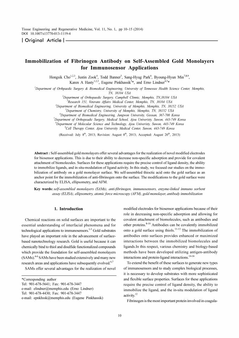
Figure 1. Schematic illustration of the concept of biosensor using
polyclonal fibronogen antibody immobilization on gold monolayer
surface; A self-assembled (SAM) gold monolayers with immobi-
lized antibodies on short thiol molecules design for immunosensor
that measuring fibrinogen concentration in small amount of blood
sample base on enzyme kinetic method in the future.
Page 3
Hongsik Cho et al.
12
2.3 Activation of Gold Monolayer and Immobilization
of Antibody
We first prepared and characterized gold substrate on silica.
Then, the bare gold substrate films were modified with thioctic
acid by submersing them in 10 mM ethanolic solutions for 6
hours. Longer immersion times did not noticeably improve the
efficiency. The substrates were then submersed in a 2 mL
solution of NHS (20 mM) and EDC (10 mM) in ultra pure water
for 20 min, followed by the antibody (poly anti-fibrinogen, 1 µg/
mL) solution in PBS for 1 hour. The residual NHS esters were
blocked by submersing the modified substrates in 1 M
ethanolamine (pH 9.0) for 20 min. After washing with ultra
pure water, the substrates were finally immersed in a 1% (w/v)
solution of BSA in 10 mM PBS pH 7.4 for 1 h.19
2.4 Ellipsometry
Ellipsometric measurements were performed using a
Gaertner Scientific ellipsometer (model: LSE) equipped with a
He-Ne laser (λ = 6328Å), set at an angle of incidence of 70o.
The gold substrate constants were derived from ellipsometric
measurements conducted at 10 or more locations on a bare gold
substrate. For the substrate coated with SAMs, the thickness of
the SAM was determined from ellipsometric measurements at
five different locations (separated by at least 0.5 cm) using the
recorded substrate constants. We assumed that the refractive
index of the film was 1.46 and the film was completely
transparent to laser beam.
2.5 Analysis Enzymatic Activities (ELISA) by TMB
Substrate Assay
After the antibody immobilized surface was washed with
PBS and Tween 20, the unreacted sites on the surface were then
blocked by reaction with a 1% solution of bovine serume
albumin (BSA). The 2nd antibody (anti-rabbit IgG) with HRP
reacted to 1st antibody during 1 hr at room temperature. The
peroxidase enzyme, in the presence of H2O2, catalysed the
oxidation of colorless 200 ul TMB substrate, (Pierce, IL, USA)
yielding to a blue colored product. After a fixed reaction time
(30 min), the reaction was stopped with 50 µL of 2M H2SO4 and
the solutions were placed into a 96-well plate and the absorbance
of the solutions was measured at 450 nm by spectrophotometer.
For this ELISA, we prepared an acrylic cage apparatus which
can securely hold the gold monolayer and has a hole that is
same size of well in 96-well plate (Fig 2).
2.6 Characterization of Gold Surface by AFM Imaging
AFM images were gathered using an MFP-3D-BIO instrument
purchased from Asylum Research (Santa Barbara, CA). All
images were collected in repulsive AC mode. The cantilevers
used were AC240TS manufactured by Olympus and purchased
from Asylum Research. The imaging of samples was done in
AC mode in air. AFM provides topographical information
about the gold surface and has the ability to detect individual
protein molecules on the surface.
2.7 Set up of Experimental Group
We first prepared and characterized ultra-smooth gold
surfaces on silica. Then, the bare gold substrate films were
modified with thioctic acid from 10 mM ethanolic solutions for
a period of 6 hr. The antibody was coupled with the carboxylic
acid group on gold surfaces via EDC and NHS activation of the
carboxylic acid group. After the primary antibody was
immobilized the surface, we detected its presence with a secondary
HRP-anti-rabbit IgG antibody and TMB. In order to determine
if the antibody was immobilized on the surface, we performed
the experiments outlined in Table 1.
3. Results and Discussion
The immunoosensors are used to detect or quantify disease-
related substances known as biomarkers in clinical diagnostics.
In immunosensor applications, antibodies are immobilized
onto the biosensor surface to capture specific biomarkers.20 The
most important consideration is the development of a technique to
immobilize antibodies onto the sensor substrate surface without
decreasing their binding affinities and binding capacities.21,22 In
this project, the resulting structures were characterized by
ellipsometry, ELISA, and AFM imaging.
3.1 Ellipsometry Analysis
In this experiment, we used two different types of SAM that
Figure 2. Diagram of the apparatus for ELISA on gold mono-
layer. This apparatus was design to support the ELISA reaction
directly onto the gold SAMs.
Page 4
Fibrinogen Antibody Immobilization on SAM
13
vary based on their chain length. Long-chain SAM (11- MUA)
has been widely used as linker molecule due to its strong van
der Waals forces among the chain molecules. This allows for it
to have a relatively high packing density in addition to well-
ordered structure. Conversely, having high densities in the
surface terminal groups leads to steric hindrance. To overcome
these limitations, short-chain SAM such as thiotic acid (alpha-
lipoic acid) has recently been suggested as an alternative linker
molecule. Short-chain SAMs offer higher sensitivity and require
shorter incubation time than long-chain SAMs, therefore, for
these experiments we used the thiolate molecules based upon
prior reports that shorter thiol molecules are better for gold
substrate immunosensors than longer thiols.16,19 The thickness
of MUA and thiotic acid monolayers were measured by
Ellipsometry. The thickness of the thioctic acid and MUA
monolayer measured by ellipsometry were 9.8 ± 1.03 Å and
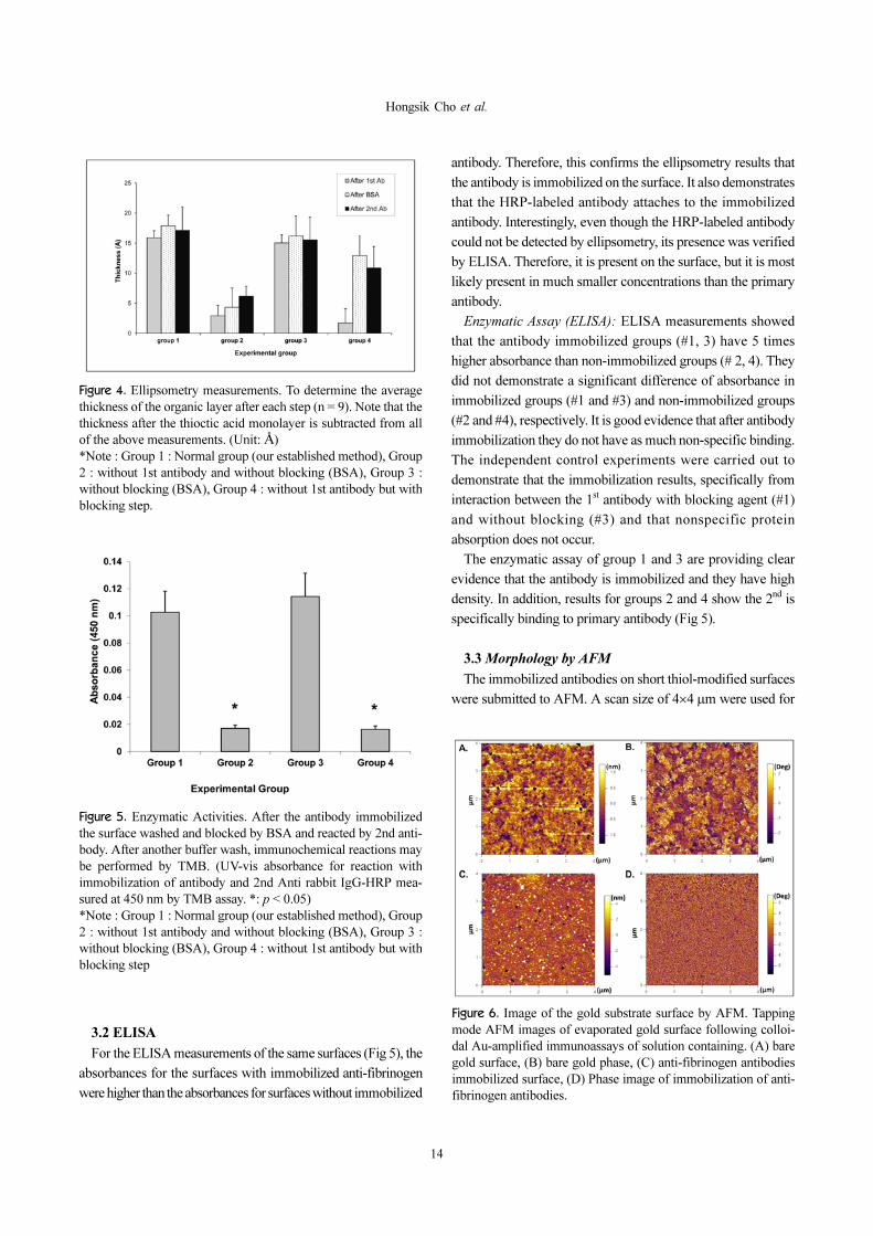
19.16 ± 1.75Å, respectively (Fig 3).
The additional thickness for only BSA (without antibody)
was 12.94 ± 3.23 Å. The thickness of the anti-fibrinogen
(without BSA) was 15.91 ± 1.14 Å. When blocking with BSA
after anti-fibrinogen, a significant difference in thickness could
not be detected. Also, the binding of the HRP-labeled antibody
did not result in a significant difference in thickness. When no
primary antibody was incubated with the surface (only
activation with EDC and NHS), the change was less than Å
(Fig 4). These ellipsometry results shown that the thiotic acid
molecule is 50% shorter than MUA and that there is no affect
on fibronogen antibodies binding efficiency.
Table 1. Experimental Procedure and Group. The Group 1 is our established method for immobilized antibody on gold substrate,
Group 2 is negative control for non specific binding to gold monolayer, Group 3 is control for 2nd Ab nonspecific binding to unre-
acted site on gold substrate, Group 4 is prepare for measurement of BSA thickness and density.
Procedure Chemical & LigandExperimental groups
Measurement1 2 3 4
7 Immunochemical reaction 2nd Ab-HRP Y Y Y Y Ellipsometry, ELISA, AFM image
6 Blocking 1% BSA Y N N Y Ellipsometry
5 Remove ramaining EDC/NHS groups Ethanolamine Y Y Y Y N/A
4 Immobilization 1st Ab (antifibrinogen) Y N Y N Ellipsometry
3 Activation of gold monolayer EDC+NHS Y Y Y Y N/A
2 Modified gold surface Thioctic acid Y Y Y Y Ellipsometry
1 Preparation of bare gold on Si Y Y Y Y Ellipsometry, AFM image
Note] To measurements the gold substrate take off and washed by DW and then dried by streamed argon gas at the point of each step. The
group 1 is our established method for immobilized antibody on gold subatrate, Group 2 is negative control for non specific binding to gold
monolayer, Group 3 is control for 2nd
Ab nonspecific binding to unreacted site on gold substrate, Group 4 is prepare for measurement of BSA
thickness and density. (2nd
Ab : anti-rabbit IgG with HRP, BSA : bovine serum albumin)
Figure 3. Change of thickness of bare gold surface after modified MUA or Thioctic Acid (A) Comparison to thickness after use shorter
thio (thioctic acid) instead of MUA (11-mercaptonudecanoic acid) using Ellipsometry, (B) Chemistry information (from The United
States Pharmacopeial Convention; USP.org)
Page 5
Hongsik Cho et al.
14
3.2 ELISA
For the ELISA measurements of the same surfaces (Fig 5), the
absorbances for the surfaces with immobilized anti-fibrinogen
were higher than the absorbances for surfaces without immobilized
antibody. Therefore, this confirms the ellipsometry results that
the antibody is immobilized on the surface. It also demonstrates
that the HRP-labeled antibody attaches to the immobilized
antibody. Interestingly, even though the HRP-labeled antibody
could not be detected by ellipsometry, its presence was verified
by ELISA. Therefore, it is present on the surface, but it is most
likely present in much smaller concentrations than the primary
antibody.
Enzymatic Assay (ELISA): ELISA measurements showed
that the antibody immobilized groups (#1, 3) have 5 times
higher absorbance than non-immobilized groups (# 2, 4). They
did not demonstrate a significant difference of absorbance in
immobilized groups (#1 and #3) and non-immobilized groups
(#2 and #4), respectively. It is good evidence that after antibody
immobilization they do not have as much non-specific binding.
The independent control experiments were carried out to
demonstrate that the immobilization results, specifically from
interaction between the 1st antibody with blocking agent (#1)
and without blocking (#3) and that nonspecific protein
absorption does not occur.
The enzymatic assay of group 1 and 3 are providing clear
evidence that the antibody is immobilized and they have high
density. In addition, results for groups 2 and 4 show the 2nd is
specifically binding to primary antibody (Fig 5).
3.3 Morphology by AFM
The immobilized antibodies on short thiol-modified surfaces
were submitted to AFM. A scan size of 4×4 µm were used for
Figure 4. Ellipsometry measurements. To determine the average
thickness of the organic layer after each step (n = 9). Note that the
thickness after the thioctic acid monolayer is subtracted from all
of the above measurements. (Unit: Å)
*Note : Group 1 : Normal group (our established method), Group
2 : without 1st antibody and without blocking (BSA), Group 3 :
without blocking (BSA), Group 4 : without 1st antibody but with
blocking step.
Figure 5. Enzymatic Activities. After the antibody immobilized
the surface washed and blocked by BSA and reacted by 2nd anti-
body. After another buffer wash, immunochemical reactions may
be performed by TMB. (UV-vis absorbance for reaction with
immobilization of antibody and 2nd Anti rabbit IgG-HRP mea-
sured at 450 nm by TMB assay. *: p < 0.05)
*Note : Group 1 : Normal group (our established method), Group
2 : without 1st antibody and without blocking (BSA), Group 3 :
without blocking (BSA), Group 4 : without 1st antibody but with
blocking step
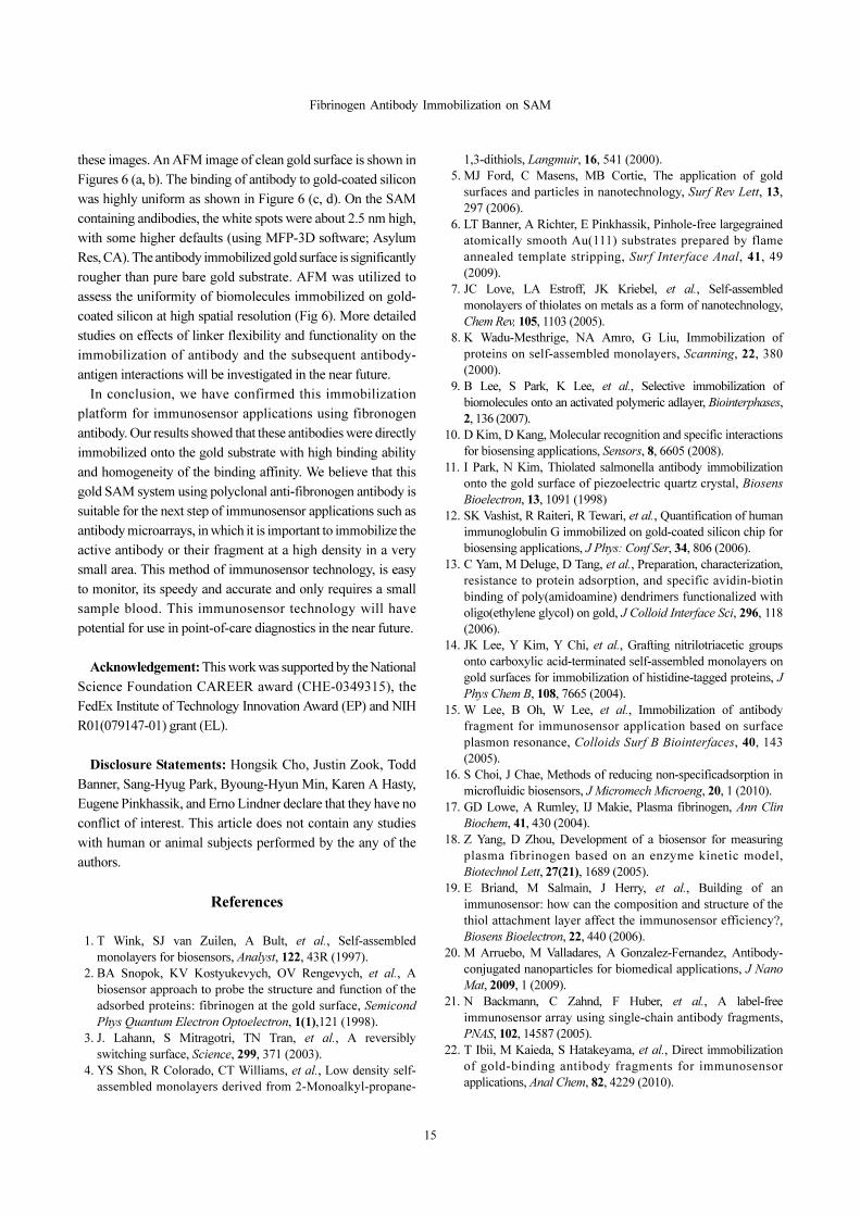
Figure 6. Image of the gold substrate surface by AFM. Tapping
mode AFM images of evaporated gold surface following colloi-
dal Au-amplified immunoassays of solution containing. (A) bare
gold surface, (B) bare gold phase, (C) anti-fibrinogen antibodies
immobilized surface, (D) Phase image of immobilization of anti-
fibrinogen antibodies.
Page 6
Fibrinogen Antibody Immobilization on SAM
15
these images. An AFM image of clean gold surface is shown in
Figures 6 (a, b). The binding of antibody to gold-coated silicon
was highly uniform as shown in Figure 6 (c, d). On the SAM
containing andibodies, the white spots were about 2.5 nm high,
with some higher defaults (using MFP-3D software; Asylum
Res, CA). The antibody immobilized gold surface is significantly
rougher than pure bare gold substrate. AFM was utilized to
assess the uniformity of biomolecules immobilized on gold-
coated silicon at high spatial resolution (Fig 6). More detailed
studies on effects of linker flexibility and functionality on the
immobilization of antibody and the subsequent antibody-
antigen interactions will be investigated in the near future.
In conclusion, we have confirmed this immobilization
platform for immunosensor applications using fibronogen
antibody. Our results showed that these antibodies were directly
immobilized onto the gold substrate with high binding ability
and homogeneity of the binding affinity. We believe that this
gold SAM system using polyclonal anti-fibronogen antibody is
suitable for the next step of immunosensor applications such as
antibody microarrays, in which it is important to immobilize the
active antibody or their fragment at a high density in a very
small area. This method of immunosensor technology, is easy
to monitor, its speedy and accurate and only requires a small
sample blood. This immunosensor technology will have
potential for use in point-of-care diagnostics in the near future.
Acknowledgement: This work was supported by the National
Science Foundation CAREER award (CHE-0349315), the
FedEx Institute of Technology Innovation Award (EP) and NIH
R01(079147-01) grant (EL).
Disclosure Statements: Hongsik Cho, Justin Zook, Todd
Banner, Sang-Hyug Park, Byoung-Hyun Min, Karen A Hasty,
Eugene Pinkhassik, and Erno Lindner declare that they have no
conflict of interest. This article does not contain any studies
with human or animal subjects performed by the any of the
authors.
References
1. T Wink, SJ van Zuilen, A Bult, et al., Self-assembled
monolayers for biosensors, Analyst, 122, 43R (1997).
2. BA Snopok, KV Kostyukevych, OV Rengevych, et al., A
biosensor approach to probe the structure and function of the
adsorbed proteins: fibrinogen at the gold surface, Semicond
Phys Quantum Electron Optoelectron, 1(1),121 (1998).
3. J. Lahann, S Mitragotri, TN Tran, et al., A reversibly
switching surface, Science, 299, 371 (2003).
4. YS Shon, R Colorado, CT Williams, et al., Low density self-
assembled monolayers derived from 2-Monoalkyl-propane-
1,3-dithiols, Langmuir, 16, 541 (2000).
5. MJ Ford, C Masens, MB Cortie, The application of gold
surfaces and particles in nanotechnology, Surf Rev Lett, 13,
297 (2006).
6. LT Banner, A Richter, E Pinkhassik, Pinhole-free largegrained
atomically smooth Au(111) substrates prepared by flame
annealed template stripping, Surf Interface Anal, 41, 49
(2009).
7. JC Love, LA Estroff, JK Kriebel, et al., Self-assembled
monolayers of thiolates on metals as a form of nanotechnology,
Chem Rev, 105, 1103 (2005).
8. K Wadu-Mesthrige, NA Amro, G Liu, Immobilization of
proteins on self-assembled monolayers, Scanning, 22, 380
(2000).
9. B Lee, S Park, K Lee, et al., Selective immobilization of
biomolecules onto an activated polymeric adlayer, Biointerphases,
2, 136 (2007).
10. D Kim, D Kang, Molecular recognition and specific interactions
for biosensing applications, Sensors, 8, 6605 (2008).
11. I Park, N Kim, Thiolated salmonella antibody immobilization
onto the gold surface of piezoelectric quartz crystal, Biosens
Bioelectron, 13, 1091 (1998)
12. SK Vashist, R Raiteri, R Tewari, et al., Quantification of human
immunoglobulin G immobilized on gold-coated silicon chip for
biosensing applications, J Phys: Conf Ser, 34, 806 (2006).
13. C Yam, M Deluge, D Tang, et al., Preparation, characterization,
resistance to protein adsorption, and specific avidin-biotin
binding of poly(amidoamine) dendrimers functionalized with
oligo(ethylene glycol) on gold, J Colloid Interface Sci, 296, 118
(2006).
14. JK Lee, Y Kim, Y Chi, et al., Grafting nitrilotriacetic groups
onto carboxylic acid-terminated self-assembled monolayers on
gold surfaces for immobilization of histidine-tagged proteins, J
Phys Chem B, 108, 7665 (2004).
15. W Lee, B Oh, W Lee, et al., Immobilization of antibody
fragment for immunosensor application based on surface
plasmon resonance, Colloids Surf B Biointerfaces, 40, 143
(2005).
16. S Choi, J Chae, Methods of reducing non-specificadsorption in
microfluidic biosensors, J Micromech Microeng, 20, 1 (2010).
17. GD Lowe, A Rumley, IJ Makie, Plasma fibrinogen, Ann Clin
Biochem, 41, 430 (2004).
18. Z Yang, D Zhou, Development of a biosensor for measuring
plasma fibrinogen based on an enzyme kinetic model,
Biotechnol Lett, 27(21), 1689 (2005).
19. E Briand, M Salmain, J Herry, et al., Building of an
immunosensor: how can the composition and structure of the
thiol attachment layer affect the immunosensor efficiency?,
Biosens Bioelectron, 22, 440 (2006).
20. M Arruebo, M Valladares, A Gonzalez-Fernandez, Antibody-
conjugated nanoparticles for biomedical applications, J Nano
Mat, 2009, 1 (2009).
21. N Backmann, C Zahnd, F Huber, et al., A label-free
immunosensor array using single-chain antibody fragments,
PNAS, 102, 14587 (2005).
22. T Ibii, M Kaieda, S Hatakeyama, et al., Direct immobilization
of gold-binding antibody fragments for immunosensor
applications, Anal Chem, 82, 4229 (2010).