Immune Activation and Autoantibodies in Humans with Long-Term Inhalation Exposure to Formaldehyde JACK D. THRASHER, Ph.D. Thrasher & Associates Northridge, California ALAN BROUGHTON, M.D., Ph.D. Antibody Assay laboratories Santa Ana, California ROBERTA MADISON, D.P.H. Department of Health Science California State University Northridge, California ABSTRACT.Four groups of patients with long-term inhalation exposure to formaldehyde (HCHO) were compared with controls who had short-term periodic exposure to HCHO. The following were determined for all groups: total white cell, lymphocyte, and T cell counts; T helper/suppressor ratios; total Tal +, IL2+, and B cell counts; antibodies to for- maldehyde-human serum albumin (HCHO-HSA) conjugate and autoantibodies. When com- pared with the controls, the patients had significantly higher antibody titers to HCHO-HSA. In addition, significant increases in Tal +, IL2+, and B cells and autoantibodies were ob- served. Immune activation, autoantibodies, and anti-HCHO-HSA antibodies are associated with long-term formaldehyde inhalation. INHALATION EXPOSURE to formaldehyde (HCHO) is associated with symptoms of irritation to mucous membranes",2 chronic health problems (e.g., asthma,2nasopharyngeal cancer,3and multiple subjective health complaints4,S). Recent observa- tions have shown that both humoral- and cell- mediated immuno:ogic mechanisms occur in hu- mans with long-term HCHO exposure. Antibodies of all isotypes to HCHO conjugated to human serum albumin (HCHO-HSA) are demonstrable in HCHO anaphylaxis,!' hemodialysis patients? mobile home, residents,4 persons with occupational exposures,S,8 office workers,9 and in persons in other environ- ments.4 In addition, changes in cell-mediated im- July/August 1990 [Vol. 45 (No.4)] munity include increases in eosinophils, basophils, and T-suppressor cells following acute re-exposure of patients with HCHO asthma.10Moreover, individ- uals with multiple subjective health complaints as- sociated with long-term HCHO inhalation have evi- dence of immune activation and the presence of - autoantibodies.4,s The patients in our study had symptoms and com- plaints related to several organs, as described previ- ously,4,S.9 which were similar to symptoms of workers with multiple chemical sensitivity,11cacosmia,12and other chemical exposures.13-1SWereport on the dif- ferences in humoral and cell-mediated immunity in humans with long-term inhalation exposure to 217
Transcript
Immune Activation and Autoantibodies in Humans with
Long-Term Inhalation Exposure to Formaldehyde
JACK D. THRASHER, Ph.D.Thrasher & Associates
Northridge, CaliforniaALAN BROUGHTON, M.D., Ph.D.Antibody Assay laboratoriesSanta Ana, CaliforniaROBERTA MADISON, D.P.H.Department of Health ScienceCalifornia State UniversityNorthridge, California
ABSTRACT.Four groups of patients with long-term inhalation exposure to formaldehyde(HCHO) were compared with controls who had short-term periodic exposure to HCHO.The following were determined for all groups: total white cell, lymphocyte, and T cellcounts; T helper/suppressor ratios; total Tal +, IL2+ , and B cell counts; antibodies to formaldehyde-human serum albumin (HCHO-HSA) conjugate and autoantibodies. When compared with the controls, the patients had significantly higher antibody titers to HCHO-HSA.In addition, significant increases in Tal +, IL2+, and B cells and autoantibodies were observed. Immune activation, autoantibodies, and anti-HCHO-HSA antibodies are associatedwith long-term formaldehyde inhalation.
INHALATION EXPOSURE to formaldehyde(HCHO) is associated with symptoms of irritation tomucous membranes",2 chronic health problems(e.g., asthma,2nasopharyngeal cancer,3and multiplesubjective health complaints4,S). Recent observations have shown that both humoral- and cellmediated immuno:ogic mechanisms occur in humans with long-term HCHO exposure. Antibodiesof all isotypes to HCHO conjugated to human serumalbumin (HCHO-HSA) are demonstrable in HCHOanaphylaxis,!' hemodialysis patients? mobile home,residents,4 persons with occupational exposures,S,8office workers,9 and in persons in other environments.4 In addition, changes in cell-mediated im-
July/August 1990 [Vol. 45 (No.4)]
munity include increases in eosinophils, basophils,and T-suppressor cells following acute re-exposureof patients with HCHO asthma.10Moreover, individuals with multiple subjective health complaints associated with long-term HCHO inhalation have evidence of immune activation and the presence of
- autoantibodies.4,sThe patients in our study had symptoms and com
plaints related to several organs, as described previously,4,S.9which were similar to symptoms of workerswith multiple chemical sensitivity,11cacosmia,12andother chemical exposures.13-1SWe report on the differences in humoral and cell-mediated immunity inhumans with long-term inhalation exposure to
217
HCHO vs. asymptomatic students (controls), whoexperienced shorHerm, periodic exposure to thechemical.
Materials and methods
Controls and patients. Five groups of subjects exposed to HCHO, who gave informed consent, wereincluded in this study.
(1.) Controls consisted of students of chiropracticmedicine (16 males, 12 females, mean age = 29 ± 9y) exposed to HCHO for 13 h/wk for 28 wk whilestudying human anatomy. Immunologic tests wereperformed 12 mo following the last classroom exposure. No measurements of HCHO concentrationswere made. It was assumed that classroom ambientconcentrations were at least 0.43 ppm.1 The studentsstated that during exposure they experienced eye,nose, and throat irritation and that there was a pungent odor of HCHO. They did not have residualhealth complaints (symptoms), and they wereasymptomatic at the time blood was taken.
(2.) Mobile home residents consisted of 19 patients (6 males, 13 females, mean age of 41 ± 20 y)who currently lived in mobile homes. The patientshad lived in their environments for 2-7 y and reported multiple symptoms.4,9Measured HCHO concentrations ranged from 0.05 to 0.5 ppm at the timeblood samples were drawn.
(3.) Office workers included 21 patients (5 males,16 females, mean age of 40 ± 10 y) who worked innew office buildings where there was inadequateventilation (closed buildings). The patients had multiple health complaints.9 It was determined frommedical histories that their symptoms commencedwith employment, waned when away from work(i.e., weekends, holidays, vacations) and becameworse upon return to work. No HCHO measurements were done; however, closed buildings haveambient concentrations ranging from 0.01 to 0.77ppm.1,16
(4.) This group included 21 patients (10 males, 11females, mean age of 35 ±17 y) who had multiplesymptoms and who had been removed from theiroriginal sources of HCHO exposure (mobile homesand/or particleboard subflooring) for at least 1 y.TheHCHO concentrations measured during their exposures ranged from 0.14to 0.81 ppm.
(5.) Occupationally exposed patients (6 males, 2females, mean age of 45 ± 11 y) had HCHO exposures from the following: biology and human anatomy classes, mortuary, pathology, physical therapy,formica furniture (particleboard), and carbon lesscopy paper. Information on six of these patients wasreported previously.s
Symptoms. All patients in this study had soughtcontinuous medical attention because of multipleorgan symptoms involving the central nervous system (eNS) (headache, memory loss, difficulty withcompleting tasks, dizziness), upper- and lowerrespiratory symptoms, skeletal-muscle complaints,and gastroenteritis. Three common symptoms were
218
expresed: (1) an initial flu-like illness from whichthey had not fully recovered, (2) chronic fatigue, and(3) an olfactory sensitivity to ambient conditionscontaining low concentrations of chemicals.4,9.11
One of the students smoked cigarettes (1 pack/d),whereas the remainder and all patients were nonsmokers. No attempt was made to correlate the in:tmunological data with histories of allergies and/oratopy. Previous efforts to make this correlation haveled to negative findings.H,g
HCHO-HSA conjugation and ELISAantibody assay.IgE, IgM, and IgG anti-HCHO-HSA antibodies weredetermined by an EliSA procedureY Conjugation ofHCHO with human serum albumin and the ELISAantibody assays were done on sera from freshlydrawn blood in accord with information publishedelsewhere,4,sexcept the HCHO-HSA conjugate wasstored at 4 0 C.
Lymphocyte surface markers. All procedures wereperformed on heparinized venous blood within 24 hfollowing collection. The total peripheral white cellcount (WBC) was performed using a Model F Coulter Counter (Coulter, FL). The total lymphocytecount was done by blood smear examination. Lymphocyte marker procedures are published elsewhere.4,s In brief, peripheral mononuclear cellswere isolated using Ficoll Hypaque density gradient.18 The percentages and absolute numbers(ABS) of lymphocyte subsets per mm3 blood weredetermined utilizing monoclonal antibodies to surface markers: LEU1 (T cells), LEU2A (T suppressorcells), LEU3A(T helper cells), LEU10 (B cells) (Beckton-Dickinson, Los Angeles, CA), and Ta1+ and1L2+ receptor cells (Coulter, Fl.). All surface markers, except Ta1+ , were identified by indirect immunoflourescence.19 Ta1+ cells were determined by adirect immunofluorescent method.20
Autoantibody screen. Antismooth muscle (ASS),anti parietal cell (APC), antibrush boarder (ABB), antimitochondrial (AMIT), and antinuclear antibodies(ANA) in the subjects' sera were detected by an indirect immunofluorescent technique and expressedas positive at a dilution of 1:20.21
Sexand ageeffectson cell numbersand antoantibodies. Each of the groups, except occupational, wereexamined to determine if either sex or age biasedthe observations on mean absolute counts and percentages of each cell type. Statistical analyses wereperformed that compared either females with malesor younger ages with older ages within each agegroup.The number of individuals in the occupationally exposed group was insufficient for statisticalevaluation for sex and age effects.
Statistical analysis.The student group was used ascontrols for all statistical tests. Each of the four patient groups were compared with the controls forthe following: (a) Z tests were performed to determine whether there was a significantly higher pro-,portion of individuals in each group with antibodytiters at or greater than 1:8 to HCHO-HSA; (b) twotailed t tests and correlation analyses were computed on grouped data to examine any relationship
Archives of Environmental Health
between age, gender, WBC, lymphocytes, anc~lymphocyte subsets in each patient group; and (c) oddsratios and 95% confidence intervals were calculatedto determine which groups were at the highest riskof having autoantibodies.
Results
Sexand ageeffectson cell numbersand autoantibodies. Gender did not affect the mean numbers andpercentages of each cell type except as describedbelow. The percentage of Ta1 cells was different inthe male office workers (p < .05) because one patient had very high absolute (2 310 cells/mm3) andpercentage Ta1cells (44%).
t tests revealed no effects of age, but the numberof T (LEU1)cells was disparate in controls (p < .05).However, correlations for age effects were not observed (r2 ranged from 0.00to 0.42).
Age had no effect on the percentage of autoantibodies. For example, APC (the most common autoantibody) for the younger vs. older individualswas 50% and 60%, respectively (mobile homes), and89% and 90%, respectively (office workers). Thenumbers with respect to sex differences were insufficient for evaluation.
As a result of the above observations, all data werepooled regardless of either sex or age for subsequent analyses.
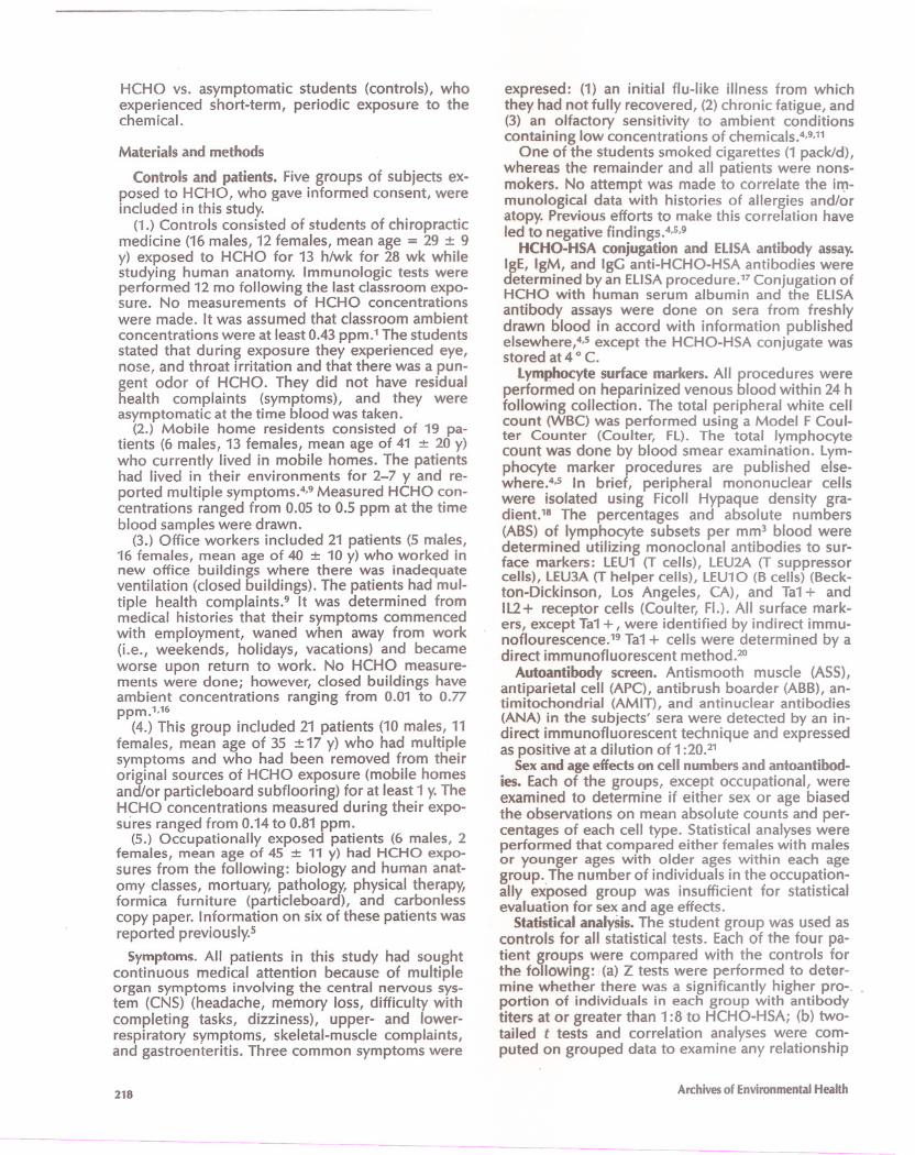
Antibody titers againstHCHO-HSA. The antibody titers against HCHO-HSA in the sera of each individual in the five groups are listed in Table 1. The controls had the lowest titers. Titers of 1:16 or greaterwere predominant in the patient groups. The proportion with positive anti-HCHO-HSA antibodieswas least in the controls (39%).The proportion withpositive titers was significantly greater (p < .01) for
each patient group when compared with the controls.
White blood cells, lymphocytes, T cells, and HIS ratios. The pooled data for WBCs, Iymphoctyes, Tcells, and HIS ratios for each group are summarizedin Table.2. The ABS for each cell type and percentages of T (LEU1), T helper (lEU3A), T suppressor(LEU2A),and HIS ratios for all groups fell within theexpected reference ranges. We performed t tests onmean ABS and percentages, using the controls ascomparison. Other than the WBCs of the officeworkers (p < .05), no significant differences fromthe controls ,for each patient group were found.
Tal +, IL2+ receptor, and B cells. The ABSand percentages of Ta1+, IL2+ receptor, and B (LEU10)cells for each group (expected reference ranges areincluded) are listed in Table 3. The mean values ofTa1+ cells for each patient group as compared withthe controls were significantly higher: mobile homeresidents (p < .001), office workers (p < .01), occupationals (p < .001),and removed patients (p < .05).The IL2+ cells were elevated in mean ABS and percentages in each of the patient groups (Table3). Theincreases were significantly higher in the mobilehome residents (p < .02) and the removed patients(p ::S .02) than in controls. The B (LEU10) cells werealso elevated in the four groups vs. controls (Table5), and they were significantly higher in the officeworkers (p < .01) and the removed patients (p <.01). The Ta1+ cells exceeded reference ranges inboth absolute numbers and percentages in allgroups, except the controls. Conversely, both IL2+and Bcells fell within the expected reference values.
Frequency of autoantibodies. The percentage ofeach autoantibody detected at a dilution of 1:20 inthe sera of the subjects in the five groups is listed in
Table 1.-lgG, IgM, and IgE Isotypes to HCHO-HSA Categorized by Each Group Exposed to HCHO
Isotypes
HCHO
Dil.IgGIgMIgE%,
Z&p*source
ratio(No.)(No.)(No.)(1:8)values
Controls
1:4232327(n = 28)
1 :855139-2:1 :16
000Mobile
1 :491017homes
1 :8200752.36(n = 20)
2:1 :169103 <.01Office
1 :410914workers
1:831473.62.36(n = 19)
2:1 :161681 <.01Occup.
1:421+5(n = 8)
1 :81021003.32:1 :16
551 <.01Removed
1 ;441122
(n = 22)
1 :834081.83.02:1 :16
1570 <.01
'Z and p values are compared with those of controls.tTwo patients did not have IgM titers performed.
July/August 1990 [Vol. 45 (No.4)] 219
Table 2.-Mean Absolute Numbers of wacs, Lymphocytes, Percentage T Cells, and HIS Ratios Found in thePeripheral Blood in Each Group
Notes: Absolute numbers are in cells/mm3 of blood. Expected ranges: Tal + (ABS = 0-160,0-4%), 112+(ABS = 0-320,0-8%), B (LEU10)cells (ABS = 50 to 400,0-15%).tp,<.OOl.:l:p,<.Ol.§p,<.05.#Not significant.IIp,<·02.
Table 4. The controls had the lowest frequency ofautoantibodies. In contrast, mobile home residentshad the highest occurrence of each of the autoantibodies. The most frequently found autoantibodywas APC. When the rate at which autoimmunity(i.e., presence of an autoantibody) was determined,
220
the controls had the lowest; the highest rate occurred in the mobile home residents.
Odds ratios for frequency and rate of occurrenceof the autoantibodies were performed on the mobile home residents and office workers vs. controls(Table 5). The odds ratio for ASS (8.2) was signifi-
Archives of Environmental Health
Discussion
Two issues should be addressed before betweengroup comparisons of the data are made. The first iswhether the students suffice as ample controls. Thesecond entails the possible effects of sex and age onthe observed differences in anti-HCHO-HSA isotypes and in Ta1 cells and autoantibodies betweenthe controls and the patients.
According to Schlesselman,22controls should befree of the disease being studied. Controls shouldalso be similar to the cases with regard to past potential for exposure. The students met both of thesecriteria. First, they were asymptomatic. Second, they
had similar risks of exposure to HCHO in either thehome or office. They did, in fact, have classroom exposure similar to that experienced by the occupational group. The major difference in the exposurebetween the students and the other patients wasone of the duration, Le., periodic vs. almost continuous, with the exception of the occupational group.
Moreover, despite the intuitive appeal of age andsex matching, there is no equivocal evidence intheory or practice that supports a general preference for this technique.22 Both t tests and correlation analyses demonstrated that both the mean absolute numbers and percentages of each cell type
Table 4.-Autoantibodies: Percentage in Each Group/ by Type of Autoantibody and by Rate
Mobile
OfficeControls
homesworkersRemoved-Occup.-(n=28)
(n=19)(n=20)(n=8)(n=3)
Typeof autoantibody
ASS14.357.9252533.3
APC3.684.2602533.3
ABB7.710.52012.533.3
AMIT021.11512.50
ANA3.610.520 033.3
Rate: Number of autoantibodies1 or moret21.189.5805066.6
2 or more7.157.9452533.3
3 or more036.815 033.3
4 or more000 00
Notes: ASS (antismooth muscle), APC (anti parietal cell), ABB (antibrush boarder), AMIT (antimitochon-drial), and ANA (antinuclear).-Numbers are insufficient to perform an odds ratio analysis:tRate (percentage) in each group with one, two, three, or four or more autoantibodies.
Table 5.-0dds Ratios for the Percentage and Rate of Autoantibodies in Mobile
Home Residents and Office Workers as compared to the controls
Mobile
Office
Autoantibody
homesworkers
ASS (odds)
8.22(95% C.I.)
33,2.02-8.7,0.5APC (odds)
14440.5(95% CI.)
1 541/14-365,4.5-ABB (odds)
2.44.3(95% CL)
16.2,0.426,0.8AMIT (odds) (95% CI.)
ttANA (odds)
3.24.8(95% CL)
40,0.2751/0.47
1 or more (odds)
31.214.7(95% CI.)
175,5.1·61.5,3.6-2 or more (odds)
17.910.6(95% C.!.)
99.7,3.3-59.5,2.0·3 or more (odds)
,(95% CLl
tt
*Confidence intervals are large as a result of small numbers in each group.tNot calculated because of zero value for control group.
July/August 1990 [Vol. 45 (No.4)] 221
cantly higher (p < .05) in the mobile home group,whereas those for APC (144 and 40.5) were significantly greater (p < .05) for both groups. Moreover,the odds ratios for the rate of the presence of autoantibodies (e.g., one, two, or three or more) werealso significantly greater in the two groups of patients than in controls (p < .05).within each group were unaffected by either sex orage. Therefore, matching sex and/or age was not anecessary requirement in this study, pe'rrhittingpooling of the data..
Although the total white cells, lymphocytes, Tcells, and H/S ratios are within expected ranges inthe five groups (Table2), the patients have evidenceof an activated cell-mediated immunity (Table 3).First, fa1 + cells are significantly elevated in theJourgroups when compared with the controls (p rangesfrom < .05 to < .001). Ta1+ expression occurs afterantigenic stimulation. Also, Ta1+ cells respond torecall antigens and, therefore, are considered antigen memory cells.20.23Moreover, circulating Ta1+cells and la-positive cells are elevated in variousautoimmune disorders.24-26 We recently demonstrated elevation of Ta1+ cells in individuals withchronic health complaints associated with HCH04.Sand isocyanate13inhalation. Because an increase incirculating Ta1-+ cells occurs in individuals undergoing chronic antigenic stimulation (Le., chemi.calsensitivity and autoimmunity), the elevation ofTa1 +cells in these patients indicates that they have achronic immune activation. Furthermore, the disparate numbers for Ta1+ cells of the removed patients in comparison with the controls lend additional support to this conclusion. These patients,along with the others, express an olfactory sensitivity to environmental conditions that elicit symptoms. Thus, higher Ta1+ cells and anti-HCHO isotypes in the removed patients are two immunologicparameters that appear associated with their ongoing health complaints.
Second, both 112+ and B (lEU10) cells of the fourgroups of patients show a trend toward elevation ascompared with the controls (Table3). The increase ISsignificant for 1l2+ cells in the mobile home residents (p <.01) and removed groups (p < .02). Also,the B cells are increased in the office workers andthose removed (p <.05 to <.01). The 1L2+ cells occur in acute immune activation and 6 cells produceantibodies.27 Therefore, the increases in these twotypes of cells support immune activation in the patients. The elevation in Ta1+ , 112+ , and 6 cells mayresult from one or both of the following: (a) immunological memory to, and antibody. productionagainst, certain environmental chemicals, and (b)the presence of autoantibodies.
Higher anti-HCHO-HSA isotypes (Le., 1:16 orgreater) are present in the patients v. controls. Oneexplanation for this difference is simply the lag timebetween the last exposure v. the time of antibodydetection. However, the higher titers of IgE;md IgMisotypes in the patients suggest that a more recent
222
exposure hasoccurred, particularly if the higher IgGtiters are considered also. In this vein, the patientscomplain of a sensitivity (both olfactory and respiratory) to environments containing low concentrations of HCHO and other chemicals. Thus, thehigher titers may indicate that their immune systemsare on constant alert, undergoing continuous activation upon encountering and recognizing environmental haptens.9.28It would be of interest to examine for other haptens to which the patients may beresponding.9
The higher antibody titers and the larger proportion of individuals with anti-HCHO isotypes in theremoved patients v. controls merit comment. 60thgroups were at least 1 y removed from their originalsource of exposure. However, the controls wereasymptomatic, whereas the patients experiencedongoing health problems associated with environmental exposures, e.g., new carpets, fresh paints,new furnishings, diesel exhaust, and perfumes.Thus, it appears that long-term low-level exposureto HCHO, and possibly other haptens, lead to immunological recognition and immune activation insensitized individuals. Apparently, shorter periodicexposure to HCHO may lead to recognition but notnecessarily immune activation. Moreover, chroniclow-level exposures to HCHO appear to effect a sensitivity to environmental chemicals.4-6,8.9Perhapsthe anti-HCHO-HSA isotypes in these patients is butone aspect of a multiple immunologic response toenvironmental exposures as observed in buildingrelated iIIness.9
It is recognized that chemicals and therapeuticdrugs are associated with a lupus-like syndrome.28•29The observations made on the patients in this studysupport this concept. The percentage of specificautoantibodies (e.g., ASS, APC, ANA, etc.) are consistently higher in the patients v. controls (Table 4).Moreover, the odds ratios for the presence of at least1, 2, or 3 autoantibodies are significantly greater inthe residents of mobile homes and office workers (p< .05) relative to controls (Table5).
Presently, autoimmune disorders have not beendiagnosed clinically in these patients. However, current investigations in progress appear to correlatethe presence of APC autoantibodies with gastritiscomplaints and antimyelin autoantibodies with CNSand PNSsymptoms.
In conclusion, measurements of changes inWBCs, T cells, anclH/S ratios in individuals with apparent chemical sensitivities appear to be inadequate immune parameters to examine. If one assumes that these individuals respond immunologically to environmental chemicals, investigations intoautoimmunity and immune activation and pertubations in the interlqukins, leukotreines, prostaglandins, and other immunologic mediators appear tobe fruitful areasfor'further research.29-32 Thus, it appears that HCHO sensitivity is a real phenomenonand requires further research.4,27-32
Archives of Environmental Health
* * * * * * * * * *We wish to thank Drs. Heuser and Baker for referring some of
the patients in this study. Valuable technical assistance was ob.tained from Mr. Gilbert Salizar and the technical staff.
Submitted for publication November 15, 1989; revised; ac·cepted for publication March 13, 1990.
Requests for reprints should be sent to Jack D. Thrasher,Ph.D., Thrasher & Associates, 11330Quail Creek Rd., Northridge,CA 91326.
* * * * * * * * * *
References
1. Breysse P.The immediate and long term effects of formaldehyde. Comments ToxicoI19OO;2:135-53.
2. Nordman H, Keskinen H, Tuppurainen M. Formaldehydeasthma - rare or overlooked? J Allergy Clin Immunol1985;75:81-99.
3. Vaughn Tl, Strader C, Davis S, Daling JR. Formaldehyde andcancers of the pharynx, sinus and nasal cavity. II. Residentialexposure. Int JCancer 1987;28:685-88.
4. Broughton A, Thrasher JD. Antibodies and altered cell mediated immunity in formaldehyde exposed humans. Comments ToxicoI1988;2:155-70.
5. Thrasher JD, Broughton A., Micevich P. Antibodies and immune profiles of individuals occupationally exposed to formaldehyde: six case reports. Am JInd Med 1988;14:479-00 ..
6. Maurice F,Rivory J-p,Larsson PH, Johansson SGO, BousquetJ. Anaphylactic shock caused by formaldehyde in a patientundergoing long-term hemodialysis. J Allergy Clin Immunol1987;77:594-97.
7. Patterson R, PateraV,Grammar LC, Harris K. Human antibodies against formaldehyde-human serum albumin or humanserum albumin in individuals exposed to formaldehyde. IntArch Allergy AppllmmunoI1986;79:53-59.
8. Wilhelmsson G, Holmstrom M. Positive formaldehyde-RASTafter prolonged formaldehyde exposure by inhalation. Lancet1987; /I (8851):54.
9. Thrasher JD, Madison R, Broughthon A, Gard Z. Buildingrelated illness and antibodies to albumin conjugates of formaldehyde, toluene diisocyanate and trimellitic anhydride.Am J Ind Med 1989;15:187-95.
10. Pross H.F, Day JH, Clark RH, Lees REM. Immunologic studiesof subjects with asthma exposed to formaldehyde and ureaformaldehyde (UFF!) off-products. / Allergy Clin Immunol1987;79:787-810.
11. Cullen MR. Workers with multiple chemical sensitivities: anoverview. Occup Med: StateArt Rev1987;2:655-61.
12. Ryan CM, Morrow LA, Hodgson M. Cacosmia and neurobehavioral dysfunction associated with occupational exposureto mixtures of organic solvents. Am J PsychoI19OO;145:144245.
13. Broughton A, Thrasher JD, Gard Z. Immunological evaluationof four arc welders exposed to fumes from ignited polyurethane (isocyanate) foam: antibodies and immune profiles. AmJ Ind Med 1988;13:463-72.
July/August1990[Vol. 45 (No.4)]
14. Bekesi IG, ROOoz J, fischbIein A, et. aL ~~. bi0chemical and clinical consequences of exposure to~inated biphenyls. In: Immunotoxicology and imI:mmoph:u_macology, Dean J et aI., eds. New York: RavenPress,t985; pip.393-~.
16. Konopinski VJ. Formaldehyde in office and commercial envi·ronments. Am Ind Hyg Assoc 1985;46:65-68.
17. Voller A. Heterogeneous enzyme-immuno assaysand theirapplications. In; Enzyme immunoassay, Maggio ET,ed. BocaRaton: CRC Press,1979; pp. 181-96.
18. Boyuma A. Isolation of mononuclear cells and granulocytesfrom human blood. Scand J Clin Lab Invest 1968; 21 (Suppl97):77-89.
19. Englemane EG,Warnke R, Fox FI, Levy R. Studies of a humanIymphocyte-T antigen recognized by monoclonal antibody.PNAS1981;78:791-95.
20. fox DA, Hussey RE,Fitzgerald KA, et al. Ta1, a noval105 KDhuman T cell activation antigen defined by a monoclonal antibody. J ImmunoI1984;133:351-54.
21. Nakamura RM, Tucker ES.Antibodies as reagent. In: Diagnosis and management by laboratory methods, Henry JD, ed.Philadelphia: WB. Saunders, 1979; p. 1184.
22. Schlesselman JJ. Case control studies design conduct andanalysis. New York: Oxford University Press, 1982; pp. 122,177.
23. Hafler DA, Fox DA, Benjamin 0, Winer HL. Antigen reactivecells are defined by Tal. J ImmunoI1985;137:414-18.
24. Halfer DA, Fox DA, Manning ME, et al. In vivo activated lymphocytes in the peripheral blood and cerebrospinal fluid ofpatients with multiple sclerosis. New Eng J Med 1985;312:1405-11.
25. Mitzutani H, Tsubakio T,Tomiyana Y,et al. Increased circulating 1a-positive T cells in patients with idiopathic thrombocytopenia purpura. Clin Exp Immunol 1987;67:191-97.
26. Jackson RA, Morris MA, Haynes BF,Eisenbarth GS. Increased1a-antigen-bearing T cells in Type I diabetes mellitus. New EngJMed 1981;306:785-88.
27. Nossal GJV.Current concepts in immunology: the basic components of the immune system. New Eng J Med 1987;316:1320-25.
28. Amos HE, Park BK. Understanding immunotoxic drug reactions. In; Immunotoxicology and immunopharmacology,Dean J.et. aI., eds. New York: Raven Press,1965; pp. 207-28.
29. Bigazzi PE.Autoimmunity induced by chemicals. Clin Toxicol1988;26:125-56.
30. Marks JG, Traullein JJ,Zwillich cw, Demers LM. Contact urti·caria and airway obstruction from carbon less copy paper.JAMA 1984;252:1038-40.
31. LaMarte FP,Merchant JA, Casale T. Acute systemic reactionsto carbonless copy paper associated with histamine release.JAMA 1988;260:242-43.
32. Stanworth DR. Current concepts in hypersensitivity. In: 1mmunotoxicology and immunopharmacology, Dean J, et aI.,eds. New York: RavenPress,1985; pp. 91-98.