WHITE PAPER PRESENTED BY PREMIER RESEARCH Immuno-Oncology Drug Development: Opportunities Beyond Hope ABSTRACT Immuno-oncology drug development is complex and requires a thorough understanding of both the immune system’s role in cancer development and the unique challenges associated with evaluating therapeutic response. ONCOLOGY
Transcript
Applying Quality by Design to the Rare Disease Population: Special Considerations 1premier-research.comImmuno-Oncology Drug Development
WHITE PAPER PRESENTED BY PREMIER RESEARCHWHITE PAPER PRESENTED BY PREMIER RESEARCH
Immuno-Oncology Drug Development: Opportunities Beyond Hope ABSTRACT
Immuno-oncology drug development is complex and requires a thorough understanding of both the
immune system’s role in cancer development and the unique challenges associated with evaluating
Applying Quality by Design to the Rare Disease Population: Special Considerations 2premier-research.comImmuno-Oncology Drug Development
WHITE PAPER PRESENTED BY PREMIER RESEARCHWHITE PAPER PRESENTED BY PREMIER RESEARCH
IntroductionOver the past decade, immuno-oncology has become one of the most promising and fastest-growing areas of cancer research and drug development. Present-day advances in immuno-oncology can be attributed to a paradigm shift in the understanding of cancer.
Up until the late 1990s and early 2000s, cancer was considered a disease of genetic origin, with hallmarks including sustained proliferation, resistance to apoptosis, the ability to promote angiogenesis, and the ability to promote invasion and metastasis. However, this view failed to take into account the dynamic nature of the interactions between the tumor and its microenvironment – not just the normal cells in the surrounding tissue, but also the immune system.
Advances in our understanding of the dual role that the immune system plays in cancer have led to the development of immunotherapies that target both the tumor and its microenvironment. In this white paper, we explore the role of the immune system in cancer development, as well as the history and challenges of developing immunotherapies for cancer.
BackgroundThe history of cancer immunotherapy dates back to the discovery of the dendritic cell in the 1970s. In the early 1990s, Bacillus Calmette-Guérin (BCG), a vaccine used to prevent tuberculosis, was approved as an immunotherapy for early bladder cancer.1 This was followed by the approval of high-dose interleukin (IL)-2 for melanoma and renal cell carcinoma and the discovery of checkpoint inhibitors.
Applying Quality by Design to the Rare Disease Population: Special Considerations 3premier-research.comImmuno-Oncology Drug Development
WHITE PAPER PRESENTED BY PREMIER RESEARCH
Recent years have seen an acceleration in the development of immuno-oncology drugs, with the approval of:
+ Sipuleucel-T for prostate cancer in 2011
+ Ipilimumab, a checkpoint inhibitor, for advanced melanoma in 2011
+ Pembrolizumab and nivolumab for advanced melanoma in 2014
+ Nivolumab for non-small-cell lung cancer (NSCLC) in 2015
+ Pembrolizumab for PD-L1+ NSCLC in 2015
Tumor immunologyA thorough understanding of the underlying tumor biology is critical for the development of immuno-oncology drugs.
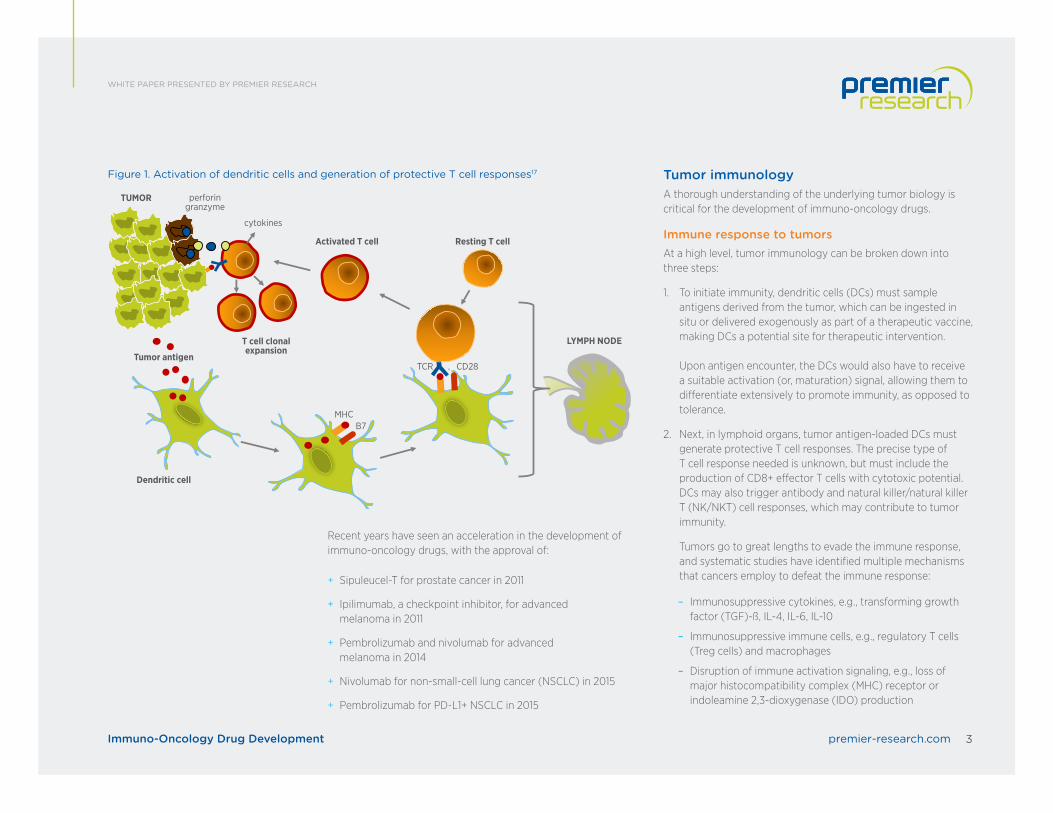
Immune response to tumorsAt a high level, tumor immunology can be broken down into three steps:
1. To initiate immunity, dendritic cells (DCs) must sample antigens derived from the tumor, which can be ingested in situ or delivered exogenously as part of a therapeutic vaccine, making DCs a potential site for therapeutic intervention. Upon antigen encounter, the DCs would also have to receive a suitable activation (or, maturation) signal, allowing them to differentiate extensively to promote immunity, as opposed to tolerance.
2. Next, in lymphoid organs, tumor antigen-loaded DCs must generate protective T cell responses. The precise type of T cell response needed is unknown, but must include the production of CD8+ effector T cells with cytotoxic potential. DCs may also trigger antibody and natural killer/natural killer T (NK/NKT) cell responses, which may contribute to tumor immunity.
Tumors go to great lengths to evade the immune response, and systematic studies have identified multiple mechanisms that cancers employ to defeat the immune response:
– Immunosuppressive immune cells, e.g., regulatory T cells (Treg cells) and macrophages
– Disruption of immune activation signaling, e.g., loss of major histocompatibility complex (MHC) receptor or indoleamine 2,3-dioxygenase (IDO) production
Figure 1. Activation of dendritic cells and generation of protective T cell responses17
Applying Quality by Design to the Rare Disease Population: Special Considerations 4premier-research.comImmuno-Oncology Drug Development
WHITE PAPER PRESENTED BY PREMIER RESEARCH
3. Finally, cancer-specific T cells must enter the tumor bed to perform their function. Here, there is the challenge of immune suppression. Presumably by skewing DC maturation, tumors may:
– Prevent immunization
– Trigger the “wrong” immune response, or
– Enable the local accumulation or expansion of Treg cells that would oppose the activity of effector T cells
Infiltration of Treg cells correlates with poor prognosis in a variety of epithelial tumor types. Tumors may downregulate their expression of MHC class I molecules or their expression of target tumor antigens. They can also produce a variety of surface molecules, such as programmed death-ligand (PD-L) 1 or PD-L2, which engage receptors on the surfaces of activated T cells (e.g., programmed cell death (PD)-1 protein), causing T cell anergy or exhaustion. Expression of such suppressive ligands can be associated with oncogenic mutations seen in many cancers, for example phosphatase and tensin homology (PTEN) loss.
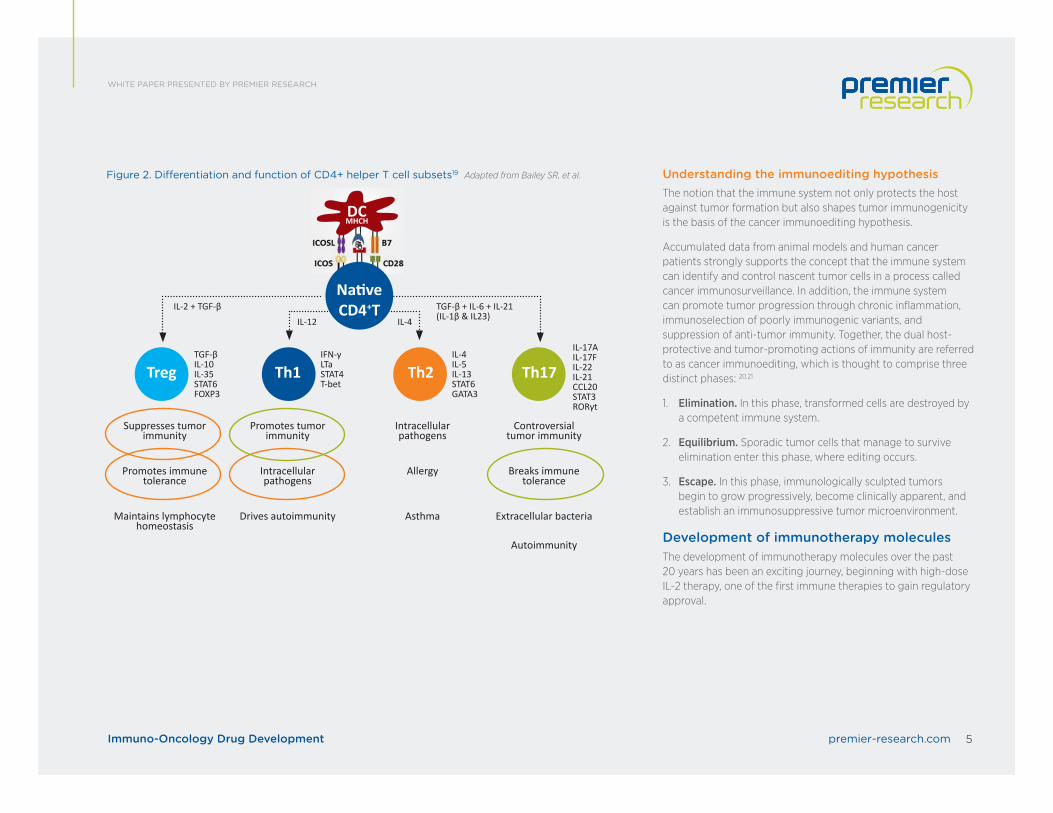
Differentiation of CD4+ helper T cell subsetsThe differentiation of CD4+ helper T cell subsets is determined by cytokines.
Many of the immuno-oncology targeted therapies activate one of the four main types of T cells:
+ Helper T cells (CD4+)
+ Cytotoxic T cells (CD8+)
+ Suppressor T cells (CD4+ CD25+ Foxp3+ Treg cells)
+ Memory T cells (CD4+ or CD8+ CCR7+ CD45RO)
CD4+ T cells, which are key regulators of the immune system, differentiate into various T helper (Th) cell lineages with distinct biological functions, depending upon how they are activated.
In the presence of IL-6, IL-21 and TGF-ß, CD4+ T cells differentiate into CD4+ T helper 17 (Th17) cells, a phenotype which is characterized by expression of the transcription factors retinoic acid receptor-related orphan receptor-gt (RORgt) and signal transducer and activator of transcription 3 (STAT3). IL-1b and IL-23 cytokines can promote and stabilize this phenotype during cell expansion. Once programmed, these Th17 cells secrete IL-17A, IL-17F, IL-21, and IL-22, which play key roles in enhancing autoimmunity and host defense.
Cytokines IL-12, IL-4, and TGF-ß and transcription factors T-bet, GATA3, and FOXP3 have been shown to regulate Th1, Th2, and Treg cell development, respectively. These distinct subsets regulate immune response to foreign, self, and tumor antigens.
Applying Quality by Design to the Rare Disease Population: Special Considerations 5premier-research.comImmuno-Oncology Drug Development
WHITE PAPER PRESENTED BY PREMIER RESEARCH
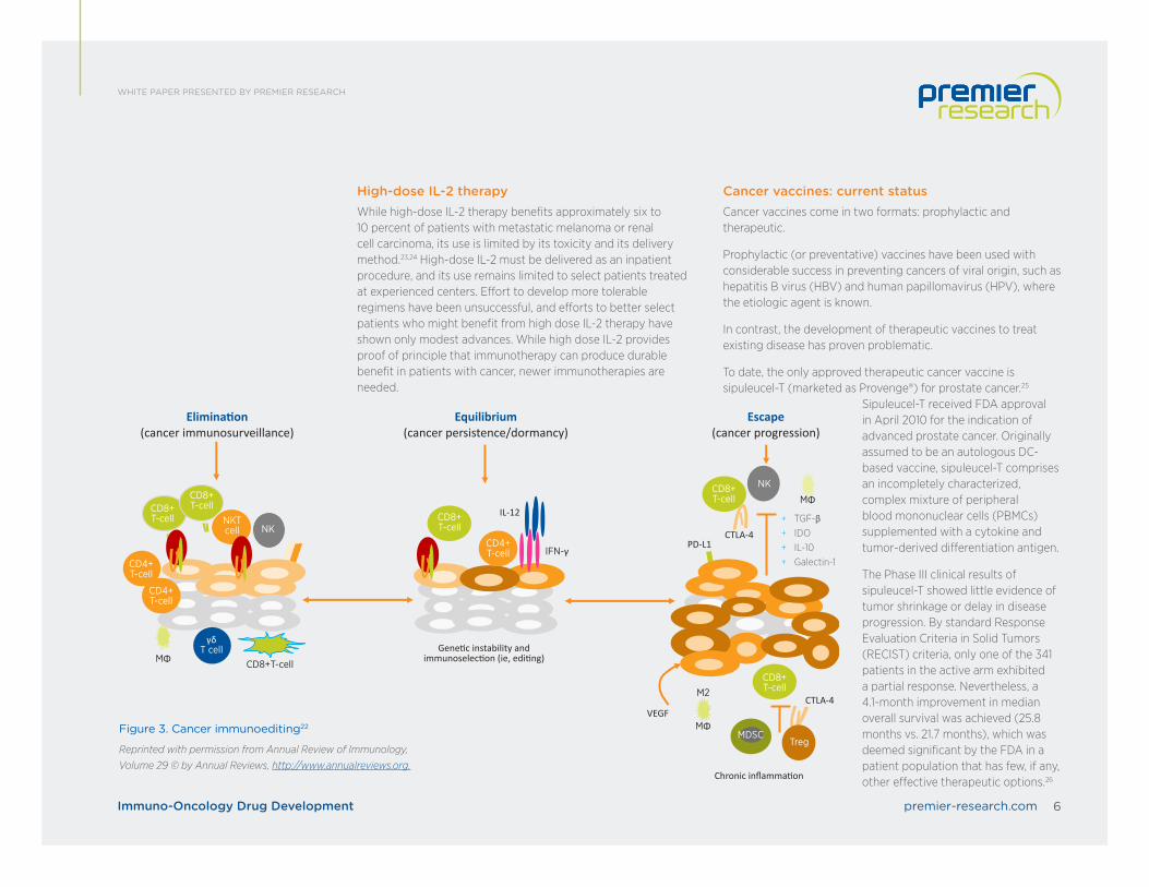
Understanding the immunoediting hypothesisThe notion that the immune system not only protects the host against tumor formation but also shapes tumor immunogenicity is the basis of the cancer immunoediting hypothesis.
Accumulated data from animal models and human cancer patients strongly supports the concept that the immune system can identify and control nascent tumor cells in a process called cancer immunosurveillance. In addition, the immune system can promote tumor progression through chronic inflammation, immunoselection of poorly immunogenic variants, and suppression of anti-tumor immunity. Together, the dual host-protective and tumor-promoting actions of immunity are referred to as cancer immunoediting, which is thought to comprise three distinct phases: 20,21
1. Elimination. In this phase, transformed cells are destroyed by a competent immune system.
2. Equilibrium. Sporadic tumor cells that manage to survive elimination enter this phase, where editing occurs.
3. Escape. In this phase, immunologically sculpted tumors begin to grow progressively, become clinically apparent, and establish an immunosuppressive tumor microenvironment.
Development of immunotherapy moleculesThe development of immunotherapy molecules over the past 20 years has been an exciting journey, beginning with high-dose IL-2 therapy, one of the first immune therapies to gain regulatory approval.
Figure 2. Differentiation and function of CD4+ helper T cell subsets19 Adapted from Bailey SR, et al.
Applying Quality by Design to the Rare Disease Population: Special Considerations 6premier-research.comImmuno-Oncology Drug Development
WHITE PAPER PRESENTED BY PREMIER RESEARCH
High-dose IL-2 therapyWhile high-dose IL-2 therapy benefits approximately six to 10 percent of patients with metastatic melanoma or renal cell carcinoma, its use is limited by its toxicity and its delivery method.23,24 High-dose IL-2 must be delivered as an inpatient procedure, and its use remains limited to select patients treated at experienced centers. Effort to develop more tolerable regimens have been unsuccessful, and efforts to better select patients who might benefit from high dose IL-2 therapy have shown only modest advances. While high dose IL-2 provides proof of principle that immunotherapy can produce durable benefit in patients with cancer, newer immunotherapies are needed.
Cancer vaccines: current statusCancer vaccines come in two formats: prophylactic and therapeutic.
Prophylactic (or preventative) vaccines have been used with considerable success in preventing cancers of viral origin, such as hepatitis B virus (HBV) and human papillomavirus (HPV), where the etiologic agent is known.
In contrast, the development of therapeutic vaccines to treat existing disease has proven problematic.
To date, the only approved therapeutic cancer vaccine is sipuleucel-T (marketed as Provenge®) for prostate cancer.25
Sipuleucel-T received FDA approval in April 2010 for the indication of advanced prostate cancer. Originally assumed to be an autologous DC-based vaccine, sipuleucel-T comprises an incompletely characterized, complex mixture of peripheral blood mononuclear cells (PBMCs) supplemented with a cytokine and tumor-derived differentiation antigen.
The Phase III clinical results of sipuleucel-T showed little evidence of tumor shrinkage or delay in disease progression. By standard Response Evaluation Criteria in Solid Tumors (RECIST) criteria, only one of the 341 patients in the active arm exhibited a partial response. Nevertheless, a 4.1-month improvement in median overall survival was achieved (25.8 months vs. 21.7 months), which was deemed significant by the FDA in a patient population that has few, if any, other effective therapeutic options.26
Applying Quality by Design to the Rare Disease Population: Special Considerations 7premier-research.comImmuno-Oncology Drug Development
WHITE PAPER PRESENTED BY PREMIER RESEARCH
Cancer vaccines: challengesThere are a variety of challenges associated with the development of therapeutic cancer vaccines:
+ The vaccine initially induces an immune reaction against the vaccine itself, not the tumor
+ The antigens are different for each tumor
+ Most immune-responsive tumors auto-vaccinate, but immune regulation prevents an effective response
Based on the foregoing issues, vaccines are unlikely to have a major anti-tumor effect in the absence of immune checkpoint control.
Evolution of immune checkpoint inhibitors in solid tumorsThe development of immune checkpoint inhibitors is an exciting turning point in immunotherapy.
Immune checkpoints refer to a plethora of inhibitory pathways in the immune system that are crucial for maintaining self-tolerance and modulating the duration and amplitude of physiological immune responses in peripheral tissues to minimize collateral tissue damage. It is now clear that tumors co-opt certain immune checkpoint pathways as a major mechanism of immune resistance, particularly against T cells that are specific for tumor antigens.27
T cell responses are regulated via multiple co-stimulatory and inhibitory interactions. T cell response to antigen is mediated by peptide-MHC recognized by the T cell receptor. The B7 family of membrane-bound ligands binds both co-stimulatory and inhibitory receptors. Targeting cytotoxic T-lymphocyte-associated antigen (CTLA)-4 and PD-1 inhibitory receptors has been a major clinical focus.27
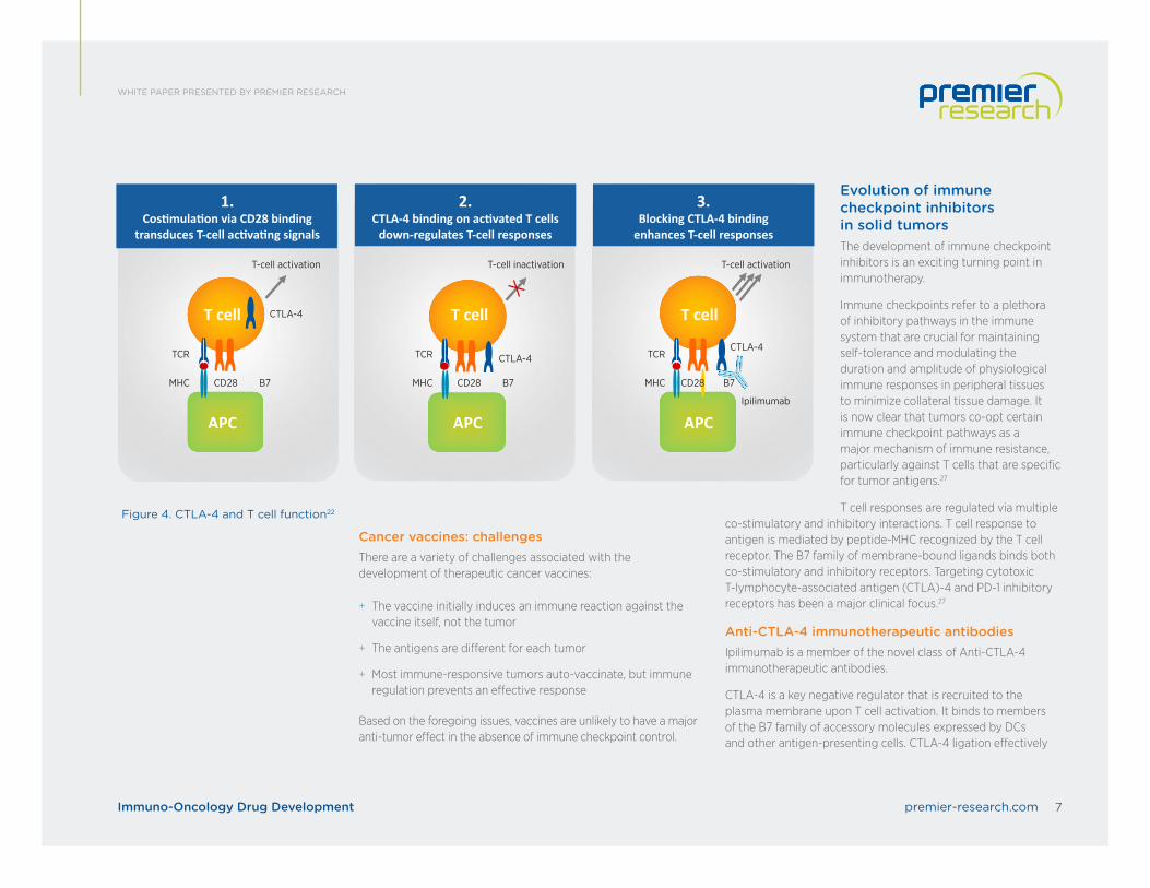
Anti-CTLA-4 immunotherapeutic antibodiesIpilimumab is a member of the novel class of Anti-CTLA-4 immunotherapeutic antibodies.
CTLA-4 is a key negative regulator that is recruited to the plasma membrane upon T cell activation. It binds to members of the B7 family of accessory molecules expressed by DCs and other antigen-presenting cells. CTLA-4 ligation effectively
Applying Quality by Design to the Rare Disease Population: Special Considerations 8premier-research.comImmuno-Oncology Drug Development
WHITE PAPER PRESENTED BY PREMIER RESEARCH
inhibits further activation and expansion, thereby controlling the progress of an immune response and attenuating the chances for chronic autoimmune inflammation. The negative regulation is overcome by use of a blocking antibody, such as ipilimumab.
The fundamental importance of CTLA-4 to controlling T cell function is well illustrated by the phenotype of CTLA-4 knockout mice, which die of an aggressive lymphoproliferative disorder at a young age. Interestingly, CTLA-4 ligation is also important for the immune-suppressive function of Tregs, further assisting to dampen T cell responses. Thus, Treg function is also thought to be blocked by anti-CTLA-4.28
The rationale for using anti-CTLA-4 in cancer therapy was to unrestrain pre-existing anti-cancer T cell responses and possibly trigger new responses. It is well known that tumor-infiltrating lymphocytes exist for melanoma (and other diseases), and that they can bear specificity for tumor antigens.
In a Phase III study, ipilimumab, with or without a glycoprotein 100 (gp100) peptide vaccine, showed improved overall survival compared to gp100 alone in patients with previously treated metastatic melanoma.10 On March 25, 2011, ipilimumab (marketed as Yervoy®) was approved for treatment of unresectable or metastatic melanoma.29
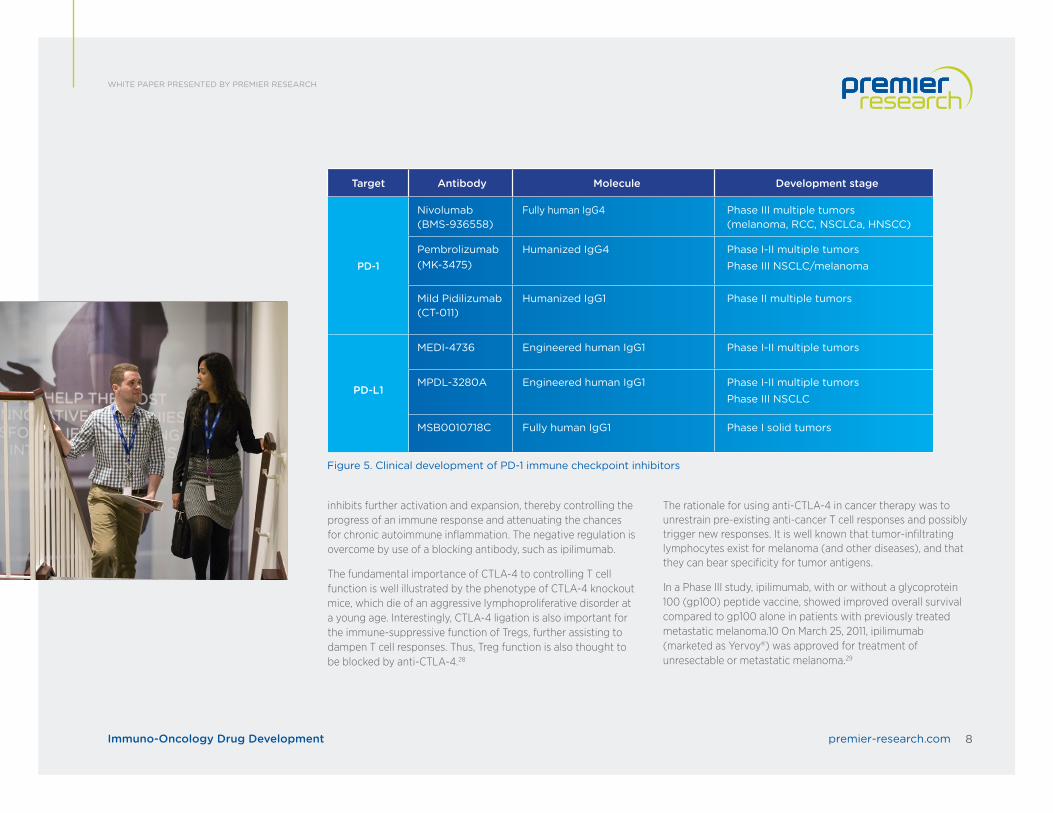
Figure 5. Clinical development of PD-1 immune checkpoint inhibitors
Target Antibody Molecule Development stage
PD-1
Nivolumab(BMS-936558)
Fully human IgG4 Phase III multiple tumors (melanoma, RCC, NSCLCa, HNSCC)
Pembrolizumab(MK-3475)
Humanized IgG4 Phase I-II multiple tumorsPhase III NSCLC/melanoma
Mild Pidilizumab(CT-011)
Humanized IgG1 Phase II multiple tumors
PD-L1
MEDI-4736 Engineered human IgG1 Phase I-II multiple tumors
MPDL-3280A Engineered human IgG1 Phase I-II multiple tumorsPhase III NSCLC
Applying Quality by Design to the Rare Disease Population: Special Considerations 9premier-research.comImmuno-Oncology Drug Development
WHITE PAPER PRESENTED BY PREMIER RESEARCH
PD-1 pathway inhibitorsBlocking the interaction between the PD-1 protein and one of its ligands, PD-L1, has been reported to have impressive anti-tumor responses.30
PD-L1 can be expressed on tumor cells either endogenously or induced by adaptive immune resistance.31,32 The interaction between PD-1 and PD-L1 results in T cell suppression (i.e., anergy, exhaustion, death). In melanoma, renal cell carcinoma, and other tumors, PD-L1 expression has been associated with adverse clinical and pathological features, such as more aggressive disease and shorter survival.33
To date, tumors that have been shown to respond to anti-PD-1 or anti-PD-L1 therapy include melanoma, renal cell carcinoma, non-small-cell lung cancer, bladder cancer, head and neck cancers, and lymphomas. Pembrolizumab (marketed as Keytruda®) and nivolumab (marketed as Opdivo®) were the first of this anti-PD-1 pathway family of checkpoint inhibitors to gain accelerated approval from the FDA for the treatment of ipilimumab-refractory melanoma.30 Pembrolizumab has also been approved as a single agent for the first-line treatment of patients with metastatic NSCLC whose tumors have high PD-L1 expression, and nivolumab has also been approved for patients with metastatic squamous NSCLC who have progressed on or after platinum-based chemotherapy.
Rationale for combination therapies Combination therapies may combine:
+ Agents that act at the effector stage (e.g., anti-PD-1 or inhibitors of immunosuppression) by re-energizing pre-existing T cells
+ Agents that act at the proliferation/activation stage (e.g., anti-CTLA-4) to not only enhance pre-existing responses, but also stimulate de novo responses
Applying Quality by Design to the Rare Disease Population: Special Considerations 10premier-research.comImmuno-Oncology Drug Development
WHITE PAPER PRESENTED BY PREMIER RESEARCH
+ Agents that act on other co-stimulatory or inhibitory pathways
+ Standard of care (e.g., chemotherapy, tyrosine kinase inhibitors, vascular endothelial growth factor inhibitors, radiation therapy)
+ Epigenetic therapy
Combining anti-CTLA-4 with anti-PD1 makes sense biologically, as the two agents remove the brakes from T cell activation at two distinct stages: proliferation (CTLA-4) and effector function (PD-1). Yet, both might be expected to exhibit similar adverse events, underscoring the need to carefully define the potential for serious toxicity.
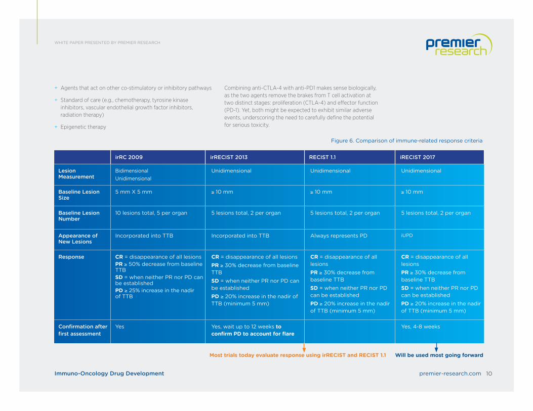
Figure 6. Comparison of immune-related response criteria
irRC 2009 irRECIST 2013 RECIST 1.1 iRECIST 2017
Lesion Measurement
BidimensionalUnidimensional
Unidimensional Unidimensional Unidimensional
Baseline Lesion Size
5 mm X 5 mm ≥ 10 mm ≥ 10 mm ≥ 10 mm
Baseline Lesion Number
10 lesions total, 5 per organ 5 lesions total, 2 per organ 5 lesions total, 2 per organ 5 lesions total, 2 per organ
Appearance of New Lesions
Incorporated into TTB Incorporated into TTB Always represents PD iUPD
Response CR = disappearance of all lesionsPR ≥ 50% decrease from baseline TTBSD = when neither PR nor PD can be establishedPD ≥ 25% increase in the nadir of TTB
CR = disappearance of all lesionsPR ≥ 30% decrease from baseline TTBSD = when neither PR nor PD can be establishedPD ≥ 20% increase in the nadir of TTB (minimum 5 mm)
CR = disappearance of all lesionsPR ≥ 30% decrease from baseline TTBSD = when neither PR nor PD can be establishedPD ≥ 20% increase in the nadir of TTB (minimum 5 mm)
CR = disappearance of all lesionsPR ≥ 30% decrease from baseline TTBSD = when neither PR nor PD can be establishedPD ≥ 20% increase in the nadir of TTB (minimum 5 mm)
Confirmation after first assessment
Yes Yes, wait up to 12 weeks to confirm PD to account for flare
Yes, 4-8 weeks
Will be used most going forwardMost trials today evaluate response using irRECIST and RECIST 1.1
Applying Quality by Design to the Rare Disease Population: Special Considerations 11premier-research.comImmuno-Oncology Drug Development
WHITE PAPER PRESENTED BY PREMIER RESEARCH
In theory, these agents could also well work in conjunction with a vaccine approach, whether exogenous or endogenous.
However, we have yet to see clinical data that supports the use of either type of vaccine.
Evidence is emerging that tumor cells can die in multiple ways, with some forms of apoptotic death leading to the enhancement of an anti-tumor immune response. So-called immunogenic cell death is characterized in part by the release of ATP and high-mobility group protein B1, which could activate local-infiltrating myeloid cells and DCs via a purinergic receptor or TLR-4, respectively. Cytotoxic agents that elicit this death fingerprint may have the ability to help induce anti-tumor immune responses and therefore be better candidates for combination therapy with immunologically active agents.
Evaluating response to immunotherapiesIt is important to note that, with anti-CTLA-4 antibodies and other immunotherapies, the disease can get worse before it gets better.
Generally, four distinct response patterns are associated with favorable overall survival:34
1. Response in baseline lesions (i.e., a typical RECIST response)
2. Stable disease with slow decline in tumor volume
3. Response following an initial increase in tumor volume
4. Response following the appearance of new lesions
A clinical challenge with ipilimumab relates to the kinetics of the anti-tumor response. In contrast to conventional cytotoxic therapies that may trigger rapid tumor shrinkage due to direct killing of cancer cells, the stimulation of T cell responses with ipilimumab may take several months to occur. Tumors may increase in size during this period, and some component of
this growth may reflect the consequences of an evolving inflammatory reaction. Indeed, as many as 10 percent of patients treated with ipilimumab who were scored with progressive disease using the modified WHO criteria for tumor size were shown to achieve disease stabilization and prolonged survival.
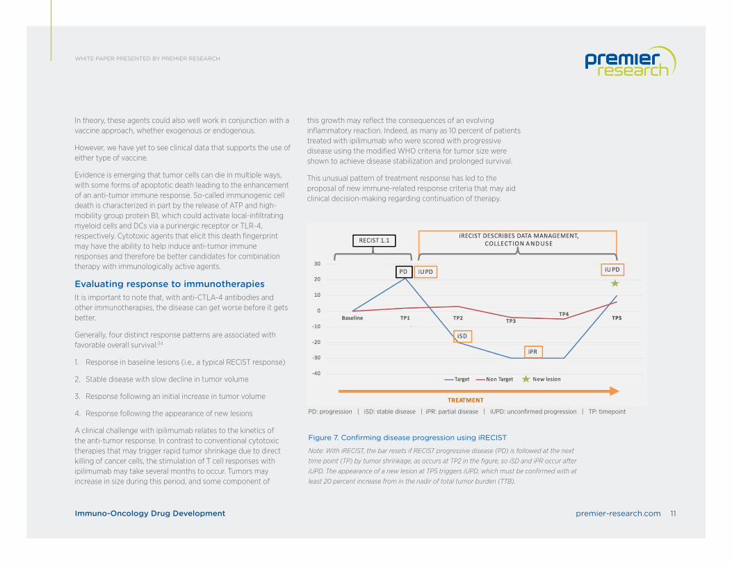
This unusual pattern of treatment response has led to the proposal of new immune-related response criteria that may aid clinical decision-making regarding continuation of therapy.
Figure 7. Confirming disease progression using iRECIST
Note: With iRECIST, the bar resets if RECIST progressive disease (PD) is followed at the next time point (TP) by tumor shrinkage, as occurs at TP2 in the figure, so iSD and iPR occur after iUPD. The appearance of a new lesion at TP5 triggers iUPD, which must be confirmed with at least 20 percent increase from in the nadir of total tumor burden (TTB).
Applying Quality by Design to the Rare Disease Population: Special Considerations 12premier-research.comImmuno-Oncology Drug Development
WHITE PAPER PRESENTED BY PREMIER RESEARCH
Introduction of iRECISTResponse and efficacy of oncology agents is measured by RECIST version 1.1 (RECIST 1.1), a set of published rules that define when cancer patients improve, stay the same, or worsen. Unfortunately, these criteria do not easily apply in immuno-oncology.
As seen with ipilimumab, novel immunotherapeutics trigger different response patterns in tumor than classic chemotherapy drugs. Only applying RECIST 1.1 to immunotherapy trials can result in:
+ Premature termination of therapy
+ Unnecessary removal of patients from clinical trials
+ Inaccurate interpretation of response
To address questions about the assessment of changes in tumor burden in immunotherapy trials, a consensus guideline – iRECIST – was developed by the RECIST working group comprised of members of industry, academia, the FDA, and the EMA. iRECIST calls for the use of modified RECIST 1.1 in cancer immunotherapy trials and describes a standardized approach to solid tumor measurements and definitions for objective change in tumor size for use in such trials.35 iRECIST also introduces a new response criteria called immune unconfirmed progression of disease (iUPD).
When to use iRECISTiRECIST has not yet been validated and should not be used as a guideline for treatment decisions. A sponsor should keep in mind that RECIST 1.1 remains the gold standard for defining treatment response-based endpoints in solid tumors for pivotal registration trials. However, iRECIST can be used in conjunction with RECIST 1.1 in later-phase studies, and may be used as primary response criteria in exploratory, early-phase studies because assessment is done via RECIST 1.1 until progression.
ConclusionImmuno-oncology drugs, particularly immune checkpoint inhibitors, can produce durable anti-tumor responses and are changing the landscape of cancer therapy. Successful development of immunotherapies requires a thorough understanding of tumor immunology, as well as careful planning that takes into account the differences between immuno-oncology drugs and conventional chemotherapy treatments, including selection of combination therapies, evaluation of disease progression, and management of immune-related adverse events.
Applying Quality by Design to the Rare Disease Population: Special Considerations 13premier-research.comImmuno-Oncology Drug Development
WHITE PAPER PRESENTED BY PREMIER RESEARCH
References1. Medscape. Bacillus Calmette-Guérin immunotherapy for bladder cancer. Available at http://emedicine.
medscape.com/article/1950803-overview.
2. Steinman RM, et al. Identification of a novel cell type in peripheral lymphoid organs of mice. J Exp Med. 1973;137:1142-1162.
3. Kirkwood JM, et al. Interferon alfa-2b adjuvant therapy of high-risk resected cutaneous melanoma: the Eastern Cooperative Oncology Group Trial EST 1684. J Clin Oncol. 1996;14:7-17.
4. Fyfe G, et al. Results of treatment of 255 patients with metastatic renal cell carcinoma who received high-dose recombinant interleukin-2 therapy. J Clin Oncol. 1995;13:688-696.
5. Atkins M. et al. High-dose recombinant interleukin 2 therapy for patients with metastatic melanoma: analysis of 270 patients treated between 1985 and 1993. J Clin Oncol. 1999;17:2105-2116.
6. Ishida Y, et al. Induced expression of PD-1, a novel member of the immunoglobulin gene superfamily, upon programmed cell death. EMBO J. 1992;11:3887-3895.
7. Nishimura H, et al. Development of lupus-like autoimmune diseases by disruption of the PD-1 gene encoding an ITIM motif-carrying immunoreceptor. Immunity. 1999;11:141-151.
8. Freeman GJ, et al. Engagement of the PD-1 immunoinhibitory receptor by a novel B7 family member leads to negative regulation of lymphocyte activation. J Exp Med. 2000;192:1027-1034.
9. Higano CS, et al. Integrated data from 2 randomized, double-blind, placebo-controlled, phase 3 trials of active cellular immunotherapy with sipuleucel-T in advanced prostate cancer. Cancer. 2009;115:3670-3679.
10. Hodi FS, et al. Improved survival with ipilimumab in patients with metastatic melanoma. N Engl J Med. 2010;363:711-723.
11. Robert C, et al. Anti-programmed-death-receptor-1 treatment with pembrolizumab in ipilimumab-refractory advanced melanoma: a randomised dose-comparison cohort of a phase 1 trial. Lancet. 2014;384:1109-1117.
12. Weber JS, et al. Nivolumab versus chemotherapy in patients with advanced melanoma who progressed after anti-CTLA-4 treatment (CheckMate 037): a randomised, controlled, open-label, phase 3 trial. Lancet Oncol. 2015;16:375-384.
13. Rizvi N, et al. Activity and safety of nivolumab, an anti-PD-1 immune checkpoint inhibitor, for patients with advanced, refractory squamous non-small-cell lung cancer (CheckMate 063): a phase 3, single-arm trial. Lancet Oncol. 2015;16:257-265.
14. Borghaei H, et al. Nivolumab versus docetaxel in advanced nonsquamous non-small-cell lung cancer. N Engl J Med. 2015;373:1627-1639.
15. Garon EB, et al. Pembrolizumab for the treatment of non-small-cell lung cancer. N Engl J Med. 2015;372:2018-2028.
16. Motzer RJ, et al. Nivolumab versus everolimus in advanced renal-cell carcinoma. N Engl J Med. 2015;373:1803-1813.
17. The Cancer Foundation. Immunotherapy in Cancer. Available at http://the-cancer-foundation.org/wp-content/uploads/2014/09/Tumor-Immunology.jpg.
18. Gajewski TF, et al. Molecular profiling to identify relevant immune resistance mechanisms in the tumor microenvironment. Curr Opinion Immunol. 2011;23(2):286-292.
19. Bailey SR, et al. Th17 cells in cancer: the ultimate identity crisis. Front Immunol. 2014;5:276.
20. Schreiber RD, Old LJ, Smyth MJ. Cancer immunoediting: integrating immunity’s roles in cancer suppression and promotion. Science. 2011;331(6024):1565-1570.
21. Mittal D, et al. New insights into cancer immunoediting and its three component phases—elimination, equilibrium and escape. Curr Opin Immunol 2014;27:16-25.
22. Vesely MD, et al. Natural innate and adaptive immunity to cancer. Annu Rev Immunol. 2011;29:235-271.
23. Atkins MB, et al. High-dose recombinant interleukin 2 therapy for patients with metastatic melanoma: analysis of 270 patients treated between 1985 and 1993. J Clin Oncol. 1999;17(7):2105-2016.
24. McDermott DF, et al. Application of IL-2 and other cytokines in renal cancer. Expert Opin Biol Ther. 2004;4:455-468.
25. National Cancer Institute. FDA Approval for Sipuleucel-T. Available at https://www.cancer.gov/about-cancer/treatment/drugs/fda-sipuleucel-T.
26. Kantoff PW, et al. Sipuleucel-T immunotherapy for castration-resistant prostate cancer. N Engl J Med. 2010;363:411-422.
27. Pardoll DM. The blockade of immune checkpoints in cancer immunotherapy. Nat Rev Cancer. 2012;12:252-264.
28. Jain N, et al. Dual function of CTLA-4 in regulatory T cells and conventional T cells to prevent multiorgan autoimmunity. PNAS. 2010;107(4):1524-1528.
29. National Cancer Institute. FDA Approval for Ipilimumab. Available at https://www.cancer.gov/about-cancer/treatment/drugs/fda-ipilimumab.
30. Mahoney KM, Freeman GJ, McDermott DF. The next immune-checkpoint inhibitors: PD-1/PD-L! blockade in melanoma. Clin Ther. 2015;37(4):764-782.
31. Topalian SL, Drake CG, Pardoll DM. Targeting the PD-1/B7-H1(PD-L1) pathway to activate anti-tumor immunity. Curr Opin Immunol. 2012;24(2):207-212.
32. Taube JM, et al. Colocalization of inflammatory response with B7-h1 expression in in human melanocytic lesions supports an adaptive resistance mechanism of immune escape. Sci Transl Med. 2012;4:127ra37.
33. Thompson RH, et al. Costimulatory B7-H1 in renal cell carcinoma patients: indicator of tumor aggressiveness and potential therapeutic target. Proc Natl Acad Sci USA. 2004;101:17174-17179.
34. Wolchok JD, et al. Guidelines for the evaluation of immune therapy activity in solid tumors: immune-related response criteria. Clin Cancer Res. 2009;15:7412-7420.
35. Seymour L, et al. on behalf of the RECIST working group. iRECIST: guidelines for response criteria for use in trials testing immunotherapeutics. Lancet. 2017;18(3):e142-152.
Nina Baluja | Senior Medical Director, Medical Services Dr. Baluja performs medical and safety monitoring on clinical research projects and provides strategic guidance on protocol development, study design, regulatory filings, and study execution. She has more than 15 years of clinical research, medical marketing, and drug safety experience in the CRO and pharmaceutical fields.
Prior to joining Premier Research in 2017, Dr. Baluja was Senior Medical Director at PRA Health Services. Before that, she oversaw global regulatory consulting at PPD and worked extensively in the pharma sector in positions that included Medical Scientific Adviser at Boehringer-Ingelheim, Associate Medical Director at Wyeth Pharmaceuticals, and Trial Delivery Manager at AstraZeneca.
Dr. Baluja’s areas of specialty include oncology, hemato-oncology, and rare diseases. She has significant clinical development regulatory experience, having prepared and participated in numerous scientific meetings with the FDA, EMA, and Health Canada.
She holds an MBBS degree from Pune University in India and a master’s in surgical oncology from Bombay University.
Luke Gill | Executive Director, Oncology, Strategic DevelopmentMr. Gill has an extensive scientific background and more than 20 years of drug development experience. Specializing in oncology, he has led numerous global CRO management teams and provided strategic assessment, management, and oversight of study enrollment and program metrics.
Prior to joining Premier Research in 2015, Mr. Gill was Director of Global Project Management for Hematology and Oncology at PPD, overseeing design and delivery of clinical development plans across multiple indications and specializing in early-phase oncology. He also served as Assistant Project Management Director at PPD, was CRO alliance program director for Merck Serono, and has held positions at Pfizer/Parke Davis, Astra, and Glaxo.
Mr. Gill holds a master’s degree in neuro and molecular pharmacology from the University of Bristol, a bachelor’s degree in biological sciences from the University of the West of England, and an MBA specializing in strategy and international enterprise from the Open University in the U.K.
About Premier Research Premier Research is a leading clinical development service provider that helps highly innovative biotech, medical device,
and specialty pharma companies transform breakthrough ideas into reality. The company has a wealth of experience
in the execution of global, regional, and local clinical development programs with a special focus on addressing unmet
needs in areas such as analgesia, dermatology, medical devices, neuroscience, oncology, pediatrics, and rare disease.
Premier Research operates in 84 countries and employs 1,000 professionals, including a strong international network of
clinical monitors and project managers, regulatory, data management, statistical, scientific, and medical experts. They
are focused on smart study design for advanced medicines that allow life-changing treatments.


![Tumor Mutational Burden Immuno-Oncology Scientific Updates · Title: Tumor Mutational Burden Immuno-Oncology Scientific Updates Author: Ritterhouse, Lauren [BSD] - PTH Created Date:](https://static.documents.pub/doc/80x56/5fc25b7302cd4e75fc4563dd/tumor-mutational-burden-immuno-oncology-scientific-updates-title-tumor-mutational.jpg)


