Kostianets et al. Diagnostic Pathology 2012, 7:161http://www.diagnosticpathology.org/content/7/1/161
RESEARCH Open Access
Immunohistochemical analysis of medullarybreast carcinoma autoantigens in differenthistological types of breast carcinomasOlga Kostianets1,2*, Stepan Antoniuk3, Valeriy Filonenko1 and Ramziya Kiyamova1
Abstract
Background: On the past decade a plethora of investigations were directed on identification of molecules involvedin breast tumorogenesis, which could represent a powerful tool for monitoring, diagnostics and treatment of thisdisease. In current study we analyzed six previously identified medullary breast carcinoma autoantigens includingLGALS3BP, RAD50, FAM50A, RBPJ, PABPC4, LRRFIP1 with cancer restricted serological profile in different histologicaltypes of breast cancer.
Methods: Semi-quantitative immunohistochemical analysis of 20 tissue samples including medullary breastcarcinoma, invasive ductal carcinoma, invasive lobular carcinoma and non-cancerous tissues obtained from patientswith fibrocystic disease (each of five) was performed using specifically generated polyclonal antibodies. Differencesin expression patterns were evaluated considering percent of positively stained cells, insensitivity of staining andsubcellular localization in cells of all tissue samples.
Results: All 6 antigens predominantly expressed in the most cells of all histological types of breast tumors andnon-cancerous tissues with slight differences in intensity of staining and subcellular localization. The mostsignificant differences in expression pattern were revealed for RAD50 and LGALS3BP in different histological typesof breast cancer and for PABPC4 and FAM50A antigens in immune cells infiltrating breast tumors.
Conclusions: This pilot study made possible to select 4 antigens LGALS3BP, RAD50, PABPC4, and FAM50A aspromising candidates for more comprehensive research as potential molecular markers for breast cancerdiagnostics and therapy.
Virtual slides: The virtual slides’ for this article can be found here: http://www.diagnosticpathology.diagnomx.eu/vs/1860649350796892
Keywords: Tumor-associated antigen, Expression pattern, Breast cancer
BackgroundBreast cancer is the most common female cancer in theworld, with high mortality rate [1,2]. Identification ofbiomarkers for early detection and new therapeutic tar-gets of breast cancer helps to continuously reduce themorbidity of this frequent pathology in women. Thisentails resolving the physiological, cellular and molecularprocesses underlying the complexity of breast tumor
* Correspondence: [email protected] of Cell Signaling, Institute of Molecular Biology and Genetics,NAS of Ukraine, 150, Zabolotnogo str., Kyiv, Ukraine2Educational and Scientific Centre “Institute of Biology”, Taras ShevchenkoNational University of Kyiv, 64, Volodymyrs’ka Str., Kyiv, UkraineFull list of author information is available at the end of the article
development and associated tumor heterogeneity [3]. Inrecent years efforts of many researchers are focused onsearch of new markers and molecules involved in breasttumorogenesis, which could be used in disease diagnos-tics, treatment and prognosis.Development and application of proteomic technolo-
gies based on detection of autoantibodies to tumor anti-gens, including serological identification of antigens byrecombinant expression cloning (SEREX) [4-6], sero-logical proteome analysis (SEPRA) [7-9], multiple affinityprotein profiling (MAPPing) [10] and high-density pro-tein microarrays [11] have lead to identification of multi-ply biomarkers of breast cancer inducing autoantibody
ral Ltd. This is an Open Access article distributed under the terms of the Creativeommons.org/licenses/by/2.0), which permits unrestricted use, distribution, andiginal work is properly cited.
Kostianets et al. Diagnostic Pathology 2012, 7:161 Page 2 of 11http://www.diagnosticpathology.org/content/7/1/161
response. The characterization of tumor markersremains a major goal in both understanding the cellularmechanisms associated with tumorogenesis and indeveloping targets for the molecular therapy and diag-nostics of cancer.Protein tumor marker reflects consistent, biologically
relevant changes in the tumor. It has been described thatnumerous autologous proteins of tumor cells, generallyknown as tumor-associated antigens (TAAs), can elicithumoral immune response in cancer patients as a resultof their aberrant expression [12], alternative splicing ofpre-messenger RNAs [13], point mutations [14], aber-rant localization, folding [15], degradation [16] and/orpost-translation alteration [17,18]. Many breast cancerantigens have been reported to be overexpressed at pro-tein level in breast tumors; these include MUC1 [19],HER2/neu [20], P53 [21], HSP-27 [22,23], GIPC-1 [24],fibulin 1 [25] and cyclins B1 [26], D1 [27] and E [28].Some tumor markers, for example, cyclin B1 also wasfound to change its location in cancer cells and localizedpredominantly in the cytosol [29], while normally it pre-sents in the nucleus.Our previous study was focused on identification of
novel TAAs of medullary breast carcinoma (MBC), arelatively rare type of ductal carcinoma, which despiteanaplastic features has favorable prognosis for patients[30-32]. High lyphocytic infiltration, indicating possiblepresence of specific antigen within tumor lesion is adistinctive feature of medullary breast carcinoma[30,33,34]. Using SEREX approach and serological plaque-spot assay we identified 41 potential antigens of medullarybreast carcinoma, and showed that 18 of them had cancerrestricted serological profile [35]. In this study proteinexpression pattern of a small subset of 6 from 18(lectin, galactoside-binding, soluble, 3 binding protein(LGALS3BP); human RAD50 S. cerevisiae homolog(RAD50); family with sequence similarity 50, member A(FAM50A); poly(A) binding protein cytoplasmic 4(PABPC4); recombination signal binding protein for im-munoglobulin kappa J region (RBPJ) and leucine rich re-peat (in FLII) interacting protein 1 (LRRFIP1)) antigenswas investigated by immunohistochemical analysis of differ-ent histological types of breast cancer and non-cancerousbreast tissues from patients with fibrocystic disease. Semi-quantitative analysis of positively stained cells, consideringintensity of staining, and subcellular localization in normal,cancer and immune cells of lymphocytic infiltrate pre-sented in some tumors using specifically generated poly-clonal antibodies have been performed.
MethodsClinicopathological dataFresh breast cancer and non-cancerous breast tissue(NCT) samples were obtained from 15 female patients
(39–75 years) with primary breast carcinoma and 5patients with fibrocystic disease (37–50 years) corres-pondingly, who underwent surgery in the Dnipropet-rovsk Clinical Oncological Center (Dnipropetrovsk,Ukraine) between 2008 and 2010. Surgical specimenswere fixed with 10% buffered formalin, and paraffin em-bedded sections were stained with hematoxilin andeosin. All the tissue sample sections were reviewed toconfirm the original diagnosis by an expert pathologist.The clinical and histopathological characteristics of thesepatients are shown in Table 1. Breast cancer casesincluded invasive ductal carcinoma (IDC) (n = 5), inva-sive lobular carcinoma (ILC) (n = 5), and medullary car-cinoma (n = 5). Clinicopathological data were obtainedfrom patient medical records and from the files kept atthe Department of Pathohistology of DnipropetrovskClinical Oncological Center. The study was approved bythe Ethics Committee of the Institute of Molecular Biol-ogy and Genetics, NAS of Ukraine and informed con-sent was obtained from all patients.
Cloning, expression and purification of recombinantproteinsSpecific cDNA of LGALS3BP, RAD50, FAM50A, RBPJ,PABPC4 and LRRFIP1 genes, isolated from MBC cDNAlibrary Br502, was cloned into pGEX4T3 and/or pET28bexpression vectors, containing glutathione-S-transferase-and 6His-tags respectively (Table 2). Expression of fusedrecombinant proteins was induced by 1 mM IPTG at37°C for 4 h in E.coli BL21 (DE3) pLysE cells trans-formed by correspondent recombinant plasmids.Proteins were affinity purified using GST-sepharose andNi-NTA-agarose according to manufacturers’ protocols.Some expressed recombinant proteins were present inthe inclusion bodies of the pellet, which were washedbefore the purification procedure according X.A. Yangprotocol [36]. Purity of proteins was analyzed by SDS-PAGE. All proteins were dialyzed against phosphate buf-fered saline (PBS), pH7.4, and then were used forimmunization of mice.
Generation of polyclonal antibodiesPolyclonal antibodies against LGALS3BP, RAD50,FAM50A, RBPJ, PABPC4 and LRRFIP1 autoantigenswere generated according the protocol described below.A primary dose of 20 mkg of each recombinant polypep-tide was solubilized in Freund’s complete adjuvant(Sigma, Aldrich, Germany) and administered intraperito-neally in six- to 8-week-old female BALB/c mice fourtimes at 2-week intervals. Then, immunized mice (withserum titer no less than 105–106) were boosted with20 mkg of antigen in PBS by intraperitoneal injection.After 3 days of the booster injection, blood was collectedfrom the mice, and the serum was separated.
Table 1 Characteristics of patients and their tissue samples
IDC (n = 5) ILC (n = 5) MBC (n = 5) NCT (n = 5)
Age (mean) 48.4 65.8 53 44.8
Age range 39-73 47-75 41-71 37-50
ER-positive 3 4 0 n/a
PR-positive 2 4 0 n/a
HER2-positive 3 2 0 n/a
Node positive 3 3 0 n/a
Ki-67 expression (range) 13-68% 8-29% n/a n/a
Kostianets et al. Diagnostic Pathology 2012, 7:161 Page 3 of 11http://www.diagnosticpathology.org/content/7/1/161
Specificity of generated polyclonal antibodies was testedby Western blotting of recombinant proteins and con-firmed by immunohystochemical analysis of MBC tumor,which was taken for generation of MBC cDNA libraryBr502 [37] with depleted against corresponding recombin-ant proteins polyclonal antibodies. Depletion of polyclonalantibody was performed as follows: antibodies diluted(1:10) in PBS were incubated overnight with correspondentrecombinant proteins (10 mkg) transferred on PVDF mem-branes by standard blotting procedure. Decreasing of theintensity of immunohistochemical staining of Br502 tumorsections, when depleted antibody were used evidenced forspecificity of generated polyclonal antibodies comparedwith undepleted ones.
Immunohistochemical analysisImmunohistochemical analysis of breast cancer sampleswith polyclonal Abs was performed according to stand-ard protocol. Briefly, representative sections of breasttumors were prepared from parafin blocks. Endogenousperoxidase was quenched with H2O2 (3%) in 0.01% PBS.After blocking of non-specific binding with avidin-biotinblocking solution (Vector Laboratories, Burlingame, CA,USA), tissue sections were incubated overnight at 4°Cwith corresponded polyclonal antibodies (1:400). Then,sections were incubated with biotinylated secondary
Table 2 Antigens used for generation of polyclonal antibodie
Antigen Full name NCBrefere
LGALS3BP Homo sapiens lectin, galactoside-binding, soluble, 3binding protein
NM_005
RAD50 Homo sapiens RAD50 homolog (S. cerevisiae) NM_005
FAM50A Homo sapiens family with sequence similarity 50,member A
NM_004
RBPJ Homo sapiens recombination signal binding proteinfor immunoglobulin kappa J region
NM_203
PABCP4 Homo sapiens poly(A) binding protein, cytoplasmic 4(inducible form)
NM_003
LRRFIP1 Homo sapiens leucine rich repeat (in FLII) interactingprotein 1
NM_004
antibodies for 2 h at room temperature at 1:400dilutions (goat anti-mouse biotinylated IgG, Sigma,Steinheim, Germany), followed by incubation withavidin-biotinperoxidase complex (Vector Laboratories,Burlingame, CA, USA) for 30 min at RT and developedwith diaminobenzidine solution. Serum from non-immune mouse was used as negative control. Immuno-histochemical staining of all breast cancer andnon-cancerous tissue slides was performed during oneexperiment in equal conditions for each of antigens ana-lyzed. Haematoxylin was used for counterstaining.Standard microscopy was performed using a Zeiss Uni-versal microscope (Zeiss, Germany), and images werecaptured using digital Axiocam software.Taking into account the fact that the expression of the
studied antigens in normal and tumor breast tissues arepoorly studied, we evaluated the staining intensity foreach antigen comparing the slides of non-cancerous andcancerous breast tissue samples of different histologicaltypes, which were processed with correspondent poly-clonal antibody under the same conditions. Tissue slideswere analyzed using a four point semi-quantitative scalefor nuclear and cytoplasmic optical staining intensity(graded as 3+ (strong), 2+ (moderate), 1+ (weak), 0 (nostaining)) and for the percentage of positively stainedcells (0 – no staining, 1 – <10% of the cells, 2 – 11 – 50%
s
Ince
DNA fragment, bp Vector MW of recombinant protein,kDa, (including tag)
567.2 1483-1686 pGEX4T3(GST-tag)
35
732.2 2552-3374 pET28b(6His-tag)
44
699.1 76-1095 pGEX4T3(GST-tag)
64
284.1 492-1850 pET28b(6His-tag)
50
819.2 927-3052 pET28b(6His-tag)
67
735.2 417-1804 pET28b(6His-tag)
51
Kostianets et al. Diagnostic Pathology 2012, 7:161 Page 4 of 11http://www.diagnosticpathology.org/content/7/1/161
of the cells, 3 – >50% of the cells). Pathologist markedbiologically representative areas, which contained amean number of 150 cells (min 72 – max 180), percent-age of positive cells was assessed as percent of cells exhi-biting reactivity as average in 10 fields at highmagnification (x400). The area of interest was selectedrandomly, excluding stromal cells and artifacts. Stainingintensity and percent reactivity were recorded as meanobserved in ten high power fields. The same slides wereviewed again by each one of 3 month later, independ-ently of the first vision.
ResultsIn our previous studies, focused on identification ofnovel TAAs of medullary breast carcinoma, we found 41antigens [37], 18 of which had cancer-restricted sero-logical profile as was shown by phage based allogenicserological screening [35]. TAAs identified represent adiverse range of cellular proteins, some of which wereshown to be implicated in cancer development, particu-larly lectin, galactoside-binding, soluble, 3 binding pro-tein (LGALS3BP) [38], double-strand break repair factorRAD50 [39], nuclear protein with unknown functionFAM50A [40], poly(A) binding protein cytoplasmic 4(PABPC4) [41], mediator of Notch signaling RBPJ [42]
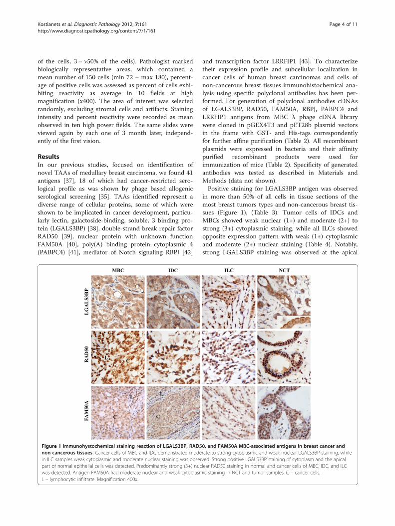
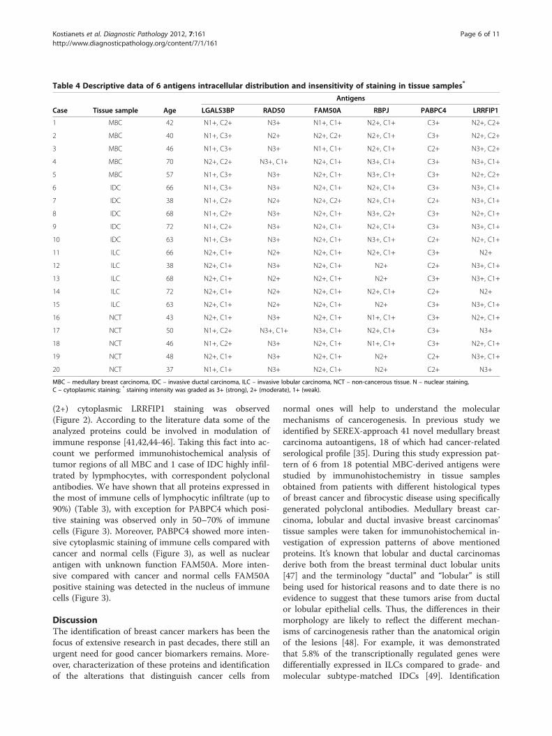
Figure 1 Immunohystochemical staining reaction of LGALS3BP, RAD5non-cancerous tissues. Cancer cells of MBC and IDC demonstrated modein ILC samples weak cytoplasmic and moderate nuclear staining was obserpart of normal epithelial cells was detected. Predominantly strong (3+) nucwas detected. Antigen FAM50A had moderate nuclear and weak cytoplasmL – lymphocytic infiltrate. Magnification 400x.
and transcription factor LRRFIP1 [43]. To characterizetheir expression profile and subcellular localization incancer cells of human breast carcinomas and cells ofnon-cancerous breast tissues immunohistochemical ana-lysis using specific polyclonal antibodies has been per-formed. For generation of polyclonal antibodies cDNAsof LGALS3BP, RAD50, FAM50A, RBPJ, PABPC4 andLRRFIP1 antigens from MBC λ phage cDNA librarywere cloned in pGEX4T3 and pET28b plasmid vectorsin the frame with GST- and His-tags correspondentlyfor further affine purification (Table 2). All recombinantplasmids were expressed in bacteria and their affinitypurified recombinant products were used forimmunization of mice (Table 2). Specificity of generatedantibodies was tested as described in Materials andMethods (data not shown).Positive staining for LGALS3BP antigen was observed
in more than 50% of all cells in tissue sections of themost breast tumors types and non-cancerous breast tis-sues (Figure 1), (Table 3). Tumor cells of IDCs andMBCs showed weak nuclear (1+) and moderate (2+) tostrong (3+) cytoplasmic staining, while all ILCs showedopposite expression pattern with weak (1+) cytoplasmicand moderate (2+) nuclear staining (Table 4). Notably,strong LGALS3BP staining was observed at the apical
0, and FAM50A MBC-associated antigens in breast cancer andrate to strong cytoplasmic and weak nuclear LGALS3BP staining, whileved. Strong positive LGALS3BP staining of cytoplasm and the apicallear RAD50 staining in normal and cancer cells of MBC, IDC, and ILCic staining in NCT and tumor samples. C – cancer cells,
Table 3 Descriptive data of 6 antigens expression in tissue samples
MBC – medullary breast carcinoma, IDC-invasive ductal carcinoma, ILC – invasive lobular carcinoma, NCT – non-cancerous tissue; n/a - normal cells were notavailable within tumor sample; 0 - no staining, 1 - <10% of the positively stained cells, 2 - 10-50% of the positively stained cells, 3 > 50% of the positively stainedcells; * - one case of IDC which was heavily infiltrated by lymphocytes; ** - PABPC4 positive staining was observed in 50-70% of immune cells.
Kostianets et al. Diagnostic Pathology 2012, 7:161 Page 5 of 11http://www.diagnosticpathology.org/content/7/1/161
part of normal epithelial cells of the ducts and lobules innon-cancerous tissues (Figure 1) and in some ILCscases.All 15 breast cancer and 5 non-cancerous tissue
samples were positive for the DNA reparation factorRAD50 (Figure 1), (Table 3). We observed moderate(2+) to strong (3+) nuclear RAD50 staining with nocytoplasmic staining apparent in normal and cancer cellsin the most cases. However, in 4 of 5 ILCs cases decreas-ing of the intensity of nuclear staining (moderate (2+))was detected.Analysis of FAM50A protein expression revealed that
more than 50% of cells were positively stained in cancerand non-cancerous tissue sets (Figure 1), (Table 3).FAM50A protein predominantly was distributed in nu-cleus and cytoplasm of cancer cells of all tumor sampleswith preferentially moderate (2+) and weak (1+) positivestaining respectively (Table 4).
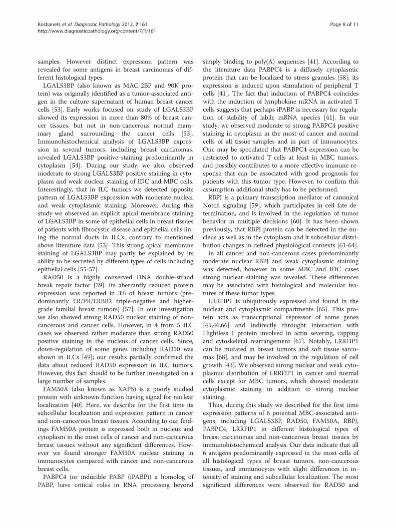
RBPJ staining was indicated in the most of cancer andnormal cells (Figure 2), (Table 3). Moderate (2+) tostrong (3+) nuclear and weak (1+) cytoplasmic staining(Table 4) was detected in MBC and IDC tissue samples.In ILC and NCT tissues samples predominantly moder-ate (2+) nuclear and additional weak (1+) cytoplasmicstaining in some cases was detected.Moderate (2+) to strong (3+) PABPC4 positive staining
was exclusively observed in cytoplasm of normal andcancer cells of non-cancerous and cancer tissue sampleswith no relation to histological type (Figure 2), (Tables 3and 4).LRRFIP1 nuclear and cytoplasmic expression was indi-
cated in more than 50% of all tissue samples (Figure 2),(Table 3). This protein was located in nucleus and cyto-plasm of both cancerous and non-cancerous tissue setswith moderate (2+) to strong (3+) nuclear and weak (1+)cytoplasmic staining, however in MBC tissues moderate
Table 4 Descriptive data of 6 antigens intracellular distribution and insensitivity of staining in tissue samples*
Antigens
Case Tissue sample Age LGALS3BP RAD50 FAM50A RBPJ PABPC4 LRRFIP1
MBC – medullary breast carcinoma, IDC – invasive ductal carcinoma, ILC – invasive lobular carcinoma, NCT – non-cancerous tissue. N – nuclear staining,C – cytoplasmic staining; * staining intensity was graded as 3+ (strong), 2+ (moderate), 1+ (weak).
Kostianets et al. Diagnostic Pathology 2012, 7:161 Page 6 of 11http://www.diagnosticpathology.org/content/7/1/161
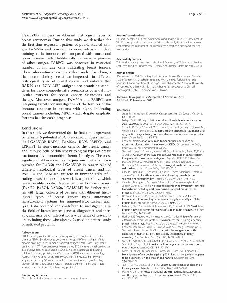
(2+) cytoplasmic LRRFIP1 staining was observed(Figure 2). According to the literature data some of theanalyzed proteins could be involved in modulation ofimmune response [41,42,44-46]. Taking this fact into ac-count we performed immunohistochemical analysis oftumor regions of all MBC and 1 case of IDC highly infil-trated by lypmphocytes, with correspondent polyclonalantibodies. We have shown that all proteins expressed inthe most of immune cells of lymphocytic infiltrate (up to90%) (Table 3), with exception for PABPC4 which posi-tive staining was observed only in 50–70% of immunecells (Figure 3). Moreover, PABPC4 showed more inten-sive cytoplasmic staining of immune cells compared withcancer and normal cells (Figure 3), as well as nuclearantigen with unknown function FAM50A. More inten-sive compared with cancer and normal cells FAM50Apositive staining was detected in the nucleus of immunecells (Figure 3).
DiscussionThe identification of breast cancer markers has been thefocus of extensive research in past decades, there still anurgent need for good cancer biomarkers remains. More-over, characterization of these proteins and identificationof the alterations that distinguish cancer cells from
normal ones will help to understand the molecularmechanisms of cancerogenesis. In previous study weidentified by SEREX-approach 41 novel medullary breastcarcinoma autoantigens, 18 of which had cancer-relatedserological profile [35]. During this study expression pat-tern of 6 from 18 potential MBC-derived antigens werestudied by immunohistochemistry in tissue samplesobtained from patients with different histological typesof breast cancer and fibrocystic disease using specificallygenerated polyclonal antibodies. Medullary breast car-cinoma, lobular and ductal invasive breast carcinomas’tissue samples were taken for immunohistochemical in-vestigation of expression patterns of above mentionedproteins. It’s known that lobular and ductal carcinomasderive both from the breast terminal duct lobular units[47] and the terminology “ductal” and “lobular” is stillbeing used for historical reasons and to date there is noevidence to suggest that these tumors arise from ductalor lobular epithelial cells. Thus, the differences in theirmorphology are likely to reflect the different mechan-isms of carcinogenesis rather than the anatomical originof the lesions [48]. For example, it was demonstratedthat 5.8% of the transcriptionally regulated genes weredifferentially expressed in ILCs compared to grade- andmolecular subtype-matched IDCs [49]. Identification
Figure 2 Immunohystochemical staining reaction of RBPJ, PABPC4, and LRRFIP1 MBC-associated antigens in breast cancer andnon-cancerous tissues. Antigen RBPJ had moderate nuclear and weak cytoplasmic staining of normal and cancer cells of NCT, MBC, and IDC,except for ILC samples which showed only moderate (2+) nuclear staining. Moderate to strong positive PABPC4 staining was detected incytoplasm of normal and cancer cells of non-cancerous and cancer tissue samples. Cancer cells of IDC and ILC, and normal cells ofnon-cancerous tissues had moderate to strong nuclear and weak cytoplasmic LRRFIP1 staining, but in MBC moderate cytoplasmic LRRFIP1staining was observed. C – cancer cells, L – lymphocytic infiltrate. Magnification 400x.
Kostianets et al. Diagnostic Pathology 2012, 7:161 Page 7 of 11http://www.diagnosticpathology.org/content/7/1/161
and characterization of molecular targets specific foreach type of breast cancer will help to design more pre-cise therapeutic and diagnostic approaches for curing ofthis type of malignancy.During this preliminary study, we investigated expres-
sion pattern of 6 MBC autoantigens, including secretoryprotein LGALS3BP, 2 nuclear proteins (RAD50,FAM50A), one cytoplasmic protein (PABPC4) and 2tratscription factors (RBPJ, LRRFIP1), in different histo-logical types of breast cancer and non-cancerous tissue
Figure 3 Immunohistochemical staining of PABPC4 (A) and FAM50A (carcinoma. Both antigens showed more intensive staining of immune cellwas detected only in a part of immune cells. FAM50A showed preferentiallby arrows. C – cancer cells, L – lymphocytic infiltrate. Magnification 400x.
samples by immunohistochemical analysis. Despite thefact that automated measurement using digital slidescanners and computer-aid methods are increasinglyused for immunohystochemical analysis, during thisstudy we used visual semi-quantitative evaluation ofMBC antigens expression pattern by expert pathologistbecause high correlation between these two approacheswas recently shown [50-52].It was shown that all analyzed proteins expressed in
the most cells of non-cancerous and cancer tissue
B) antigens in cells of lymphocytic infiltrate of medullary breasts compared with cancer cells. Positive cytoplasmic PABPC4 stainingy nuclear staining in immune cells. Positively stained cells are indicated
Kostianets et al. Diagnostic Pathology 2012, 7:161 Page 8 of 11http://www.diagnosticpathology.org/content/7/1/161
samples. However distinct expression pattern wasrevealed for some antigens in breast carcinomas of dif-ferent histological types.LGALS3BP (also known as MAC-2BP and 90K pro-
tein) was originally identified as a tumor-associated anti-gen in the culture supernatant of human breast cancercells [53]. Early works focused on study of LGALS3BPshowed its expression in more than 80% of breast can-cer tissues, but not in non-cancerous normal mam-mary gland surrounding the cancer cells [53].Immunohistochemical analysis of LGALS3BP expres-sion in several tumors, including breast carcinomas,revealed LGALS3BP positive staining predominantly incytoplasm [54]. During our study, we also, observedmoderate to strong LGALS3BP positive staining in cyto-plasm and weak nuclear staining of IDC and MBC cells.Interestingly, that in ILC tumors we detected oppositepattern of LGALS3BP expression with moderate nuclearand weak cytoplasmic staining. Moreover, during thisstudy we observed an explicit apical membrane stainingof LGALS3BP in some of epithelial cells in breast tissuesof patients with fibrocystic disease and epithelial cells lin-ing the normal ducts in ILCs, contrary to mentionedabove literature data [53]. This strong apical membranestaining of LGALS3BP may partly be explained by itsability to be secreted by different types of cells includingepithelial cells [55-57].RAD50 is a highly conserved DNA double-strand
break repair factor [39]. Its aberrantly reduced proteinexpression was reported in 3% of breast tumors (pre-dominantly ER/PR/ERBB2 triple-negative and higher-grade familial breast tumors) [57]. In our investigationwe also showed strong RAD50 nuclear staining of non-cancerous and cancer cells. However, in 4 from 5 ILCcases we observed rather moderate than strong RAD50positive staining in the nucleus of cancer cells. Since,down-regulation of some genes including RAD50 wasshown in ILCs [49]; our results partially confirmed thedata about reduced RAD50 expression in ILC tumors.However, this fact should to be further investigated on alarge number of samples.FAM50A (also known as XAP5) is a poorly studied
protein with unknown function having signal for nuclearlocalization [40]. Here, we describe for the first time itssubcellular localization and expression pattern in cancerand non-cancerous breast tissues. According to our find-ings FAM50A protein is expressed both in nucleus andcytoplasm in the most cells of cancer and non-cancerousbreast tissues without any significant differences. How-ever we found stronger FAM50A nuclear staining inimmunocytes compared with cancer and non-cancerousbreast cells.PABPC4 (or inducible PABP (iPABP)) a homolog of
PABP, have critical roles in RNA processing beyond
simply binding to poly(A) sequences [41]. According tothe literature data PABPC4 is a diffusely cytoplasmicprotein that can be localized to stress granules [58]; itsexpression is induced upon stimulation of peripheral Tcells [41]. The fact that induction of PABPC4 coincideswith the induction of lymphokine mRNA in activated Tcells suggests that perhaps iPABP is necessary for regula-tion of stability of labile mRNA species [41]. In ourstudy, we observed moderate to strong PABPC4 positivestaining in cytoplasm in the most of cancer and normalcells of all tissue samples and in part of immunocytes.One may be speculated that PABPC4 expression can berestricted to activated T cells at least in MBC tumors,and possibly contributes to a more effective immune re-sponse that can be associated with good prognosis forpatients with this tumor type. However, to confirm thisassumption additional study has to be performed.RBPJ is a primary transcription mediator of canonical
Notch signaling [59], which participates in cell fate de-termination, and is involved in the regulation of tumorbehavior in multiple decisions [60]. It has been shownpreviously, that RBPJ protein can be detected in the nu-cleus as well as in the cytoplasm and it subcellular distri-bution changes in defined physiological contexts [61-64].In all cancer and non-cancerous cases predominantly
moderate nuclear RBPJ and weak cytoplasmic stainingwas detected, however in some MBC and IDC casesstrong nuclear staining was revealed. These differencesmay be associated with histological and molecular fea-tures of these tumor types.LRRFIP1 is ubiquitously expressed and found in the
nuclear and cytoplasmic compartments [65]. This pro-tein acts as transcriptional repressor of some genes[45,46,66] and indirectly throught interaction withFlightless 1 protein involved in actin severing, cappingand cytoskeletal rearrangement [67]. Notably, LRRFIP1can be mutated in breast tumors and soft tissue sarco-mas [68], and may be involved in the regulation of cellgrowth [43]. We observed strong nuclear and weak cyto-plasmic distribution of LRRFIP1 in cancer and normalcells except for MBC tumors, which showed moderatecytoplasmic staining in addition to strong nuclearstaining.Thus, during this study we described for the first time
expression patterns of 6 potential MBC-associated anti-gens, including LGALS3BP, RAD50, FAM50A, RBPJ,PABPC4, LRRFIP1 in different histological types ofbreast carcinomas and non-cancerous breast tissues byimmunohistochemical analysis. Our data indicate that all6 antigens predominantly expressed in the most cells ofall histological types of breast tumors, non-canceroustissues, and immunocytes with slight differences in in-tensity of staining and subcellular localization. The mostsignificant differences were observed for RAD50 and
Kostianets et al. Diagnostic Pathology 2012, 7:161 Page 9 of 11http://www.diagnosticpathology.org/content/7/1/161
LGALS3BP antigens in different histological types ofbreast carcinomas. During this study we described forthe first time expression pattern of poorly studied anti-gen FAM50A and observed its more intensive nuclearstaining in the immune cells compared with cancer andnon-cancerous cells. Additionally increased expressionof other antigen PABPC4 was observed in restrictednumber of immune cells infiltrating breast tumors.These observations possibly reflect molecular changesthat occur during breast carcinogenesis in differenthistological types of breast cancer and indicate thatRAD50 and LGALS3BP antigens are promising candi-dates for more comprehensive research as potential mo-lecular markers for breast cancer diagnostics andtherapy. Moreover, antigens FAM50A and PABPC4 areintriguing targets for investigation of the features of theimmune response in patients with highly infiltratingbreast tumors including MBC, which despite anaplasticfeatures has favorable prognosis.
ConclusionsIn this study we determined for the first time expressionpatterns of 6 potential MBC-associated antigens, includ-ing LGALS3BP, RAD50, FAM50A, RBPJ, PABPC4, andLRRFIP1, in non-cancerous cells of the breast, cancerand immune cells of different histological types of breastcarcinomas by immunohistochemical analysis. The mostsignificant differences in expression pattern wererevealed for RAD50 and LGALS3BP in cancer cells ofdifferent histological types of breast cancer and forPABPC4 and FAM50A antigens in immune cells infil-trating breast tumors. This work is a pilot study, whichmade possible to select 4 potential breast cancer markers(FAM50, PABC4, RAD50, LGALS3BP) for further stud-ies with larger cohorts of patients with different histo-logical types of breast cancer using automatedmeasurement systems for immunohistochemical ana-lysis. Data obtained can contribute to investigations inthe field of breast cancer genesis, diagnostics and ther-apy, and may be of interest for a wide range of research-ers including those who already focused on precise studyof indicated proteins.
AbbreviationsSEREX: Serological identification of antigens by recombinant expressioncloning; SEPRA: Serological proteome analysis; MAPPing: Multiple affinityprotein profiling; TAAs: Tumor-associated antigens; MBC: Medullary breastcarcinoma; NCT: Non-cancerous breast tissue; IDC: Invasive ductal carcinoma;ILC: Invasive lobular carcinoma; LGALS3BP: Lectin, galactoside-binding,soluble, 3 binding protein; RAD50: Human RAD50 S. cerevisiae homolog;PABPC4: Poly(A) binding protein, cytoplasmic 4; FAM50A: Family withsequence similarity 50, member A; RBPJ: Recombination signal bindingprotein for immunoglobulin kappa J region; LRRFIP1: Transcription factorleucine rich repeat (in FLII) interacting protein 1.
Competing interestsThe authors declare that they have no competing interests.
Authors’ contributionsOK and SA carried out the experiments and analysis of results obtained. OK,VF, RG participated in the design of the study, analysis of obtained resultsand drafted the manuscript. All authors have read and approved the finalmanuscript.
AcknowledgementsThis work was supported by the National Academy of Sciences of Ukraineand State Fund of Fundamental Research of Ukraine (grant №F40.69-2011).
Author details1Department of Cell Signaling, Institute of Molecular Biology and Genetics,NAS of Ukraine, 150, Zabolotnogo str., Kyiv, Ukraine. 2Educational andScientific Centre “Institute of Biology”, Taras Shevchenko National Universityof Kyiv, 64, Volodymyrs’ka Str., Kyiv, Ukraine. 3Dnipropetrovsk ClinicalOncological Center, Dnipropetrovsk, Ukraine.
Received: 30 August 2012 Accepted: 14 November 2012Published: 26 November 2012
References1. Siegel R, Naishadham D, Jemal A: Cancer statistics. CA Cancer J Clin 2012,
62(1):10–29.2. Ferlay J, Shin H-R, Bray F: Estimates of world wide burden of cancer in
2008: GLOBOCAN 2008. Int J Cancer 2010, 127(12):2893–2917.3. Connolly D, Yang Z, Castaldi M, Simmons N, Oktay MH, Coniglio S, Fazzari MJ,
Verdier-Pinard P, Montagna C: Septin 9 isoform expression, localization andepigenetic changes during human and mouse breast cancer progression.Breast Cancer Res 2011, 13(4):R76.
4. Chen YT: Identification of human tumor antigens by serologicalexpression cloning: an online review on SEREX. Cancer Immunol 2004,http://www.cance-rimmunity.org/SEREX/.
5. Stockert E, Jager E, Chen YT, Scanlan MJ, Gout I, Karbach J, Arand M, KnuthA, Old LJ: A survey of the humoral immune response of cancer patientsto a panel of human tumor antigens. J Exp Med 1998, 187:1349–1354.
6. Devitt G, Meyer C, Wiedemann N, Eichmuller S, Kopp-Schneider A,Haferkamp A, Hautmann R, Zoller M: Serological analysis of human renalcell carcinoma. Int J Cancer 2006, 118:2210–2219.
7. Canelle L, Bousquet J, Pionneau C, Deneux L, Imam-Sghiouar N, Caron M,Joubert-Caron R: An efficient proteomics-based approach for thescreening of autoantibodies. J Immunol Methods 2005, 299:77–89.
8. Canelle L, Bousquet J, Pionneau C, Hardouin J, Choquet-Kastylevsky G,Joubert-Caron R, Caron M: A proteomic approach to investigate potentialbiomarkers directed against membrane-associated breast cancerproteins. Electrophoresis 2006, 27:1609–1616.
9. Hardouin J, Lasserre JP, Sylvius L, Joubert-Caron R, Caron M: Cancerimmunomics: from serological proteome analysis to multiple affinityprotein profiling. Ann N Y Acad Sci 2007, 1107:223–230.
10. Balboni I, Chan SM, Kattah M, Tenenbaum JD, Butte AJ, Utz PJ: Multiplexedprotein array plat- forms for analysis of autoimmune diseases. Annu RevImmunol 2006, 24:391–418.
11. Hudson ME, Pozdnyakova I, Haines K, Mor G, Snyder M: Identification ofdifferentially expressed proteins in ovarian cancer using high-densityprotein microarrays. Proc Natl Acad Sci U S A 2007, 104:17494–17499.
12. Chen YT, Scanlan MJ, Sahin U, Tureci O, Gure AO, Tsang S, Williamson B,Stockert E, Pfreundschuh M, Old LJ: A testicular antigen aberrantlyexpressed in human cancers detected by autologous antibodyscreening. Proc Natl Acad Sci U S A 1997, 94:1914–1918.
13. Wang ET, Sandberg R, Luo S, Khrebtukova I, Zhang L, Mayr C, Kingsmore SF,Schroth GP, Burge CB: Alternative isoform regulation in human tissuetranscriptomes. Nature 2008, 456:470–476.
14. Winter SF, Minna JD, Johnson BE, Takahashi T, Gazdar AF, Carbone DP:Development of antibodies against p53 in lung cancer patients appearsto be dependent on the type of p53 mutation. Cancer Res 1992,52:4168–4174.
15. Tan HT, Low J, Lim SG, Chung MC: Serum autoantibodies as biomarkersfor early cancer detection. FEBS J 2009, 276(23):6880–6904.
16. Utz PJ, Anderson P: Posttranslational protein modifications, apoptosis,and the bypass of tolerance to autoantigens. Arthritis Rheum 1998,41:1152–1160.
Kostianets et al. Diagnostic Pathology 2012, 7:161 Page 10 of 11http://www.diagnosticpathology.org/content/7/1/161
17. Dittmer F, Pohlmann R, Von FK: The phosphorylation pattern ofoligosaccharides in secreted procathepsin D is glycosylation site-specificand independent of the expression of mannose 6- phosphate receptors.J Biol Chem 1997, 272:852–858.
18. Hakomori S: Glycosylation defining cancer malignancy: new wine in anold bottle. Proc Natl Acad Sci U S A 2002, 99:10231–10233.
20. Goodell V, Waisman J, Salazar LG, de la Rosa C, Link J, Coveler AL, Childs JS,Fintak PA, Higgins DM, Disis ML: Level of HER2/neu protein expression inbreast cancer may affect the development of endogenousHER-2/neu-specific immunity. Mol Cancer Ther 2008, 7(3):449–454.
21. Yamashita H, Nishio M, Toyama T, Sugiura H, Zhang Z, Kobayashi S, Iwase H:Coexistence of HER2 over-expression and p53 protein accumulation is astrong prognostic molecular marker in breast cancer. Breast Cancer Res2004, 6(1):R24–R30.
22. Thor A, Benz C, Moore D, Goldman E, Edgerton S, Landry J, Schwartz L,Mayall B, Hickey E, Weber LA: Stress response protein (srp-27)determination in primary human breast carcinomas: clinical, histologic,and prognostic correlations. J Natl Cancer Inst 1991, 83(3):170–178.
23. Love S, King RJ: A 27 kDa heat shock protein that has anomalousprognostic powers in early and advanced breast cancer. Br J Cancer 1994,69(4):743–748.
24. Yavelsky V, Rohkin S, Shaco-Levy R, Tzikinovsky A, Amir T, Kohn H, DelgadoB, Rabinovich A, Piura B, Chan G, Kalantarov G, Trakht I, Lobel L: Nativehuman autoantibodies targeting GIPC1 identify differential expression inmalignant tumors of the breast and ovary. BMC Cancer 2008, 8:247.
25. Greene LM, Twal WO, Duffy MJ, McDermott EW, Hill AD, O’Higgins NJ,McCann AH, Dervan PA, Argraves WS, Gallagher WM: Elevated expressionand altered processing of fibulin-I protein in human breast cancer. Br JCancer 2003, 88(6):871–878.
26. Suzuki H, Graziano DF, McKolanis J, Finn OJ: T cell-dependent antibodyresponses against aberrantly expressed cyclin B1 protein in patientswith cancer and premalignant disease. Clin Cancer Res 2005,11(4):1521–1526.
27. Arnold A, Papanikolaou A: Cyclin D1 in breast cancer pathogenesis. J ClinOncol 2005, 23(18):4215–4224.
28. Nielsen NH, Arnerlöv C, Emdin SO, Landberg G: Cyclin E overexpression, anegative prognostic factor in breast cancer with strong correlation tooestrogen receptor status. Br J Cancer 2001, 74(6):874–880.
29. Kao H, Marto JA, Hoffmann TK, Shabanowitz J, Finkelstein SD, Whiteside TL,Hunt DF, Finn OJ: Identification of cyclin B1 as a shared human epithelialtumor-associated antigen recognized by T cells. J Exp Med 2001,194(9):1313–1323.
30. Ridolfi RL, Rosen PP, Port A, Kinne D, Miké V: Medullary carcinoma of thebreast: a clinicopathologic study with 10 year follow-up. Cancer 1977,40:1365–1385.
31. Wargotz ES, Silverberg SG: Medullary carcinoma of the breast: aclinicopathologic study with appraisal of current diagnostic criteria.Hum Pathol 1988, 19:1340–1346.
32. Marginean F, Rakha EA, Ho BC, Ellis IO, Lee AH: Histological features ofmedullary carcinoma and prognosis in triple-negative basal-likecarcinomas of the breast. Mod Pathol 2010, 23:1357–1363.
33. Hsu SM, Raine L, Nayak RN: Medullary carcinoma of breast: animmunohistochemical study of its lymphoid stroma. Cancer 1981,48:1368–1376.
34. Guo X, Fan Y, Lang R, Gu F, Chen L, Cui L, Pringle GA, Zhang X, Fu L: Tumorinfiltrating lymphocytes differ in invasive micropapillary carcinoma andmedullary carcinoma of breast. Mod Pathol 2008, 21:1101–1107.
35. Kostianets O, Shyian M, Demidov S, Antoniuk S, Gout I, Filonenko V,Kiyamova R: Serological analysis of SEREX-defined medullary breastcarcinoma-associated antigens. J Cancer Investig 2012, 7:519–527.
36. Yang XA, Dong XY, Li Y, Wang YD, Chen WF: Purification and refolding ofa novel cancer/testis antigen BJ-HCC-2 expressed in the inclusion bodiesof Escherichia coli. Protein Expr Purif 2004, 33(2):332–338.
37. Kiyamova R, Kostianets O, Malyuchik S, Filonenko V, Usenko V, Gurtovyy V,Khozayenko Y, Antonuk S, Old L, Gout I: Identification of tumor-associatedantigens from medullary breast carcinoma by a modified SEREXapproach. Mol Biotechnol 2010, 46(2):105–112.
38. Grassadonia A, Tinari N, Iurisci I, Piccolo E, Cumashi A, Innominato P,D’Egidio M, Natoli C, Piantelli M, Iacobelli S: 90 K (Mac-2 BP) and galectinsin tumor progression and metastasis. Glycoconj J 2004, 19(7–9):551–556.
39. Dolganov GM, Maser RS, Novikov A, Tosto L, Chong S, Bressan DA, PetriniJH: Human Rad50 is physically associated with human Mre11:identification of a conserved multiprotein complex implicated inrecombinational DNA repair. Mol Cell Biol 1996, 16:4832–4841.
40. Mazzarella R, Pengue G, Yoon J, Jones J, Schlessinger D: Differentialexpression of XAP5, a candidate disease gene. Genomics 1997,45(1):216–219.
41. Yang H, Duckett CS, Lindsten T: iPABP, an inducible poly(A)-bindingprotein detected in activated human T cells. Mol Cell Biol 1995,15:6770–6776.
42. Tanigaki K, Han H, Yamamoto N, Tashiro K, Ikegawa M, Kuroda K, Suzuk A,Nakano T, Honjo T: Notch-RBP-J signaling is involved in cell fatedetermination of marginal zone B cells. Nat Immun 2002, 3:443–450.
43. Arakawa R, Bagashev A, Song L, Maurer K, Sullivan KE: Characterization ofLRRFIP1. Biochem Cell Biol 2010, 88(6):899–906.
44. Ullrich A, Sures I, D’Egidio M, Jallal B, Powell TJ, Herbst R, Dreps A, Azam M,Rubinstein M, Natoli C, Shawver LK, Schlessinger J, Iacobelli S: The secretedtumor-associated antigen 90 K is a potent immune stimulator. J BiolChem 1994, 269:18401–18407.
45. Khachigian LM, Santiago FS, Rafty LA: GC factor 2 represses platelet-derived growth factor a-chain gene transcription and is itself induced byarterial injury. Circ Res 1999, 84:1258–1267.
46. Suriano AR, Sanford AN, Kim N, Oh M, Kennedy S, Henderson MJ,Dietzmann K, Sullivan KE: GCF2/LRRFIP1 represses tumor necrosis factoralpha expression. Mol Cell Biol 2005, 25(20):9073–9081.
47. Simpson P, Reis-Filho J, Gale T, Lakhani S: Molecular evolution of breastcancer. J Pathol 2005, 205:248–254.
48. Turashvili G, Bouchal J, Ehrmann J, Fridman E, Skarda J, Kolar Z: Novelimmunohistochemical markers for the differentiation of lobular andductal invasive breast carcinomas. Biomed Pap Med Fac Univ PalackyOlomouc Czech Repub 2007, 151(1):59–64.
49. Weigelt B, Geyer FC, Natrajan R, Lopez-Garcia MA, Ahmad AS, Savage K,Kreike B, Reis-Filho JS: The molecular underpinning of lobular histologicalgrowth pattern: a genome-wide transcriptomic analysis of invasivelobular carcinomas and grade- and molecular subtype-matched invasiveductal carcinomas of no special type. J Pathol 2010, 220(1):45–57.
50. Rizzardi AE, Johnson AT, Vogel RI, Pambuccian SE, Henriksen J, Skubitz AP,Metzger GJ, Schmechel SC: Quantitative comparison ofimmunohistochemical staining measured by digital image analysisversus pathologistvisual scoring. Diagn Pathol 2012, 7:42.
51. Laurinaviciene A, Dasevicius D, Ostapenko V, Jarmalaite S, Lazutka J,Laurinavicius A: Membrane connectivity estimated by digital imageanalysis of HER2 immunohistochemistry is concordant with visualscoring and fluorescence in situ hybridization results: algorithmevaluation on breast cancer tissue microarrays. Diagn Pathol 2011, 6:87.
52. Krecsák L, Micsik T, Kiszler G, Krenács T, Szabó D, Jónás V, Császár G, Czuni L,Gurzó P, Ficsor L, Molnár B: Technical note on the validation of a semi-automated image analysis software application for estrogen andprogesterone receptor detection in breast cancer. Diagn Pathol 2011, 6:6.
53. Iacobelli S, Arno E, D’Orazio A, Coletti G: Detection of antigens recognizedby a novel monoclonal antibody in tissue and serum from patients withbreast cancer. Cancer Res 1986, 46:3005–3010.
54. Lee JH, Zhang X, Shin BK, Lee ES, Kim I: Mac-2 binding protein andgalectin-3 expression in mucinous tumours of the ovary: an annealingcontrol primer system and immunohistochemical study. Pathology 2009,41(3):229–333.
55. Rosenberg I, Cherayil BJ, Isselbacher KJ, Pillai S: Mac-2-bindingglycoproteins putative ligands for a cytosolic beta-galactoside lectin.J Biol Chem 1991, 266(28):18731–18736.
56. Koths K, Taylor E, Halenbeck R, Casipit C, Wang A: Cloning andcharacterization of a human Mac-2-binding protein, a new member ofthe superfamily defined by the macrophage scavenger receptorcysteine-rich domain. J Biol Chem 1993, 268:14245–14249.
57. Bartkova J, Tommiska J, Oplustilova L, Aaltonen K, Tamminen A, Heikkinen T,Mistrik M, Aittomäki K, Blomqvist C, Heikkilä P, Lukas J, Nevanlinna H, Bartek J:Aberrations of the MRE11-RAD50-NBS1 DNA damage sensor complex inhuman breast cancer: MRE11 as a candidate familial cancer-predisposinggene. Mol Oncol 2008, 2:296–316.
Kostianets et al. Diagnostic Pathology 2012, 7:161 Page 11 of 11http://www.diagnosticpathology.org/content/7/1/161
58. Burgess HM, Richardson WA, Anderson RC, Salaun C, Graham SV, Gray NK:Nuclear relocalisation of cytoplasmic poly(A)-binding proteins PABP1and PABP4 in response to UV irradiation reveals mRNA-dependentexport of metazoan PABPs. J Cell Sci 2011, 124:3344–3355.
59. Alimirah F, Panchanathany R, Davisy FJ, Cheny J, Choubey D: Restoration ofp53 expression in human cancer cell lines upregulates the expression ofNotch1: implications for cancer cell fate determination after genotoxicstress. Neoplasia 2007, 9:427–434.
60. Hu XB, Feng F, Wang YC, Wang L, He F, Dou GR, Liang L, Zhang HW, LiangYM, Han H: Blockade of Notch Signaling in Tumor-Bearing Mice MayLead to Tumor Regression, Progression, or Metastasis, Depending onTumor Cell Types. Neoplasia 2009, 11(1):32–38.
61. Sakai T, Furukawa T, Iwanari H, Oka C, Nakano T, Kawaichi M, Honjo T: Lossof immunostaining of the RBP-J kappa transcription factor upon F9 celldifferentiation induced by retinoic acid. J Biochem 1995, 118(3):621–628.
62. Zhou S, Hayward SD: Nuclear localization of CBF1 is regulated byinteractions with the SMRT corepressor complex. Mol Cell Biol 2001,21(18):6222–6232.
63. Krejcí A, Bray S: Notch activation stimulates transient and selectivebinding of Su(H)/CSL to target enhancers. Genes Dev 2007,21(11):1322–1327.
64. Wacker SA, Alvarado C, von Wichert G, Knippschild U, Wiedenmann J, Clauss K,Nienhaus GU, Hameister H, Baumann B, Borggrefe T, Knöchel W, Oswald F:RITA, a novel modulator of Notch signalling, acts via nuclear export ofRBP-J. EMBO J 2011, 30(1):43–56.
65. Soler G, Nusbaum S, Varet B, Macintyre EA, Vekemans M, Romana SP,Radford-Weiss I: LRRFIP1, a new FGFR1 partner gene associated with8p11 myeloproliferative syndrome. Leukemia 2009, 23:1359–1361.
66. Reed AL, Yamazaki H, Kaufman JD, Rubinstein Y, Murphy B, Johnson AC:Molecular cloning and characterization of a transcription regulator withhomology to GC-binding factor. J Biol Chem 1998, 273(34):21594–21602.
67. Liu YT, Yin HL: Identification of the binding partners for flightless I, Anovel protein bridging the leucine-rich repeat and the gelsolinsuperfamilies. J Biol Chem 1998, 273(14):7920–7927.
68. Sjöblom T, Jones S, Wood LD, Parsons DW, Lin J, Barber TD, Mandelker D,Leary RJ, Ptak J, Silliman N, Szabo S, Buckhaults P, Farrell C, Meeh P,Markowitz SD, Willis J, Dawson D, Willson JK, Gazdar AF, Hartigan J, Wu L,Liu C, Parmigiani G, Park BH, Bachman KE, Papadopoulos N, Vogelstein B,Kinzler KW, Velculescu VE: The consensus coding sequences of humanbreast and colorectal cancers. Science 2006, 314(5797):268–274.
doi:10.1186/1746-1596-7-161Cite this article as: Kostianets et al.: Immunohistochemical analysis ofmedullary breast carcinoma autoantigens in different histological typesof breast carcinomas. Diagnostic Pathology 2012 7:161.
Submit your next manuscript to BioMed Centraland take full advantage of:
• Convenient online submission
• Thorough peer review
• No space constraints or color figure charges
• Immediate publication on acceptance
• Inclusion in PubMed, CAS, Scopus and Google Scholar
• Research which is freely available for redistribution
Submit your manuscript at www.biomedcentral.com/submit