IMMUNOHISTOCHEMICAL APPROACHES TO THE DIAGNOSIS OF UNDIFFERENTIATED MALIGNANT TUMORS Mark R. Wick, M.D. From the Division of Surgical Pathology & Cytopathology, University of Virginia Health System, Charlottesville, Virginia, USA Contact information: Dr. Wick—Room 3020 University Hospital, University of Virginia Health System, 1215 Lee Street, Charlottesville, VA 22908-0214, USA (Telephone 434-242-2410; FAX 434-924-9617; E-mail—[email protected]; www.markwickmd.com)
Transcript
IMMUNOHISTOCHEMICAL APPROACHES TO THE DIAGNOSIS OF UNDIFFERENTIATED MALIGNANT
TUMORS
Mark R. Wick, M.D.
From the Division of Surgical Pathology & Cytopathology, University of Virginia Health System, Charlottesville, Virginia, USA Contact information: Dr. Wick—Room 3020 University Hospital, University of Virginia Health System, 1215 Lee Street, Charlottesville, VA 22908-0214, USA (Telephone 434-242-2410; FAX 434-924-9617; E-mail—[email protected]; www.markwickmd.com)
Wick Page 2
Not uncommonly, pathologists are confronted with neoplasms that have few distinguishing microscopic characteristics and, therefore, appear to be "undifferentiated." The differential diagnostic considerations in such cases are many, and special studies are almost always necessary to reach a definite conclusion. Making a diagnostic separation between two differentiated but histologically similar neoplasms represents an additional challenge in surgical pathology that requires ancillary laboratory analyses. This presentation will address approaches to these dilemmas that are particularly suited to the use of immunodiagnosis. The following discussion is not intended to be exhaustive or all-inclusive; rather, the author’s aim is to provide a framework for the contemporary use of immunohistochemistry in the interpretation of solid tumors. REAGENTS FOR THE EVALUATION OF UNDIFFERENTIATED NEOPLASMS An extensive array of antibodies is available to the surgical pathologist to facilitate characterization of tumors without histologically specific features. A highly select panel of immunostains, based on the histopathologic impression of the tumor, can be extremely useful to narrow the diagnostic considerations, if not definitively identify the tumor. The antibodies utilized for this purpose in our laboratory are described briefly below. Immunohistochemical Reagents Intermediate Filaments Cytokeratins are constituents of the intermediate filaments (IFs) of epithelial cells expressed in various combinations depending on the epithelial type and the degree of differentiation. This class of IFs remains among the most commonly studied determinants in immunohistochemistry. Cytokeratin positivity helps corroborate a diagnosis of carcinoma and typically excludes the possibility of sarcoma, malignant lymphoma, or melanoma (1,2). Possible exceptions include synovial sarcoma, chordoma, Ewing's sarcoma, and epithelioid sarcoma; some smooth muscle tumors may also react for keratin. Monoclonal antibodies are now available to a wide range of keratin proteins (40-67 kDa). To maximize cytokeratin detection, proteolysis or microwave-mediated epitope retrieval in citrate buffer is mandatory before application of primary antibodies to rehydrated paraffin sections. Since, in most cases, the question is whether or not any cytokeratin is present in a given neoplasm, combinations or "cocktails" of monoclonal antibodies may be prepared to evaluate the widest range of kilodalton weights. Antikeratin "cocktails" are the most useful in the diagnosis of poorly differentiated epithelial tumors, but monospecific keratin antibodies also have distinct advantages in selected circumstances. For example, Merkel cell carcinoma of the skin--an example of a small round cell undifferentiated malignancy--regularly expresses keratin 20, whereas its differential diagnostic simulators generally do not (3). Of course, one must be assured that the anticytokeratin preparations being utilized are specific. This point is particularly important with respect to the immunodetection of IFs as a group (cytokeratin, vimentin, desmin, glial fibrillary acidic protein [GFAP], and neurofilament protein), since all share a common peptide sequence (4). Vimentin is an IF that is present in most mesenchymal neoplasms and a variety of other classes of neoplasms (e.g., endometrial carcinoma) (5-7). It is highly valuable as a marker of adequate tissue fixation and processing, in which positive and appropriate vimentin staining provides certainty of the ability of the tissue to react with antibodies in general (8). Desmin is another IF present in mesenchymal lesions, found principally in myogenous tumors such as rhabdomyosarcoma and leiomyosarcoma (9).
Wick Page 3
The neural IFs include neurofilament proteins and GFAP. The first of these is restricted to neuronal and neuroendocrine cellular proliferations (10); however, it is suboptimally preserved in formalin-fixed specimens. GFAP-immunoreactivity characterizes glial neoplasms such as astrocytomas and ependymomas (11). Expression of this reactant appears to be maintained even in poorly-differentiated tumors of the nervous system. CD45 CD45 is a surface antigen expressed by virtually all hematolymphoid proliferations, and monoclonal antibodies for this marker are reliably specific (12,13). The utility of CD45 is enhanced by concomitant staining with panels of antibodies to cytokeratin and S100 protein. These reagents are helpful in the resolution of such problems as whether or not a polygonal or small cell undifferentiated lesion is a carcinoma, lymphoma, or melanoma. Epithelial Membrane Antigen Since its description in 1979 (14), epithelial membrane antigen (EMA) has become a widely used determinant in diagnostic immunocytochemistry; it is also known as MUC-1. EMA represents a complex membrane glycoprotein originally isolated from milk fat globules; it is unrelated to the keratin family (15-17). If membrane-based immunoreactivity is required to define a positive result, monoclonal antibodies to this discriminant are useful in determining the epithelial nature of undifferentiated tumors (18). The tissue distribution of EMA is largely similar to that of keratin, with some caveats; in particular, not all epithelia are positive. Specifically, hepatocellular carcinomas, adrenocortical carcinomas, and malignant germ cell neoplasms lack EMA (16,19). Also, EMA may be seen in some nonepithelial lesions. Large cell anaplastic lymphoma, plasmacytoma, some T -cell lymphomas (20,21), epithelioid sarcoma, synovial sarcoma, and meningioma may also show potential EMA-reactivity (22). MOC-31 MOC-31 is a 41-kDa glycoprotein that is cell membrane-based; it is widely distributed in epithelial cells and tumors in many tissue sites (23,24). Monoclonal antibodies to this determinant have most often been used in the diagnostic distinction between serosal adenocarcinoma and mesothelioma (which typically lacks MOC-31) (25), but they also fail to label a selected small group of epithelial malignancies that includes hepatocellular carcinomas, germ cell tumors, and renal cell carcinomas (26). Placental Alkaline Phosphatase The isoenzyme of alkaline phosphatase that is expressed by the normal placenta (PLAP) is also evident as an oncofetal antigen in some genitourinary, gastrointestinal, and pulmonary carcinomas (19,27). Moreover, it is nearly universally seen in germ cell tumors (28-30). Immunostains for PLAP therefore have their greatest application in separating germ cell neoplasms from somatic tumors. We have found anti-PLAP to be an extremely useful screening reagent for possible germ cell differentiation in undifferentiated tumors. Anti-PLAP consistently identifies both seminoma and embryonal carcinoma (29,30). Both of those tumors differ from most other epithelial malignancies in that they generally lack EMA reactivity. Seminoma also is devoid of keratin reactivity in 80% of cases, whereas embryonal carcinoma is keratin-positive. CD30 is helpful in the latter distinction, because it is present in embryonal carcinoma and is only rarely if ever seen in seminoma (30,31). In contrast to germ cell tumors, most PLAP-positive somatic tumors uniformly express EMA and lack CD30 (19). Therefore, a panel of stains that includes pankeratin, EMA, CD30, and PLAP is useful in distinguishing among these pleomorphic neoplasms.
Wick Page 4
Carcinoembryonic Antigen Carcinoembryonic antigen (CEA) has enjoyed its greatest recognition in clinical medicine as a serologic indicator of the growth of colorectal cancer. Immunohistochemically, CEA is strongly expressed in colorectal adenocarcinoma, but it may be found in many other epithelial tumors as well (32-34). Monoclonal antibodies to CEA represent prototypic epitope-specific probes, which recognize a small portion of a large antigen (32,34). Different carcinomas express a common portion of the CEA molecule but may also produce mutually exclusive epitopes that are tissue restricted (34). CEA also continues to be an effective discriminant between metastatic carcinoma in the pleura and malignant mesothelioma. CD15 & CD30 CD15 is a hematopoietic differentiation antigen shared by granulocytes, monocytes, Reed-Sternberg cells, and subsets of neoplastic B-cells and T-lymphocytes (35,36). Several epithelial cell lines (most of which are glandular or neuroendocrine) also express CD15 (35), and it may be detected immunohistochemically in their malignant tumor counterparts. Reactivity for CD15 is applicable to the separation of metastatic adenocarcinomas from histologically similar lesions such as malignant mesotheliomas, because it is lacking in the latter tumors (37). When used in combination with leukocyte common antigen (CD45), CD 15 also provides information on the cell lineage of lymphoreticular neoplasms; CD15-reactive tumors in that tumor class predominantly include Hodgkin's disease and T-cell non-Hodgkin lymphomas (36). CD30 is likewise relatively restricted in distribution to activated lymphoid cells, Reed-Sternberg cells, a subset of large-cell lymphomas, and particular malignant germ cell tumors (38). It is useful in helping to recognize “syncytial” Hodgkin lymphoma, embryonal carcinoma of the gonads, and anaplastic large-cell T-cell lymphoma (39). Importantly, all variants of classical Hodgkin lymphoma (HL) are CD45- and CD30+; that represents a singular immunophenotype among hematopoietic lesions, and it allows one to separate HL from histologically-similar non-Hodgkin lymphomas. Calretinin Calretinin is a calcium-binding protein that is expressed by mesothelial cells and malignant mesotheliomas (40). Calretinin is a recognized marker for separating epithelioid mesothelioma from adenocarcinoma in paraffin sections and effusion cytology samples (41-44). A limited number of cases (approximately 70%) of sarcomatoid mesotheliomas also show calretinin expression, separating those lesions from a spectrum of other serosal spindle cell neoplasms (45). S100 Protein S100 protein is a calcium-flux determinant that is expressed by normal melanocytes, Langerhans' histiocytes, cartilaginous cells, adipocytes, Schwann cells, astrocytes, oligodendroglia, ependyma, eccrine sweat glands, reticulum cells, salivary glands, and myoepithelial cells (46-49). It has its greatest use in the diagnosis of solid tumors in the identification of malignant melanoma, clear-cell sarcoma (“melanoma of soft parts”), glioma, and malignant peripheral nerve sheath tumors. One pitfall when interpreting diagnostic immunostains for S-100 protein is the diversity of potentially reactive cell types, as listed above. Hence, such stains should not be used alone in the characterization of any given neoplasm. For example, we have found that a surprisingly large number of carcinomas are invested with a reactive intratumoral Langerhans' histiocyte population. The S100 protein-reactivity that the latter cells display may be misinterpreted as tumoral positivity, possibly resulting in an erroneous diagnosis of malignant melanoma.
Wick Page 5
Also, it has become evident that certain poorly differentiated epithelial malignancies have the ability to express S100 protein. Carcinomas of the breast, genitourinary tract, pancreas, salivary glands, and sweat glands are most notable for this antigenic expression (46-48). Failure to include antibodies to cytokeratin and EMA in assessments of such lesions may result in diagnostic misadventures. Other Melanocyte Markers Several other monoclonal antibodies with selectivity for melanocytic determinants have entered general practice as well. These include HMB-45 (50), antityrosinase, MART-1 (Melan-A), and PNL2 (51,52)). Such markers exhibit excellent labeling of formalin-fixed tissues and show absolute or nearly complete specificity for melanocytes. In our experience and that of others (49-51), HMB-45, PNL2, and anti-tyrosinase do not label carcinomas or lymphomas. On the other hand, MART -1 reactivity can be seen in most adrenocortical carcinomas and other steroidogenic neoplasms (50,52). Chromogranin-A Chromogranin A (CGA) is a protein that is indigenous to the matrices of neurosecretory granules. It has a relatively ubiquitous distribution in neuroendocrine tissues, e.g., those of the anterior pituitary gland, thyroid C-cell system, parathyroid glands, paraganglion system, adrenal medulla, and pancreatic islets (53,54). Accordingly, antibodies to CGA are exceedingly specific in the delineation of neuroendocrine differentiation in epithelial tumors. However, because CGA reactivity is directly related to the relative number of cytoplasmic endocrine granules in any given neoplasm, this marker is somewhat insensitive. For example, roughly 30% of small-cell neuroendocrine carcinomas and neuroblastomas exhibit labeling with anti-CG, but it is distinctly unusual for primitive neuroectodermal tumors to do so (55). Synaptophysin Synaptophysin is an integral membrane glycoprotein of presynaptic vesicles that is detectable in a wide range of normal and neoplastic neuroendocrine cells (55-59). In light of its subcellular associations, one might assume that synaptophysin would have a tissue distribution which is synonymous to that of the chromogranins; however, that is not true (59). The vesicles that contain synaptophysin can colocalize with neurofilaments or epithelial filaments, showing expression independent of other neuronal differentiation markers. A sizable proportion of neuroendocrine neoplasms will label for CGA but not for synaptophysin, and the converse of that relationship also holds true. Thus, antisynaptophysin and antichromogranin should be conceptualized as complementary reagents. CD56 CD56 is synonymous with the neural-cell adhesion molecule, as expressed on the surfaces of neuroendocrine epithelia, some Schwann cells, and selected neuroepithelial elements and tumors (55,60). A small subset of hematopoietic and gonadal-stromal cells also can be labeled for this molecule (61). In the setting of undifferentiated neoplasia, CD56 is a sensitive screening marker for neuroendocrine carcinoma and neuroectodermal tumors. It labels a much greater proportion of such lesions than anti-CGA or antisynaptophysin do. CD99 The membranocytoplasmic protein known as CD99 is the same molecule that has been called "MIC-2" or "p30/32 protein" (62). The function of this moiety remains uncertain but is known to be expressed in virtually all primitive neuroectodermal tumors (PNETs) and Ewing's sarcomas (55,63). The specificity of CD99 antibodies for a neuroectodermal lineage is not absolute, because they may also label a minority (~15%) of alveolar rhabdomyosarcomas as well as the great
Wick Page 6
majority (90%) of lymphoblastic lymphomas. CD99 is also observed in up to 20% of neuroendocrine carcinomas in some anatomic sites (64). That is an important fact, because it may obscure the difference between NEC and PNET in selected instances; this is particularly true in light of the potential for keratin reactivity that both of these lesions have. FLI-1 (Friend Leukemia Virus Integration Molecule-1) FLI-1 is a protooncogene in the ETS family of transcription factor loci. In normal cells, this nuclear moiety is involved in the regulation of cell growth and differentiation, especially in elements of the hematopoietic system and in endothelia (64a). Thus, one of the applications of FLI-1 is as an immunohistologic marker for vascular proliferations. In addition, the FLI-1 gene is often paired chimerically with the EWS locus in the t(11;22) chromosomal translocation that typifies Ewing’s sarcoma/primitive neuroectodermal tumor (ES/PNET) (64b). In the setting of small-cell undifferentiated malignancies, therefore, nuclear reactivity for FLI-1 argues for the presence of the latter neoplasm (64c). On the other hand, classical and olfactory neuroblastomas (esthesioneuroblastomas) do not label for FLI-1 (64d). NB (Neuroblastoma)-84 NB84 is a monoclonal antibody raised against neuroblastomatous tumor tissue. It binds to a 57 kD moiety that has not yet been characterized functionally (64e). Among malignant small cell neoplasms, NB84-reactivity is present in almost all neuroblastomas, regardless of whether they are undifferentiated or differentiated. In addition, immunoreactivity can be observed in PNET (20% of cases) or desmoplastic small round-cell tumor (50%) (64f). Other histopathological entities in the small-cell group are NB84-negative. Muscle-Specific Actin The molecular family of actin proteins includes some moieties that are confined to cells with muscular differentiation, whereas others are seen in epithelia as well. In the diagnosis of mesenchymal neoplasms, monospecific antibodies to the former are highly desirable. One such reagent, HHF-35, demonstrates an excellent level of specificity and sensitivity for smooth and striated muscular proliferations, including leiomyosarcomas and rhabdomyosarcomas (65,66). As such, it is a valuable adjunct to anti-desmin antibodies in the immunohistologic detection of myogenic tumors. CD34 CD34, or the human hematopoietic progenitor cell antigen, is recognized by several monoclonal antibodies including My10, QBEND10, and BI-3C5 (67). It is a 110 kDa protein which, as its name suggests, is expressed by embryonic cells of the hematopoietic system. They include lymphoid and myelogenous elements and also endothelial cells (68). In the setting of soft tissue tumors, CD34 is a potential indicator of vascular differentiation. It is highly sensitive for endothelial differentiation, regardless of tumor grade, and recognizes >85% of angiosarcomas and Kaposi's sarcomas (67-69). Nevertheless, the specificity of CD34 is a problem, inasmuch as it has been reported in some leiomyosarcomas, peripheral nerve sheath tumors, and epithelioid sarcomas, which could potentially simulate variants of angiosarcoma or hemangioendothelioma (68). In addition, CD34 is so commonly present in dermatofibrosarcoma protuberans, spindle cell lipomas, gastrointestinal stromal tumors, and solitary fibrous tumors that it is regularly used as an adjunct for the diagnosis of those tumors (70). Therefore, as endothelial markers, antibodies to CD34 are best used in a panel of reagents that is designed to account for these other diagnostic possibilities. CD31 The platelet-endothelial cell adhesion molecule-1 (PECAM -1) is also known as CD31. It is a 130-kDa transmembrane glycoprotein shared by vascular lining cells, megakaryocytes, platelets,
Wick Page 7
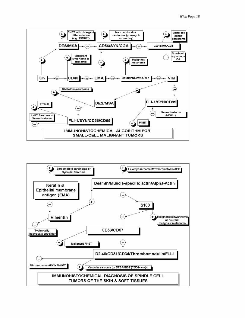
and selected other hematopoietic elements (67). This marker is highly restricted to endothelial neoplasms among all tumors of the soft tissue. In our hands, virtually 100% of angiosarcomas are CD31-positive, regardless of grade or subtype (71), and the same statement applies to hemangioma and hemangioendothelioma variants. It must be acknowledged, however, that Kaposi's sarcoma is labeled more consistently for CD34 than for CD31. Thrombomodulin (CD141) Thrombomodulin (CD141) is a 75-kDa membrano-cytoplasmic glycoprotein that is distributed among endothelial cells, mesothelial cells, osteoblasts, mononuclear phagocytic cells, and selected epithelia (72). CD141 may be present in some metastatic carcinomas and is seen in most mesotheliomas (73), both of which may be confused with epithelioid angiosarcomas. Therefore, it should be interpreted with that caveat in mind and in the context of staining results for other vascular markers. Nevertheless, CD141 has proved to be a highly sensitive indicator of endothelial differentiation, particularly in poorly-differentiated vascular malignancies. Kaposi's sarcoma is likewise consistently immunoreactive for this determinant (74). CD117 CD117 is a cell-membrane determinant that is also known as c-kit protein or Steele factor. It is potentially expressed by a wide variety of tumor types, including gastrointestinal stromal tumors (GISTs), mast-cell neoplasms, seminomas, small-cell carcinomas, primitive neuroectodermal tumors, granulocytic sarcomas, and melanomas (75). Among those lesions, CD117 probably has its greatest diagnostic use in the context of spindle-cell mesenchymal neoplasms because of the relative specificity of that marker for GIST (76). Podoplanin Podoplanin is recognized by antibody D2-40. It is a mucin-type glycoprotein that is principally localized to cell membranes of endothelia, mesothelium, osteocytes, glandular myoepithelial cells, ependyma, and reticulum-dendritic cells of lymphoid organs (77). In the context of undifferentiated or morphologically-ambiguous neoplasms, podoplanin is best used as an adjunct marker for angiosarcoma and mesothelioma. In particular, we have found that it is superior to calretinin in labeling sarcomatoid mesotheliomas (78). Finally, podoplanin appears to be an effective discriminant between seminoma—which is immunoreactive for that marker (79)—and embryonal carcinoma, which is generally D2-40-negative. APPROACHES TO GENERIC CLASSIFICATION OF SOLID TUMORS Small Round Cell Neoplasms Small cell neoplasms that cause potential diagnostic confusion include rhabdomyosarcoma, Ewing's sarcoma, neuroblastoma, PNET (peripheral neuroepithelioma), malignant lymphoma, and small cell carcinomas. The anatomic locations and microscopic details of these tumors and other aspects of their clinical presentations have a strong bearing on the relative likelihood of respective diagnoses, and immunohistochemical analyses may be tailored according to such considerations. Small-Cell Tumors of Soft Tissue Rhabdomyosarcomas are typified by their immunoreactivity for desmin and muscle-specific actin. When used in combination, antibodies to these determinants yield virtually absolute sensitivity for myogenic sarcomas and allow for exclusion of other diagnostic possibilities (65). Similarly, small cell lymphomas exhibit CD45 in a uniform manner, whereas other neoplasms in this category are devoid of reactivity (12), with extremely rare exceptions (80). Neuroblastomas and neuroectodermal tumors may express synaptophysin, CD56, and NB34 (81). Rhabdomyosarcomas also share potential reactivity for CD56, but, excluding rare examples of
Wick Page 8
PNET with divergent differentiation (82), primitive striated muscle sarcomas are distinguished from small cell neurogenic neoplasms by their desmin or actin positivity as mentioned above. A particularly diagnostically challenging member of this tumor group is, in fact, Ewing's sarcoma/PNET. Our approach is to make this unqualified diagnosis when a small cell neoplasm of soft tissue is reactive for CD99, with or without vimentin, synaptophysin, or CD56, and in the absence of desmin, actin, S100 protein, and CD45. Some PNETs may also synthesize keratin in an aberrant fashion (81), and, as mentioned earlier, roughly 20% are NB84-positive. A special variant of PNET is represented by desmoplastic small round-cell tumor, inasmuch as it regularly demonstrates coexpression of cytokeratin and desmin (64c). Small-Cell Carcinomas Small cell carcinomas may exhibit neuroendocrine, squamous, or glandular differentiation. However, a determination of cell lineage is often difficult on conventional microscopy of these lesions, prompting the use of discriminating immunostains. CD56, synaptophysin, and CGA are restricted to neuroendocrine neoplasms in this morphologic class of lesions; nevertheless, it should be reemphasized that they are seen in only 30-50% of cases (83). Thus, most of the time, immunohistologic studies will not successfully corroborate a neuroendocrine lineage for small cell carcinoma even though it is really present, and one could seriously question whether or not such analyses are cost-effective. On the other hand, small cell adenocarcinomas usually display MOC-31, whereas small cell squamous tumors do not. All three forms of small cell carcinoma express cytokeratin reactivity, separating them from most small cell sarcomas of soft tissue. There is some potential overlap between PNET and small cell neuroendocrine carcinoma in regard to the possibility of conjoint staining for CD99 and keratin in those neoplasms (64). Nevertheless, PNET typically shows only scattered keratin-reactivity in the tumor cells, and it is diffusely distributed throughout the cytoplasm. In contrast, small-cell carcinomas are globally keratin-positive and staining for that protein typically manifests a perinuclear “dot”-like pattern (55). Spindle-Cell Neoplasms Spindle cell ("sarcomatoid") carcinomas have been described in a diversity of organs, including the skin, aerodigestive tract, kidney, bladder, female genital tract, lung, and other sites, and must be distinguished from melanomas and true sarcomas in such locations (84. In addition, spindle cell mesenchymal malignancies of soft tissue can be difficult to separate from one another diagnostically. Spindle-Cell Carcinomas In mucosal or organ-based locations, any spindle cell tumor that displays reactivity for cytokeratin or EMA can be defined as carcinomatous (84). Most sarcomatoid carcinomas also coexpress vimentin, but the latter protein should be regarded as nonspecific when seen in conjunction with any other intermediate filament. Spindle-Cell Amelanotic Malignant Melanomas Melanomas that are composed exclusively of fusiform cells retain the general immunohistochemical attributes of other malignant melanocytic neoplasms, with one exception. For unknown reasons, spindle cell melanomas react poorly or not at all with HMB-45, PLN2, MART-1, and antityrosinase, making the conjoint use of antibodies to S100, vimentin, EMA, and cytokeratin paramount in this diagnostic context (85). Positivity for either of the last two of these determinants excludes the possibility of malignant melanoma. Spindle-Cell Sarcomas of Soft Tissue Malignant spindle cell neoplasms of soft tissue include fibrosarcoma, leiomyosarcoma, malignant peripheral nerve sheath tumor, monophasic synovial sarcoma, malignant fibrous
Wick Page 9
histiocytoma (MFH), gastrointestinal stromal tumor (GIST), and Kaposi's sarcoma (70). Among these, fibrosarcoma is characterized by its reactivity for vimentin, to the exclusion of desmin, actin, S100 protein, CD56, cytokeratin, and EMA. In addition, it lacks endothelial markers. Leiomyosarcomas are unique in their diffuse reactivity for desmin and muscle-specific actin but may also express S100 protein and CD57. Malignant peripheral nerve sheath tumors generally demonstrate only the last two of these four determinants, and GIST expresses CD117 with or without CD34. Synovial sarcoma represents the sole lesion in this category that is capable of cytokeratin and EMA synthesis; it may also express CD99 (7). The immunohistologic features of Kaposi's sarcomas are controversial. Some investigators have obtained consistent labeling of such neoplasms for CD34 or CD3l (67), whereas others have observed this finding to be less than universal. Fortunately, conventional histologic analysis and clinical correlation usually allow for confident identification of that lesion. Epithelioid (Large-Polygonal Cell) Neoplasms Metastatic carcinoma and melanoma, large cell malignant lymphoma, "syncytial" Hodgkin's disease, epithelioid sarcoma, clear cell sarcoma, epithelioid angiosarcoma, epithelioid leiomyosarcoma, and epithelioid malignant peripheral nerve sheath tumor are the differential diagnostic possibilities when dealing with polygonal cell malignancies in lymph nodes and soft tissues. Lymph Nodal Lesions In lymph nodes, epithelioid neoplasms most commonly represent large cell lymphomas or metastases of visceral carcinomas and malignant melanomas. The separation of these differential diagnostic possibilities principally involves the use of antibodies to CD45, cytokeratin, EMA, and S100 protein. Carcinomas are reactive for cytokeratin, with or without EMA, whereas only lymphomas exhibit CD45 (12). It is important to avoid equating CD15 reactivity with the presence of Reed-Sternberg cells, because anaplastic carcinomas and anaplastic non-Hodgkin lymphomas may also display that determinant (35,36); moreover, like Hodgkin lymphomas, the latter two tumor types lack CD45. Similarly, EMA is rarely present in some Hodgkin's and non-Hodgkin's lymphomas (86). Malignant melanomas and some poorly differentiated carcinomas exhibit S100 protein, but only the latter tumors generally express cytokeratin. Soft Tissue Lesions An epithelioid malignancy of soft tissue is relatively unlikely to represent metastatic carcinoma or melanoma, unless widespread dissemination by those neoplasms is clinically obvious. Similarly, primary non-Hodgkin's lymphomas of soft tissues do exist but they are quite rare. Indeed, in this tissue compartment, epithelioid sarcoma, epithelioid angiosarcoma, epithelioid malignant Schwannoma, epithelioid leiomyosarcoma, and clear cell sarcoma are the principal differential diagnostic considerations (7). Epithelioid sarcoma is a polyphenotypic neoplasm that may express vimentin, S100 protein, and even desmin, overlapping with the immunohistologic attributes of other mesenchymal tumors (87-89). However, it is unique among polygonal cell sarcomas in its consistent expression of cytokeratin; EMA reactivity is also observed in 75% of epithelioid sarcomas (87). Epithelioid angiosarcoma exhibits vimentin positivity, reactivity for CD31, thrombomodulin, and CD34 (68,70,71,73). Its immunoprofile usually includes a lack of keratin positivity, but some examples of epithelioid angiosarcoma have demonstrated aberrant keratin expression (90). Clear cell sarcoma is the only one of these lesions that labels for HMB-45, PNL2, and tyrosinase. It displays S100 protein and vimentin as well, but lacks cytokeratin, EMA, desmin,
Wick Page 10
CD31, and CD34 (7,89). HMB-45, tyrosinase, and PNL2 are important in distinguishing between clear cell sarcoma and epithelioid malignant peripheral nerve sheath tumors, which are typically S100 protein-positive but nonreactive for more specialized melanocytic markers (91). Lastly, only epithelioid leiomyosarcoma expresses desmin and muscle-specific actin among the large polygonal-cell sarcomas. Another possible diagnostic entity in this category is GIST, which may assume the form of an undifferentiated polyhedral-cell neoplasm. It is distinctive in its immunoreactivity for CD117, with or without CD34 (92). Pleomorphic Neoplasms Tumors that are capable of assuming extremely bizarre, pleomorphic cellular shapes include malignant fibrous histiocytoma (MFH), pleomorphic liposarcoma, pleomorphic rhabdomyosarcoma, "dedifferentiated" sarcomas of various types, malignant peripheral nerve sheath tumor, and metastatic carcinomas or melanomas. Metastatic Carcinomas and Melanomas Metastases of carcinomas and melanomas with a pleomorphic microscopic appearance are delineated by their reactivity for cytokeratin or melanocyte-selective markers, respectively. These immunophenotypes are not shared by any other pleomorphic malignancies. Pleomorphic Soft Tissue Tumors Malignant peripheral nerve sheath tumors are unique among pleomorphic soft tissue sarcomas, because they are the only lesions in this class that are capable of diffuse expression of S100 protein, with or without CD56 (7). Pleomorphic rhabdomyosarcomas are globally positive for desmin and muscle-specific actin; in addition, they express myoglobin or myogenin, both of which are proteins that are restricted to striated muscle (7,70). Of the latter, myogenin is strictly limited to the nuclei. Although it is a relatively insensitive marker in other forms of rhabdomyosarcoma, myoglobin assumes some importance among pleomorphic neoplasms, because "dedifferentiated" leiomyosarcomas also exhibit desmin and actin reactivity but are devoid of myoglobin (7). Pleomorphic MFH lacks all antigens except vimentin; pleomorphic liposarcoma differs from this profile in showing focal Sl00 protein positivity in signet ring or multivacuolated tumor lipoblasts. Mesotheliomas vs. Simulants Both in the pleura and the peritoneum, malignant mesotheliomas can be confused with metastatic carcinoma or melanoma in the small-cell, spindle-cell, epithelioid, or pleomorphic subgroups (93). Moreover, primary serosal sarcomas also enter differential diagnosis (94). In these contexts, mesotheliomas have a distinctive immunophenotype that separates them from other possible diagnostic entities. One sees global reactivity for keratin, and the majority of mesothelial tumors also express calretinin, thrombomodulin, or podoplanin. In contrast, they lack MOC31, CEA, CD15, PLAP, S100 protein, CD34, CD31, specialized melanocytic markers, and neuroendocrine-neuroectodermal determinants (95,96). CONCLUSION Throughout this review, an integrated multiparametric approach to the immunohistologic diagnosis of poorly differentiated and morphologically ambiguous malignant tumors has been emphasized. This is indeed an effective process, as reflected by its beneficial results in many medical centers. Nonetheless, one should not lose sight of the fact that surgical pathology is still based on skill in morphologic interpretation; one cannot achieve success as a diagnostic immunohistochemist without having a sound foundation in histopathologic analysis. Also, we do not mean to imply that other methods of detailed pathologic assessment- histochemistry and electron microscopy- have been relegated to a historical perspective. They are still valuable in the
Wick Page 11
evaluation of selected neoplasms and often supply information that is synergistic with that of immunohistology. Also, immunohistochemistry is not itself being outmoded by more "molecular" techniques, which are being increasingly integrated currently into surgical pathology. One should rather anticipate that all these methods will be employed selectively in the future at the discretion of the pathologist, to provide rapid and comprehensive solutions for problem cases.
The current status of diagnostic immunohistology is in flux, and probably always will be, as
more protein markers of cellular differentiation become available for clinical use. My belief is that the algorithmic approach to diagnosis presented herein will soon become the cornerstone of immunohistochemistry in anatomic pathology, not only in regard to undifferentiated malignancies but in reference to other tumor classes as well. That is true because this method compensates for biological variation in a logical fashion. In the next decade, pathologists will hopefully see more uniform standardization of technique, with more consistency in the performance of antibody reagents. Moreover, we must strive to make validation of technique (e.g., of diagnostic immunohistochemistry (DIHC) by in-situ hybridization or gene profiling) a uniform part of quality assurance in the clinical laboratory. As mentioned above, it is unlikely that DIHC will be supplanted as the principal means of studying undifferentiated neoplasms, because it represents the most rapid and cost-effective method for melding morphologic analysis with evaluation of neoplastic protein expression.
Wick Page 12
REFERENCES 1. Miettinen, M. (1993) Keratin immunohistochemistry: update on applications and pitfalls. Pathol. Annu. 28, 113-143. 2. Battifora, H. (1988) Diagnostic uses of antibodies to keratins. Prog. Surg. Pathol. 8, 1-16. 3. Chan, J. K. C., Suster, S., Wenig, B. M., Tsang, W. Y., Chan, J. B., and Lau, A. L. (1997) Cytokeratin 20 immunoreactivity distinguishes Merkel cell (primary cutaneous neuroendocrine) carcinomas and salivary gland small cell carcinomas from small cell carcinomas of various sites. Am. J. Surg. Pathol. 21, 226-234. 4. Pruss, R. M., Mirsky, R., Raff, M. C., Thrope, R., Dowling, A. J., and Anderton, B. H. (1981) All classes of intermediate filaments share a common antigenic determinant defined by a monoclonal antibody. Cell 27, 419-428. 5. Caselitz, J., Janner, M., Breitbart, E., Weber, K., and Osborn, M. (1983) Malignant melanomas contain only the vimentin type of intermediate filaments. Virchows Arch. A. 400,43-51. 6. Gustmann, C., Altmannsberger, M., Osborn, M., Griesser, H., and Feller, A. C. (1991) Cytokeratin expression and vimentin content in large cell anaplastic lymphomas and other non-Hodgkin's lymphomas. Am. J. Pathol. 138, 1413-1422. 7. Swanson, P. E., and Wick, M. R. (1995) Soft tissue tumors, in Diagnostic Immunopathology, 2nd ed. (Colvin, R., Bhan, A., and McCluskey, R., eds.), Lippincott-Raven, Philadelphia, pp. 599-632. 8. Battifora, H. (1991) Assessment of antigen damage in immunohistochemistry: the vimentin internal control. Am. J. Clin. Pathol. 96, 669-671. 9. Truong, L. D., Rangdaeng, S., Cagle, P. T., Ro, J. Y., Hawkins, H., and Font, R. L. (1990) The diagnostic utility of desmin: a study of 584 cases and review of the literature. Am. J. Clin. Pathol. 93, 305-314. 10. Trojanowski,J. Q., Lee, V. M. Y., and Schlaepfer, W. W. (1984) An immunohistochemical study of human central and peripheral nervous system tumors with monoclonal antibodies against neurofilaments and glial filaments, Hum. Pathol. 15, 248-257. 11. Morrison, C. D. and Prayson, R. A. (2000) Immunohistochemistry in the diagnosis of neoplasms of the central nervous system Semin. Diagn. Pathol. 17, 204-215. 12. Kurtin, P. J. and Pinkus, G. S. (1985) Leukocyte common antigen-a diagnostic discriminant between hematopoietic and nonhematopoietic neoplasms in paraffin sections using monoclonal antibodies: correlation with immunologic studies and ultrastructural localization. Hum. Pathol. 16, 353-365. 13. Wick, M. R. (1988) Monoclonal antibodies to leukocyte common antigen, in Monoclonal Antibodies in Diagnostic Immunohistochemistry (Wick, M. R. and Siegal, G. P., eds.), Marcel Dekker, New York, pp. 285-307. 14. Heyderman, E., Steele, K., and Ormerod, M. G. (1979) A new antigen on the epithelial membrane: its immunoperoxidase localization in normal and neoplastic tissue. J. Clin. Pathol. 32, 35-44. 15. Sloane, J. P. and Ormerod, M. G. (1981) Distribution of epithelial membrane antigen in normal and neoplastic tissues and its value in diagnostic pathology, Cancer 47, 178-185. 16. Pinkus, G. S. and Kurtin, P. J. (1985) Epithelial membrane antigen- a diagnostic discriminant in surgical pathology. Hum. Pathol. 16, 929-938. 17. Swanson, P. E., Manivel, J. C., Scheithauer, B. W., and Wick, M. R. (1989) Epithelial membrane antigen in human sarcomas: anirnmunohistochemical study. Surg. Pathol. 2,313-322.
Wick Page 13
18. DeLellis RA, Dayal Y (1987): The role of immunohistochemistry in the diagnosis of poorly-differentiated malignant neoplasms. Semin. Oncol. 14, 173-192. 19. Wick, M. R., Swanson, P. E., and Manivel, J. C. (1987) Placental-like alkaline phosphatase reactivity in human tumors: an immunohistochemical study of 520 cases. Hum. Pathol. 18, 946-954. **20. Elgin, J., Phillips, J. G., Reddy, V. V., Gibbs, P.O., and Listinsky, C. M. (1999) Hodgkin's and non-Hodgkin's lymphoma: spectrum of morphologic and immunophenotypic overlap. Ann. Diagn. Pathol. 3, 263-275. (This reference emphasizes the morphological and immunohistological difficulties in separating non-Hodgkin from Hodgkin lymphoma in selected cases) 21. Salter, D. M., Krajewski, A. S., Miller, E. P., and Dewar, A. E. (1985) Expression of leukocyte common antigen and epithelial membrane antigen in plasmacytic malignancies. J. Clin. Pathol. 38, 843-844. 22. Schnitt, S. J. and Vogel, H. (1986) Meningiomas: diagnostic value of immunoperoxidase staining for epithelial membrane antigen. Am. J. Surg. Pathol. 10, 640-649. 23. Ruitenbeek, C. T., Gouw, A. S. H., and Poppema, S. (1994) Immunocytology of body cavity fluids: MOC-31, a monoclonal antibody discriminating between mesothelial and epithelial cells. Arch. Pathol. Lab. Med. 118, 265-269. 24. Sosolik, R. C., McGaughy, V. R., and DeYoung, B. R. (1997) Anti-MOC31: a potential addition to the pulmonary adenocarcinoma versus mesothelioma immunohistochemistry panel. Mod. Pathol. 10,716-719. 25. Chenard-Neu, M. P., Kabou, A., Mechine, A., Brolly, F., Orion, B., and Bellocq, J. P. (1998) Immunohistochemistry in the differential diagnosis of mesothelioma and adenocarcinoma: evaluation of 5 new antibodies and 6 traditional antibodies. Ann. Pathol. 18,460-465. 26. DeYoung, B. R. and Wick, M. R. (2000) Immunohistologic evaluation of metastatic carcinomas of unknown origin: an algorithmic approach. Semin. Diagn. Pathol. 17, 184-193. 27. Bollinger DJ, Wick MR, Dehner LP, et al.: Peritoneal malignant mesothelioma versus serous papillary adenocarcinoma: a histochemical and immunohistochemical comparison. Am J Surg Pathol 1989; 13: 659-670. 28. Manivel, J. C., Jessurun, J., Wick, M. R., et al. (1987) Placental alkaline phosphatase immunoreactivity in testicular germ cell neoplasms. Am. J. Surg. Pathol. 11,21-30. 29. Niehans, G. A., Manivel, J. C., Copland, G. T., et al. (1988) Immunohistochemistry of germ cell and trophoblastic neoplasms. Cancer 62, 1113-1123. 30. Suster, S., Moran, C. A., Dominguez-Malagon, H., and Quevedo-Blanco, P. (1998) Germ cell tumors of the mediastinum and testis: a comparative immunohistochemical study of 120 cases. Hum. Pathol. 29,737-742. 31. Cheville, J. C., Rao, S., Iczkowshi, K. A., Lohse, C. M., and Pankratz, V. S. (2000) Cytokeratin expression in seminoma of the human testis. Am. J. Clin. Pathol. 113,583-588. 32. Colcher, D., Hand, P. H., Nuti, M., and Schlom, J. (1983) Differential binding to human mammary and nonmammary tumors of monoclonal antibodies reactive with carcinoembryonic antigen. Cancer Invest. 1, 127-138. 33. Wick, M. R. (1988) Monoclonal antibodies to carcinoembryonic antigen, in Monoclonal Antibodies in Diagnostic Immunohistochemistry (Wick, M. R. and Siegal, G. P., eds.), Marcel Dekker, New York, pp. 539-567.
Wick Page 14
34. Sheahan, K., O'Brien, M. J., Burke, B., et al. (1990) Differential reactivities of carcinoembronic antigen (CEA) and CEA-related monoclonal and polyclonal antibodies in common epithelial malignancies. Am. J. Clin. Pathol. 94, 157-164. 35. Sheibani, K., Battifora, H., Burke, J. S., and Rappaport, H. (1986) Leu-M 1 antigen in human neoplasms: an immunohistologic study of 400 cases. Am. J. Surg. Pathol. 10, 227-236. 36. Arber, D. A. and Weiss, L. (1993) CD15: a review. Appl. Immunohistochem. 1, 17-30. 37. Wick, M. R., Mills, S. E., and Swanson, P. E. (1990) Expression of "myelomonocytic" antigens in mesotheliomas and adenocarcinomas involving the serosal surfaces. Am. J. Clin. Pathol. 94, 18-26. 38. Al-Shamkhani A (2004): The role of CD30 in the pathogenesis of hematopoietic malignancies. Curr. Opin. Pharmacol. 4, 355-359. **39. Ulbright TM (2005): Germ cell tumors of the gonads: a selective review emphasizing problems in differential diagnosis, newly-appreciated, and controversial issues. Mod. Pathol. 10 (Suppl), S61-S79. (This review considers the major diagnostic features and morphologic pitfalls in the diagnosis of germ-cell tumors with potentially-undifferentiated appearances). 40. Dei Tos, A. P. and Doglioni, C. (1998) Calretinin: a novel tool for diagnostic immunohistochemistry. Adv. Anat. Pathol. 5, 61-66. 41. Gotzos, V., Wintergerst, E. S., Musy, J. P., Spichtin, H. P., and Genton, C. Y. (1999) Selective distribution of calretinin in adenocarcinomas of the human colon and adjacent tissues. Am. J. Surg. Pathol. 23,701-711. 42. Chhieng, D. C., et al. (2000) Calretinin staining pattern aids in the differentiation of mesothelioma from adenocarcinoma in serous effusions. Cancer 90, 194-200. 43. Kitazume, H., et al. (2000) Cytologic differential diagnosis among reactive mesothelial cells, malignant mesothelioma, and adenocarcinoma: utility of combined E-cadherin and calretinin immunostaining. Cancer 90, 55-60. 44. Ordonez, N. G. (1998) Value of calretinin immunostaining in differentiating epithelial mesothelioma from lung adenocarcinoma. Mod. Pathol. 11, 929-933. 45. Atanoos, R. L., Dojcinov, S. D., Webb, R., and Gibbs, A. R. (2000) Antimesothelial markers in sarcomatoid mesothelioma and other spindle cell neoplasms. Histopathology 37,224-231. 46. Drier, J., Swanson, P. E., Cherwitz, D. L., and Wick, M. R. (1987) S100 protein immunoreactivity in poorly-differentiated carcinomas: immunohistochemical comparison with malignant melanoma. Arch. Pathol. Lab. Med. 111, 447-452. 47. Wick, M. R., Ockner, D. M., Mills, S. E., Ritter, J. H., and Swanson, P. E. (1998) Homologous carcinomas of the breasts, salivary glands, and skin. Am. J. Clin. Pathol. 109, 75-84. 48. Matsushima, S., Mori, M., Adachi, Y., Matsukuma, A., and Sugimachi, K. (1994) S100 protein-positive breast carcinomas: an immunohistochemical study. J. Surg. Oncol. 55, 108-113. 49. Loeffel, S. C., Gillespie, G. Y., Mirmiran, A., Sawhney, D., Askin, F. B., and Siegal, G. P. (1985) Cellular immunolocalization of S100 protein within fixed tissue sections by monoclonal antibodies. Arch. Pathol. Lab. Med. 109, 117-122. **50. Yaziji H, Gown, A. M. (2003): Immunohistochemical markers of melanocytic tumors. Int. J. Surg. Pathol. 11, 11-15. (This paper is a practical and yet comprehensive review of commonly-used immunohistochemical markers for melanocytic neoplasms.) 51. Rochaix P, Lacroix-Triki M, Lamant L, et al.: PNL2, a new monoclonal antibody directed against a fixative-resistant melanocyte antigen. Mod. Pathol. 16, 481-490.
Wick Page 15
52. Kaufmann, 0., Koch, S., Burghardt, J., Andring, H., and Dietel, M. (1998) Tyrosinase, Melan-A, and KBA62 as markers for the immunohistochemical identification of metastatic amelanotic melanomas on paraffin sections. Mod. Pathol. 11,740-746. 53. Lloyd, R. V. and Wilson, B. S. (1983) Specific endocrine tissue marker defined by a monoclonal antibody. Science 222, 628-630. 54. Wilson, B. S. and Lloyd, R. V. (1984) Detection of chromogranin in neuroendocrine cells with a monoclonal antibody. Am. J. Pathol. 115, 458-468. **55. Wick, M. R. (2000) Immunohistology of neuroendocrine and neuroectodermal tumors. Semin. Diagn. Pathol. 17, 194-203. (This review outlines the typical immunoprofiles of neuroendocrine carcinomas and non-epithelial neuroectodermal neoplasms.) 56. Miettinen, M. (1987) Synaptophysin and neurofilament protein as markers for neuroendocrine tumors. Arch. Pathol. Lab. Med. 111, 813-818. 57. Gould, V. E., Lee, I., Wiedenmann, B., et al. (1986) Synaptophysin: a novel marker for neurons, certain neuroendocrine cells, and their neoplasms. Hum. Pathol. 17, 979-983. 58. Buffa, R., Rindi, G., Sessa, F., et al. (1987) Synaptophysin immunoreactivity and small clear vesicles in neuroendocrine cells and related tumors. Mol. Cell. Probes 1,367-381. 59. Stirdsberg, M. (1995) The use of chromogranin, synaptophysin, and islet amyloid polypeptide as markers for neuroendocrine tumors. Ups. J. Med. Sci. 100, 169-199. 60. Miettinen M (1993): Immunohistochemistry in tumor diagnosis. Ann. Med. 25, 221-233. 61. Assaf C, Gellrich S, Whittaker S, et al. (2006): CD56+ lymphoproliferative disorders of the skin. J. Clin. Pathol. [Online] doi:10.1136/jcp.2006.042135 (Accessed 6/25/07). 62. Smith MJ, Goodfellow PJ, Goodfellow PN (1993): The genomic organization of the human pseudoautosomal gene MIC2 and the detection of a related locus. Hum. Mol. Genet. 2, 417-422. 63. Dehner, L. P. (1993) Primitive neuroectodermal tumor and Ewing's sarcoma. Am. J. Surg. Pathol. 17, 1-13. 64. Lumadue, J. A., Askin, F. B., and Perlman, E. J. (1994) MIC2 analysis of small cell carcinoma. Am. J. Clin. Pathol. 102, 692-694. 64a. Zhang L, Eddy A, Teng YT, et al.: An immunological renal disease in transgenic mice that overexpress FLI-1, a member of the ETS familiy of transcription factor genes. Mol. Cell Biol. 1995; 15: 6961-6970. 64b. Sebire NJ, Gibson S, Rampling D, et al.: Immunohistochemical findings in embryonal small round cell tumors with molecular diagnostic confirmation. Appl Immunohistochem Mol Morphol 2005; 13: 1-5. 64c. Rossi S, Orvieto E, Furlanetto A, et al.: Utility of the immunohistochemical detection of FLI-1 expression in round-cell and vascular neoplasms using a monoclonal antibody. Mod. Pathol. 2004; 17: 547-552. 64d. Kumar S, Perlman E, Pack S, et al.: Absence of EWS/FLI1 fusion in olfactory neuroblastomas indicates these tumors do not belong to the Ewing’s sarcoma family. Hum. Pathol. 1999; 30: 1356-1360. 64e. Thomas JO, Nijjar J, Turley H, Micklem K, Gatter KC: NB84: a new monoclonal antibody for the recognition of neuroblastoma in routinely-processed material. J. Pathol. 1991; 163: 69-75. 64f. Miettinen M, Chatten J, Paetau A, Stevenson A: Monoclonal antibody NB84 in the differential diagnosis of neuroblastoma and other small round-cell tumors. Am. J. Surg. Pathol. 1998; 22: 327-332.
Wick Page 16
65. Tsukada, T., McNutt, M. A., Ross, R., and Gown, A. M. (1987) HHF35, a muscle actin-specific monoclonal antibody. II. Reactivity in normal, reactive, and neoplastic human tissues. Am. J. Pathol. 127, 389-402. 66. Rangdaeng, S. and Truong, L. D. (1991) Comparative immunohistochemical staining for desmin and muscle-specific actin: a study of 576 cases. Am. J. Clin. Pathol. 96, 32- 45. 67. Miettinen, M., Lindenmayer, A. E., and Chanbal, A. (1994) Endothelial cell markers CD31, CD34, and BNH9 antibody to H- and Y - antigens: evaluation of their specificity and sensitivity in the diagnosis of vascular tumors and comparison with von Wi11ebrand factor. Mod. Pathol. 7,82-90. 68. Traweek, S. T., Kandalaft, P. L., Mehta, P., and Battifora, H. (1991) The human hematopoietic progenitor cell antigen (CD34) in vascular neoplasms. Am. J. Clin. Pathol. 96, 25-31. 69. Cohen, P. R., Rapini, R. P., and Farhood, A. I. (1993) Expression of the human hematopoietic progenitor cell antigen CD34 in vascular and spindle cell tumors. J. Cutan. Pathol. 20, 15-20. **70. Suster, S. (2000) Recent advances in the application of immunohistochemical markers for the diagnosis of soft tissue tumors. Semin. Diagn. Pathol. 17, 225-235. (This paper is an excellent review of immunohistochemistry as applied to mesenchymal malignancies.) 71. DeYoung, B. R., Wick, M. R., Fitzgibbon, J. F., et al. (1993) CD31: an immunospecific marker for endothelial differentiation in human neoplasms. Appl. Immunohistochem. 1,97-100. 72. Karmochkine, M. and Boffa, M. C. (1997) Thrombomodulin: physiology and clinical applications. Rev. Med. Interne 18, 119-125. 73. Ordonez, N. G. (1997) Value of thrombomodulin immunostaining in the diagnosis of mesothelioma. Histopathology 31, 25-30. 74. Appleton, M. A., Attanoos, R. L., and Jasani, B. (1996) Thrombomodulin as a marker of vascular and lymphatic tumors. Histopathology 29, 1531-157. 75. Tsuura Y, Hiraki H, Watanabe K, et al. (1994): Preferential localization of c-kit product in tissue mast cells, basal cells of skin, epithelial cells of breast, small cell lung carcinoma and seminoma/dysgerminoma in humans: immunohistochemical study on formalin-fixed, paraffin-embedded tissues. Virchows Arch. 424,135-141. 76. Hornick JL, Fletcher CDM (2002): Immunohistochemical staining for KIT (CD117) in soft tissue sarcomas is very limited in distribution. Am. J. Clin. Pathol. 117, 188-193. 77. Ordonez NG (2006): Podoplanin: a novel diagnostic immunohistochemical marker. Adv. Anat. Pathol. 13, 83-88. 78. Padgett DM, Cathro HC, Wick MR, Mills SE (In press, 2007): Podoplanin as a marker for sarcomatoid mesothelioma. Am. J. Surg. Pathol. 79. Lau SK, Weiss LM, Chu PG (2007): D2-40 immunohistochemistry in the differential diagnosis of seminoma and embryonal carcinoma: a comparative immunohistochemical study with KIT (CD117) and CD30. Mod. Pathol. 20, 320-325. 80. Nandedkar, M. A., Palazzo, J., Abbondanzo, S. L., Lasota, J., and Miettinen, M. (1998) CD45 (leukocyte common antigen) immunoreactivity in metastatic undifferentiated and neuroendocrine carcinoma: a potential diagnostic pitfall. Mod. Pathol. 11, 1204-1210. **81. Devoe, R. and Weidner, N. (2000) Immunohistochemistry of small round-cell tumors. Semin. Diagn. Pathol. 17,216-224. (This review is a concise and yet comprehensive summary of immunohistochemical findings in small round-cell malignancies.) 82. Parham, D. M., Dias, P., Kelly, D. R., Rutledge, J. C., and Houghton, P. (1992) Desmin-positivity in primitive neuroectodermal tumors of childhood. Am. J. Surg. Pathol. 16,483-492.
Wick Page 17
83. Guinee, D. G., Jr., Fishback, N. F., Koss, M. N., et al. (1994) The spectrum ofimmunohistochemical staining of small cell lung carcinoma in specimens from transbronchial and open-lung biopsies. Am. J. Clin. Pathol. 102, 406-414. 84. Wick, M. R. and Swanson, P. E. (1993) "Carcinosarcomas"-current perspectives and an historical review of nosological concepts. Semin. Diagn. Pathol. 10, 118-127. 85. Wick, M. R., Fitzgibbon, J. F., and Swanson, P. E. (1993) Cutaneous sarcomas and sarcomatoid neoplasms of the skin. Semin. Diagn. Pathol. 10, 148-158. 86. Elgin, J., Phillips, J. G., Reddy, V. V., Gibbs, P.O., and Listinsky, C. M. (1999) Hodgkin's and non-Hodgkin's lymphoma: spectrum of morphologic and immunophenotypic overlap. Ann. Diagn. Pathol. 3, 263-275. 87. Manivel, J. C., Wick, M. R., Dehner, L. P., and Sibley, R. K. (1987) Epithelioid sarcoma: an immunohistochemical study. Am. J. Clin. Pathol. 87, 319-326. 88. Laskin WB, Miettinen M (2003): Epithelioid sarcoma: new insights based on an extended immunohistochemical analysis. Arch. Pathol. Lab. Med. 127, 1161-1168. 89. Papas-Corden, P. C., Zarbo, R. J., Gown, A. M., and Crissman, J. D. (1989) Immunohistochemical characterization of synovial, epithelioid, and clear-cell sarcomas. Surg. Pathol. 2, 43-58. 90. Swanson, P. E., Dehner, L. P., Sirgi, K. E., and Wick, M. R. (1994) Cytokeratin-immunoreactivity in malignant tumors of bone and soft tissue. Appl. lmmunohistochem. 2,103-112. 91. Wick, M. R., Swanson, P. E., Scheithauer, B. W., and Manivel, J. C. (1987) Malignant peripheral nerve sheath tumor: an immunohistochemical study of 62 cases. Am. J. Clin. Pathol. 87, 425-433. 92. Sarlomo-Rikala, M., Kovatich, A. J., Barusevicius, A., et al. (1998) CDl17: a sensitive marker for gastrointestinal stromal tumors that is more specific than CD34. Mod. Pathol. 11, 728-734. 93. Wick MR, Mills SE (2000): Mesothelial proliferations: an increasing morphologic spectrum. Am. J. Clin. Pathol. 113, 619-622. 94. Miettinen M, Limon J, Niezabitowski A, Lasota J (2001): Calretinin and other mesothelioma markers in synovial sarcoma: analysis of antigenic similarities and differences with malignant mesothelioma. Am. J. Surg. Pathol. 25, 610-617. 95. Moran, C. A., Wick, M. R., and Suster, S. (2000) The role of immunohistochemistry in the diagnosis of malignant mesothelioma. Semin. Diagn. Pathol. 17, 178-183. **96. Ordonez NG (2007): What are the current best immunohistochemical markers for the diagnosis of epithelioid mesothelioma? A review and update. Hum. Pathol. 38, 1-16. (This summary review paper provides a compendium of immunohistochemical data on mesothelioma, by an experienced author.)


![3 LASOP Case 3 2006.ppt [Read-Only]lasop.com/pgs/hdouts/2006-03_Case3.pdfmeningioma (WHO grade II) • Microcystic gliomas • Hemangioblastomas • Myxoid schwannomas. Microcystic](https://static.documents.pub/doc/80x56/5e3bd740d005aa51c76678a8/3-lasop-case-3-2006ppt-read-onlylasopcompgshdouts2006-03case3pdf-meningioma.jpg)