Inside this Issue IMMUNOHISTOCHEMISTRY n e w s Thermo Scientific SP1 and SP 3 Rabbit Monoclonal Antibodies: Real World Advantages in Breast Cancer Analysis, page 2 Comprehensive IHC for Breast Pathology, page 4 Toward Standardization in Immunohistochemistry, page 7 Hadi Yaziji, M.D. Medical Director, Vitro Molecular Laboratories President, Ancillary Pathways Miami, FL Pathology Corner: An Essential Tool in the Diagnosis of Malignant Mesothelioma, page 10 Ferda Filiz, MD QC Scientist, Thermo Fisher Scientific Fremont, CA FEATURE ARTICLE Allen M. Gown, M.D. Medical Director and Chief Pathologist PhenoPath Laboratories Seattle, WA Volume 1, Issue 2 Featuring Rabbit Monoclonal Calponin-1 (RM-2102), E-Cadherin (RM-2100), and more. page 6 New Products: SP 1 Estrogen Receptor Clone: SP1 Cat# RM-9101 SP 3 Her- 2 Clone: SP 3 Cat #: RM- 9 10 3
Transcript
Inside this Issue
IMMUNOHISTOCHEMISTRYn e w s
Thermo Scientific SP1 and SP3 Rabbit Monoclonal Antibodies: Real World Advantages in Breast Cancer Analysis, page 2
Comprehensive IHC for Breast Pathology, page 4
Toward Standardization in Immunohistochemistry, page 7
Hadi Yaziji, M.D.Medical Director, Vitro Molecular LaboratoriesPresident, Ancillary PathwaysMiami, FL
Pathology Corner: An Essential Tool in the Diagnosis of Malignant Mesothelioma, page 10
Ferda Filiz, MDQC Scientist, Thermo Fisher ScientificFremont, CA
FEATURE ARTICLE
Allen M. Gown, M.D. Medical Director and Chief Pathologist PhenoPath Laboratories Seattle, WA
Volume 1, Issue 2
Featuring Rabbit Monoclonal Calponin-1 (RM-2102), E-Cadherin (RM-2100), and more. page 6
New Products:
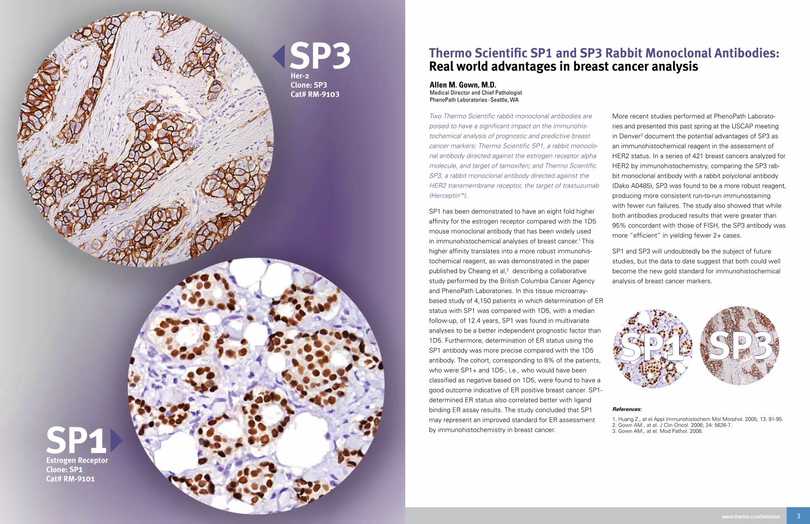
SP1Estrogen Receptor Clone: SP1 Cat# RM-9101
SP3Her-2 Clone: SP3 Cat #: RM-9103
SP3SP1
More recent studies performed at PhenoPath Laborato-
ries and presented this past spring at the USCAP meeting
in Denver3 document the potential advantages of SP3 as
an immunohistochemical reagent in the assessment of
HER2 status. In a series of 421 breast cancers analyzed for
HER2 by immunohistochemistry, comparing the SP3 rab-
bit monoclonal antibody with a rabbit polyclonal antibody
(Dako A0485), SP3 was found to be a more robust reagent,
producing more consistent run-to-run immunostaining
with fewer run failures. The study also showed that while
both antibodies produced results that were greater than
95% concordant with those of FISH, the SP3 antibody was
more “efficient” in yielding fewer 2+ cases.
SP1 and SP3 will undoubtedly be the subject of future
studies, but the data to date suggest that both could well
become the new gold standard for immunohistochemical
analysis of breast cancer markers.
References: 1. Huang Z., at el Appl Immunohistochem Mol Morphol. 2005; 13: 91-95. 2. Gown AM., at el. J Clin Oncol. 2006; 24: 5626-7. 3. Gown AM., at el. Mod Pathol. 2008.
�www.thermo.com/labvision
Two Thermo Scientific rabbit monoclonal antibodies are
poised to have a significant impact on the immunohis-
tochemical analysis of prognostic and predictive breast
cancer markers: Thermo Scientific SP1, a rabbit monoclo-
nal antibody directed against the estrogen receptor alpha
molecule, and target of tamoxifen; and Thermo Scientific
SP3, a rabbit monoclonal antibody directed against the
HER2 transmembrane receptor, the target of trastuzumab
(Herceptin™).
SP1 has been demonstrated to have an eight fold higher
affinity for the estrogen receptor compared with the 1D5
mouse monoclonal antibody that has been widely used
in immunohistochemical analyses of breast cancer.1 This
higher affinity translates into a more robust immunohis-
tochemical reagent, as was demonstrated in the paper
published by Cheang et al,2 describing a collaborative
study performed by the British Columbia Cancer Agency
and PhenoPath Laboratories. In this tissue microarray-
based study of 4,150 patients in which determination of ER
status with SP1 was compared with 1D5, with a median
follow-up, of 12.4 years, SP1 was found in multivariate
analyses to be a better independent prognostic factor than
1D5. Furthermore, determination of ER status using the
SP1 antibody was more precise compared with the 1D5
antibody. The cohort, corresponding to 8% of the patients,
who were SP1+ and 1D5-, i.e., who would have been
classified as negative based on 1D5, were found to have a
good outcome indicative of ER positive breast cancer. SP1-
determined ER status also correlated better with ligand
binding ER assay results. The study concluded that SP1
may represent an improved standard for ER assessment
by immunohistochemistry in breast cancer.
Allen M. Gown, M.D. Medical Director and Chief PathologistPhenoPath Laboratories-Seattle, WA
Thermo Scientific SP1 and SP3 Rabbit Monoclonal Antibodies: Real world advantages in breast cancer analysis
Her-2Clone: SP3Cat# RM-9103
Estrogen Receptor Clone: SP1Cat# RM-9101
SP1
SP3
�www.thermo.com/labvision
�www.thermo.com/labvision
The ability of pathologists to accurately and confidently assess a patient’s tumor is limited by the tests available from their laboratories. To fully understand all of the
diagnostic and prognostic characteristics of a patient’s
tumor, pathologists must have a comprehensive array of
sensitive, specific and accurate tests at their disposal.
As research identifies useful new targets and better
antibodies, laboratories may not always have the best
tests available. To ensure laboratories have the most
advanced technology for their antibody panels we have
arranged our portfolio into panels based on tumor origin.
Utilizing the panel approach, we are systematically
reviewing our clinical portfolio by tumor origin,
incorporating robust clones for each target. Building on
a foundation of rabbit monoclonal antibodies, shown
to be more sensitive and specific than current mouse
monoclonal antibodies, we have finalized the first panel
in our series.1, 3, 5 We are pleased to announce the
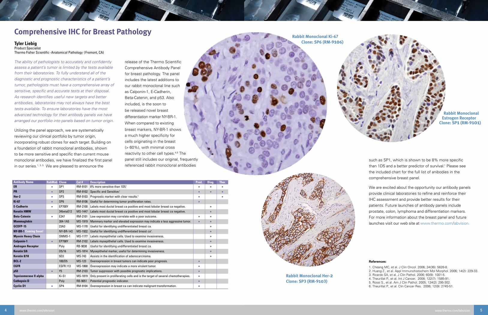
release of the Thermo Scientific
Comprehensive Antibody Panel
for breast pathology. The panel
includes the latest additions to
our rabbit monoclonal line such
as Calponin-1, E-Cadherin,
Beta-Catenin, and p53. Also
included, is the soon to
be released novel breast
differentiation marker NY-BR-1.
When compared to existing
breast markers, NY-BR-1 shows
a much higher specificity for
cells originating in the breast
(> 60%), with minimal cross
reactivity to other cell types.4,6 The
panel still includes our original, frequently
referenced rabbit monoclonal antibodies
Antibody Name RabMab Clone Cat # Description Pred. Diag. Ther.ER • SP1 RM-9101 8% more sensitive than 1D5.1 • • •PR • SP2 RM-9102 Specific and Sensitive.2 • • •Her-2 • SP3 RM-9103 Prognostic marker with clear results.3 • •Ki-67 • SP6 RM-9106 Useful for determining tumor proliferation rates. •E-Cadherin • EP700Y RM-2100 Labels most ductal breast ca positive and most lobular breast ca negative. •Keratin HMW ��betaE12 MS-1447 Labels most ductal breast ca positive and most lobular breast ca negative. •Beta-Catenin • E247 RM-2101 Low expression may correlate with a poor outcome. • •Mammaglobin 304-1A5 MS-1919 Mammary marker and elevated expression may indicate a less aggressive tumor. • •GCDFP-15 23A3 MS-1170 Useful for identifying undifferentiated breast ca. •NY-BR-1 Coming Soon! NY-BR-1#2 MS-1932 Useful for identifying undifferentiated breast ca4. •Myosin Heavy Chain SMMS-1 MS-1177 Labels myoepithelial cells. Used to examine invasiveness. •Calponin-1 • EP798Y RM-2102 Labels myoepithelial cells. Used to examine invasiveness. •Androgen Receptor Poly RB-9030 Useful for identifying undifferentiated breast ca. •Keratin 5/6 D5/16 MS-1814 Myoepithelial marker, useful for determining invasiveness. •Keratin 8/18 5D3 MS-743 Assists in the identification of adenocarcinoma. •BCL-2 100/D5 MS-123 Overexpression in breast tumors can indicate poor prognosis. •EGFR EGFR.113 MS-1868 Overexpression may indicate a more virulent tumor. •p53 • Y5 RM-2103 Tumor suppressor with possible prognostic implications. •Topoisomerase II alpha Ki-S1 MS-1819 Only present in proliferating cells and is the target of several chemotherapies. •Cathepsin D Poly RB-9051 Potential prognostic indicator. •Cyclin D1 • SP4 RM-9104 Overexpression in breast ca can indicate malignant transformation. •
Comprehensive IHC for Breast PathologyTyler LiebigProduct Specialist Thermo Fisher Scientific-Anatomical Pathology ( Fremont, CA)
such as SP1, which is shown to be 8% more specific
than 1D5 and a better predictor of survival.1 Please see
the included chart for the full list of anibodies in the
comprehensive breast panel.
We are excited about the opportunity our antibody panels
provide clinical laboratories to refine and reinforce their
IHC assessment and provide better results for their
patients. Future launches of antibody panels include
prostate, colon, lymphoma and differentiation markers.
For more information about the breast panel and future
launches visit our web site at www.thermo.com/labvision.
“enhancing” reagents, variable types of chromogens, vari-
able counterstains, variable mounting media and variable
interpretation of scoring of the final IHC reaction product by
the pathologist. In case the reader missed the count, the
word “variable” was mentioned twenty-two times in this
paragraph.
To begin with, there is an intrinsic difficulty in achieving
standardized testing in IHC, imposed by the mere nature of
surgical specimen: unlike blood or urine samples, it is
unrealistic to ‘reproduce’ a tissue sample in case the origi-
nal sample fails the test. This fact makes it more difficult to
achieve standardization. One might legitimately ask: why is
standardization needed in IHC? Isn’t the entire interpreta-
tion of H&E stained slides is largely subjective and hasn’t
been standardized in decades? The answer is simple: The
better IHC will evolve as a technology, the more oncolo-
gists will rely on it to obtain information to treat their cancer
patients. In other words, the scope of clinical applications
of IHC in the ‘old days’ has been largely limited to comple-
menting H&E in answering diagnostic questions. Increas-
ingly, however, the previous success of IHC as a reliable
tool for estrogen receptor testing1 has led to a plethora of
IHC predictive assays. It is reasonable to assume that this
trend will continue for the next 10 years and perhaps longer
until other proteomic technologies can reliably replace IHC.
The next logical question is, “Is it possible to achieve
standardization in IHC and how?” The answer, to the best
of my knowledge, is “yes” but with difficulty and a lot of
Towards Standardization in ImmunohistochemistryHadi Yaziji, M.D. Medical Director, Vitro Molecular LaboratoriesPresident, Ancillary Pathways – Miami, FL
Continued on page 8
E-cadherin (uvomorulin,
cell-CAM120/80) is a
calcium-dependent cell
adhesion molecule
expressed predominantly
in epithelial tissues. It
plays an important role in
the growth and develop-
ment of cells via the mechanisms of control of tissue
architecture and the maintenance of tissue integrity.
Numerous studies have demonstrated that reduction and/
or loss of E-cadherin expression in carcinomas correlates
positively with the potential of these tumors for invasion
and metastasis. E-cadherin is used in distinguishing lobular
carcinoma in-situ (LCIS) from ductal carcinoma in-situ
(DCIS) and that of invasive lobular carcinoma (ILC) from
invasive ductal carcinoma (IDC) in indeterminate cases.
New Antibodies to Strengthen Your Routine Clinical Diagnostic Portfolio
*Research Use Only
Suggested References:
1. Douglas—Jones A., at el. Histopathology. 2005; 47(2): 202-8.2. Jacobs TW, at el. Am J Surg Pathol. 2001; 25(2): 229-36.3. Dolled-Filhart M., at el. Cancer Res. 1996; 66: 5487-94.
Antibody Clone RabMab Cat # Description
Beta-Catenin E247 • RM-2101 Low expression may correlate with a poor outcome.
P53 Y5 • RM-2103 Tumor suppressor with possible prognostic implications.
NY-BR-1 NY-BR-1#2 MS-1932 Useful for identifying metastatic breast ca.
Dynamin 1* MS-1931 Study of neuron and synaptic vesicle release.
NE
W
Featuring Rabbit Monoclonal Calponin-1 (RM-2102) and E-Cadherin (RM-2100)
Calponin, a thin filament-associated
protein is implicated in the regula-
tion and modulation of smooth
muscle contraction. It is capable of
binding to actin, calmodulin, tropo-
nin C and tropomyosin. Calponin
is expressed in smooth muscle
and tissues containing significant
amounts of smooth muscle. Two isoforms of calponin exist
whose molecular weights are 34kDa and 29kDa. Expres-
sion of the 29kDa form is primarily restricted to muscle of
the urogenital tract. The expression of calponin has also
been demonstrated in myoepithelial cells from benign and
malignant breast lesions. It stains smooth muscle, myoepi-
thelial cells, myofibroblasts, keratinocytes and nerve fibers.
It identifies myoepithelial cells in breast lesions, and helps
differentiate breast collagenous spherulosis (positive) from
adenoid cystic carcinoma. Adenoid cystic carcinoma in
salivary gland tumors is calponin positive. Calponin is used
to differentiate non-invasive from invasive breast cancer.
Standardize Your Laboratory with Thermo Scientific Anatomical Pathology System Solutions
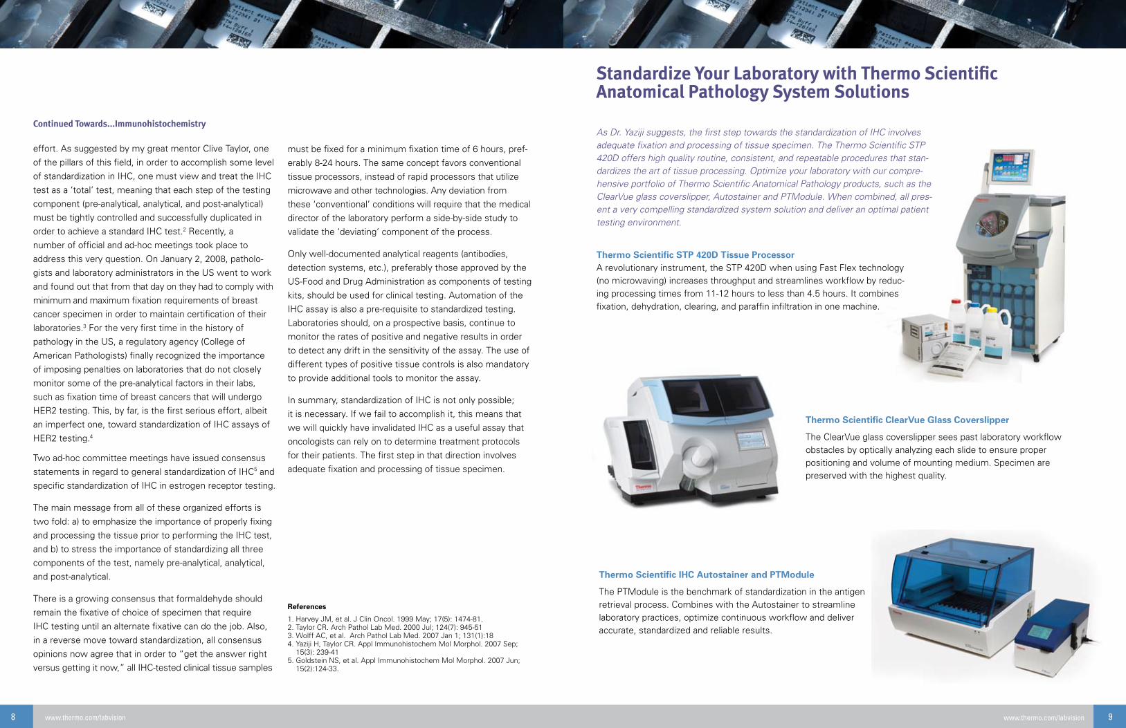
Thermo Scientific ClearVue Glass Coverslipper
The ClearVue glass coverslipper sees past laboratory workflow obstacles by optically analyzing each slide to ensure proper positioning and volume of mounting medium. Specimen are preserved with the highest quality.
Thermo Scientific IHC Autostainer and PTModule
The PTModule is the benchmark of standardization in the antigen retrieval process. Combines with the Autostainer to streamline laboratory practices, optimize continuous workflow and deliver accurate, standardized and reliable results.
Thermo Scientific STP 420D Tissue Processor A revolutionary instrument, the STP 420D when using Fast Flex technology (no microwaving) increases throughput and streamlines workflow by reduc-ing processing times from 11-12 hours to less than 4.5 hours. It combines fixation, dehydration, clearing, and paraffin infiltration in one machine.
As Dr. Yaziji suggests, the first step towards the standardization of IHC involves adequate fixation and processing of tissue specimen. The Thermo Scientific STP 420D offers high quality routine, consistent, and repeatable procedures that stan-dardizes the art of tissue processing. Optimize your laboratory with our compre-hensive portfolio of Thermo Scientific Anatomical Pathology products, such as the ClearVue glass coverslipper, Autostainer and PTModule. When combined, all pres-ent a very compelling standardized system solution and deliver an optimal patient testing environment.
10 www.thermo.com/labvision
Malignant Mesothelioma (MM) is an uncommon tumor
arising from the serosal surfaces of the various body cavi-
ties, the most common location being the pleura. It shows
epithelial, sarcomatous, and biphasic mixed differentiation.
Epithelial MM with its various histological patterns, needs
to be differentiated from adenocarcinomas and other epi-
thelial tumors. Sarcomatous MM, often being composed
of solid sheets of pleomorphic spindle cells, will resemble
a spindle cell or pleomorphic sarcoma. Immunohisto-
chemistry (IHC) is the “sine qua none” to demonstrate
mesothelial, epithelial or sarcomatous differentiation in the
diagnosis of mesothelioma, that rendered obsolete many
previously used tests. Current markers generally have very
good sensitivity and specificity for the differential diagno-
sis of MM, adenocarcinomas of lung, and tumor of other
origin. However, it yields a lower accuracy for the differen-
(from a long list) to include in their differential diagnosis of
MM. There is growing body of evidence about the need to
establish evidence-based guidelines to determine which
epithelial and mesothelial markers need to be included in
the differential diagnosis of antibody panels.5
Below are the most reliable Thermo Scientific Mesothelial
and Epithelial markers. Please visit our website for more
information www.thermo.com/labvision.
Pathology Corner: An essential tool in the diagnosis of Malignant Mesothelioma
tial diagnosis of MM and other epithelial neoplasms, such as
squamous cell carcinoma, and carcinoma of kidney, bladder,
ovary, and other sites.1
In addition, MM epithelial-type can be very difficult to distin-
guish from reactive mesothelial hyperplasia with cytological
atypia.2 Suggested IHC panel for this includes keratin AE1/AE3, EMA, p53, and Desmin. Strong Desmin expression is
indicative of a reactive process.
Mesothelial markers except Calretinin show very low
sensitivity and specificity for the diagnosis of sarcomatous
MM.3 These technical limitations, along with garden variety histological patterns and wide range of differentiation, have promoted the use of antibody panels rather than the use of one or two markers for the diagnosis of MM.4 Different
experts have varying preferences as to how many antibodies
* +/- indicates present in some tumor cells; +++, present in most tumor cells; and EMA, epithelial membrane antigen.
Antibody Name Clone Cat#
Calretinin SP13 RM-9113
CK5/6 D5/16 B4 MS-1814
Wilm’s Tumor protein (WT1) Poly RB-9267
Mesothelin 5B2 MS-1320
Mesothelioma HBME-1 MS-1494
CEA CD66e RB-368
TTF-1 8G7G3/1 MS-699
CD-15 MMA MS-1259
Keratin, Pan Ab-1 AE1/AE3 MS-343
TAG-72/CA 72-4 Ab-1 B72.3 MS-138
TABLE-1 shows the most useful mesothelial and epithelial markers available for the diagnosis of MM and its differ-ential diagnosis from selected carcinomas and sarcomas, adopted from Marchevsky.5
References:
1. Marchevsky AM, Wick MR. Appl Immunohistochem Mol Morphol. 2007;15:140–144. 2. Attanoos RL et al. Histopathology. 2003; 43: 231-383. Lucas DR, et al. Histopathology. 2003;42:270–279.4. Yaziji H., et al. Mod Pathol. 2006;19:514–523.5. Marchevsky AM, Arch Pathol Lab Med-Vol l32, March 2008
11www.thermo.com/labvision
TABLE-2 indicates the distinction between benign me-sothelial reactions and malignant mesothelioma, adopted from Marchevsky.5
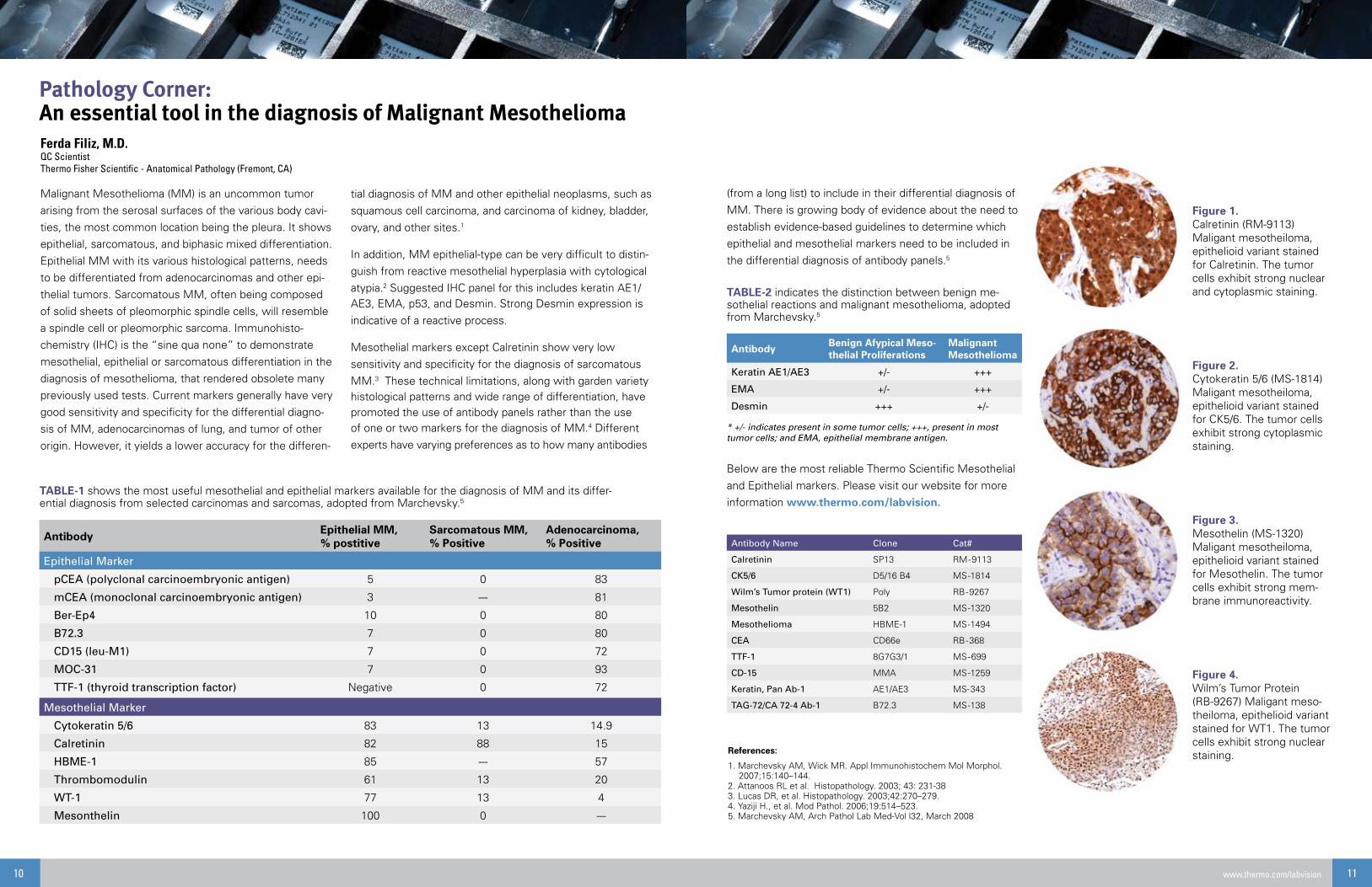
Figure 1.Calretinin (RM-9113)Maligant mesotheiloma, epithelioid variant stained for Calretinin. The tumor cells exhibit strong nuclear and cytoplasmic staining.
Figure 2.Cytokeratin 5/6 (MS-1814)Maligant mesotheiloma, epithelioid variant stained for CK5/6. The tumor cells exhibit strong cytoplasmic staining.
Figure 3.Mesothelin (MS-1320)Maligant mesotheiloma, epithelioid variant stained for Mesothelin. The tumor cells exhibit strong mem-brane immunoreactivity.
Figure 4.Wilm’s Tumor Protein (RB-9267) Maligant meso-theiloma, epithelioid variant stained for WT1. The tumor cells exhibit strong nuclear staining.