28
Immunohistochemistry (IHC) Handbook
Learn more | novusbio.comLearn more | novusbio.com
Novus-lu-2945 Immunohistochemistry (IHC) Handbook
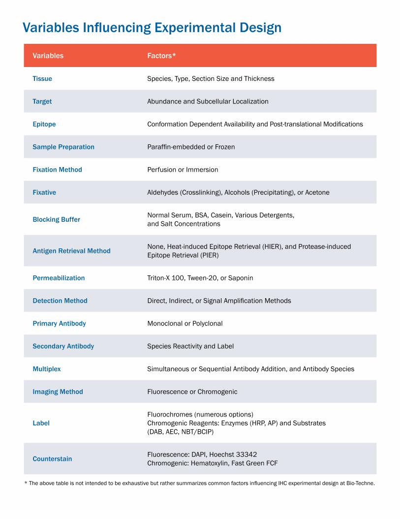
Variables Influencing Experimental Design
Variables Factors*
Tissue Species, Type, Section Size and Thickness
Target Abundance and Subcellular Localization
Epitope Conformation Dependent Availability and Post-translational Modifications
Sample Preparation Paraffin-embedded or Frozen
Fixation Method Perfusion or Immersion
Fixative Aldehydes (Crosslinking), Alcohols (Precipitating), or Acetone
Blocking Buffer Normal Serum, BSA, Casein, Various Detergents, and Salt Concentrations
Antigen Retrieval Method None, Heat-induced Epitope Retrieval (HIER), and Protease-induced Epitope Retrieval (PIER)
Permeabilization Triton-X 100, Tween-20, or Saponin
Detection Method Direct, Indirect, or Signal Amplification Methods
Primary Antibody Monoclonal or Polyclonal
Secondary Antibody Species Reactivity and Label
Multiplex Simultaneous or Sequential Antibody Addition, and Antibody Species
Imaging Method Fluorescence or Chromogenic
LabelFluorochromes (numerous options)Chromogenic Reagents: Enzymes (HRP, AP) and Substrates (DAB, AEC, NBT/BCIP)
Counterstain Fluorescence: DAPI, Hoechst 33342Chromogenic: Hematoxylin, Fast Green FCF
* The above table is not intended to be exhaustive but rather summarizes common factors influencing IHC experimental design at Bio-Techne.

Introduction to IHC
Sample Preparation and Fixation Key Differences of Sample Preparation Standard Fixatives What is the Difference Between Paraformaldehyde, Formaldehyde, and Formalin? Sample Preparation and Sectioning Protocols
Epitope (Antigen) Retrieval Is Antigen Retrieval Necessary? HIER Protocol PIER Protocol
Blocking Non-Specific Binding Endogenous Activity and Reactive Epitopes
Antibodies and Detection Methods Key Differences of Antibody Clonality Detection Methods Signal Amplification Methods Which Detection Method is Best for Multiplex IHC?
IHC Controls Positive Tissue Control Negative Tissue Control Tissue Artifact Control Antibody Controls
Complementary Techniques Western Blotting RNA in situ Hybridization
IHC Staining Protocol Deparaffinization and Rehydration Protocol Permeabilization IF Staining Protocol Chromogenic Staining Protocol
IHC Workflow
Troubleshooting Guide No or Poor Signal High Background Poor Tissue Morphology Uneven or Non-specific Staining
Reference Buffers
Bio-Techne Support Products for IHC
1
2-5
6-7
8-9
10-11
12-13
14
15-17
18-19
20-22
23
24
TABLE OF CONTENTS

Learn more | novusbio.com
Introduction
1
Immunohistochemistry (IHC) uses antibodies to detect cell and tissue proteins and provide semi-quantitative data about target protein expression, distribution, and localization. Tissues are sectioned from fixed embedded (e.g. IHC-Paraffin or plastic) or frozen blocks (e. g. IHC-Frozen), and the sections are then probed with primary antibodies against the antigens of interest. Target expression can be evaluated with the corresponding labeled primary antibody (direct detection) or, more commonly, with the addition of labeled secondary antibodies (indirect detection). The label, either fluorescent or enzymatic, is used to visualize the antigen-antibody complex.
In this multi-step application, staining may be influenced by multiple variables and requires the careful optimization of new assays for each target antigen. For example, when investigating a high abundance protein in formaldehyde-fixed tissue, the IHC protocol may include a heat-induced antigen retrieval step and use a directly labeled primary antibody. In another example, the optimal protocol for staining a low abundance protein in a methanol fixed, frozen liver section may require blocking of endogenous biotin and a signal amplification technique.
Because a number of variables can introduce artifacts or interfere with a successful outcome, appropriate controls are necessary for accurate interpretation of IHC results. This brief guide is intended to serve as a reference for researchers to understand, perform, and troubleshoot various IHC protocols during the development and optimization of new IHC assays.
TissueSection Antigen of interest is stained
and imaged with microscopeNuclear staining
of islet cells
Substrate
Signal
Secondary Antibody
Primary Antibody
Enzyme
Antigen
STAINING VISUALIZATION
Basics of an IHC Experiment

Learn more | novusbio.comLearn more | novusbio.com 2
IHC can be broadly classified into two forms based on the type of tissue processing involved: IHC- formalin-fixed, paraffin-embedded (FFPE) and IHC-frozen (Fr). Often the preservation method is closely associated with the type of fixation. Formalin-fixed tissues are commonly paraffin-embedded following fixation, while frozen tissue sections can be fixed with formaldehyde or alcohol prior to or following cryosectioning.
Sample Preparation and Fixation
IHC-FFPE IHC-FrFixation Performed before embedding
into paraffin wax.Can be performed before or after cryo-sectioning.
Common Fixative Formaldehyde Formaldehyde or AlcoholsSectioning Microtome, 4-10 μm sections Cryostat/Cryotome, 5-20 μm sectionsStorage Ideally fresh sections should be cut after
4 weeks due to loss of antigenic epitopes.Fresh cut sections should not be used after 1 month. For long term storage (several years), coating of slide in paraffin is recommended.
Short-term-1 year at -80 oC.
Major Advantage Ease of handling and preserves structural morphology. Blocks can be stored long term.
Preserves enzyme & antigen function. Useful for study of post-translationally modified protein, DNA, or RNA.
Major Limitations Variable fixation times. Fixation can mask epitopes.
Less optimal for studying structural morphology. Ice crystals can impact tissue structure.
Key Differences of Sample Preparation
Fixation preserves the histologically relevant morphology of tissues and antigenicity of target molecules. For the best results, tissue should undergo rapid and uniform fixation. Whole animal perfusion (Figure 1A), using an animal’s vascular network to disperse the fixative solution, is the preferred method for tissues from small animals including mice and rats. Alternatively, dissected tissue can be directly immersed in the fixative immediately after tissue collection (Figure 1B). For fixation byimmersion, tissue should be cut into 10 mm thick pieces and then immersed in fixative solution, 50-100X the tissue volume, for effective diffusion of the fixative.
A B
Figure 1. Fixation Methods: Perfusion (A) vs. Immersion (B)

Learn more | novusbio.com 23
Sample Preparation and Fixation
Standard Fixatives
4% formaldehyde in phosphate-buffered saline (PBS) is the most common fixative for preserving protein targets in tissues. Formaldehyde reacts with amino groups in proteins to form methylene bridges that crosslink proteins in tissue sections. These molecular cross-links can mask protein epitopes from antibody binding and may require the addition of an antigen retrieval step for proper IHC staining. Additionally, formaldehyde-mediated tissue fixation has been shown to induce translocation of phosphorylation-dependent epitopes from the membrane to the cytoplasm.
The predominant alcohols used for fixation are ≥70% methanol and ≥80% ethanol. Alcohols work by removing and replacing water molecules in tissue, which can destabilize hydrophobic bonds and alter the tertiary structure of proteins. This also causes the precipitation of soluble proteins, making alcohol-mediated fixation more appropriate for detection of membrane bound proteins.
Aldehydes
Alcohols
No single fixation condition works for all target molecules and tissues. Finding the optimal fixative and fixation time for each antigen is critical to preserve tissue morphology and antibody binding capacity. Any issues arising from incomplete fixation such as autolysis cannot be reversed or fully rectified in later steps.
What is the difference between paraformaldehyde, formaldehyde, and formalin? Paraformaldehyde (PFA) is the polymerized form of formaldehyde and is not itself a fixing agent. Formaldehyde can be prepared by dissolving PFA in PBS using heat and sodium hydroxide (NaOH). Formalin refers to a saturated formaldehyde solution and some commercial formalin solutions include methanol as a stabilizer to prevent formaldehyde polymerization. A 10% formalin solution is equivalent to a 3.7% formaldehyde solution.
Acetone is also used as a strong dehydrant and precipitant, typically applied to sections of snap-frozen tissues. Acetone fixation is generally mild and may be followed by fixation with alcohols or formaldehyde.
Acetone

Learn more | novusbio.comLearn more | novusbio.com 2 4
Sample Preparation and Sectioning
01
PROTOCOL: Formalin-fixed, paraffin-embedded (FFPE)
02
03
04
05
Fix the tissue of interest by immersing it in 10% neutral buffered formalin (4% formaldehyde) for 4-24 hours at room temperature. Fixation time and temperature depends on tissue type/size. After fixation, wash the tissues 3x in PBS.
Dehydrate by full immersion of tissue in the following solutions (2x for 30 minutes each):a) 70% Ethanolb) 95% Ethanolc) 100% Ethanold) Xylene
Embed the tissue in molten paraffin. After the paraffin solidifies keep the blocks at 4°C until sectioning.
Note: Paraffin melts at 57°C.
Keep FFPE blocks chosen for sectioning tissue face down in an ice water bath, to hydrate the tissue and avoid cracking during sectioning. Certain tissues (e.g. liver or spleen) require this to be repeated after 10-20 cut sections.
Use a microtome to cut the paraffin tissue blocks into 4-10 µm thick sections and transfer them to a 37°C water bath with distilled water.
Pick up the floating tissue section using a clean histological slide (coated with gelatin or poly-L-lysine to improve adhesion of tissue sections) and allow mounted tissue sections to dry for about 30 min on a 37°C hot plate followed by baking them for 2-3 hours in a 40°C oven. Slides can be safely stored at room temperature until ready for staining. Storage of cut slides for longer than 1 month is usually not recommended.
06
01
PROTOCOL: 4% Formaldehyde solution in PBS
02
03
04
05
For 1 L of 4% formaldehyde, add 100 mL of 10X PBS and 700ml distilled water to a glass beaker on a stir plate in a ventilated hood. Heat while stirring to approximately 60°C. Make sure that temperature of the solution does not exceed 60°C.
Add 40 g of paraformaldehyde powder to the heated PBS solution.
Note: The powder will not immediately dissolve into solution. Slowly raise the pH by adding 1 N NaOH dropwise from a pipette until the solution clears.
Once the paraformaldehyde is dissolved, the solution should be cooled and filtered.
Adjust the volume of the solution to 1 L with distilled water.
Recheck the pH, and adjust it with small amounts of dilute HCl to approximately 6.9.
Note: The solution can be stored at 2-8°C for up to one month.

Learn more | novusbio.com5
Sample Preparation and Sectioning Protocols
01
PROTOCOL: Method II - Cryopreservation & sectioning of alcohol fixed tissues
02
03
After dissection, immediately snap freeze tissue with isopentane cooled by dry ice. To do this, fill the styroform box with dry ice and pour isopentane over it so that chunks of dry ice immersed into isopentane half the way through. Place a metal cryostat chuck on top of the dry ice/isopentane slurry and then mount piece of tissue on top of the chuck and cover it with OCT.
Wait until tissue in OCT is completely frozen (OCT turns from clear to opaque white solid material) and transfer chuck with frozen tissue into the cryostat. Allow the temperature of the tissue to equilibrate to the temperature in the cryostat.
Cut the tissue in 5-20 µm thick sections. Mount tissue sections onto gelatin or poly-L-lysine coated slides: place tissue sections onto cold slides and then warm the slides up to thaw tissue sections so that they can permanently adhere to the slides. Slides can be safely stored for 6-12 months at -80°C until ready for fixing. Uncut tissue can be restored at -80°C.
Remove slides from freezer and fix with cold fixative (acetone or methanol) for 10 minutes. Proceed to staining.04
01
PROTOCOL: Method I - Frozen samples (whole animal or dissected tissue)
02
03
Fixation:
Whole animal - Perfuse the animal with warm saline solution to flush the blood out of vasculature andimmediately follow this by perfusion with freshly prepared 4% formaldehyde.
Dissected tissue - Immerse tissue in 4% formaldehyde (50-100x the tissue volume) for 4-24 hours at room temperature. Wash tissue with PBS. Fixation temperature and time may require optimization depending on the tissue type and size.
Cryoprotection:
Whole animal - Continue perfusion with a 10% sucrose solution.
Dissected tissue - Immerse dissected tissue in a 10% sucrose solution overnight at 4°C. Tissue will sink in sucrose solution upon reaching equilibration.
Embed tissue in OCT cryostat sectioning medium and store at -80°C until ready for sectioning. Tissue can be safely stored for up to 1 month.
When ready for sectioning, move the embedded tissue directly into the cryostat and use OCT medium to mount it to the chuck. Allow the temperature of the tissue to equilibrate with the cryostat.
Cut the tissue in 5-20 µm thick sections. Mount tissue sections onto gelatin or poly-L-lysine coated slides by placing the cold sections onto warm slides. Slides can be safely stored for 6-12 months at -80°C until ready for staining.
04
05
Learn more | novusbio.comLearn more | novusbio.com 6
Epitope (Antigen) Retrieval
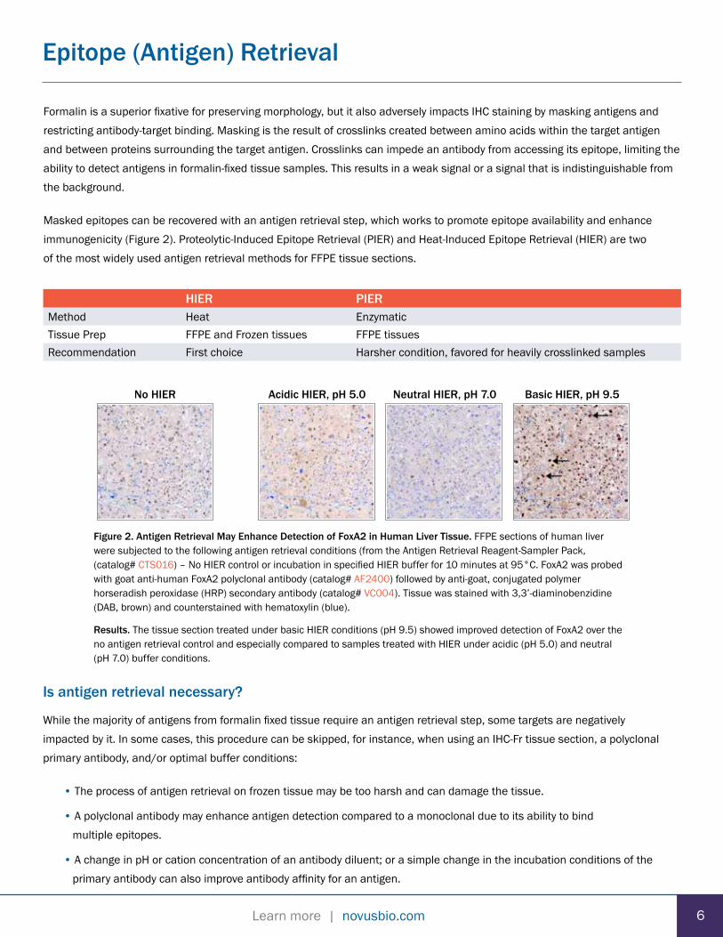
HIER PIERMethod Heat EnzymaticTissue Prep FFPE and Frozen tissues FFPE tissuesRecommendation First choice Harsher condition, favored for heavily crosslinked samples
Formalin is a superior fixative for preserving morphology, but it also adversely impacts IHC staining by masking antigens and restricting antibody-target binding. Masking is the result of crosslinks created between amino acids within the target antigen and between proteins surrounding the target antigen. Crosslinks can impede an antibody from accessing its epitope, limiting the ability to detect antigens in formalin-fixed tissue samples. This results in a weak signal or a signal that is indistinguishable from the background.
Masked epitopes can be recovered with an antigen retrieval step, which works to promote epitope availability and enhance immunogenicity (Figure 2). Proteolytic-Induced Epitope Retrieval (PIER) and Heat-Induced Epitope Retrieval (HIER) are two of the most widely used antigen retrieval methods for FFPE tissue sections.
Is antigen retrieval necessary?
While the majority of antigens from formalin fixed tissue require an antigen retrieval step, some targets are negatively impacted by it. In some cases, this procedure can be skipped, for instance, when using an IHC-Fr tissue section, a polyclonal primary antibody, and/or optimal buffer conditions:
• The process of antigen retrieval on frozen tissue may be too harsh and can damage the tissue.
• A polyclonal antibody may enhance antigen detection compared to a monoclonal due to its ability to bind multiple epitopes.
• A change in pH or cation concentration of an antibody diluent; or a simple change in the incubation conditions of the primary antibody can also improve antibody affinity for an antigen.
Figure 2. Antigen Retrieval May Enhance Detection of FoxA2 in Human Liver Tissue. FFPE sections of human liver were subjected to the following antigen retrieval conditions (from the Antigen Retrieval Reagent-Sampler Pack, (catalog# CTS016) – No HIER control or incubation in specified HIER buffer for 10 minutes at 95°C. FoxA2 was probed with goat anti-human FoxA2 polyclonal antibody (catalog# AF2400) followed by anti-goat, conjugated polymer horseradish peroxidase (HRP) secondary antibody (catalog# VC004). Tissue was stained with 3,3’-diaminobenzidine (DAB, brown) and counterstained with hematoxylin (blue).
Results. The tissue section treated under basic HIER conditions (pH 9.5) showed improved detection of FoxA2 over the no antigen retrieval control and especially compared to samples treated with HIER under acidic (pH 5.0) and neutral (pH 7.0) buffer conditions.
No HIER Neutral HIER, pH 7.0Acidic HIER, pH 5.0 Basic HIER, pH 9.5

Learn more | novusbio.com7
Epitope Retrieval Protocols
PROTOCOL: PIER
First select one of 2 PIER buffer options:
• Trypsin Working Solution, 0.05%
• Proteinase K Working Solution, 20 μg/ml
Cover sections with chosen PIER buffer.
Incubate for 10-20 minutes at 37°C in humidified chamber.
Allow sections to cool at room temperature for 10 minutes.
01
02
03
04
PROTOCOL: HIER
05
First select one of 3 HIER buffer options:
• Citrate Buffer - 10mM Citric Acid, 0.05% Tween 20, pH 6.0(also see: Pre-mixed Citrate Buffer from Novus# NB900-62075)
• Tris Buffered Saline (TBS) - 50mM TBS, 0.05% Tween 20, pH 9.0
• EDTA Buffer - 1mM EDTA, 0.05% Tween 20, pH 8.0
Pre-heat retrieval solution in a staining dish in a vegetable steamer until temperature reaches 95-100°C.
A microwave or pressure cooker can be used as alternative heating source in place of the steamer.
Immerse slides in the staining dish.
Place the lid loosely on the staining dish and incubate it for 20-40 minutes in the steamer.
Remove the staining dish from the steamer and place it on the lab bench at room temperature.
Allow the slides to cool for 20-30 minutes before proceeding with the staining procedure.
Note: For aldehyde fixed frozen tissue, try using the Citrate Buffer and reduce the incubation period to 5 to 7 minutes.
01
02
03

Learn more | novusbio.comLearn more | novusbio.com 8
Blocking Non-Specific Binding
Antibody-based applications rely on the specific binding of an antibody to the target epitope for generating accurate expression data. The same forces that govern specific interactions can also contribute to non-specific binding including hydrophobic interactions, ionic interactions, and hydrogen bonding. Common buffers to block non-specific interactions are serum, BSA, casein, or commercial buffers (Figure 3). If blocking with normal serum, the species of the animal serum should be the same as the host of the secondary antibody. For example, use goat serum if using a goat anti-mouse secondary.
+ Blocking No Blocking
Figure 3. Blocking with Serum Reduces Background Staining. FFPE sections of human liver was either blocked with normal donkey serum for 15 minutes at room temperature or left untreated. FoxA2 was then detected using goat anti-human FoxA2 polyclonal antibody (catalog# AF2400) followed by donkey anti-goat, conjugated polymer HRP secondary antibody (catalog# VC004). Tissue was stained with DAB (brown) and counterstained with hematoxylin (blue).
Results. Non-specific binding is significantly decreased and FoxA2 staining is easier to identify in samples that were pretreated with serum.
Blocking other reactive epitopes and quenching endogenous enzymatic reactions in tissue samples prior to the primary antibody incubation step also prevents non-specific binding and false positive staining. Autofluorescence can impact imaging for immunofluorescence (IF), especially for tissue samples with elevated levels of flavins or porphyrins. Non-specific staining observed when using common detection reagents and tips on how to minimize high background signal are listed in Table 1 on the next page.
Tip: The detection method should be compatible with the sample tissue. For detecting a low abundance protein in tissues with high levels of endogenous biotin, consider using a polymer based signal amplification method.
The choice of blocking buffer is also contingent on the method of detection used. For instance, if using an alkaline-phosphatase (AP) conjugated secondary antibody, the blocking serum should be diluted in tris-buffered saline (TBS). PBS will interfere with the alkaline phosphatase reaction.

Learn more | novusbio.com
Blocking Non-Specific Binding
Endogenous Activity Reactive Epitopes
Peroxidase
• Tissues with High Activity - Kidney, liver, or vascularareas with red blood cells, lysosomal membranesespecially in active phagocytic cells
• Affected Step– Chromogenic detection with HRP
• Block – Treat tissue with 3-10% hydrogen peroxide inmethanol prior to incubation with HRP conjugatedsecondary antibody.
• Background Control – Before the antibody incubationstep, incubate the sample with DAB substrate. Thepresence of endogenous peroxidase will be associatedwith the deposition of brown color.
Phosphatase
• Tissues with High Activity - Intestine, kidney, lymphoid
• Affected Step – Chromogenic detection with AP
• Block – Treat tissue section with 1mM Levamisole priorto incubation with AP conjugated secondary antibody.For intestinal sections, block with 1% acetic acid.
• Background Control – Before the antibody incubationstep, incubate the sample with nitro bluetetrazolium/5-bromo-4-chloro-3-indolyl phosphate(NBT/BCIP) substrate. The presence of endogenousphosphatases will be associated with the depositionof blue color.
Biotin
• Reactive Tissues/Conditions - Liver, kidney, spleen,heart, brain, and lung or frozen tissues
• Affected Step – Chromogenic detection using biotinconjugated reagents
• Block – Treat tissue with avidin prior to incubation withbiotin-conjugated reagents. Then treat sample withbiotin to block additional biotin binding sites on theavidin molecule.
• Background Control – Before the antibodyincubation step, incubate the sample with avidin-biotincomplex or streptavidin (SA)-HRP, then DAB. Thepresence of endogenous biotin will be associated withthe deposition of brown color.
Autofluorescence
• Tissues/Conditions with Endogenous Fluorescence –Pancreas, brain, red blood cells, other pigmented celltypes, lipofuscin, extracellular matrix components, oraldehyde fixation
• Affected Step – IF detection, most severe in theshorter visible fluorescence wavelengths
• Block – For aldehyde autofluorescence, block withsodium borohydride in PBS. Choose fluorochromesemitting at unaffected wavelengths.
• Background Control - Prior to staining, examine tissuesections under fluorescent microscope usingappropriate filter sets.
9
Table 1. Endogenous Activity and Reactive Epitopes
Learn more | novusbio.comLearn more | novusbio.com
Antibodies and Detection Methods
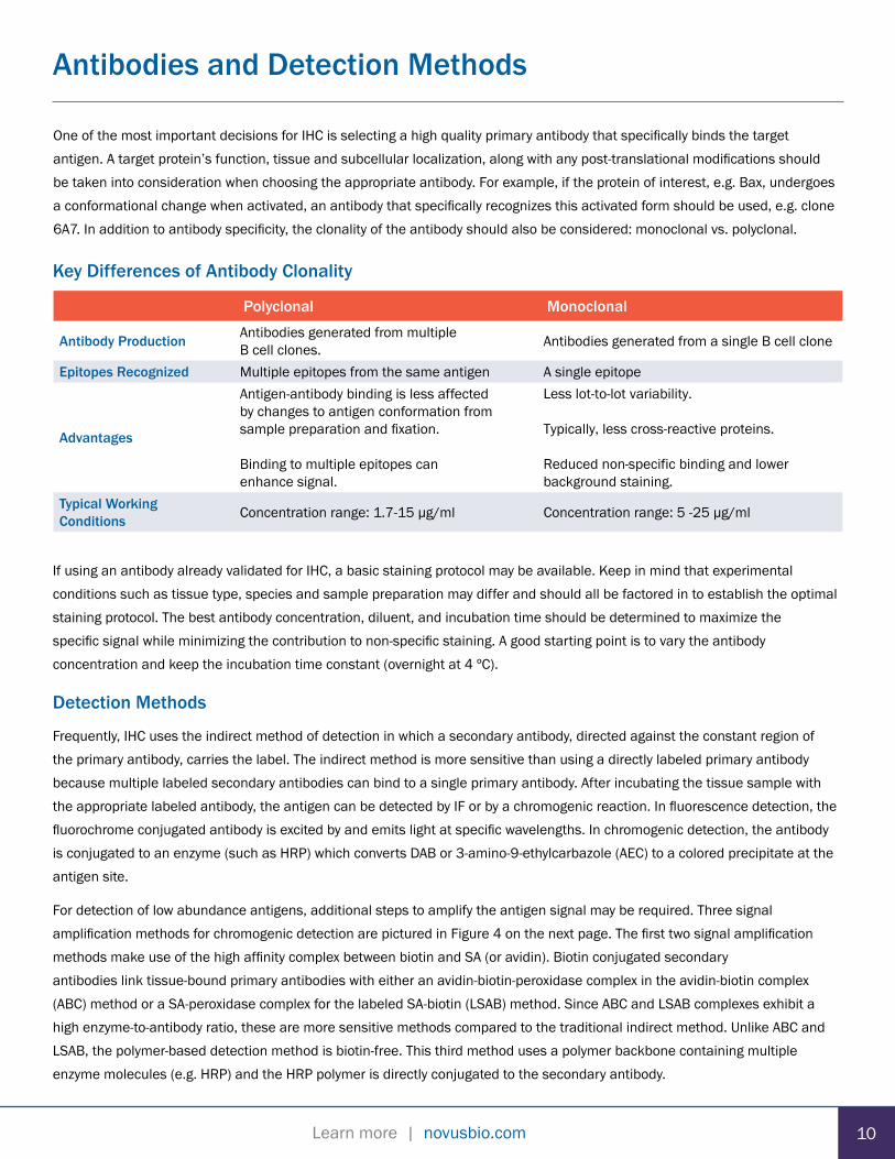
One of the most important decisions for IHC is selecting a high quality primary antibody that specifically binds the target antigen. A target protein’s function, tissue and subcellular localization, along with any post-translational modifications should be taken into consideration when choosing the appropriate antibody. For example, if the protein of interest, e.g. Bax, undergoes a conformational change when activated, an antibody that specifically recognizes this activated form should be used, e.g. clone 6A7. In addition to antibody specificity, the clonality of the antibody should also be considered: monoclonal vs. polyclonal.
10
Polyclonal Monoclonal
Antibody Production Antibodies generated from multiple B cell clones. Antibodies generated from a single B cell clone
Epitopes Recognized Multiple epitopes from the same antigen A single epitope
Advantages
Antigen-antibody binding is less affected by changes to antigen conformation from sample preparation and fixation.
Binding to multiple epitopes can enhance signal.
Less lot-to-lot variability.
Typically, less cross-reactive proteins.
Reduced non-specific binding and lower background staining.
Typical Working Conditions Concentration range: 1.7-15 µg/ml Concentration range: 5 -25 µg/ml
Key Differences of Antibody Clonality
If using an antibody already validated for IHC, a basic staining protocol may be available. Keep in mind that experimental conditions such as tissue type, species and sample preparation may differ and should all be factored in to establish the optimal staining protocol. The best antibody concentration, diluent, and incubation time should be determined to maximize the specific signal while minimizing the contribution to non-specific staining. A good starting point is to vary the antibody concentration and keep the incubation time constant (overnight at 4 ºC).
Detection Methods
Frequently, IHC uses the indirect method of detection in which a secondary antibody, directed against the constant region of the primary antibody, carries the label. The indirect method is more sensitive than using a directly labeled primary antibody because multiple labeled secondary antibodies can bind to a single primary antibody. After incubating the tissue sample with the appropriate labeled antibody, the antigen can be detected by IF or by a chromogenic reaction. In fluorescence detection, the fluorochrome conjugated antibody is excited by and emits light at specific wavelengths. In chromogenic detection, the antibody is conjugated to an enzyme (such as HRP) which converts DAB or 3-amino-9-ethylcarbazole (AEC) to a colored precipitate at the antigen site.
For detection of low abundance antigens, additional steps to amplify the antigen signal may be required. Three signal amplification methods for chromogenic detection are pictured in Figure 4 on the next page. The first two signal amplification methods make use of the high affinity complex between biotin and SA (or avidin). Biotin conjugated secondary antibodies link tissue-bound primary antibodies with either an avidin-biotin-peroxidase complex in the avidin-biotin complex (ABC) method or a SA-peroxidase complex for the labeled SA-biotin (LSAB) method. Since ABC and LSAB complexes exhibit a high enzyme-to-antibody ratio, these are more sensitive methods compared to the traditional indirect method. Unlike ABC and LSAB, the polymer-based detection method is biotin-free. This third method uses a polymer backbone containing multiple enzyme molecules (e.g. HRP) and the HRP polymer is directly conjugated to the secondary antibody.

Learn more | novusbio.com
Faster with less stepsthan indirect detection.
Greater sensitivity and more flexibility comparedto direct detection.
Greater sensitivity compared to traditional indirect detection.
Greater sensitivity compared to ABC and LSAB method.
Fewer steps than ABC andLSAB method.
LSAB Polymer-based
Signal Amplification Methods
ABCIndirectDirect
Reporter enzymeor fluorochrome
Labeled primary antibody
Antigen
Reporter enzymeor fluorochrome
Primary antibody
Antigen
Labeled secondary antibody
Primary antibody
Antigen
Biotinylated secondary antibody
Reporter enzyme
Biotin
Avidin/SA
Primary antibody
Antigen
Biotinylated secondary antibody
Reporter enzyme
Biotin
SA
Primary antibody
Antigen
Labeled secondary antibody
Reporter enzyme
Polymer backone
Advantages
Greater sensitivity than traditional indirect detection.
Smaller complex size over ABC facilitates better tissue penetration.
Figure 4. Types and Advantages of Detection Methods
Antibodies and Detection Methods
Which detection method is best for multiplex IHC?
Fluorescence detection allows for simultaneous detection of a large number of antigens due to the availability of fluorochromes that span the visible and adjacent (ultra-violet and near infrared) spectral regions. Many fluorochromes have narrow emission spectra and when carefully chosen in combination can minimize spectral overlap. Since colored precipitates from chromogenic reactions have broad bright-field spectra, the potential for chromogenic overlap is high and can obscure results for co-localized antigens. Hence, the chromogenic method is generally limited to detecting up to 3 targets and is not recommended for visualiz-ing antigens located in same subcellular region. Multiplexed experiments are also easier with direct detection because assays require less steps and reagents. Cross-reactivity of labeled secondary antibodies is a potential complication with the indirect detection method that can be minimized by choosing primary antibodies from different host species.
11

Learn more | novusbio.comLearn more | novusbio.com
IHC Controls
Due to the number of variables that can affect IHC staining, the inclusion of proper controls in each experiment is important for identifying the source of potential staining issues and for validating the results.
Positive Tissue Control
A tissue sample known to express the target antigen can be used as a positive control. This positive control should be run in each IHC experiment to confirm the presence of the target antigen. It is especially critical to ensure there were no problems in the IHC protocol itself.
Commonly used positive controls:
• Tissue or experimental condition with proven positive signal• Samples from transgenic animals that overexpress the antigen
Negative Tissue Control
A tissue sample that does not express the target antigen can be used as a negative tissue control. This negative control should be run in each IHC experiment to determine the degree of background staining caused by non-specific interactions. Background staining should be negligible and not resemble specific staining.
Commonly used negative controls:
• Tissue or experimental condition with proven negative signal• Samples from knockdown or knockout tissues
Tissue Artifact Control
Background staining may be more pronounced in particular tissues. Since high background can mask positive signal from low abundance antigens or result in artifacts being mistaken for specific staining, IHC data can be misinterpreted. Before commencing staining, tissues should be examined under a bright-field or fluorescence microscope (for chromogenic or fluorescent labels, respectively) to ensure the signal is not due to the inherent properties of the tissue. For example, lipofuscin is an endogenous pigment that accumulates in lysosomes of postmitotic cells which mimics positive staining for both bright-field and fluorescence microscopy.
13 12

Learn more | novusbio.com
IHC Controls
Antibody Controls
The specificity of the antigen-antibody interaction is critical for accurate detection of the antigen. Due to the complex natureof tissue and the multifactorial effect of sample preparation on the staining procedure, antibody controls help to confirm the presence of the antigen of interest. A few recommended antibody controls include:
Figure 5. Primary Antibody Control in Human Kidney. Neprilysin/CD10 was detected in a FFPE section of human kidney using goat anti-human Neprilysin/CD10 polyclonal antibody (catalog# AF1182). Tissue was stained with anti-goat HRP-DAB Cell & Tissue Staining Kit (brown; catalog# CTS008) and counterstained with hematoxylin (blue). Neprilysin/CD10 staining (brown) is lost if the primary antibody is absent from the diluent in a parallel experiment.
+ Primary antibody No Primary antibodyNo Primary Antibody Control is a control in which the primary antibody is absent from the diluent during the primary antibody incubation step. This control will show the contribution of all other components, such as the secondary antibody and detection reagents, to background staining (Figure 5).
Isotype Control should be used to confirm the specificity of the primary antibody. For this control, the primary antibody should be replaced with a non-immune immunoglobulin of the same isotype and at the same concentration as the primary antibody. Example isotypes are IgG1, IgG2a, IgG2B, and IgM. All other conditions and protocol steps should remain the same.
This control will demonstrate if the staining by the primary antibody is a result of an interaction with the antigen binding site (paratope) or due to non-specific interaction with the immunoglobulin molecule.
Absorption Control is also used to determine if the primary antibody binds the antigen of interest. For this control, the primary antibody is first inactivated by preincubation with the immunogen (antigen) and then used in place of the primary antibody in the IHC protocol. The antigen to antibody solution should be prepared at a molar ratio of 10:1 to fully saturate the antibody and then incubated in the IHC antibody diluent overnight at 4°C.
The optimal immunogen is small purified peptides. A whole protein is more likely to interact with other proteins in the tissue sample, even if bound to the primary antibody, and may lead to false positive staining. Also, some epitopes are more universal and may be present in multiple proteins. In this case, all antibody-antigen binding sites may be occupied in an absorption control, which would prevent other proteins with the same epitope from interacting with the antibody and give a false negative result. Thus, absorption control results should be analyzed with discretion.
1413

Learn more | novusbio.comLearn more | novusbio.com 14
Complementary Techniques
Western Blotting
Western blots are frequently used as a companion technique to confirm antibody specificity and provide a more quantitative analysis of antigen levels. However, the target molecule conformation may differ between the samples for IHC and Western blots. The epitope recognized by the primary antibody may not be available under both assay conditions. For example, a protein is typically denatured and reduced for Western blots, whereas the same protein in an IHC tissue section retains a more native-like structure. Using a polyclonal primary antibody, which recognizes multiple epitopes, may help mitigate this problem.
RNA in situ Hybridization
In situ hybridization (ISH) is a technique that employs probes to detect specific RNA sequences in heterogeneous cell populations such as fixed tissue sections. When used alongside IHC, it provides a more comprehensive overview of the complex molecular mechanisms involved in biological pathways. Since both techniques share similar procedures including sample fixation and signal amplification, dual ISH-IHC methods have been developed to collect RNA and protein expression data on the same tissue sample (Figure 6).
Figure 6. Fluorescent Dual ISH-IHC: ER1 mRNA (green) was detected in FFPE sections of human breast cancer tissue with a RNAscope probe (Advanced Cell Diagnostics). HER2 (red) was detected using goat anti-human ErbB2/Her2 polyclonal antibody (catalog# AF1129). Tissue was counterstained with DAPI (blue).
Typical Areas of Research
1. Viral Detection and Host Response
2. Transcriptional and Translational Regulatory Events
3. Comparison of Gene and Protein Expression in Cancer Biopsies
4. Secretory Proteins
5. Stem Cell Pluripotency & Differentiation
15 14
Learn more | novusbio.com
IHC Staining Protocol
01
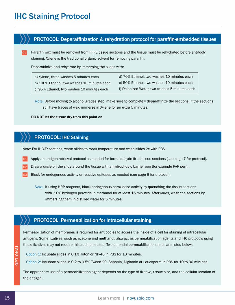
PROTOCOL: Deparaffinization & rehydration protocol for paraffin-embedded tissues
Paraffin wax must be removed from FFPE tissue sections and the tissue must be rehydrated before antibody staining. Xylene is the traditional organic solvent for removing paraffin.
Deparaffinize and rehydrate by immersing the slides with:
Note: Before moving to alcohol grades step, make sure to completely deparaffinize the sections. If the sections still have traces of wax, immerse in Xylene for an extra 5 minutes.
DO NOT let the tissue dry from this point on.
a) Xylene, three washes 5 minutes eachb) 100% Ethanol, two washes 10 minutes eachc) 95% Ethanol, two washes 10 minutes each
d) 70% Ethanol, two washes 10 minutes eache) 50% Ethanol, two washes 10 minutes eachf) Deionized Water, two washes 5 minutes each
PROTOCOL: IHC Staining
01
02
Apply an antigen retrieval protocol as needed for formaldehyde-fixed tissue sections (see page 7 for protocol).
Draw a circle on the slide around the tissue with a hydrophobic barrier pen (for example PAP pen).
Block for endogenous activity or reactive epitopes as needed (see page 9 for protocol).
Note: If using HRP reagents, block endogenous peroxidase activity by quenching the tissue sections with 3.0% hydrogen peroxide in methanol for at least 15 minutes. Afterwards, wash the sections by immersing them in distilled water for 5 minutes.
PROTOCOL: Permeabilization for intracellular staining
Permeabilization of membranes is required for antibodies to access the inside of a cell for staining of intracellular antigens. Some fixatives, such as acetone and methanol, also act as permeabilization agents and IHC protocols using these fixatives may not require this additional step. Two potential permeabilization steps are listed below:
Option 1: Incubate slides in 0.1% Triton or NP-40 in PBS for 10 minutes.
Option 2: Incubate slides in 0.2 to 0.5% Tween 20, Saponin, Digitonin or Leucoperm in PBS for 10 to 30 minutes.
The appropriate use of a permeabilization agent depends on the type of fixative, tissue size, and the cellular location of the antigen.
03
1615
Note: For IHC-Fr sections, warm slides to room temperature and wash slides 2x with PBS.
OP
TIO
NA
L

Learn more | novusbio.comLearn more | novusbio.com
PROTOCOL: Deparaffinization & rehydration protocol for paraffin-embedded tissues
16
IHC Staining Protocol
PROTOCOL: IHC Staining
04 Block any non-specific binding by incubating the tissue sections with 5% animal serum in PBS + 0.3% Triton X-100 (PBS-T) for 30 minutes at room temperature.
Add the primary antibody diluted in 1% animal serum in PBS-T and incubate at room temperature for 1-2 hours. Continue the incubation overnight at 4°C in a humidified chamber.
Reminder: Keep in mind, the species of the primary antibody is raised in should differ from the source of the tissue sample.
Note: Use the recommended dilution of the antibody specified on the datasheet. If not specified, use 3-fold serial dilutions: 2 μg/mL, 6 μg/mL and 18 μg/mL.
Wash sections twice with 1% serum in PBS-T for 10 minutes each.
Proceed with either the IF or chromogenic staining protocol.
05
06
Important note: When examining slides using a fluorescence microscope avoid long exposures at the fluorochrome’s excitation wavelength to prevent photo-bleaching.
01
PROTOCOL: IF Staining
02
03
04
05
Add a fluorochrome-conjugated secondary antibody and incubate at room temperature for 1 -2 hours. Use the recommended dilution of the antibody as specified on the data sheet.
Note: The secondary antibody should be directed against the host species of the primary antibody. For help on selecting secondary antibodies, see our Secondary Antibody Handbook.
Wash sections twice with 1% serum PBS-T for 10 minutes each.
For nuclear staining, add DAPI solution (~2.9 μM) or other DNA binding dye (e.g. Hoechst 33342) and incubate 2-5 minutes at room temperature. Rinse 1X with PBS. DAPI has an absorption maximum at 358 nm and an emission maximum of 461 nm.
Apply a drop of mounting media containing a fluorescence anti-fade agent. Carefully place a coverslip on it and remove the excess mounting media, if necessary. Circle the edges of the coverslip with clear fingernail polish to prevent the cells from drying. Allow nail polish to air dry.
Note: Some mounting media solutions have DAPI already added and will harden after exposure to air, eliminating the need to seal the edges of the coverslip.
Examine the cells under a fluorescence microscope using the appropriate excitation and emission filters and image as required. Slides can be stored between -20°C and 4°C in a dark slide box or slide book.
07
16 16

Learn more | novusbio.com
IHC Staining Protocol
01
02
03
04
05
06
Add a biotin conjugated secondary antibody and incubate at room temperature for 1 hour. Use the recommended dilution of the antibody as specified on the data sheet.
Note: The secondary antibody should be directed against the host species of the primary antibody. For help on selecting secondary antibodies, see our Secondary Antibody Handbook.
Wash sections twice with 1% serum PBS-T for 10 minutes each.
Add ABC-HRP reagent and incubate at room temperature for 1 hour. Follow manufacturer’s guidelines for reagent preparation.
Prepare a working solution of DAB and apply to tissue sections. Monitor the reaction as the chromogenic reaction turns the epitope sites brown (time of color development may vary from few seconds to 10 minutes). Proceed to the next step when the intensity of the signal is appropriate for imaging.
Wash sections twice in PBS for 10 minutes each.
Wash the sections twice in distilled water for 2 minutes each.
To counterstain nuclei, use Hematoxylin according to the manufacturer’s instructions. Note: If using an aqueous chromogen instead of DAB (i.e. AEC, Fast Red, etc.), skip the following dehydration step and mount in aqueous media instead of organic mounting media.
Dehydrate tissue sections by moving slides through the following solutions twice for 2 minutes each:
a) 95% Ethanolb) 100% Ethanolc) Xylene
Add mounting media to slides and top with coverslips. The DAB reaction is permanent and stable and can be analyzed under a bright-field microscope at any time.
07
08
PROTOCOL: Chromogenic Staining
Important note: DAB is a carcinogen. Always wear gloves and work in a fume hood when working with DAB. Deactivate and clean work area after use according to manufacturer’s instructions.
1717
09

Learn more | novusbio.comLearn more | novusbio.com 17
Workflow of IHC Sample Preparation
Dissectedtissue
Immerse in4% PFA
Snap freeze tissue in dry ice/isopentane slurry
Section tissue andmount on slide
Fix tissue in alcohol
Section tissue andmount on slide
Whole animal perfusionwith 4% PFA
Freezetissue
Embed tissuein para�n
Section tissue andmount on slide
Depara�nizeand rehydrate
Section tissue andmount on slide
Freezetissue
-80°C
Antigenretrieval
18 18

Learn more | novusbio.com
Workflow of IHC Staining
ChromogenicDetection
FluorescenceDetection
Signal
Block with serumor BSA
Probe withprimary antibody
Addchromogen
Counterstainwith hematoxylin
Counterstainwith DAPI
Image with �uorescence microscope
Image with bright-�eld microscope
Incubatewith labeledsecondaryantibody
Substrate
Blocking of reactive epitopes and quenching
of endogenousenzymatic reactions may
also be necessary.
Blocking of reactiveepitopes and quenching
of endogenousenzymatic reactions may
also be necessary.
1919

Learn more | novusbio.comLearn more | novusbio.com 19
Troubleshooting Guide
Issue Cause Recommendation
Antigen Not Present Protein isn’t expressed in tissue or under experimental conditions.
Check protein expression by Western Blot or in situ hybridization.
Tissue Fixation
Over-fixation can cause epitope masking.
Under-fixation can cause heavy edge staining with little to no positive signal in middle of your specimen. It can also result in autolysis and target protein degradation.
Reduce the time or concentration of the fixative. Apply an antigen retrieval step.
Increase the time or concentration of the fixative. Alternatively, try a different fixative.
Paraffin-Embedding Epitope altered and not recognized by primary antibody. Switch to using frozen sections.
Antigen Retrieval Epitope masking prevents binding of primary antibody.
Increase treatment time or change procedure (either new buffer or different method).
Permeabilization Inadequate penetration of antibodies and buffers into the tissue sections.
Use 0.5-1.0% Triton (or Tween-20) detergent in the buffers.
Antibody ApplicationEpitope isn’t saturated with antibody and/or hasn’t reached binding equilibrium.
Increase the concentration or incubation time of the primary or secondary antibody.
Antibody Compatibility
Primary antibody isn’t reactive in the tissue sample.
Primary antibody binds an epitope only exposed when protein is unfolded.
Incompatible primary and secondary antibody.
Confirm the species reactivity of the primary antibody.
Confirm the antibody can be used for assays in which the protein is in its native conformation.
Verify that the secondary antibody will interact with the species of the primary antibody.
Reagents Missing reagents or added improperly. Repeat staining procedure and verify all reagents are added in correct order.
Microscope Adjustments (Fluorescence)
Fluorochrome isn’t effectively excited or emission isn’t captured by filters.
Verify fluorochrome is compatible with filter sets and appropriate settings are used. Also, increase the camera exposure time.
No or Poor Signal
No Antigen Staining Good Antigen Staining
20 20

Learn more | novusbio.com
Issue Cause Recommendation
Tissue Fixation Over-fixation can cause strong non-specific staining.
Reduce the time or concentration of the fixative.
Blocking Step Inadequate blocking. Increase the incubation time or concentration of serum in the blocking buffer.
Endogenous Activity/Reactivity
Tissue may have high autofluorescence, biotin, peroxidase activity, etc.
Check slides prior to staining and use the appropriate blocking/quenching steps. Try a more compatible detection method.
Antibody Application
Primary or secondary antibody concentration too high.
Non-specific binding of primary or secondary antibody.
Titrate antibodies to find the optimal signal to background staining.
Increase the amount or incubation period of washes. Reduce the secondary antibody incubation period.
Antibody Compatibility Secondary antibody is non-specifically binding the tissue sample.
Select a secondary antibody pre-absorbed against the species of the experimental sample. For example, a secondary antibody adsorbed against mouse immunoglobulin or serum is recommended when staining mouse tissue.
Detection Method
DAB overstaining.
Endogenous peroxidases are activating the DAB reaction.
Endogenous biotin is activating the SA-biotin complex using the ABC signal amplification method.
Reduce the time of incubation with DAB chromogen.
Quench with hydrogen peroxide.
Block endogenous biotin.
Slide Condition
Reagents sticking to old or poorly prepared slides.
Use freshly prepared or purchased slides.
Avoid letting the tissue dry during the staining procedure.
Tissue dried out.
Buffer Conditions Background from ionic interactions. Increase the ionic strength of the blocking and antibody buffers.
Spectral Overlap(Fluorescence)
Multiple fluorochromes have overlapping emission spectra.
Select compatible fluorochrome conjugates.
Troubleshooting Guide
High Background Low Background
High Background
2121

Learn more | novusbio.comLearn more | novusbio.com 21
Troubleshooting Guide
Issue Cause Recommendation
Tissue Preparation
Tissue section appears torn or folded. Air bubbles under section.
Thick sections cause poor resolution of tissue morphology.
Frozen sections - Ice crystals may have destroyed morphology.
Re-cut sections using a sharp blade or adjust the cutting speed. Ignore damaged areas when analyzing the results.
Cut thinner tissue sections. Repeat procedure or try paraffin embedded procedure.
Tissue Fixation
Under-fixation can lead to tissue damage.
Under-fixation leads to tissue sections falling off slides (more common with frozen sections).
Increase fixation time or fixative/tissue ratio. Try alternative fixing reagent.
Increase the fixation time or use alternative fixative. Use freshly prepared, adequately charges slides.
Antigen Retrieval Methods are too harsh and damage tissue. Determine optimal conditions (buffers andincubation periods). Try a different method.
Issue Cause Recommendation
Tissue Preparation Thickness of tissue section is variable. Re-cut sections using a sharp blade.
Tissue Fixation
Under-fixation with alcohols can produce staining artifacts.
Delay in fixation causes antigen diffusion.
Increase fixing time. Try a different fixative.
Fix tissues immediately. Try a cross-linking fixative over an organic (alcohol fixative).
Deparaffinization Incomplete removal of paraffin wax. Use fresh xylene.
Antibody Compatibility Primary antibody may bind epitopes on other antigens.
Switch from a polyclonal to a monoclonal antibody. Try a different monoclonal antibody.
Permeabilization Cell membrane damage and membrane proteins removed.
Use detergent at a lower concentration or apply a less stringent detergent (Tween-20).
Slide Condition Tissue dried out. Avoid letting the tissue dry during the staining procedure.
Poor tissue morphology
Poor Tissue Morphology
Uneven or Non-specific Staining
Good tissue morphology
22 22

Learn more | novusbio.com
Reference Buffers
Sample Preparation and Fixation Buffers
10X PBS Solution
NaCl...............................................80g KCl...................................................2g Na2HPO4-7H20........................11.5g KH2PO4...........................................2g Distilled water..............up to 1000 ml
Mix to dissolve. Adjust pH to 7.4 w/ 1N NaOH.
10% Sucrose Solution
Sucrose..........................................10g 10x PBS.......................................10mL Distilled water................up to 100 ml
Mix to dissolve. Filter Sterilize.
Antigen Retrieval Buffers
HIER Buffer 1: Citrate Buffer (10mM Citric Acid pH 6.0) Citric acid (anhydrous)............................................1.92 g Distilled water.............................................up to 1000 ml
Mix to dissolve. Adjust pH to 6.0 w/ 1N NaOH.
HIER Buffer 2: TBS (50mM TBS pH 9.0) Tris..............................................................................6.1 g Sodium Chloride........................................................8.8 g Distilled water.............................................up to 1000 ml
Mix to dissolve. Adjust pH to 9.0 w/ HCl. Mix well.
HIER Buffer 3: EDTA Buffer (1mM EDTA pH 8.0) EDTA (Sigma, Cat# E-5134)....................................0.37 g Distilled water.............................................up to 1000 ml
Mix to dissolve. Adjust pH to 8.0 using 1N NaOH. Mix well. Store this solution at room temperature for 3 months or at 4oC for longer storage.
PIER Buffer 1: (Trypsin Working Solution, 0.05%) Trypsin stock solution (0.5%).....................................1 ml Calcium chloride stock solution 1%..........................1 ml Distilled Water...................................................up to 8 ml
Adjust pH to 7.8 with 1N NaOH.
PIER Buffer 2: (Proteinase K Working Solution, 20 μg/ml) Proteinase K Stock Solution (20X)............................1 ml TE Buffer, pH8.0.......................................................19 ml
Mix well.
2323

Learn more | novusbio.comLearn more | novusbio.com 23
Bio-Techne Support Products for IHC
Tissue Control Slides
Novus Biologicals provides over 250 FFPE tissue sections from healthy and diseased organs for rapid identification of proteinexpression and cellular localization. These slides can serve as a reference to help validate IHC assays and troubleshoot potential staining issues.
Tissue MicroArrays
Novus offers over 100 tissue microarray (TMA) slides containing multiple FFPE tissue sections on each slide, with samples available from human, rat, mouse and primate tissue. TMAs enable a high-throughput approach to histology and each tissue core is subjected to the same experimental conditions. Select from normal and/or diseased states including matched cancerous and normal tissue samples for evaluating differential antigen expression.
Benefits
• Save time by testing up to 75 tissue samples at once
• Improve uniformity of experiments
• Gain access to scarce tissue samples
• Compatible for use with automated staining systems
Signal Amplification Detection ReagentsUtilizing secondary antibodies conjugated with a polymer HRP can increase the sensitivity of IHC assays (up to 50-fold in comparison to conventional IHC methods). Plus, the amount of primary antibody needed for IHC staining is substantially reduced.
Human Colon Tissue Slide (Normal)
Human Lung Tissue Slide(Squamous Cell Carcinoma)
Cat# NBP2-30177 Cat# NBP2-30284
Colon Tissue Slide(Adenocarcinoma)
Cat# S165W0
Human Placenta Tissue Slide (Normal)
Cat# S143W0
Anti-rabbit, mouse secondary antibody [HRP polymer]
Cat# VC002
Phospho-38 (T180/Y182)Mouse Embryo Gut
Anti-rabbit, secondary antibody [HRP polymer]
Cat# VC003
SKGHuman Kidney
Anti-mouse, secondary antibody [HRP polymer]
Cat# VC001
Alkaline PhosphataseHuman Liver (Cancer)
24 24

Learn more | novusbio.comBR_IHCGuide_011617
Global [email protected] bio-techne.com/find-us/distributors TEL +1 612 379 2956North America TEL 800 343 7475 Europe | Middle East | Africa TEL +44 (0)1235 529449China [email protected] TEL +86 (21) 52380373
bio-techne.com
Tocri-2945
For research use or manufacturing purposes only. Trademarks and registered trademarks are the property of their respective owners.
R&D Systems develops and manufactures high-quality proteins and serves as a world leader in immunoassays. R&D Systems also produces quality antibodies, antibody arrays, stem cell and cell culture products, and cell selection and detection products, serving the life science and diagnostics industry. rndsystems.com Novus Biologicals licenses, manufactures, and markets antibodies to over 20,000 unique targets to support a wide array of research areas. Novus is built on honesty, collaboration and strong relationships and continues to provide quality tools that accelerate research. Every product is backed by our 100% guarantee. novusbio.com Tocris Bioscience is the leading supplier of high performance tools for life science research. The Tocris range of small molecules and peptides includes novel and exclusive receptor ligands, ion channel modulators, enzyme inhibitors, caged compounds, fluorescent probes, and screening libraries. tocris.com ProteinSimple develops and commercializes proprietary systems and consumables for protein analysis that ultimately help reveal new insights into the true nature of proteins. Making protein analysis more quantifiable and affordable, their comprehensive portfolio of tools includes Simple Western™ and Simple Plex™ platforms that quantify protein expression and iCE and MFI® systems that probe the structure and purity of protein-based therapeutics. proteinsimple.com


