SPECIALHANDLINGINSTRUCTIONS• Store ELISA Washing Buffer, Blocking Buffer (2X NAP‐Blocker) & ELISA Substrate at
4°C. • Store ELISA standard, ELISA samples and antibodies at ‐20°C. • All other reagents can be stored at room temperature. • Briefly centrifuge all small vials before opening to prevent waste of reagents.
OBJECTIVES• To understand the principle of the Enzyme Linked ImmunoSorbent Assay (ELISA),
an immunoquantification technique. • To run ELISA using the supplied buffers, antibodies and detection reagent. • To establish its importance in identifying disease conditions.
INTRODUCTIONThe study of the immune system and the mechanism how the body protects itself against foreign molecules is known as Immunology. Mammalian immune systems produce molecules, called antibodies that recognize foreign molecules in the host system. Molecules that cause your body to mount an immune response are called antigens, and may include components of infectious agents like bacteria, viruses, and fungi. Within days, millions of antibodies that recognize the antigen are formed and attach themselves to their target antigens flagging them for destruction by other cells of the immune system.
Antibodies have incredible specificity to locate and attach themselves to their targets and make the foreign particles recognizable to other cells of the immune system, so that they can be destroyed. Antibodies have become a valuable research and diagnostic tool, used in biotechnology and disease diagnosis/treatment.
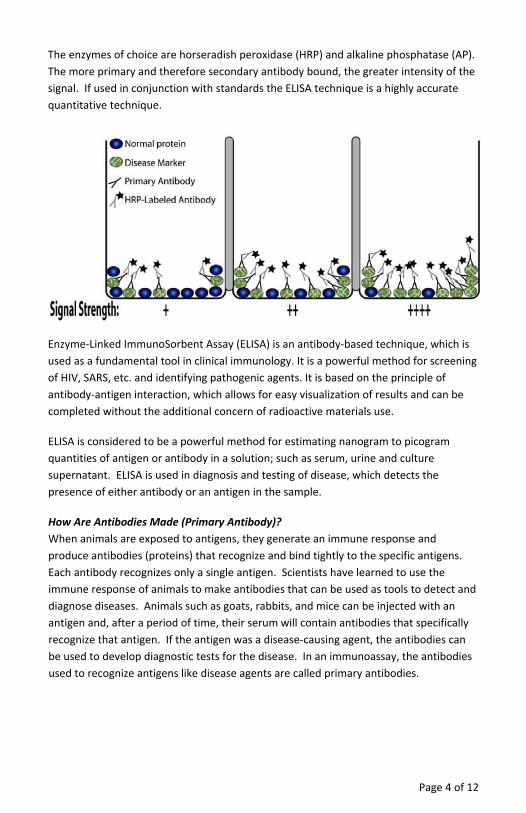
The key feature of ELISA is the use of immunodetection to identify a specific protein, for example a protein marker for a disease. The proteins are immobilized in a protein binding well and non‐specific sites are then blocked. The blocking step is used to increase the specificity of the ELISA technique by preventing non‐specific interactions. If the wells are not blocked then the antibodies can stick to non‐specific proteins due to their charge. To prevent this, the wells are incubated with a protein mixture and the proteins block the charges that would attract the antibodies. Several blocking agents are used, including dried milk powder, bovine serum albumin and casein; however modern blocking agents use synthetic and/or non‐animal proteins to prevent any cross reaction with the animal antibodies. An example of a non animal blocker is the provided NAP‐Blocker™.
Once blocked, the wells can be probed with a primary antibody, an antibody specific for the protein of interest. Once bound the antibody is visualized, either with a specific tag coupled to the primary antibody or with a secondary antibody. The secondary antibody is a general antibody that recognizes the constant domain of immunoglobulin G and is species specific. So, if the primary antibody is a mouse antibody, the secondary antibody used will recognize all mouse antibodies. If a secondary antibody is used then this will carry the tag that allows visualization of the protein (see figure below).
The most common tags used are enzymes that catalyze a substrate to produce either light that is detected with radiography film, or color that is visualized on the membrane.
Page 4 of 12
The enzymes of choice are horseradish peroxidase (HRP) and alkaline phosphatase (AP). The more primary and therefore secondary antibody bound, the greater intensity of the signal. If used in conjunction with standards the ELISA technique is a highly accurate quantitative technique.
Enzyme‐Linked ImmunoSorbent Assay (ELISA) is an antibody‐based technique, which is used as a fundamental tool in clinical immunology. It is a powerful method for screening of HIV, SARS, etc. and identifying pathogenic agents. It is based on the principle of antibody‐antigen interaction, which allows for easy visualization of results and can be completed without the additional concern of radioactive materials use.
ELISA is considered to be a powerful method for estimating nanogram to picogram quantities of antigen or antibody in a solution; such as serum, urine and culture supernatant. ELISA is used in diagnosis and testing of disease, which detects the presence of either antibody or an antigen in the sample.
How Are Antibodies Made (Primary Antibody)? When animals are exposed to antigens, they generate an immune response and produce antibodies (proteins) that recognize and bind tightly to the specific antigens. Each antibody recognizes only a single antigen. Scientists have learned to use the immune response of animals to make antibodies that can be used as tools to detect and diagnose diseases. Animals such as goats, rabbits, and mice can be injected with an antigen and, after a period of time, their serum will contain antibodies that specifically recognize that antigen. If the antigen was a disease‐causing agent, the antibodies can be used to develop diagnostic tests for the disease. In an immunoassay, the antibodies used to recognize antigens like disease agents are called primary antibodies.
Page 5 of 12
Enzyme Labeled Antibodies (Secondary Antibodies) Secondary antibodies recognize and bind to primary antibodies in immunoassays (e.g. ELISA, Western blots). Secondary antibodies are prepared in the same manner as primary antibodies and the antigens are antibodies from a different species, normally a fragment containing the constant (conserved) domain.
Specific enzymes, such as horseradish peroxidase (HRP) and alkaline phosphatase (AP), are then chemically coupled to the constant domain of the antibody, away from the antigen binding domain. The enzymes are able to catalyze a chemical substrate to produce either a chemiluminescence (light) or colorimetric (color) product that can be detected. This experiment uses HRP and a colorimetric substrate known as 3,3’,5,5’‐tetramethylbenzidine (TMB).
In this study, students carry out a pseudo‐diagnostic experiment to examine 3 clinical samples and compare them to known standards to determine the severity of the disease. A real world example of this test is the screening for HIV antibodies, ELISA is used to test for the presence of HIV antibodies and to determine the levels of the antibodies. In this case ELISA is used to a) detect for the presence of HIV and b) determine the severity of the infection, HIV or AIDS.
TEACHER’SPREEXPERIMENTSETUP1. Make a 1X ELISA Washing Buffer solution by adding 40ml 10X ELISA Washing Buffer
to a 0.5‐liter container. Bring up to final volume of 400ml with DI water
The ELISA Washing Buffer is PBS (Phosphate buffered saline) supplemented with a mild detergent to aid in membrane washing.
2. The morning of the experiment, gently shake the supplied 20ml Blocking Buffer (2X NAP‐Blocker) bottle to mix and then mix equal volumes of Blocking Buffer (2X NAP‐Blocker) with 20ml 1X ELISA Washing Buffer. Supply each group with 6ml Blocking Buffer (NAP‐Blocker) in an appropriately labeled tube.
3. Add 2ml 1X Blocking Buffer to the ELISA Standard vial. Soak for 5 minutes with periodically vortexing to dissolve completely.
4. Label 6 tubes with “10U Standards” and aliquot in 330μl ELISA Standard. Supply each group with one vial.
5. Add 1ml 1X Blocking Buffer to each of the simulated ELISA Sample vials. Soak for 5 minutes with periodically vortexing to dissolve completely.
6. Label three sets of 6 tubes for each ELISA sample and aliquot 150μl of each ELISA sample. Supply each group with one vial of each sample.
Page 6 of 12
7. Add 300μl sterile water to the BE Antibody 1 (Primary Antibody) and BE Antibody 4 (HRP Secondary) vials. Soak the antibody for 5 minutes with periodically vortexing to dissolve the antibody completely.
8. Label two sets of 6 tubes for each antibody, label “primary” and “secondary” and aliquot 50μl antibody solution into each corresponding vial. Supply each group with one vial of each antibody.
9. Label six tubes with “ELISA Substrate” and aliquot 1.6ml ELISA Substrate into each tube. Supply each group with one tube.
MATERIALSFOREACHGROUPSupply each group with the following components. Components shared by the whole class and should be kept on a communal table.
NOTE: Place the Assay Strip Holder on a shaker. For incubation and shaking of Assay Strips, instruct students to position the Assay Strips in the Strip Holder after addition of reagents.
Page 7 of 12
PROCEDURE1. Prepare the ELISA Standard by making appropriate dilutions. First label 4 tubes 0U,
2.5U, 5U and 7.5U. These numbers refer to relative units of infection. The tube labeled “0” is the blank for the experiment. The supplied ELISA Standard is 10 Relative Units (10U).
2. Add the amount of Blocking Buffer and then ELSA Standard to the tubes labeled 0U, 2.5U, 5U and 7.5U as indicated in the table below. Vortex or vigorously shake to mix.
Blocking Buffer ELISA Standard (10U)
0U 150μl 0μl
2.5U 110μl 40μl
5U 75μl 75μl
7.5U 40μl 110μl
3. Bind Antigen: Place Assay Strips on a clean surface. Bind the antigens to the ELISA strips by adding 50μl standards and patient samples to the wells as shown in the table below. The assay is performed in duplicate.
NOTE: When adding any reagent to the well ensure the pipette tip is at the bottom of the well. This prevents artifacts occurring due to residual reagents remaining on well walls.
Well # Assay Strip 1 Assay Strip 2
1 0U Standard (Blank) 0U Standard (Blank)
2 2.5U Standard 2.5U Standard
3 5U Standard 5U Standard
4 7.5U Standard 7.5U Standard
5 10U Standard 10U Standard
6 Sample 1 Sample 1
7 Sample 2 Sample 2
8 Sample 3 Sample 3
4. Carefully, place the Assay Strips in the Strip Holder, according to your teacher’s instructions, and incubate with shaking for 0.5‐1 hour at room temperature.
5. After incubation, discard liquid from wells by inverting plate and gently shaking out the liquid over a sink or tray.
Page 8 of 12
6. Wash each well by adding 300μl 1X ELISA Washing Buffer and return to the Strip Holder and shake for 5 minutes.
7. After 5 minutes, discard ELISA Washing Buffer from wells as before and repeat the step 6 once more.
8. Blocking Stage: Add 200μl 1X Blocking Buffer (NAP‐Blocker) into each well and incubate with shaking for 0.5‐1 hour at room temperature.
If necessary, this is a convenient stopping point. Wrap the ELISA plate in plastic film, such as saran wrap or cling film, and store in a fridge. Do not store longer than a week.
9. After incubation, discard Blocking Buffer (NAP‐Blocker) and wash each well with 300μl of ELISA Washing Buffer as before. Repeat wash once.
10. During wash stages, prepare ELISA Primary Antibody. Add 1ml of 1X Blocking Buffer (NAP‐Blocker) to the tube containing 50μl of Primary Antibody. Vortex to mix well.
11. ELISA Primary Antibody: Add 50μl of the Primary Antibody solution to each well and incubate with shaking for 0.5‐1 hour at room temperature. Ensure the pipette touches the bottom of the well on addition of antibody.
If necessary, this is a convenient stopping point. Wrap the ELISA plate in plastic film, such as saran wrap or cling film, and store in a fridge. Do not store longer than overnight.
12. After incubation, discard antibody solution and wash each well with 300μl of ELISA Washing Buffer as before. Repeat wash three times.
13. During wash stages, prepare ELISA Secondary Antibody. Add 1ml of Blocking Buffer (NAP‐Blocker) to the tube containing 50μl of Secondary Antibody (HRP Labeled). Vortex to mix well.
14. Add 50μl of the Secondary Antibody solution to each well and incubate with shaking for 0.5‐1 hour at room temperature. Ensure the pipette touches the bottom of the well on addition of antibody.
If necessary, this is a convenient stopping point. Wrap the ELISA plate in plastic film, such as saran wrap or cling film, and store in a fridge. Do not store longer than overnight.
15. After incubation, discard antibody and wash each well with 300μl of ELISA Washing Buffer as before. Repeat wash three times.
Page 9 of 12
16. Add 100μl ELISA Substrate to each well and shake for 10‐30 minutes at room temperature. Monitor the plate to observe color change.
17. A blue color develops, examine the wells and record the intensity of blue color of the sample wells compared to the ELISA Standards.
18. OPTIONAL: If a microplate reader is available, read the plate at a wavelength of 650nm, using the first well of each strip as a blank. Record your results.
RESULTS,ANALYSIS&ASSESSMENT1. What does ELISA stand for?
ELISA stands for Enzyme‐Linked Immunosorbent Assay.
2. Why is secondary antibody labeled with enzymes used in this immunoassay?
Enzymes provide a way to see whether the primary antibody has attached to its target (antigen) in the microplate well. Primary and secondary antibodies are invisible, so a detection method is necessary. The enzyme HRP is linked to the secondary antibody. HRP reacts with a colorless substrate in a chemical reaction that turns blue. If the secondary antibody is present in the well, the color change indicates a positive result.
3. What is an example of a disease that attacks the human immune system?
Diseases that attack the immune system includes autoimmune diseases e.g., rheumatoid arthritis, lupus, asthma, and AIDS etc.
4. Describe the results after adding the substrate into micro‐wells containing the diseased samples:
A blue color developed depending on the severity of the disease condition.
5. Using the relative units of infection, plot a standard curve of Absorbance against Relative Units. What is the level of infection of each patient?
6. Describe the severity of the disease as seen in the three simulated clinical samples?
Students explain using the data from their results.
7. Why it is important to wash the wells after every step?
Washing removes any proteins that have not bound to the micro‐wells and any antibodies that have not bound to their targets, thus preventing unbound proteins (either antigen or antibodies) from giving false positive result.
Page 10 of 12
Page 11 of 12
8. Describe why enzymes are used in immunoassays.
Enzymes are used as they are able to rapidly catalyze substrates to produce either a chemiluminescence or colorimetric product, allowing the quantity of antibody, and therefore antigen, to be calculated.
TROUBLESHOOTINGProblem Possible Cause Solution
High Background Insufficient Washing
1. Ensure all wash steps are carried out for at least five minutes.
2. Increase number and length of wash steps.
Too much signal, all wells turned dark blue
Insufficient Washing
1. Ensure all wash steps are carried out for at least five minutes.
OBJECTIVES• To understand the principle of the Enzyme Linked ImmunoSorbent Assay (ELISA),
an immunoquantification technique. • To run ELISA using the supplied buffers, antibodies and detection reagent. • To establish its importance in identifying disease conditions.
INTRODUCTIONThe study of the immune system and the mechanism how the body protects itself against foreign molecules is known as Immunology. Mammalian immune systems produce molecules, called antibodies that recognize foreign molecules in the host system. Molecules that cause your body to mount an immune response are called antigens, and may include components of infectious agents like bacteria, viruses, and fungi. Within days, millions of antibodies that recognize the antigen are formed and attach themselves to their target antigens flagging them for destruction by other cells of the immune system.
Antibodies have incredible specificity to locate and attach themselves to their targets and make the foreign particles recognizable to other cells of the immune system, so that they can be destroyed. Antibodies have become a valuable research and diagnostic tool, used in biotechnology and disease diagnosis/treatment.
The key feature of ELISA is the use of immunodetection to identify a specific protein, for example a protein marker for a disease. The proteins are immobilized in a protein binding well and non‐specific sites are then blocked. The blocking step is used to increase the specificity of the ELISA technique by preventing non‐specific interactions. If the wells are not blocked then the antibodies can stick to non‐specific proteins due to their charge. To prevent this, the wells are incubated with a protein mixture and the proteins block the charges that would attract the antibodies. Several blocking agents are used, including dried milk powder, bovine serum albumin and casein; however modern blocking agents use synthetic and/or non‐animal proteins to prevent any cross reaction with the animal antibodies. An example of a non animal blocker is the provided NAP‐Blocker™.
Once blocked, the wells can be probed with a primary antibody, an antibody specific for the protein of interest. Once bound the antibody is visualized, either with a specific tag coupled to the primary antibody or with a secondary antibody. The secondary antibody is a general antibody that recognizes the constant domain of immunoglobulin G and is species specific. So, if the primary antibody is a mouse antibody, the secondary antibody used will recognize all mouse antibodies. If a secondary antibody is used then this will carry the tag that allows visualization of the protein (see figure below).
The most common tags used are enzymes that catalyze a substrate to produce either light that is detected with radiography film, or color that is visualized on the membrane.
Page 3 of 12
The enzymes of choice are horseradish peroxidase (HRP) and alkaline phosphatase (AP). The more primary and therefore secondary antibody bound, the greater intensity of the signal. If used in conjunction with standards the ELISA technique is a highly accurate quantitative technique.
Enzyme‐Linked ImmunoSorbent Assay (ELISA) is an antibody‐based technique, which is used as a fundamental tool in clinical immunology. It is a powerful method for screening of HIV, SARS, etc. and identifying pathogenic agents. It is based on the principle of antibody‐antigen interaction, which allows for easy visualization of results and can be completed without the additional concern of radioactive materials use.
ELISA is considered to be a powerful method for estimating nanogram to picogram quantities of antigen or antibody in a solution; such as serum, urine and culture supernatant. ELISA is used in diagnosis and testing of disease, which detects the presence of either antibody or an antigen in the sample.
How Are Antibodies Made (Primary Antibody)? When animals are exposed to antigens, they generate an immune response and produce antibodies (proteins) that recognize and bind tightly to the specific antigens. Each antibody recognizes only a single antigen. Scientists have learned to use the immune response of animals to make antibodies that can be used as tools to detect and diagnose diseases. Animals such as goats, rabbits, and mice can be injected with an antigen and, after a period of time, their serum will contain antibodies that specifically recognize that antigen. If the antigen was a disease‐causing agent, the antibodies can be used to develop diagnostic tests for the disease. In an immunoassay, the antibodies used to recognize antigens like disease agents are called primary antibodies.
Page 4 of 12
Enzyme Labeled Antibodies (Secondary Antibodies) Secondary antibodies recognize and bind to primary antibodies in immunoassays (e.g. ELISA, Western blots). Secondary antibodies are prepared in the same manner as primary antibodies and the antigens are antibodies from a different species, normally a fragment containing the constant (conserved) domain.
Specific enzymes, such as horseradish peroxidase (HRP) and alkaline phosphatase (AP), are then chemically coupled to the constant domain of the antibody, away from the antigen binding domain. The enzymes are able to catalyze a chemical substrate to produce either a chemiluminescence (light) or colorimetric (color) product that can be detected. This experiment uses HRP and a colorimetric substrate known as 3,3’,5,5’‐tetramethylbenzidine (TMB).
In this study, students carry out a pseudo‐diagnostic experiment to examine 3 clinical samples and compare them to known standards to determine the severity of the disease. A real world example of this test is the screening for HIV antibodies, ELISA is used to test for the presence of HIV antibodies and to determine the levels of the antibodies. In this case ELISA is used to a) detect for the presence of HIV and b) determine the severity of the infection, HIV or AIDS.
MATERIALSFOREACHGROUPSupply each group with the following components. Components shared by the whole class and should be kept on a communal table.
NOTE: Place the Assay Strip Holder on a shaker. For incubation and shaking of Assay Strips, instruct students to position the Assay Strips in the Strip Holder after addition of reagents.
Page 5 of 12
PROCEDURE1. Prepare the ELISA Standard by making appropriate dilutions. First label 4 tubes 0U,
2.5U, 5U and 7.5U. These numbers refer to relative units of infection. The tube labeled “0” is the blank for the experiment. The supplied ELISA Standard is 10 Relative Units (10U).
2. Add the amount of Blocking Buffer and then ELSA Standard to the tubes labeled 0U, 2.5U, 5U and 7.5U as indicated in the table below. Vortex or vigorously shake to mix.
Blocking Buffer ELISA Standard (10U)
0U 150μl 0μl
2.5U 110μl 40μl
5U 75μl 75μl
7.5U 40μl 110μl
3. Bind Antigen: Place Assay Strips on a clean surface. Bind the antigens to the ELISA strips by adding 50μl standards and patient samples to the wells as shown in the table below. The assay is performed in duplicate.
NOTE: When adding any reagent to the well ensure the pipette tip is at the bottom of the well. This prevents artifacts occurring due to residual reagents remaining on well walls.
Well # Assay Strip 1 Assay Strip 2
1 0U Standard (Blank) 0U Standard (Blank)
2 2.5U Standard 2.5U Standard
3 5U Standard 5U Standard
4 7.5U Standard 7.5U Standard
5 10U Standard 10U Standard
6 Sample 1 Sample 1
7 Sample 2 Sample 2
8 Sample 3 Sample 3
4. Carefully, place the Assay Strips in the Strip Holder, according to your teacher’s instructions, and incubate with shaking for 0.5‐1 hour at room temperature.
5. After incubation, discard liquid from wells by inverting plate and gently shaking out the liquid over a sink or tray.
Page 6 of 12
6. Wash each well by adding 300μl 1X ELISA Washing Buffer and return to the Strip Holder and shake for 5 minutes.
7. After 5 minutes, discard ELISA Washing Buffer from wells as before and repeat the step 6 once more.
8. Blocking Stage: Add 200μl 1X Blocking Buffer (NAP‐Blocker) into each well and incubate with shaking for 0.5‐1 hour at room temperature.
If necessary, this is a convenient stopping point. Wrap the ELISA plate in plastic film, such as saran wrap or cling film, and store in a fridge. Do not store longer than a week.
9. After incubation, discard Blocking Buffer (NAP‐Blocker) and wash each well with 300μl of ELISA Washing Buffer as before. Repeat wash once.
10. During wash stages, prepare ELISA Primary Antibody. Add 1ml of 1X Blocking Buffer (NAP‐Blocker) to the tube containing 50μl of Primary Antibody. Vortex to mix well.
11. ELISA Primary Antibody: Add 50μl of the Primary Antibody solution to each well and incubate with shaking for 0.5‐1 hour at room temperature. Ensure the pipette touches the bottom of the well on addition of antibody.
If necessary, this is a convenient stopping point. Wrap the ELISA plate in plastic film, such as saran wrap or cling film, and store in a fridge. Do not store longer than overnight.
12. After incubation, discard antibody solution and wash each well with 300μl of ELISA Washing Buffer as before. Repeat wash three times.
13. During wash stages, prepare ELISA Secondary Antibody. Add 1ml of Blocking Buffer (NAP‐Blocker) to the tube containing 50μl of Secondary Antibody (HRP Labeled). Vortex to mix well.
14. Add 50μl of the Secondary Antibody solution to each well and incubate with shaking for 0.5‐1 hour at room temperature. Ensure the pipette touches the bottom of the well on addition of antibody.
If necessary, this is a convenient stopping point. Wrap the ELISA plate in plastic film, such as saran wrap or cling film, and store in a fridge. Do not store longer than overnight.
15. After incubation, discard antibody and wash each well with 300μl of ELISA Washing Buffer as before. Repeat wash three times.
Page 7 of 12
16. Add 100μl ELISA Substrate to each well and shake for 10‐30 minutes at room temperature. Monitor the plate to observe color change.
17. A blue color develops, examine the wells and record the intensity of blue color of the sample wells compared to the ELISA Standards.
18. OPTIONAL: If a microplate reader is available, read the plate at a wavelength of 650nm, using the first well of each strip as a blank. Record your results.
Page 8 of 12
RESULTS,ANALYSIS&ASSESSMENT1. What does ELISA stand for?


![ENZYME-LINKED IMMUNOSORBENT ASSAY [ELISA]¡Enzyme-linked immunosorbent assay. ¡Is a biochemical plate-based assay technique designed for detecting and quantifying substances such](https://static.documents.pub/doc/80x56/5f4f5b992afa395c6303586c/enzyme-linked-immunosorbent-assay-elisa-enzyme-linked-immunosorbent-assay-is.jpg)
