Immunotoxins and Neurotrophins: Novel Strategies for the Efficient Expression of Recombinant Proteins Dem Fachbereich Chemie der Universität Kaiserslautern zur Erlangung des akademischen Grades “Doktor der Naturwissenschaften” vorgelegte Dissertation DISSERTATION (D 386) Zhuoyu Li Betreuer: Prof. Dr. Wolfgang E. Trommer Prof. Dr. Jingming Yuan Kaiserslautern 2002
Transcript
Immunotoxins and Neurotrophins: Novel Strategies for the Efficient Expression of Recombinant Proteins
Dem Fachbereich Chemie der Universität Kaiserslautern
zur Erlangung des akademischen Grades
“Doktor der Naturwissenschaften” vorgelegte Dissertation
DISSERTATION (D 386)
Zhuoyu L i
Betreuer: Prof. Dr. Wolfgang E. Trommer Prof. Dr. Jingming Yuan
Kaiserslautern 2002
Die vorliegende Arbeit entstand zwischen Dezember 1997 und Juni 2001 im Fachbereich Chemie,
Abteilung Biochemie der Unversität Kaiserslautern, Deutschland und Biotechnologie Zentrum der
Universität Shanxi, Taiyuan, China.
Promotionskommission:
Vorsitzender: Prof. Dr. Dr. D Schrenk
1. Berichterstatter: Prof. Dr. W. E. Trommer
2. Berichterstatter: Prof. Dr. J. Yuan
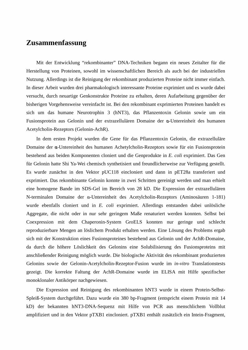
Zusammenfassung
Mit der Entwicklung “ rekombinanter” DNA-Techniken begann ein neues Zeitalter für die
Herstellung von Proteinen, sowohl im wissenschaftlichen Bereich als auch bei der industriellen
Nutzung. Allerdings ist die Reinigung der rekombinant produzierten Proteine nicht immer einfach.
In dieser Arbeit wurden drei pharmakologisch interessante Proteine exprimiert und es wurde dabei
versucht, durch neuartige Genkonstrukte Proteine zu erhalten, deren Aufarbeitung gegenüber der
bisherigen Vorgehensweise vereinfacht ist. Bei den rekombinant exprimierten Proteinen handelt es
sich um das humane Neurotrophin 3 (hNT3), das Pflanzentoxin Gelonin sowie um ein
Fusionsprotein aus Gelonin und der extrazellulären Domaine der � -Untereinheit des humanen
Acetylcholin-Rezeptors (Gelonin-AchR).
In dem ersten Projekt wurden die Gene für das Pflanzentoxin Gelonin, die extrazelluläre
Domaine der � -Untereinheit des humanen Achetylcholin-Rezeptors sowie für ein Fusionsprotein
bestehend aus beiden Komponenten cloniert und die Genprodukte in E. coli exprimiert. Das Gen
für Gelonin hatte Shi Ya-Wei chemisch synthetisiert und freundlicherweise zur Verfügung gestellt.
Es wurde zunächst in den Vektor pUC118 eincloniert und dann in pET28a transferiert und
exprimiert. Das rekombinante Gelonin konnte in zwei Schritten gereinigt werden und man erhielt
eine homogene Bande im SDS-Gel im Bereich von 28 kD. Die Expression der extrazellulären
N-terminalen Domaine der � -Untereinheit des Acetylcholin-Rezeptors (Aminosäuren 1-181)
wurde ebenfalls cloniert und in E. coli exprimiert. Allerdings entstanden dabei unlösliche
Aggregate, die nicht oder in nur sehr geringem Maße renaturiert werden konnten. Selbst bei
Coexpression mit dem Chaperonin-System GroELS konnten nur geringe und schlecht
reproduzierbare Mengen an löslichem Produkt erhalten werden. Eine Lösung des Problems ergab
sich mit der Konstruktion eines Fusionsproteines bestehend aus Gelonin und der AchR-Domaine,
da durch die höhere Löslichkeit des Gelonins eine Solubilisierung des Fusionsproteins mit
anschließender Reinigung möglich wurde. Die biologische Aktivität des rekombinant produzierten
Gelonins sowie der Gelonin-Acetylcholin-Rezeptor-Fusion wurde im in-vitro Translationstests
gezeigt. Die korrekte Faltung der AchR-Domaine wurde im ELISA mit Hilfe spezifischer
monoklonaler Antikörper nachgewiesen.
Die Expression und Reinigung des rekombinanten hNT3 wurde in einem Protein-Selbst-
Spleiß-System durchgeführt. Dazu wurde ein 380 bp-Fragment (entspricht einem Protein mit 14
kD) der bekannten hNT3-DNA-Sequenz mit Hilfe von PCR aus menschlichem Vollblut
amplifiziert und in den Vektor pTXB1 eincloniert. pTXB1 enthält zusätzlich ein Intein-Fragment,
an dem eine Chitin-Bindungsproteindomaine (CBD) anfusioniert ist. Ein ähnliches Konstrukt,
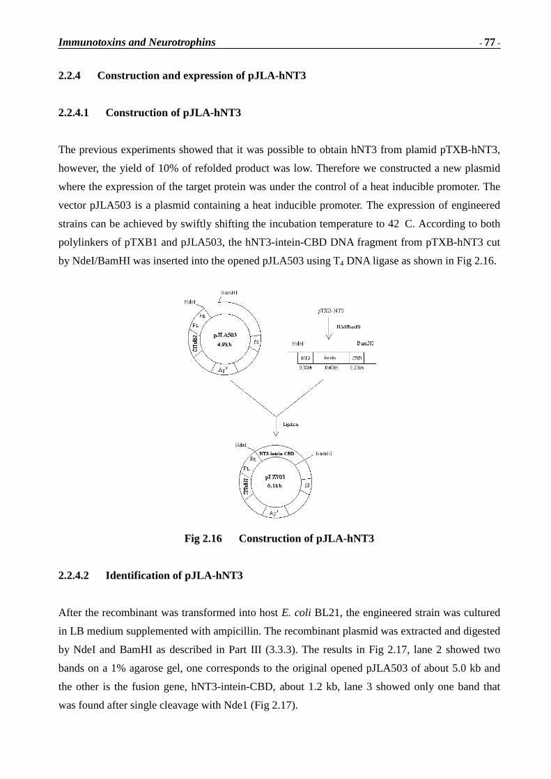
pJLA-hNT3, wurde zusätzlich hergestellt, bei dem die Genexpression unter einen
Hitzeschockpromotor gestellt ist. Von beiden Konstrukten konnte das Zielprotein
hNT3-intein-CBD entweder nach Induktion mit IPTG oder nach Hitzeschock erhalten werden. Das
exprimierte Protein akkumulierte in Aggregatform und wurde nach Denaturierung und
Renaturierung als lösliches Fusionsprotein auf einer Chitin-Affinitätssäule gereinigt. Nach
Spaltung in Gegenwart von DTT wurde ein 14 kD Protein erhalten, das dem hNT3 entspricht.
Sowohl das hNT3 als auch das Fusionsprotein hNT3-intein-CBD zeigten dieselbe biologische
Aktivität basierend auf Wachstumsassays an dorsalen Ganglien in Hühnerembryonen.
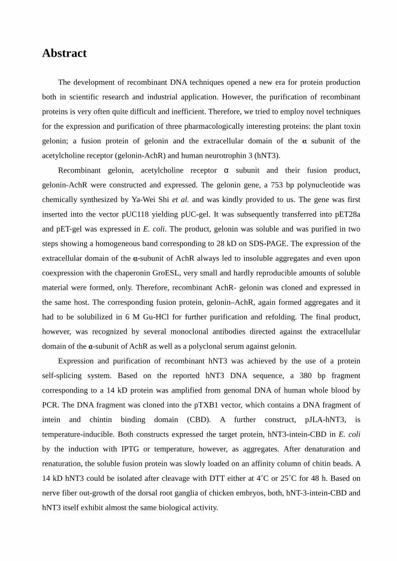
Abstract
The development of recombinant DNA techniques opened a new era for protein production
both in scientific research and industrial application. However, the purification of recombinant
proteins is very often quite difficult and inefficient. Therefore, we tried to employ novel techniques
for the expression and purification of three pharmacologically interesting proteins: the plant toxin
gelonin; a fusion protein of gelonin and the extracellular domain of the � subunit of the
acetylcholine receptor (gelonin-AchR) and human neurotrophin 3 (hNT3).
Recombinant gelonin, acetylcholine receptor α subunit and their fusion product,
gelonin-AchR were constructed and expressed. The gelonin gene, a 753 bp polynucleotide was
chemically synthesized by Ya-Wei Shi et al. and was kindly provided to us. The gene was first
inserted into the vector pUC118 yielding pUC-gel. It was subsequently transferred into pET28a
and pET-gel was expressed in E. coli. The product, gelonin was soluble and was purified in two
steps showing a homogeneous band corresponding to 28 kD on SDS-PAGE. The expression of the
extracellular domain of the � -subunit of AchR always led to insoluble aggregates and even upon
coexpression with the chaperonin GroESL, very small and hardly reproducible amounts of soluble
material were formed, only. Therefore, recombinant AchR- gelonin was cloned and expressed in
the same host. The corresponding fusion protein, gelonin–AchR, again formed aggregates and it
had to be solubilized in 6 M Gu-HCl for further purification and refolding. The final product,
however, was recognized by several monoclonal antibodies directed against the extracellular
domain of the � -subunit of AchR as well as a polyclonal serum against gelonin.
Expression and purification of recombinant hNT3 was achieved by the use of a protein
self-splicing system. Based on the reported hNT3 DNA sequence, a 380 bp fragment
corresponding to a 14 kD protein was amplified from genomal DNA of human whole blood by
PCR. The DNA fragment was cloned into the pTXB1 vector, which contains a DNA fragment of
intein and chintin binding domain (CBD). A further construct, pJLA-hNT3, is
temperature-inducible. Both constructs expressed the target protein, hNT3-intein-CBD in E. coli
by the induction with IPTG or temperature, however, as aggregates. After denaturation and
renaturation, the soluble fusion protein was slowly loaded on an affinity column of chitin beads. A
14 kD hNT3 could be isolated after cleavage with DTT either at 4˚C or 25˚C for 48 h. Based on
nerve fiber out-growth of the dorsal root ganglia of chicken embryos, both, hNT-3-intein-CBD and
hNT3 itself exhibit almost the same biological activity.
Contents Part I Immunotoxins…….……………………………………………………...…….1
1A Gene clone, expression and char acter istics………………………….1
3.5.4 The inhibition of protein synthesis in vitro………………………….104
3.6 Literature………………………………………………………………………105
Abbreviations………………………………………………………………………………..106
Acknowledgements………………………………………………………………………..109
Curr iculum Vitae………………………………………………………………………….110
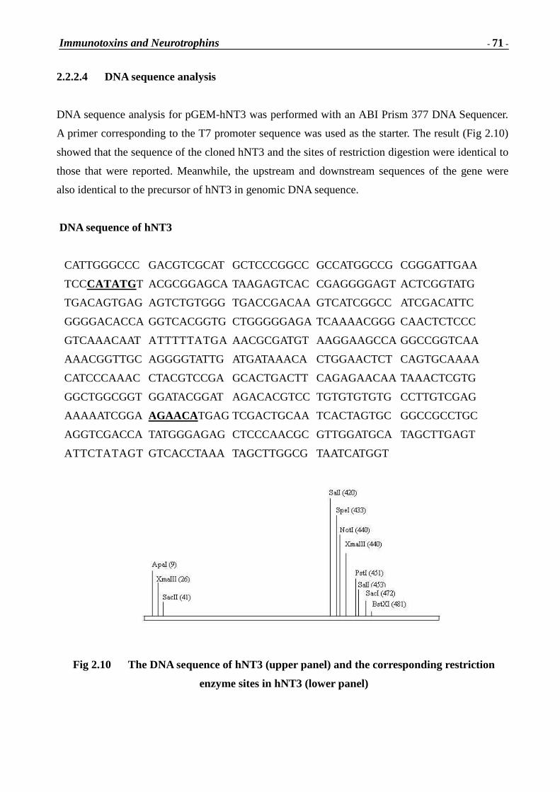
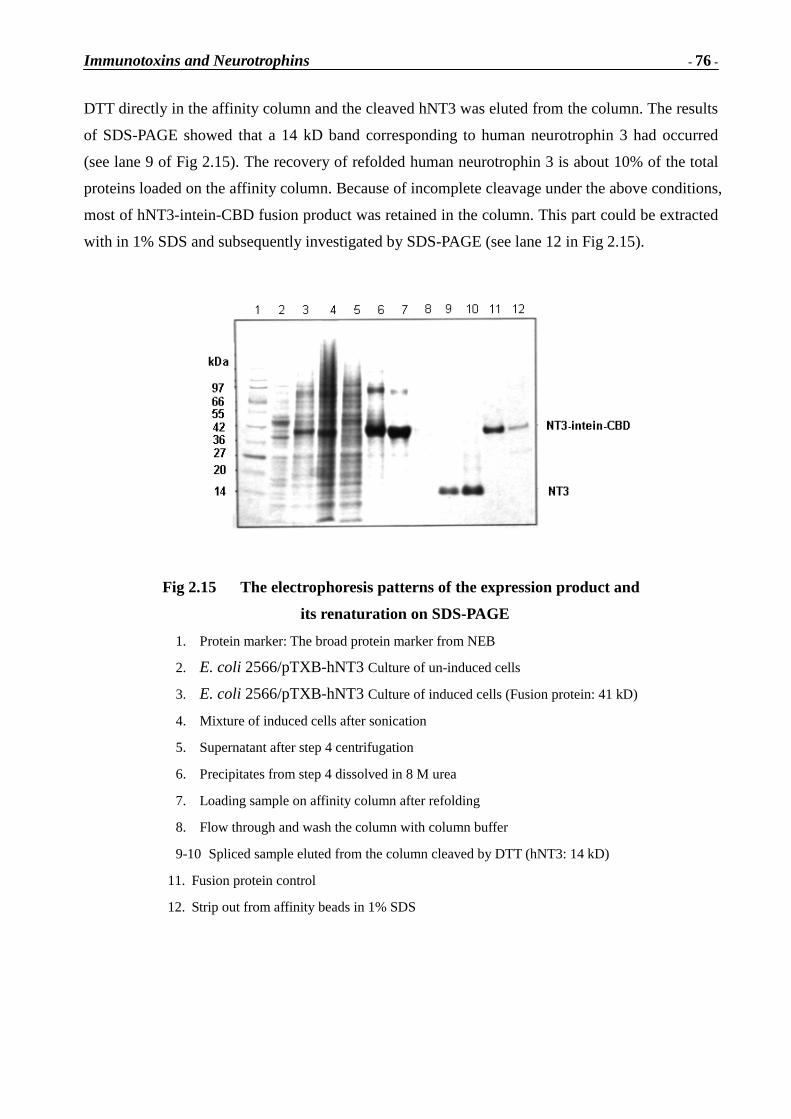
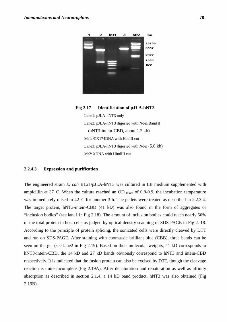
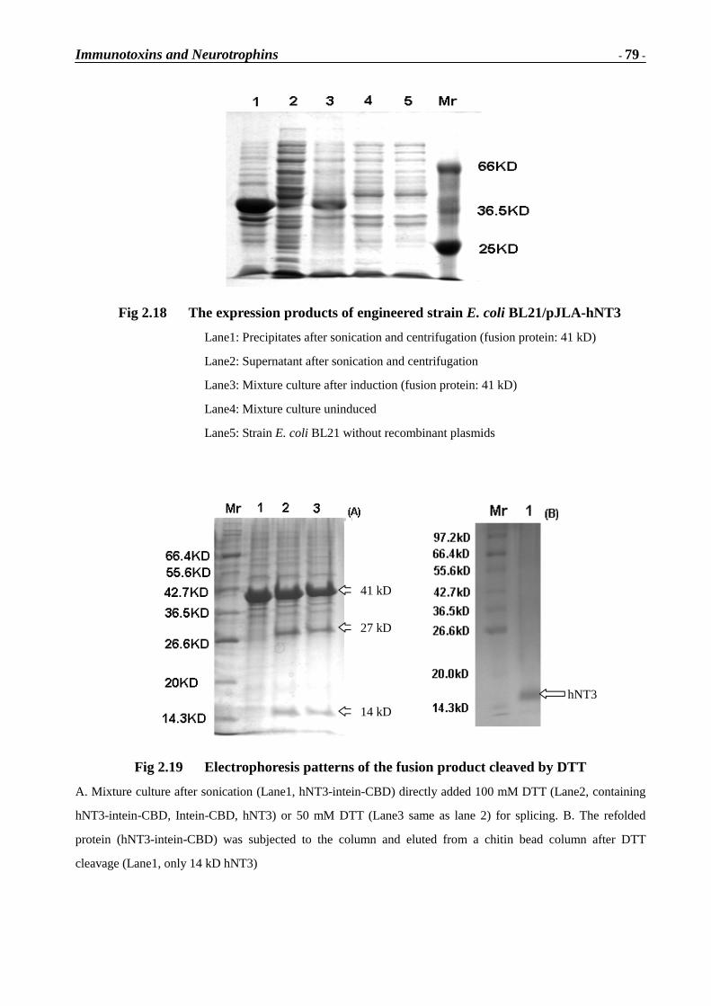
Immunotoxins and Neurotrophins
- 1 -
Part I Immunotoxins
Conjugates composed of tissue-specific monoclonal antibodies and toxins have found wide interest
as potential anti-cancer drugs. Far less common is the similar approach to use conjugates of
antigens with toxins for the treatment of autoimmune diseases. In this case, the auto-antigen would
direct the toxin to its target, antigen-specific lymphocytes, which would be selectively eliminated
resulting in an antigen-specific suppression of the immune system. This approach, as initially
suggested by Géza Filipp, was successfully taken in the laboratory of Trommer in Kaiserslautern
[1] in the case of experimental autoimmune Myasthenia gravis (EAMG). Myasthenic rats were
treated with a conjugate composed of the acetylcholine receptor (AchR) and the plant toxin
gelonin leading to a substantial recovery of functional receptors in the neuromuscular endplates of
these animals. Neither the receptor nor the toxin alone had comparable effects. However, the
antibody titer against the receptor raised during treatment. A possible reason could have been
immune reactions against pathophysiologically irrelevant parts of the receptor from Torpedo which
had been used in the conjugates [1]. Hence, in the current work, recombinant fragments of the � -subunit, which contains the main immunogenic region (MIR) will be employed.
1A Gene clone, expression and character istics of gelonin
1A.1 Ribosome inactivating proteins
Ribosome inactivating proteins (RIPs) are bio-macromolecules that specifically interfere with
eukaryotic protein translation. Most plant and bacterial RIPs exert their effects through
catalytically and irreversibly inactivating the 60S subunit of eukaryotic ribosomes [2]. Plant RIPs
can be categorized into two groups: type-I RIPs, most of them being glycoproteins such as gelonin,
bryodin or trichosanthin, are single-chain proteins with molecular weight of about 30 kD. However,
type-II RIPs, such as ricin or abrin, consist of two polypeptide chains (A and B) which are linked
by a disulfide bridge. The enzymatic property of type-II RIPs is associated with the A chain, while
the B chain is like a lectin, which facilitates entry of the toxin into intact cells by binding
non-specifically to galactose moieties on the cell surface [3, 4, 5].
Immunotoxins and Neurotrophins
- 2 -
1A.1.1 Toxicity
The potent cytotoxicity of heterodimeric ricin was first described over a century ago. The
mechanism by which RIPs inactivate 60S ribosomal subunits was elucidated by Endo and his
colleagues in 1987 [1, 6]. The mechanism was first deduced from ricin A chain, but has not been
shown to operate for all plant RIPs and for bacterial toxins such as Shigella toxin and the
Shiga-like toxins from certain enteropathogenic strains of E. coli. Ricin A chain is an RNA specific
N-glycosidase that hydrolytically cleaves a single N-glycosidic bond from among over four
thousand nucleoside bases present in 28S rRNA (4800 bases). A specific adenine residue is
removed (A-4324 in the case of rat liver 28S rRNA) resulting in the inability of the ribosome to
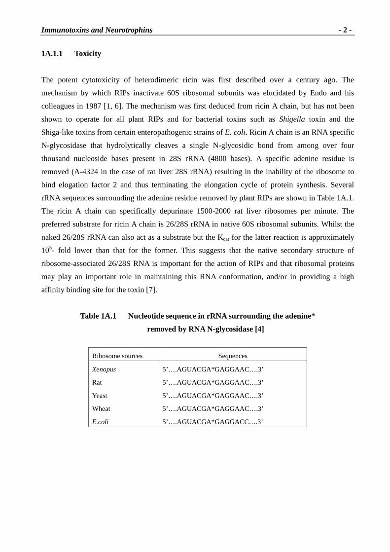
bind elogation factor 2 and thus terminating the elongation cycle of protein synthesis. Several
rRNA sequences surrounding the adenine residue removed by plant RIPs are shown in Table 1A.1.
The ricin A chain can specifically depurinate 1500-2000 rat liver ribosomes per minute. The
preferred substrate for ricin A chain is 26/28S rRNA in native 60S ribosomal subunits. Whilst the
naked 26/28S rRNA can also act as a substrate but the Kcat for the latter reaction is approximately
105- fold lower than that for the former. This suggests that the native secondary structure of
ribosome-associated 26/28S RNA is important for the action of RIPs and that ribosomal proteins
may play an important role in maintaining this RNA conformation, and/or in providing a high
affinity binding site for the toxin [7].
Table 1A.1 Nucleotide sequence in rRNA sur rounding the adenine*
removed by RNA N-glycosidase [4]
Ribosome sources Sequences
Xenopus
Rat
Yeast
Wheat
E.coli
5’….AGUACGA*GAGGAAC….3’
5’….AGUACGA*GAGGAAC….3’
5’….AGUACGA*GAGGAAC….3’
5’….AGUACGA*GAGGAAC….3’
5’….AGUACGA*GAGGACC….3’
Immunotoxins and Neurotrophins
- 3 -
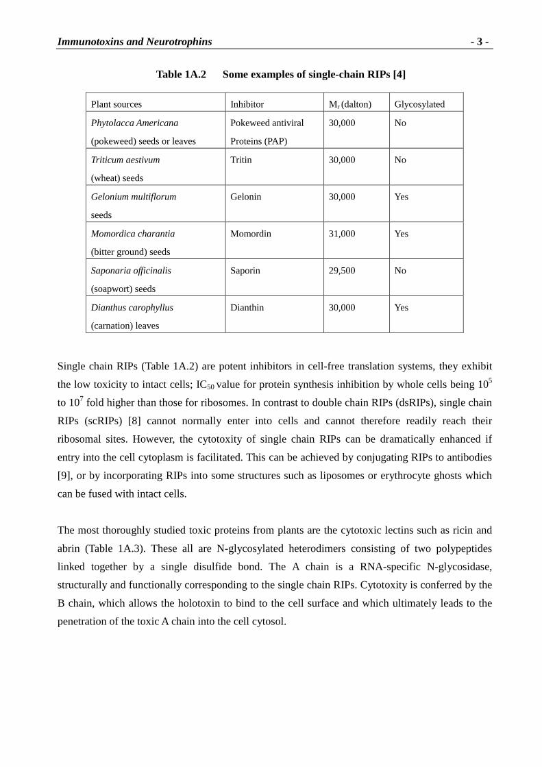
Table 1A.2 Some examples of single-chain RIPs [4]
Single chain RIPs (Table 1A.2) are potent inhibitors in cell-free translation systems, they exhibit
the low toxicity to intact cells; IC50 value for protein synthesis inhibition by whole cells being 105
to 107 fold higher than those for ribosomes. In contrast to double chain RIPs (dsRIPs), single chain
RIPs (scRIPs) [8] cannot normally enter into cells and cannot therefore readily reach their
ribosomal sites. However, the cytotoxity of single chain RIPs can be dramatically enhanced if
entry into the cell cytoplasm is facilitated. This can be achieved by conjugating RIPs to antibodies
[9], or by incorporating RIPs into some structures such as liposomes or erythrocyte ghosts which
can be fused with intact cells.
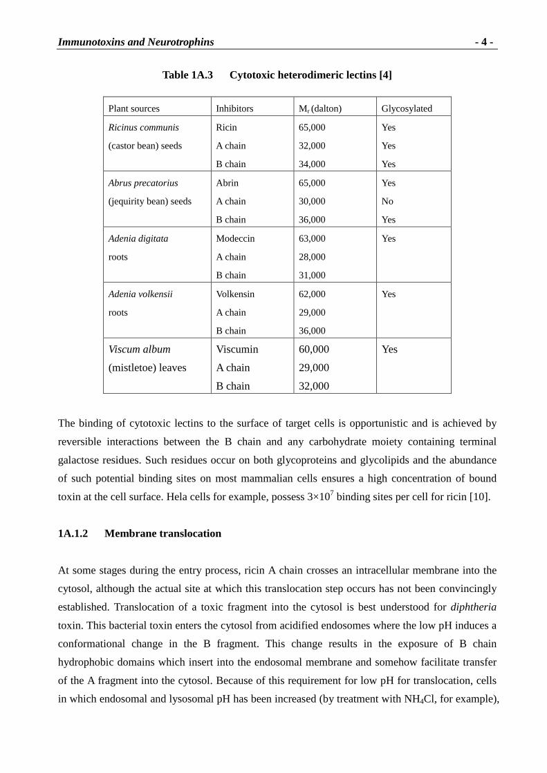
The most thoroughly studied toxic proteins from plants are the cytotoxic lectins such as ricin and
abrin (Table 1A.3). These all are N-glycosylated heterodimers consisting of two polypeptides
linked together by a single disulfide bond. The A chain is a RNA-specific N-glycosidase,
structurally and functionally corresponding to the single chain RIPs. Cytotoxity is conferred by the
B chain, which allows the holotoxin to bind to the cell surface and which ultimately leads to the
penetration of the toxic A chain into the cell cytosol.
Plant sources Inhibitor Mr (dalton) Glycosylated
Phytolacca Americana
(pokeweed) seeds or leaves
Pokeweed antiviral
Proteins (PAP)
30,000 No
Triticum aestivum
(wheat) seeds
Tritin 30,000 No
Gelonium multiflorum
seeds
Gelonin 30,000 Yes
Momordica charantia
(bitter ground) seeds
Momordin 31,000 Yes
Saponaria officinalis
(soapwort) seeds
Saporin 29,500 No
Dianthus carophyllus
(carnation) leaves
Dianthin 30,000 Yes
Immunotoxins and Neurotrophins
- 4 -
Table 1A.3 Cytotoxic heterodimer ic lectins [4]
Plant sources Inhibitors Mr (dalton) Glycosylated
Ricinus communis
(castor bean) seeds
Ricin
A chain
B chain
65,000
32,000
34,000
Yes
Yes
Yes
Abrus precatorius
(jequirity bean) seeds
Abrin
A chain
B chain
65,000
30,000
36,000
Yes
No
Yes
Adenia digitata
roots
Modeccin
A chain
B chain
63,000
28,000
31,000
Yes
Adenia volkensii
roots
Volkensin
A chain
B chain
62,000
29,000
36,000
Yes
Viscum album
(mistletoe) leaves
Viscumin
A chain
B chain
60,000
29,000
32,000
Yes
The binding of cytotoxic lectins to the surface of target cells is opportunistic and is achieved by
reversible interactions between the B chain and any carbohydrate moiety containing terminal
galactose residues. Such residues occur on both glycoproteins and glycolipids and the abundance
of such potential binding sites on most mammalian cells ensures a high concentration of bound
toxin at the cell surface. Hela cells for example, possess 3×10⁷ binding sites per cell for ricin [10].
1A.1.2 Membrane translocation
At some stages during the entry process, ricin A chain crosses an intracellular membrane into the
cytosol, although the actual site at which this translocation step occurs has not been convincingly
established. Translocation of a toxic fragment into the cytosol is best understood for diphtheria
toxin. This bacterial toxin enters the cytosol from acidified endosomes where the low pH induces a
conformational change in the B fragment. This change results in the exposure of B chain
hydrophobic domains which insert into the endosomal membrane and somehow facilitate transfer
of the A fragment into the cytosol. Because of this requirement for low pH for translocation, cells
in which endosomal and lysosomal pH has been increased (by treatment with NH4Cl, for example),
Immunotoxins and Neurotrophins
- 5 -
or mutant cell lines defective in the acidification of endosomes, are resistant to diphtheria toxin.
Although the endosome has also been implicated as the intracellular site of ricin translocation, it
appears that the endocytosis of ricin continues beyond the endosomal stage before translocation
takes place. Treatments which increase endosomal pH do not reduce ricin toxicity, and the lag time
between cellular exposure to ricin and measurable decrease in protein synthetic activity (60-90 min)
is considerably longer than that for the bacterial toxin. Several studies using immunoelectron
microscopy have shown that endocytosed ricin is first delivered to the endosomes and a fraction
subsequently appears within the Golgi complex, in particular the trans-Golgi network [11]. There
is now considerable experimental evidence that ricin must reach the Golgi complex in order to
exert its cytotoxic effects. A number of treatments that induce morphological changes as the
disruption of the Golgi by brefeldin A, inhibit the cytotoxicity of ricin and pseudomonas exotoxin
A, while having no effect on the cytotoxicity of diphtheria toxin [12].
Ricin B chain is required for the efficient translocation of ricin A chain into the cytosol, beyond its
role in the initial cell surface binding of the holotoxin [13]. This B chain requirement is not
absolute however, since many immunotoxins containing ricin A chain alone are potently cytotoxic.
Clearly the A chain can cross a membrane in the absence of B chain, which argues against the B
chain having a direct function in the translocation step. It is possible that the B chain facilitates
translocation indirectly by delivering the A chain to a translocationally-competent compartment. If
this is the case, the association with the B chain might effectively prevent the A chain from
inserting into membranes until such a compartment has been reached. The intracellular role of ricin
B chain envisaged above could result from the B chain binding to intracellular
galactose-containing components (receptor), particularly in the trans-Golgi. Such galactose binding
could reduce the amount of ricin recycled back to the cell surface, it might allow ricin to move
from the Golgi to the ER as discussed above, or it could have a more direct role in membrane
translocation within the Golgi complex. In these situations, the galactose binding sites are clearly
important for the B chain’s intracellular role in cellular intoxication [14, 15].
1A.1.3 Antiviral activity
The first single chain RIP to be isolated and studied was pokeweed antiviral protein (PAP). It was
known that leaf extracts from several plants, including Phytolacca americana (pokeweed), when
mixed with a suspension of tobacco mosaic virus, prevent the mechanical transmission of viral
infection to other plants. The pokeweed antiviral factor was purified and identified as a single
chain RIP. PAP also prevented animal virus replication in mammalian cells, where it was found to
Immunotoxins and Neurotrophins
- 6 -
inhibit protein synthesis by virally infected cells at PAP concentrations which did not affect normal
cells. All RIPs are known to be more toxic to virus-infected cells than to non-infected cells,
apparently because infection permeabilizes the host and allows the toxins to penetrate into the
cytosol [16].
From early studies on the antiviral activity of plant extracts it was concluded that while these
extracts prevented viral infection in other plant species, they did not prevent infection of the very
plants from which they derived. This suggested that the antiviral principle did not act directly on
the virus but that its effectiveness was due to some action on the infected plant. Now, it has been
clearly shown that pokeweed ribosomes are sensitive to PAP [17]. A study of the intracellular
location of PAP has shown that this RIP is heavily sequestered in the cell wall of pokeweed cells
[18], a location consistent with its proposed anti-viral role. Thus viral infection of pokeweed cells
might provoke a damage limitation exercise in which the infected cells become permeable to their
own extra-cellular RIP that enters the cytosol and depurinates the pokeweed ribosomes and thereby
prevents viral replication.
1A.1.4 Ribosome specificities
The active site was identified by x-ray analysis of the ricin A chain and confirmed by site-directed
mutagenesis. This region is highly conserved in RIPs (Table 1A.4). Their ribosomal substrate
specificities however, can be very different, for example, mammalian and yeast ribosomes are very
sensitive to ricin A chain, plant ribosomes much less so, and prokaryotic ribosomes are completely
insensitive. In spite of the fact that the consensus sequence around the target adenine is also highly
conserved in all 23, 26, 28S rRNAs, the sensitivity of a single type of ribosome to different RIPs
also varies markedly, for example, wheat germ ribosomes are relatively insensitive to ricin A chain
but are 1000 to 10,000 times more sensitive to the single chain RIP dianthin.
The most dramatic variation in sensitivity which has emerged recently is that E. coli ribosomes
were believed to be completely insensitive to all RIPs. Indeed, E. coli has been successfully used
as host to produce recombinant ricin A chain and abrin A chain [19]. These proteins were produced
cytoplasmically where they accounted for up to 10% of the total bacterial protein without affecting
bacterial growth. Mirabilis antiviral protein (MAP) cytoplasmically in E. coli resulted in severely
inhibited growth of the host, caused by the recombinant product, and the yield of product was very
low. Subsequently MAP was shown to inhibit protein synthesis by E. coli ribosomes in vitro, in
contrast to the effect of the ricin or abrin A chain [20]. More recently it was shown that both PAP
Immunotoxins and Neurotrophins
- 7 -
and dianthin likewise inhibit protein synthesis by E. coli ribosomes. These single chain RIPs are
able to specifically depurinate E. coli 23S rRNA at the expected site (Table 1A.1). The adenine
residue -glycosidically removed from 23S rRNA by the RIPs is known to be a key binding residue
for EF-G and EF-Tu [21].
Table 1A.4 Compar ison of active site region of plant toxins [4]
RIP Residue position Amino acid sequence
Ricin A chain 172-185 CIQMISEA*ARFQYI
Abrin A chain 158-171 IIQMVSEA*ARFRYI
PAP 170-183 AIQMVSEA*ARFKYI
MAP 163-176 AIQMVSEA*ARFKYI
Saporin 171-184 AIQMTAEA*ARFRYI
Diarithin 152-165 AIQMTAEA*ARFRYI
1A.1.5 Gelonin
Gelonin extracted from seeds of the plant Gelonium multiflorum belongs to type-I ribosome
inactivating proteins. Gelonin is glycosylated with terminal mannose residues. Due to lack of the
B-chain domain binding the cell surface, it normally does not enter intact cells. Even if it enters
cells via a different mechanism, e.g. as a conjugate, it exhibits a different intracellular distribution,
compared with type-II RIPs. Gelonin is assumed to be trapped in the endosomal/lysosomal
compartment, a location explaining its relatively low toxicity. In cell-free systems, however,
gelonin has the powerful N-glycosidase activity on eukaryotic ribosomes by releasing
adenine-4324 from a vital region of the 28S rRNA unit [20].
Table 1A.5 The amino acid sequence of gelonin precursor
10 20 30 40 50
MKGNMKVYWI KIAVATWFCC T T I V L G ST A R I FSL PT N D EE ETSK TL GL DT
60 70 80 90 100
V SFST K GAT Y ITYVNFLNEL RV K L K PEGN S H GI PL L RK K C DDPGK CFV LV
110 120 130 140 150
A LSNDNGQLA EIA IDV TSVY V V GY QV RNRS Y FFK DA PDA A Y EGL FK N T I K
Immunotoxins and Neurotrophins
- 8 -
160 170 180 190 200
TRL H FGGSY P SLEGEKAYRE T T D L G I E P L R I GI K K L D EN A I D N Y K PT EI A
210 220 230 240 250
SSL LV V I QM V SEA A RFTFIE NQI RNNFQQR I R PA N N T I SL ENKWGKLSFQ
260 270 280 290 300
I RTSGA NGM F SEAVELERAN GKKY Y V TAVD QV K PK I A L L K FV DK DPK TSL
310 316
A A EL I I QN Y E S L V G F D
*This sequence is from Swiss-prot P33186. The residue G from 47 is the N-terminus of recombinant gelonin
and K at the stop of 297 is the C-terminus.
1A.1.5.1 The pr imary structure
Gelonin consists of 251 amino acid residues, similar to other scRIPs such as monorcharin (247 aa)
and the A-chain of dsRIPs for instance ricin A-chain (267 aa). Though the conserved amino acid
residues among them are less than 40%, their structure is the same from the results of different
RIPs cross-reaction with specific antiserum. In vivo, an inactive precursor of gelonin is first
biosynthesized and then transported to safe compartments within the cell. Finally, the functional
gelonin is produced by glycosylation with the post-translational modification. The primary
structure of the precursor is as shown above Table 1A.5 [21].
1A.1.5.2 The conformation
Hosur et al. published a three-dimensional structure of gelonin at 1.8 Å resolution [22], which is
basically identical to the conformation of ricin A and � -momorcharin (Fig 1A.1).
Fig 1A.1 Superposition of gelonin, r icin A and � -momorchar in [22]
The stereoview shows C�tracing: bold, gelonin; Mediun, ricin A; light, α-momorcharin. Every 10th residue of
gelonin is marked. Differences are mostly in the loop regions.
Immunotoxins and Neurotrophins
- 9 -
As shown in Fig 1A.1, structurally, gelonin belongs to the (� +�
) class of proteins. With respect to
the �
strands, �
1, �
4, �
5, �
6, �
7 and �
8 form a mixed �
sheet, and �
2, �
3 as well as �
9, �
10 form two
antiparallel beta ribbons, while the helix regions, except for short segments of type 310 (123 to 125
and 237 to 239) are all of the � -helix type. Thus, there are two distinct structural domains in the
gelonin molecule, one large and a small one. The former consists of residues 3-32 and 40-187
while the latter is composed of residues 33-39 and 188-247.
1A.1.5.3 Catalytic mechanism
Active site From the varies studies as X-ray diffraction, site-directed mutagenesis and chemical
modification etc, the active site residues of gelonin are Tyr74, Arg169, Gly111, Glu166, Tyr113
and Trp198. There is a number of hydrogen bonding interactions between these residues, such as
Arg169/Glu166. In addition, the water molecules also participate in the catalytic reaction, there are
at least two hydrogen bonds from water to protein atoms.
Glycosylation Gelonin is a glycoprotein with terminal mannose residues. It is known that
Asn82 and Asn189 are two potential glycosylation sites in the N-terminal glycosylation of gelonin.
Catalytic mechanism As a glycosidase, it is the cleavage of the N-glycosidic bond of a specific
adenine in 28S rRNA from the 60S subunit of eukaryotic ribosomes. It has been shown that the
true substrate is a ribo-oligonuclotide hairpin in which the double helical stem is at least three base
pairs long and the connecting hairpin loop contains the sequence GAGA. The first adenine will be
removed by RIPs. The removal of adenine is a hydrolysis reaction and involves addition of a water
molecule to the substrate. Based on the structure, two kinds of molecular catalytic mechanisms
have been suggested. Both include an oxycarbonium ion intermediate which is generated by
different chemical steps. However, in both methods a water molecular is required for nucleophilic
attack on the C1’ atom of the intermediate [23].
1A.1.6 Research goals
Toxins as generally utilized in immunoconjugates belong to a group of enzymes that inhibit the
protein biosynthesis [24]. Gelonin, a plant toxin from Gelonium multiflorum has successfully been
employed in such approaches including its application in antigen-specific immune suppression [1].
However, it is not easily available from natural sources. Recombinant gelonin has been reported
[21] but the clone is not available from the authors. In their work the cDNA was obtained from
Immunotoxins and Neurotrophins
- 10 -
highly fragmented mRNA isolated from the seeds of Gelonium multiflorum. Based on the
published sequence of gelonin, Ya-Wei Shi et al. synthesized the gene chemically in four
fragments which were kindly provided to us [25]. In these constructs some of the bases had been
substituted for the corresponding nucleotides in the published DNA sequence in order to yield
optimized codons for the same amino acids in E. coli. These DNA fragments will now be fused
and used for the expression of gelonin.
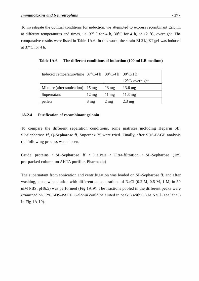
1A.2 Results and discussion
1A.2.1 Gene clone of gelonin
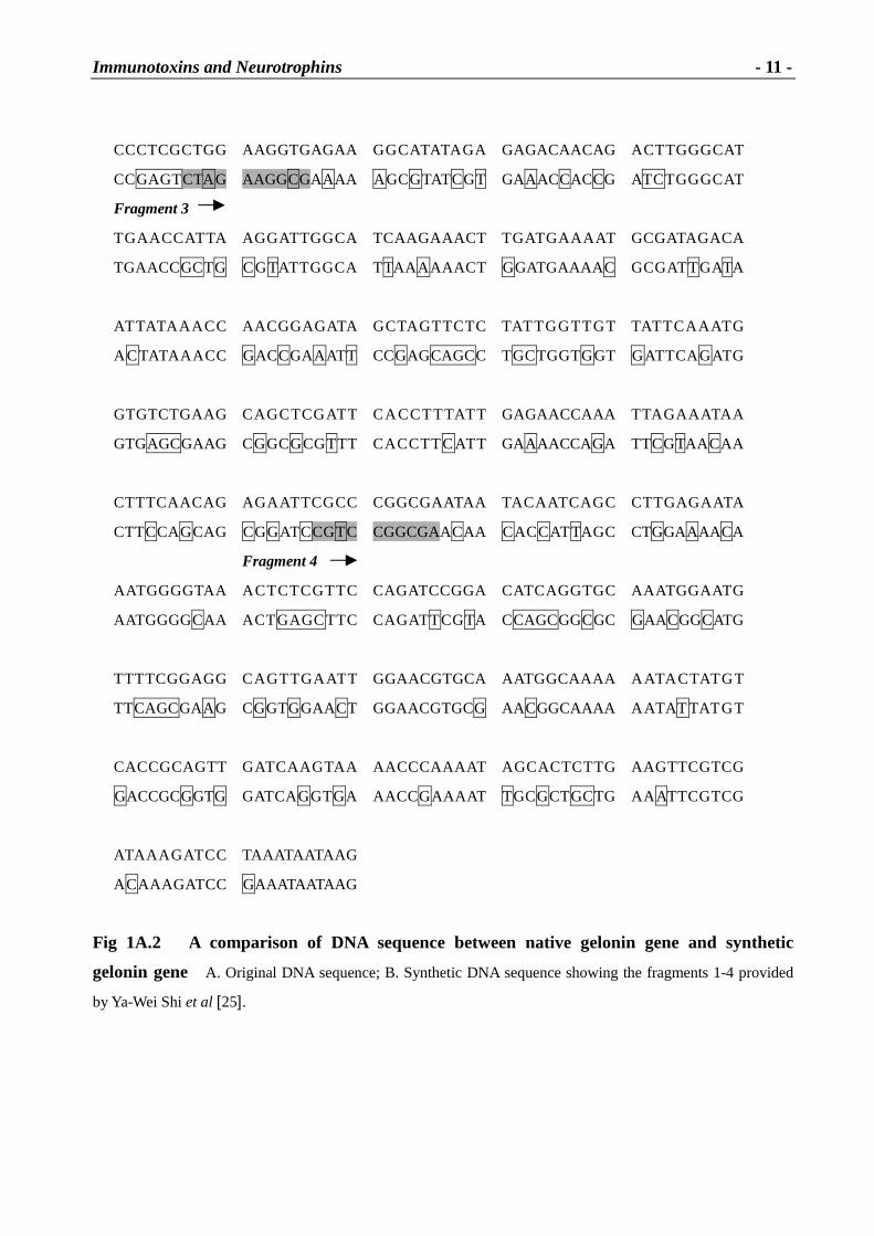
The gene coding for gelonin was kindly provided by Ya-Wei Shi et al. in 4 consecutive fragments
as shown in Fig 1A.2. Ya-Wei Shi et al. also provided the two recombinant plasmids pUC-gel I and
pUC-gel II containing gelonin fragments 1+2 and 3+4 respectively [25].
A. DNA-
B DNA- ATG
GGCCT GGA C
GGCCT GGA T
ACCGTGAGCT
ACCGTGAGCT
TTA GCA CTA A
TCAGCACCAA
AGGTGCCACT
AGGCGCCACC
Fragment 1
TATAT TA CCT
TATAT TA CCT
A CGTGA ATTT
ATGTGA A CTT
CTTGA ATGAG
CCTGAACGAA
CTA CGA GTTA
CTGCGTGTGA
AATTGAAACC
AA CTGAAACC
CGAAGGTAAC
GGAAGGCAAC
AGCCATGGAA
AGCCATGGCA
TCCCATTGCT
TTCCGCTGCT
GCGCAAAAAA
GCGTAAAAAA
TGTGATGATC
TGCGATGATC
CTGGAAAGTG
CGGGCAAATG
TTTCGTTTTG
CTTCGTGCTG
GTA GCGCTTT
GTGGCGCTGA
CAAATGACAA
GCAA CGATAA
TGGACAGTTG
CGGCCAGCTA
Fragment 2
GCGGAAATAG
GCGGAAATTG
CTATA GATGT
CGAT T GATGT
TA CA A GTGTT
GACCAGCGTG
TATGTGGTGG
TATGTGGTGG
GCTATCA A GT
GCTATCA GGT
AAGAAACAGA
GCGTAACCGT
T CT TA CTTCT
A GCTAT TTCT
TTA A A GATGC
TCAAAGATGC
TCCA GATGCT
GCCGGATGCG
GCTTACGAAG
GCGTATGAAG
GCCTCTTCAA
GCCTGTTCAA
A A A CA CA ATT
AA ACA CCATT
AAAACAAGAC
AAAACCCGTC
TTCATTTTGG
TGCATTTTCGG
CGGCAGCTAT
CGGCAGCTAT
Immunotoxins and Neurotrophins
- 11 -
CCCTCGCTGG
CCGAGTCTA G
AAGGTGAGAA
AAGGCGAAAA
GGCATATA GA
A GCGTATCGT
GAGACAACAG
GAAACCACCG
A CTTGGGCAT
ATCTGGGCAT
Fragment 3
TGA A CCATTA
TGAACCGCTG
AGGATTGGCA
CGTATTGGCA
TCAAGAAACT
TTAA A AAACT
TGATGA A A AT
GGATGAAAAC
GCGATAGACA
GCGATTGATA
ATTATA A A CC
A CTATAA A CC
AACGGAGATA
GACCGA A ATT
GCTA GTTCTC
CCGAGCAGCC
TATTGGTTGT
TGCTGGTGGT
TATTCA A ATG
GATTCA GATG
GTGTCTGAAG
GTGAGCGAAG
CA GCTCGATT
CGGCGCGTTT
CA CCTTTATT
CA CCTT CATT
GAGAACCAAA
GAAAACCAGA
TTA GA A ATA A
TTCGTAA CAA
CTTTCAACAG
CTTCCA GCAG
A GA ATTCGCC
CGGATCCGTC
CGGCGAATAA
CGGCGAACAA
TA CA ATCA GC
CACCATTAGC
CTTGAGA ATA
CTGGAAAACA
Fragment 4
AATGGGGTAA
AATGGGGCAA
A CTCTCGTTC
A CTGAGCTTC
CAGATCCGGA
CAGATTCGTA
CATCAGGTGC
CCAGCGGCGC
AAATGGAATG
GAACGGCATG
TTTTCGGAGG
TTCAGCGAA G
CA GTTGA ATT
CGGTGGAACT
GGAACGTGCA
GGAACGTGCG
AATGGCAAAA
AACGGCAAAA
A ATA CTATGT
A ATAT TATGT
CACCGCAGTT
GACCGCGGTG
GATCA A GTA A
GATCA GGTGA
AACCCAAAAT
AACCGAAAAT
A GCACTCTTG
TGCGCTGCTG
AAGTTCGTCG
AA ATTCGTCG
ATA A A GATCC
A CAAAGATCC
TAAATAATAAG
GAAATAATAAG
Fig 1A.2 A compar ison of DNA sequence between native gelonin gene and synthetic
gelonin gene A. Original DNA sequence; B. Synthetic DNA sequence showing the fragments 1-4 provided
by Ya-Wei Shi et al [25].
Immunotoxins and Neurotrophins
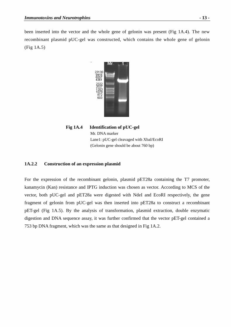
- 12 -
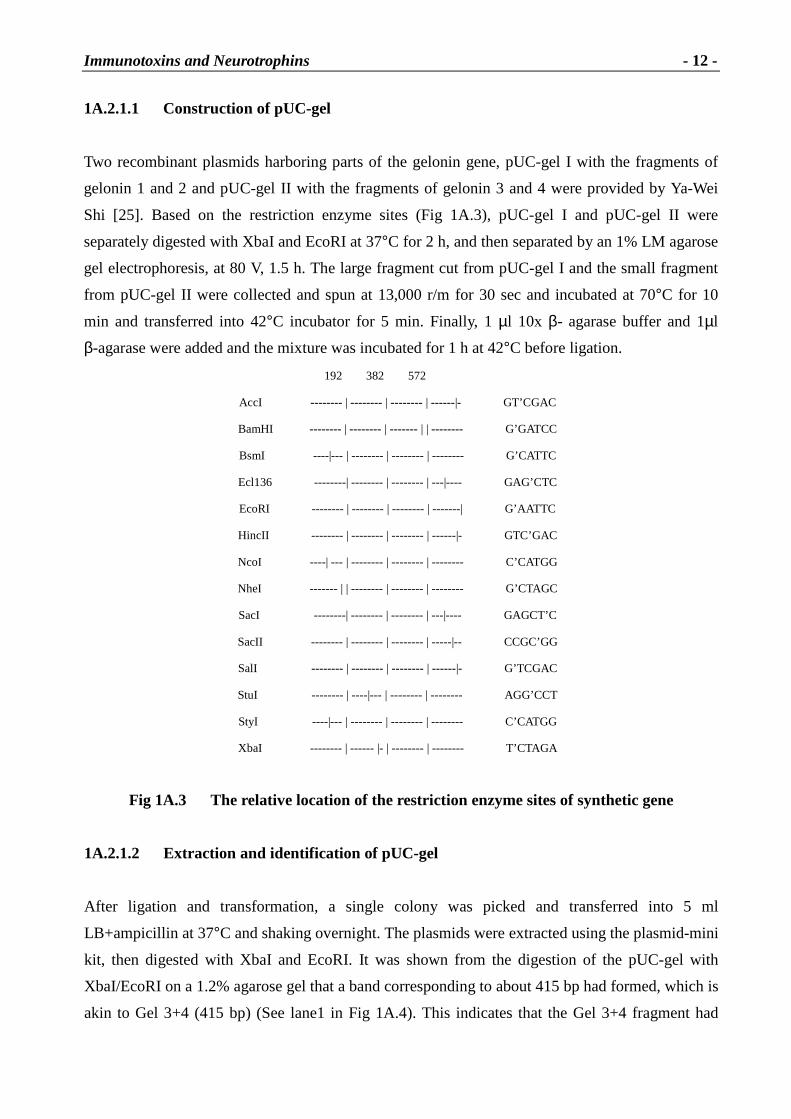
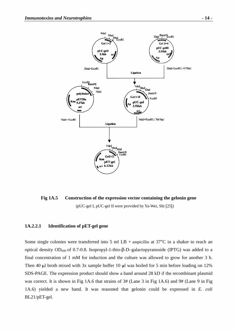
1A.2.1.1 Construction of pUC-gel
Two recombinant plasmids harboring parts of the gelonin gene, pUC-gel I with the fragments of
gelonin 1 and 2 and pUC-gel II with the fragments of gelonin 3 and 4 were provided by Ya-Wei
Shi [25]. Based on the restriction enzyme sites (Fig 1A.3), pUC-gel I and pUC-gel II were
separately digested with XbaI and EcoRI at 37°C for 2 h, and then separated by an 1% LM agarose
gel electrophoresis, at 80 V, 1.5 h. The large fragment cut from pUC-gel I and the small fragment
from pUC-gel II were collected and spun at 13,000 r/m for 30 sec and incubated at 70°C for 10
min and transferred into 42°C incubator for 5 min. Finally, 1 µl 10x β- agarase buffer and 1µl
β-agarase were added and the mixture was incubated for 1 h at 42°C before ligation.
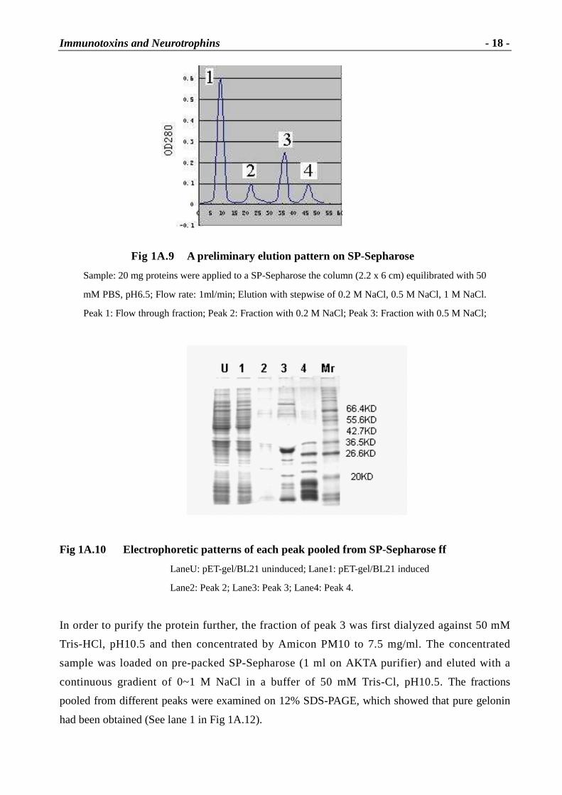
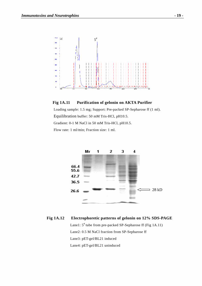
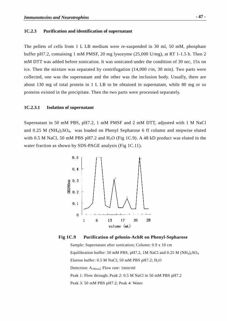
Fig 1A.12 Electrophoretic patter ns of gelonin on 12% SDS-PAGE
Lane1: 5# tube from pre-packed SP-Sepharose ff (Fig 1A.11)
Lane2: 0.5 M NaCl fraction from SP-Sepharose ff
Lane3: pET-gel/BL21 induced
Lane4: pET-gel/BL21 uninduced
28 kD
5#
Immunotoxins and Neurotrophins
- 20 -
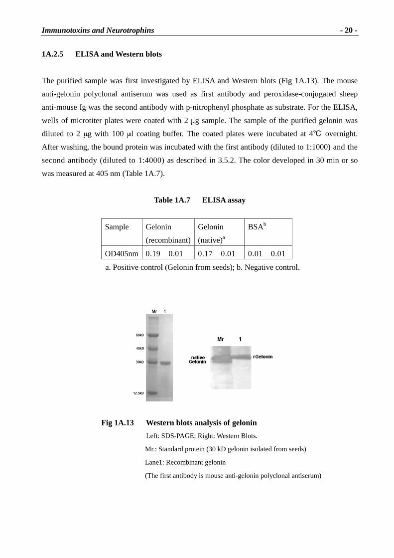
1A.2.5 ELISA and Western blots
The purified sample was first investigated by ELISA and Western blots (Fig 1A.13). The mouse
anti-gelonin polyclonal antiserum was used as first antibody and peroxidase-conjugated sheep
anti-mouse Ig was the second antibody with p-nitrophenyl phosphate as substrate. For the ELISA,
wells of microtiter plates were coated with 2 � g sample. The sample of the purified gelonin was
diluted to 2 � g with 100 � l coating buffer. The coated plates were incubated at 4� overnight.
After washing, the bound protein was incubated with the first antibody (diluted to 1:1000) and the
second antibody (diluted to 1:4000) as described in 3.5.2. The color developed in 30 min or so
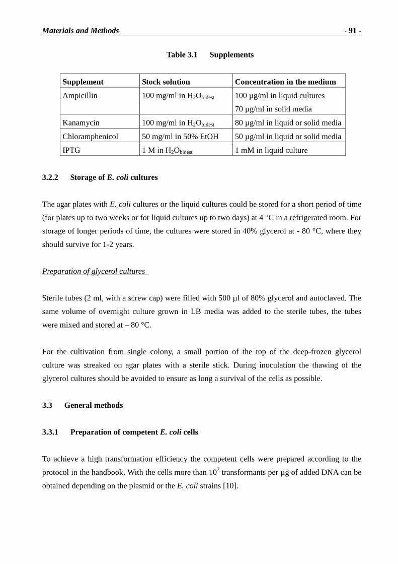
was measured at 405 nm (Table 1A.7).
Table 1A.7 ELISA assay
Sample Gelonin
(recombinant)
Gelonin
(native)a
BSAb
OD405nm 0.19�0.01 0.17�0.01 0.01�0.01
a. Positive control (Gelonin from seeds); b. Negative control.
Fig 1A.13 Western blots analysis of gelonin
Left: SDS-PAGE; Right: Western Blots.
Mr.: Standard protein (30 kD gelonin isolated from seeds)
Lane1: Recombinant gelonin
(The first antibody is mouse anti-gelonin polyclonal antiserum)
Immunotoxins and Neurotrophins
- 21 -
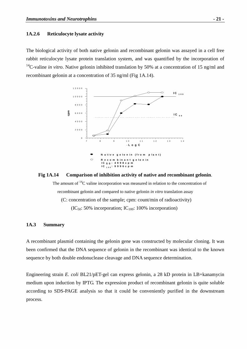
1A.2.6 Reticulocyte lysate activity
The biological activity of both native gelonin and recombinant gelonin was assayed in a cell free
rabbit reticulocyte lysate protein translation system, and was quantified by the incorporation of 14C-valine in vitro. Native gelonin inhibited translation by 50% at a concentration of 15 ng/ml and
recombinant gelonin at a concentration of 35 ng/ml (Fig 1A.14).
Fig 1A.14 Compar ison of inhibition activity of native and recombinant gelonin.
The amount of 14C valine incorporation was measured in relation to the concentration of
recombinant gelonin and compared to native gelonin in vitro translation assay
(C: concentration of the sample; cpm: count/min of radioactivity)
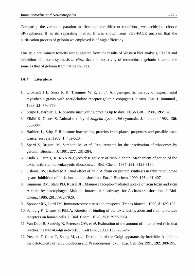
Fig 1B.1 depicts the structures of the subunits comprising these three branches of the AchR family
Immunotoxins and Neurotrophins
- 25 -
���������. The N-terminal extracellular domain consists of about 220 amino acids and contains
two homologus disulfide-linked loops. In most AchR subunits there is an N-glycosylation site at
141, some contain additional glycosylation sites in the large extracellular domain, and all contain
at least one such site �� �� �� �.
1B.1.1 Muscle AchRs
In 1973, the first purification of milligram amounts of acetylcholine receptor from electrophorus
electric organ resulted in the discovery of EAMG (experimental autoimmune Myasthenia gravis)
when it was found that rabbits immunized with that AchR became weak and died [10]. At that time,
muscle AchRs were well characterized electrophysiologically as Ach-gated cation channels, and
snake venom toxins such as � -bungarotoxin were just coming into use for localizing, quantifying,
and affinity purifying AchRs.
The structure of muscle-type AchRs found in the Torpedo electric organ has been characterized in
some details [4, 11]. The nicotinic acetylcholine receptor (AchR) found at the neuromuscular
junction is the autoantigen involved in the autoimmune disease Myasthenia gravis (MG). Owing to
its dual importance as a model autoantigen and as a model neurotransmitter receptor, its general
and antigenic structures have been extensively studied over the last 26 years. The AchR molecule
is a transmembrane glycoprotein (Mr ~290 kD) consisting of five homologous subunits in the
stoichimetry �2
� � � (embryonic) or �2
� � � (adult), which form the action channel. The � subunit
carries in its N-terminal extracellular domain the main immunogenic region (MIR), a group of
conformationally dependent epitopes that seems to be a major target for the anti-AchR antibodies
in MG patients [12, 13].
AchR subunits are thought to be organized like barrel staves around the central ion channel in the
order � � � � �, as shown in Fig 1B.2. The binding sites of acetylcholine (Ach) are formed at the
interfaces between � and � or � subunits. Because amino acids from both subunits contribute to
each of the two Ach binding sites, the properties of each of the Ach binding sites are somewhat
different. Both sites must have agonist bound for the ion channel to have a high probability of
flickering open for 1 or 2 ms. An antagonist bound to either site prevents activation.
Electron diffraction studies of Torpedo electric organ AchRs have reached a resolution of 4.6 Å
[11]. The AchR is about 80 Å in diameter and 120 Å long and 65 Å extending on the extracellular
surface, 40 Å crossing the lipid bilayer, and 15 Å extending beneath the bilayer into the cytoplasm.
Immunotoxins and Neurotrophins
- 26 -
The extracellular vestibule of the channel is about 25 Å in diameter surrounded by walls about 25
Å thick. The actual lumen of the channel through the lipid bilayer is quite narrow. The gate of the
channel is thought to be at its cytoplasmic end [13].
Fig 1B.2 The ar rangement of subunits of muscle AchRs [3]
Nicotinic acetylcholine receptors, as well as other members of the ligand-gated ion channel
superfamily, present a very simple repertoire of functional properties. The AchR recognizes the
neurotransmitter Ach and upon binding, the intrinsically coupled ion channel is opened,
augmenting in turn the possibility of cations to cross the lipid membrane. Thus, after channel
opening, a new ionic concentration is found at the aqueous solution bathing the opposed faces of
the lipid bilayer of the cell. In particular, the extracellular liquid presents now a higher
concentration of K+ (efflux) and the cytoplasmic compartment has a higher content of Na+ (influx).
When the concentration reaches equilibrium a membrane depolarization is produced.
Depolarization of the membrane provokes a specific physiological response by each cell that is
involved. For instance, if the muscle membrane depolarization is large enough, an action potential
propagates from the neuromuscular junction all over the muscle fiber. During the propagating
action potential, the release of Ca2+ ions from intracellular stores in the muscle cell is stimulated.
The final response in the muscle is the contraction of its myofibrils [14].
All these biologically relevant AchR properties are triggered by the binding of the neurotransmitter
Ach. Upon binding, the receptor protein undergoes a conformational change. Several lines of
experimental evidence suggest that the AchR may exist in a minimum of four interconvertible
states. The tetrahedral diagram indicating the existence of at least four receptor states is shown in
Fig 1B.3. In the absence of agonists, most Torpedo receptors (~80%) are in the resting state (Fig
1B.3, R). An additional about 20% of receptors is in the desensitized state (Fig 1B.3, D). The
Immunotoxins and Neurotrophins
- 27 -
resting state is defined by the existence of an activatable closed ion channel. In the presences of
agonists, the receptor is activated (Fig 1B.3, A) in the microsecond to millisecond time frame. The
state presents an open ion channel and a low affinity for agonists (apparent dissociation constant
Kd between 10 � M and 1 mM). Concomitantly, in the prolonged presence of agonists the activated
receptor is commuted to an intermediate state (Fig 1B.3, I) in the 1-100 ms time scale and further
to a D state in the second to minutes time frame. No energy sources nor an ionic gradient is needed
to induce the conformational shift from the R to the D state [14].
Fig 1B.3 Diagram showing the dynamic of multiple conformational
states of the nicotinic acetylcholine receptor [14]
In the absence of the neurotransmitter Ach, shown here as empty circles, the AchR is in the resting
state (R), a conformation state where the ion channel is closed. The closed ion channel can be
opened upon binding of two Ach molecules to AchR. This active state (A) presents low affinity for
agonists. The transition from the R to A state is a fast process that proceeds in the microsecond and
millisecond time regime. In the prolonged presence of agonists, the AchR becomes refractive to
the agonist’s pharmacological action and thus, to the activation of its ion channel. In the Torpedo
AchR there exists two refractive closed ion channel states, the intermediate (I) and the desensitized
(D) state. Both states show high affinity for agonists and some antagonists. The transition from the
A to the I state is a slow process that is produced in 1-100 ms time range. The transition from the A
to D state has a much slower time course.
Immunotoxins and Neurotrophins
- 28 -
1B.1.2 Research goals
The acetylcholine receptor (AchR) is a trans-membrane glycoprotein, consisting of 2� , �
, � , �
subunits. In muscle tissue of adults, the � -subunit is substituted by the � -subunit. A mature � -subunit has 437 amino acid residues containing the so-called main immunogenic region (MIR)-
a binding site of a polysaccharide. MIR located at residues 66 to 76 of � -subunit and contains an
Asn residue as the binding site of polysaccharide. MIR, as the target of AchR antibodies, plays an
important role in the autoimmune disease Myasthenia gravis. In order to investigate the
mechanism of Myasthenia gravis, researchers have attempted to obtain the relevant proteins by
gene technology. Unfortunately, the amount of AchR or its domain when expressed in engineering
bacteria usually is very low or the product occurs in the form of inclusion bodies. For improving
the efficiency of refolding and solubility of recombinant proteins, two recombinant vectors of
co-expressing the AchR � -subunit fragment and the chaperonin GroESL will be performed. In this
part, plasmid pPR506 which contains the gene of the � -subunit domain of AchR and plasmid
pGE60 containing the gene of the chaperonin GroESL will be co-transferred into E. coli DH5� and
co-expressed by the induction with IPTG. The receptor fragment could be coupled chemically to
gelonin and be employed in the treatment of Myasthenia gravis.
1B.2 Results and discussion
1B.2.1 Identification of the recombinant plasmids
The construction of pPR506 is shown in Fig 1B.4. To verify co-transformation of pPR506 and
pGE60, the two plasmids were transferred singly or doubly into E. coli DH5� . Afterwards, the
plasmids were extracted and analyzed by 1% agarose electrophoresis (Fig 1B.5). The gel
confirmed indicated that the molecular weights of pPR506 and pGE60 of about 2.3 kb (Fig 1B.5;
A) and 6.0 kb (Fig 1B.5; C) respectively, while the co-transformed plasmid contained both pPR506
and pGE60 (Fig 1B.5; B) as deduced from their electrophoretic patterns.
Immunotoxins and Neurotrophins
- 29 -
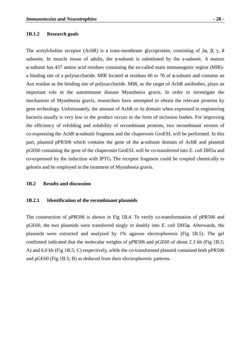
Fig 1B.4 Construction of plasmid pPR506
The target gene is a DNA fragment of AchR � -subunit residues from 1-120
By DNA sequence analysis, the fragment sequence in pPR506 is identical to the DNA sequence
deduced from the known amino acid residues of the AchR � -subunit domain. GAA encoding Glu is
the first amino acid of the domain and TCC encoding Ser is located at the 3'-terminus [15].
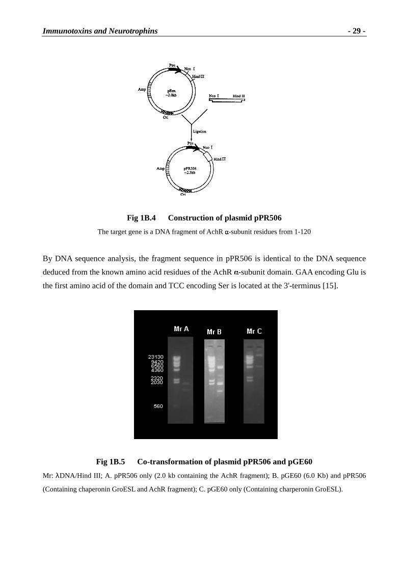
Fig 1B.5 Co-transformation of plasmid pPR506 and pGE60
Mr: λDNA/Hind III; A. pPR506 only (2.0 kb containing the AchR fragment); B. pGE60 (6.0 Kb) and pPR506
(Containing chaperonin GroESL and AchR fragment); C. pGE60 only (Containing charperonin GroESL).
Immunotoxins and Neurotrophins
- 30 -
1B.2.2 Co-expression of the recombinant plasmids in E. coli
The engineering strain E. coli DH5� /pPR506+pGE60 was cultured in LB medium which contained
both ampicillin (Amp) and chloromycetin (Chl) at 37˚C, 220 r/m until the optical density (OD600nm)
of the culture reached about 0.5~0.6. Subsequently, IPTG was added for induction and
fermentation was continued for another 4 h at the same temperature. Pellets were harvested by
centrifugation (5000 r/m, 30 min) and sonicated in PBS. The supernatant was analyzed by
SDS-PAGE. Electrophoretic analysis of the culture products related that the expression of pPR506
had yield only a 13 kD band equal to the AchR � -subunit domain and the amount of expression
was also very low and those of pGE60 had two bands at 60 kD and 10 kD equal to the products of
GroEL and GroES respectively, while the co-expression product displayed four bands, of which
the 60 kD band could result from GroEL and 10 kD band from GroES as well as a 13 kD band that
is likely to be the � -subunit fragment (Fig 1B.6). The new band at 23 kD could be the complex
which contains 13 kD � -subunit fragment from the result of immunoblotting assay that both bands
of 13 kD and 23 kD were positive (Fig 1B.7).
Fig 1B.6 SDS-PAGE analysis for expression product
Lane1 and lane2: Co-expression products (AchR fragment and charperonin GroESL)
Lane3 and Lane4: Expression products by DH5� /pPR506 (Only AchR fragment)
Lane5: Expression products of DH5� (Control, no plasmids presented)
Lane6: Expression products by DH5� /pGE60 (Only charperonin GroEL/GroES)
Immunotoxins and Neurotrophins
- 31 -
1B.2.3 Analysis of Western blots
After centrifugation of the sonicated mixture, the supernatant was run on 12% SDS-PAGE, the
components on the gel were transferred into a nitrocellulose membrane by sandwich
electrophoresis (Fig 1B.6, Lane1, 2, 3, 4 were transferred onto membrane). The membrane was
incubated with the primary antibody (mAb35; Part I·1C.2.3.4) and the second antibody (rabbit
anti-rat Ig 1:1000) respectively. Finally, the membrane treated as above was developed by
BCIP/NBT until the color of the target protein occurred and re-transferred into PBS to be fixed
immediately. Upon repetition of the experiments, the western blot was not always positive.
1B.2.4 Pur ification of products
After the supernatant was concentrated by Centriprep, not only some low molecular weight
proteins were partly extruded but also the sample was concentrated about 6 folds. The total protein
of cells was approximately 62 mg under the condition of the experiments. The concentrated sample
(about 5 mg) was loaded on Superose 12 HR10/30 on FPLC. The four eluted peaks were pooled
and run on SDS-PAGE respectively (Fig1B.8).
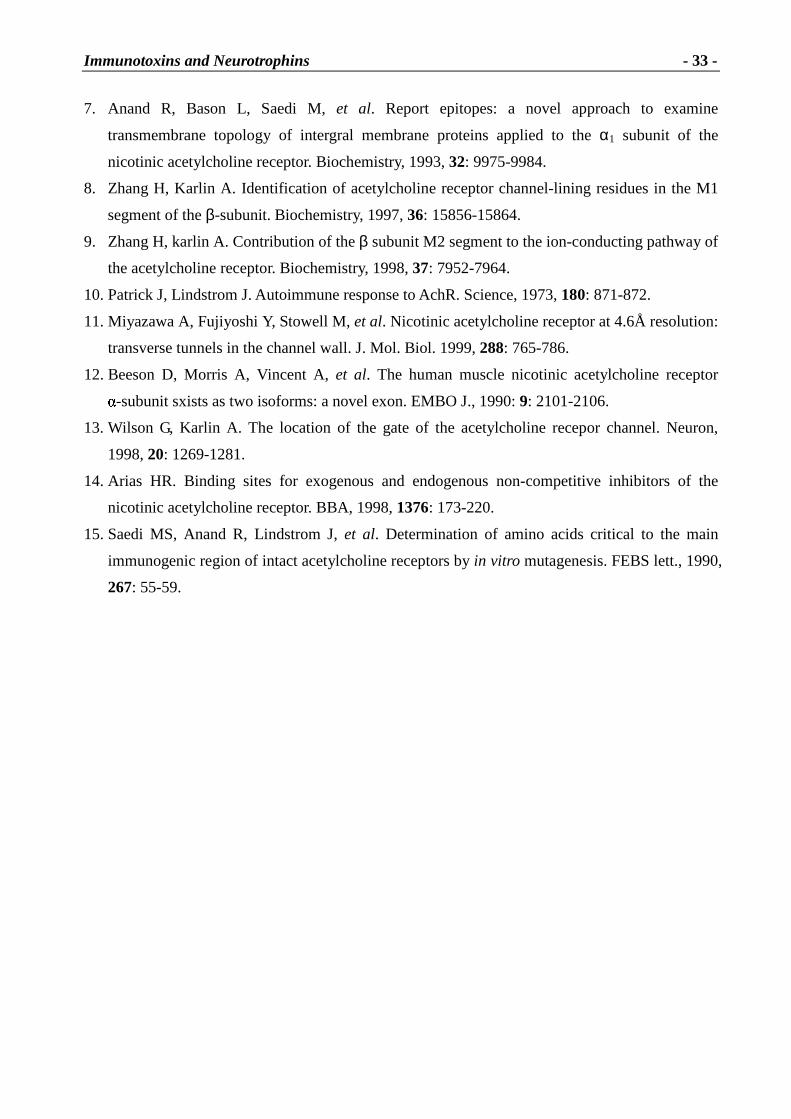
Fig 1B.8 Electrophoretic patterns of each
peak from HPLC by SDS-PAGE
(The co-expression products of AchR fragment and
GroESL were purified with Superose 12 HR 10/30.)
L Lane1: Peak1, 60 kD (GroEL)
Lane2: Peak2, 23 kD and 13 kD (AchR fragment)
Lane3: Peak3, 10 kD (GroES).
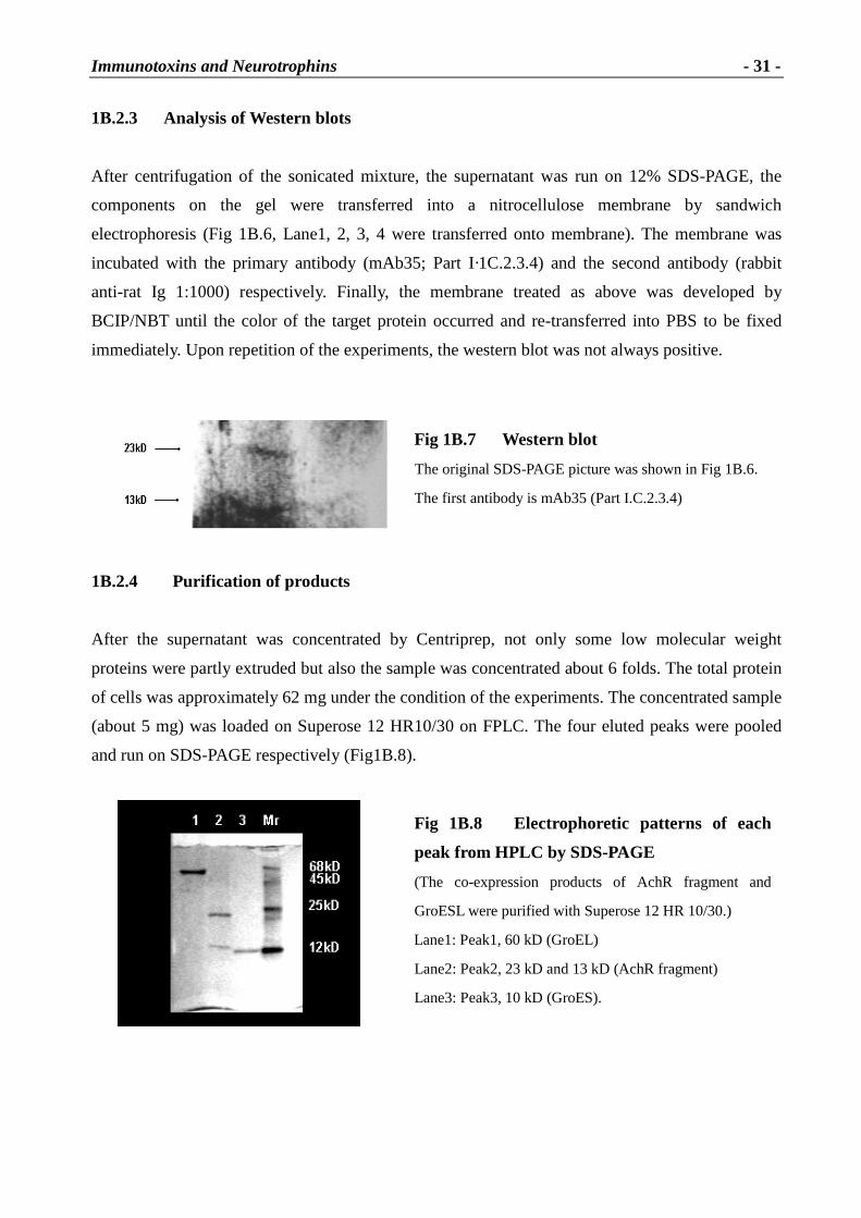
Fig 1B.7 Western blot
The original SDS-PAGE picture was shown in Fig 1B.6.
The first antibody is mAb35 (Part I.C.2.3.4)
Immunotoxins and Neurotrophins
- 32 -
1B.3 Summary
As described in previous work, the expression products of the acetylcholine receptor � -subunit or
its domain in bacteria usually resulted in inclusion bodies consisting of insoluble aggregates of the
proteins. It was then necessary to denature and renature the aggregates to obtain native protein.
However, it was reported that molecular chaperonins such as GroESL may facilitate renaturation of
denatured proteins to their native conformation. It was shown from our experiments that GroESL
slightly improved the correct expression of the acetylcholine receptor � -subunit fragment. Based
on the molecular weight of acetylcholine receptor � -subunit domain and GroES, it is very likely
that the 13 kD band in the SDS-gel belongs to the � -subunit fragment. The 23 kD would account
for a complex between the � subunit fragment and GroES, but there is no further evidence for such
a complex besides the Western blot (Fig 1B.7).
Our experiments clearly show that co-expression of the � -subunit fragment of AchR together with
GroESL leads to a soluble form of the receptor. However, the yields are very low and not
reproducible. The amount of receptor fragment was certainly not sufficient for chemical
cross-linking with gelonin. Hence, we tried to express the conjugate as a fusion protein as
described in Part I· C
1B.4 L iterature
1. Lindstrom J. Neuronal nicotinic acetylcholine receptors. In: Narahashi T, editor. Ion channels.
New York: Plenum press, 1996: p377-450.
2. Lindstrom J. Purification and cloning of nicotinic acetylcholine receptors. In Aeneric S, Brioni
D, editors. Neuronoal nicotinic receptors: pharmacology and therapeutic opportunities. New
York, John Wiley and sons; 1999.
3. Lindstrom J. Acetylcholine receptors and Myasthenia. Muscle and Nerve, 2000, 23: 453-477.
4. Karlin A, Akabas M. Toward a structure basis for the function of nicotinic acetylcholine
receptorsand their cousins. Neuron, 1995, 15: 1231-1244.
5. LeNovere N, Changeux JP. Molecular evolution of the nicotinc acetylcholine receptor: an
example of multi-gene family in excitable cells. J. Mol. Evol., 1995, 40:155-172.
6. LeNovere N, Corringer JP, Changeux JP. Improved secondary structure predictions for a
nicotinic receptor subunit: incoporation of solvent accessibility and experimental data into a
8. Zhang H, Karlin A. Identification of acetylcholine receptor channel-lining residues in the M1
segment of the β-subunit. Biochemistry, 1997, 36: 15856-15864.
9. Zhang H, karlin A. Contribution of the β subunit M2 segment to the ion-conducting pathway of
the acetylcholine receptor. Biochemistry, 1998, 37: 7952-7964.
10. Patrick J, Lindstrom J. Autoimmune response to AchR. Science, 1973, 180: 871-872.
11. Miyazawa A, Fujiyoshi Y, Stowell M, et al. Nicotinic acetylcholine receptor at 4.6Å resolution:
transverse tunnels in the channel wall. J. Mol. Biol. 1999, 288: 765-786.
12. Beeson D, Morris A, Vincent A, et al. The human muscle nicotinic acetylcholine receptor � -subunit sxists as two isoforms: a novel exon. EMBO J., 1990: 9: 2101-2106.
13. Wilson G, Karlin A. The location of the gate of the acetylcholine recepor channel. Neuron,
1998, 20: 1269-1281.
14. Arias HR. Binding sites for exogenous and endogenous non-competitive inhibitors of the
to AchRs [14, 15, 16], (3) thymectomy is often beneficial [17], (4) 10-15% of MG patients exhibit
thymoma [18], (5) the thymus is not hyperplastic or thymomatous in EAMG where the
autoimmune response is initiated with exogenous AchR [19], (6) transplant of fragments of MG
patient thymus to severe combined immunodeficiency (SCID) mice can transfer MG to the mice
[20]. However, the thymus is probably not the sole site for induction or maintenance of the
autoimmune response to AchR, thymectomy is not uniform or complete in its beneficial effect on
MG.
EAMG has been induced in many species by immunization with purified Torpedo electric organ
AchR [21]. AchR is quite immunogenic and EAMG has been induced by AchR [22]. Adjuvants are
usually used, but it is possible to induce EAMG with AchR even in the absence of adjuvants [23].
Among MG patients, the absolute concentration of antibodies to AchR does not correlate closely
with severity [24], but generally patients with only ocular manifestation have lower autoantibody
concentrations than do patients with generalized MG, and changes in an individual’s autoantibody
concentrations usually parallels changes in their clinical state [25]. The basic mechanisms by
which autoantibodies to AchR impair neuromuscular transmission appear to be very similar in MG
and chronic EAMG [26, 27].
1C.1.2.1 Cellular immune mechanisms in MG and EAMG
EAMG is caused by an antibody mediated autoimmune response, it is not surprising that
development of EAMG depends on the presence of B-cells. B-cell-deficient mutant mice are
resistant to induction of EAMG, but develop a normal T-lymphocyte-mediated immune response
and are susceptible to passively transferred EAMG mediated by a mAb to the MIR [28]. The T-cell
response in these mice lacking B-cells developed more slowly than it would have with antigen
presentation by B-cells as well as professional antigen-presenting cells. Antigen-specific B-cells
can be very efficient at presenting peptides from the native antigens that they bind with high
affinity.
Development of EAMG and MG depends on T-lymphocytes to cooperate with B-lymphocytes in
developing autoantibody-producing plasma cells, but which T-cell types are involved has been
more difficult to determine. CD4+ helper lymphocytes are necessary for the production of
antibodies to AchR in both EAMG and MG [29]. Using various methods with human MG patient
lymphocytes, different AchR epitopes have been identified. In various strains of rats and mice,
different prominent epitopes have also been identified [30]. Some of these have been used to
Immunotoxins and Neurotrophins
- 40 -
induce tolerance and reduce induction of EAMG [31]. The dependence of these epitopes on the
genetics of the antigen-presenting proteins, T-cell receptors, and other components of the cellular
immune processing, recognition, and regulatory systems makes inbred rodent strains exquisitely
sensitive to immune regulation by AchR peptides and small mutations of these peptides. However,
it is unclear whether such subtleties will prove useful in predicting or manipulating the immune
response in the outbred human populations that comprise MG patients.
1C.1.2.2 Development of specific immunosuppressive therapies for EAMG and MG
There is currently no specific immunosuppressive therapy or cure for MG. Nonspecific
immunosuppressive therapy of MG with prednisone and other drugs, as well as by thymectomy
combined with symptomatic therapy using inhibitors of acetylcholinesterase, permits substantial
control of MG and has greatly reduced its mortality. There are significant side effects associated
with prolonged nonspecific immunosuppressive therapy. Because so much is known about the
antigenic structure of muscle AchRs and the pathological mechanisms of MG, it would seem
reasonable to hope for a specific immunosuppressive therapy, because it has been thought that MG
is the best characterized autoimmune disease [32]. Successful specific immunosuppression of MG
might be valuable not only for its benefit to MG patients, but also as a model for what might be
applied to others autoimmune diseases with well characterized autoantigens.
Some exotic approaches to inhibit autoantibodies to the MIR have been reported as potential
therapies for MG. Tzartos and coworkers proposed to use monovalent Fab fragments of mAbs to
the MIR to compete for binding of autoantibodies [33]. Due to lacking the Fc region, they cannot
fix complement and cause disruption of the postsynaptic membrane. Because they are monovalent,
they can not crosslink AchRs and cause antigenic modulations, as can bivalent F(ab)2 fragments
[34]. Fab fragments can prevent antigenic modulation of AchRs in cultured cells caused by mAbs
to the MIR or by MG patient sera [15]. Single chain Fv fragments of MIR mAbs and MG patient
autoantibodies have been expressed in bacteria, humanized, and selected for high affinity [35].
Unfortunately, Fab is very quickly cleared from circulation.
In addition there are other approaches taken for specific immunosuppression, such as Torpedo
AchR coupled with toxic drugs in order to try to kill B-lymphocytes that interact with it. It was
reported by Trommer that an AchR-conjugate i.e. AchR-gelonin obtained by chemical coupling
could be employed for selective elimination of specific lymphocytes involved in triggering and
maintenance of EAMG in vitro. The plant toxin gelonin was used which catalytically inhibits the
Immunotoxins and Neurotrophins
- 41 -
elongation step of protein synthesis. In their study, it was indicated that a marked improvement of
clinical symptoms as well as a significant increase in functional AchR had occurred as compared to
treatment with AchR or gelonin alone or untreated rats with EAMG as determined 6-10 weeks later
[3].
1C.1.3 Research goals
Myasthenia gravis is an autoimmune disorder characterized by weakness and fatigability of
skeletal muscles. The pathogenesis of MG in human and experimental MG in animals (EAMG)
results from a reduction of the available acetylcholine receptors at neuromuscular junctions due to
an antibody-mediated autoimmune response. Although treatment of MG with general
immunosuppressive agents is reasonably effective, it may have numerous adverse side effects.
Ideally, treatment of MG should eliminate the specific pathogenic autoimmune response to AchRs,
without otherwise suppressing the immune system. The present study aims at a novel strategy for
specific immunotherapy of MG
AchR-toxin conjugates have been employed for selective elimination of specific lymphocytes
involved in triggering and maintenance of EAMG in vivo using the plant toxin ricin. Ricin as well
as gelonin catalytically inhibits the elongation step of protein synthesis. In the previous chapter
(Part I· 1B), we have described attempts for coexpression of the extracellular domain of the � -subunit of AchR with the chaperonin GroESL. It was then planned to couple the receptor
fragment chemically to gelonin. However, since the yields of soluble protein were very low, the
expression of a fusion protein composed of gelonin and AchR receptor fragment will be tried and
its biological activity be tested.
1C.2 Results and discussion
1C.2.1 Cloning gelonin-AchR fragment gene
Based on our earlier work and the relevant data obtained, a strategy of recombinants pET-GA and
pJLA-GA was employed as shown in Fig 1C.3. The beginning two plasmids pUC-gel I and
pUC-gel II-AchR were provided by Ya-Wei Shi [25, 1A.4]. In order to obtain a gelonin-AchR
fusion gene, the gel II-AchR fragment from pUC-gel II-AchR was inserted into the opened
pUC-gel I by double enzymic digestion with XbaI/EcoRI and ligation, to be a fusion construct,
named pUC-GA. After identification of the construct, a band of 1196 bp equal to the fragment
Immunotoxins and Neurotrophins
- 42 -
expected has been found on the gel which indicated that the recombinant was the one we
anticipated (Fig 1C.4).
Fig 1C.3 Construction of expression vector for gelonin-AchR
(pUC-gel I, pUC-gel II-AchR were provided by Ya-Wei Shi et al [25, in 1A.4])
Immunotoxins and Neurotrophins
- 43 -
Table 1C.1 Reaction system of enzymatic digestion of pUC-gel I /pUC-gel I I -AchR
Compounds Volume
PUC-gel I or pUC-gel II-AchR 30 µl
XbaI 2 µl
EcoRI 2 µl
10x buffer2# 4 µl
H2Obidest 2 µl
* The reaction mixture was incubated at 37�
for 2 h
Table 1C.2 L igation system for pUC-GA
Reaction mixture Volume
PUC-gel I (opened) 0.5 µl
Gel3+4+AchR (1196 bps) 6 µl
10xT4 DNA ligase buffer 1 µl
T4 DNA ligase 1 µl
H2Obidest 1.5 µl
*The reaction mixture was incubated at 26˚C for 2 h and continuously at 16˚C overnight
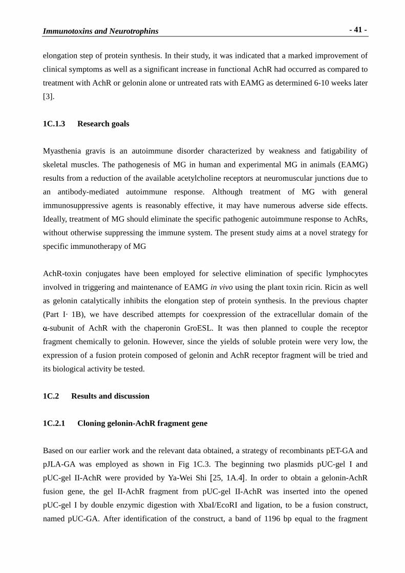
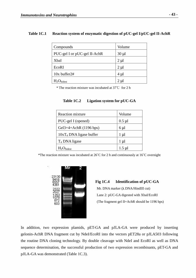
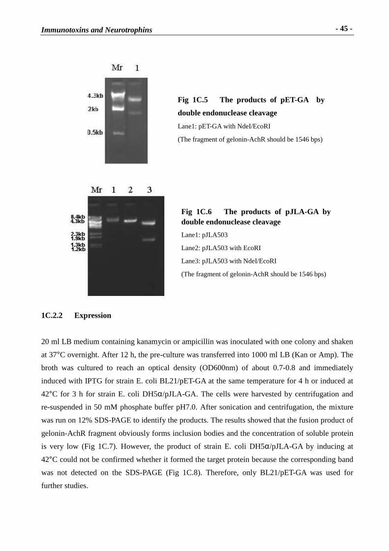
In addition, two expression plamids, pET-GA and pJLA-GA were produced by inserting
gelonin-AchR DNA fragment cut by NdeI/EcoRI into the vectors pET28a or pJLA503 following
the routine DNA cloning technology. By double cleavage with NdeI and EcoRI as well as DNA
sequence determination, the successful production of two expression recombinants, pET-GA and
pJLA-GA was demonstrated (Table 1C.3).
Fig 1C.4 Identification of pUC-GA
Mr. DNA marker (� DNA/HindIII cut)
Lane 2: pUC-GA digested with XbaI/EcoRI
(The fragment gel II+AchR should be 1196 bps)
Immunotoxins and Neurotrophins
- 44 -
Table 1C.3 The ligation system for pET-GA and pJLA-GA
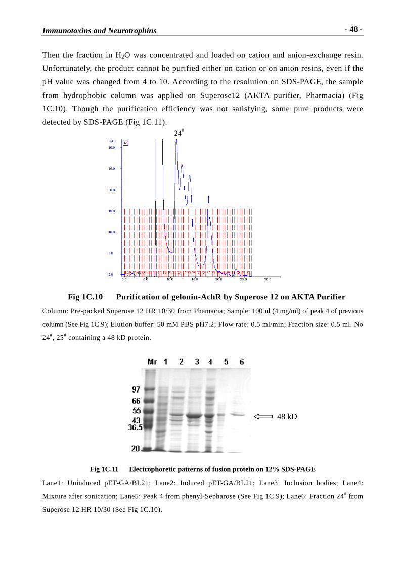
Fig 1C.15B Electrophoretic patter ns of refolded gelonin-AchR fr agment protein
eluted from Q-Sepharose column
Lane1: Fraction of refolded gelonin-AchR fragment
Lane2: Mixture after sonication.
Method 2
The denatured inclusion bodies were mixed with the final concentration of 20 mM Tris-HCl pH8.0,
5 mM DTT, 2 mM EDTA, 2 mM GSH and 0.2 mM GSSG by rapid votexing at room temperature
to a final concentration of 400 � g /ml protein and 1 M GuHCL. Incubation was performed at 4ºC
for about 16 h. Normally more than 200 � g refolded protein could be obtained from 1 mg
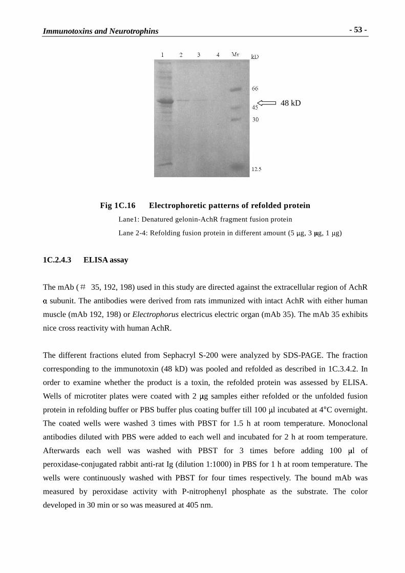
denatured protein as Fig 1C.16.
48 kD
Immunotoxins and Neurotrophins
- 53 -
Fig 1C.16 Electrophoretic patter ns of refolded protein
Lane1: Denatured gelonin-AchR fragment fusion protein
Lane 2-4: Refolding fusion protein in different amount (5 � g, 3 � g, 1 � g)
1C.2.4.3 ELISA assay
The mAb (� 35, 192, 198) used in this study are directed against the extracellular region of AchR � subunit. The antibodies were derived from rats immunized with intact AchR with either human
muscle (mAb 192, 198) or Electrophorus electricus electric organ (mAb 35). The mAb 35 exhibits
nice cross reactivity with human AchR.
The different fractions eluted from Sephacryl S-200 were analyzed by SDS-PAGE. The fraction
corresponding to the immunotoxin (48 kD) was pooled and refolded as described in 1C.3.4.2. In
order to examine whether the product is a toxin, the refolded protein was assessed by ELISA.
Wells of microtiter plates were coated with 2 � g samples either refolded or the unfolded fusion
protein in refolding buffer or PBS buffer plus coating buffer till 100 � l incubated at 4°C overnight.
The coated wells were washed 3 times with PBST for 1.5 h at room temperature. Monoclonal
antibodies diluted with PBS were added to each well and incubated for 2 h at room temperature.
Afterwards each well was washed with PBST for 3 times before adding 100 � l of
peroxidase-conjugated rabbit anti-rat Ig (dilution 1:1000) in PBS for 1 h at room temperature. The
wells were continuously washed with PBST for four times respectively. The bound mAb was
measured by peroxidase activity with P-nitrophenyl phosphate as the substrate. The color
developed in 30 min or so was measured at 405 nm.
48 kD
Immunotoxins and Neurotrophins
- 54 -
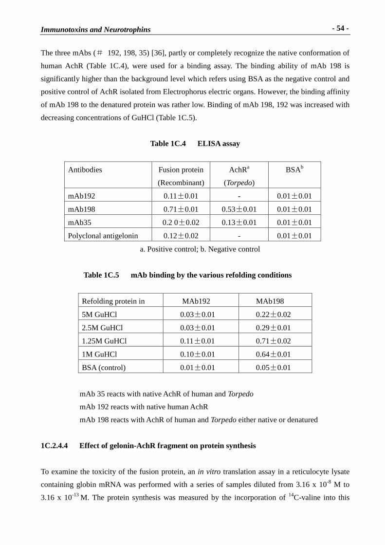
The three mAbs (� 192, 198, 35) [36], partly or completely recognize the native conformation of
human AchR (Table 1C.4), were used for a binding assay. The binding ability of mAb 198 is
significantly higher than the background level which refers using BSA as the negative control and
positive control of AchR isolated from Electrophorus electric organs. However, the binding affinity
of mAb 198 to the denatured protein was rather low. Binding of mAb 198, 192 was increased with
decreasing concentrations of GuHCl (Table 1C.5).
Table 1C.4 ELISA assay
Antibodies Fusion protein
(Recombinant)
AchRa
(Torpedo)
BSAb
mAb192 0.11�0.01 - 0.01�0.01
mAb198 0.71�0.01 0.53�0.01 0.01�0.01
mAb35 0.2 0�0.02 0.13�0.01 0.01�0.01
Polyclonal antigelonin 0.12�0.02 - 0.01�0.01
a. Positive control; b. Negative control
Table 1C.5 mAb binding by the var ious refolding conditions
Refolding protein in MAb192 MAb198
5M GuHCl 0.03�0.01 0.22�0.02
2.5M GuHCl 0.03�0.01 0.29�0.01
1.25M GuHCl 0.11�0.01 0.71�0.02
1M GuHCl 0.10�0.01 0.64�0.01
BSA (control) 0.01�0.01 0.05�0.01
mAb 35 reacts with native AchR of human and Torpedo
mAb 192 reacts with native human AchR
mAb 198 reacts with AchR of human and Torpedo either native or denatured
1C.2.4.4 Effect of gelonin-AchR fragment on protein synthesis
To examine the toxicity of the fusion protein, an in vitro translation assay in a reticulocyte lysate
containing globin mRNA was performed with a series of samples diluted from 3.16 x 10⁻⁸ M to
3.16 x 10⁻¹³ M. The protein synthesis was measured by the incorporation of 14C-valine into this
Immunotoxins and Neurotrophins
- 55 -
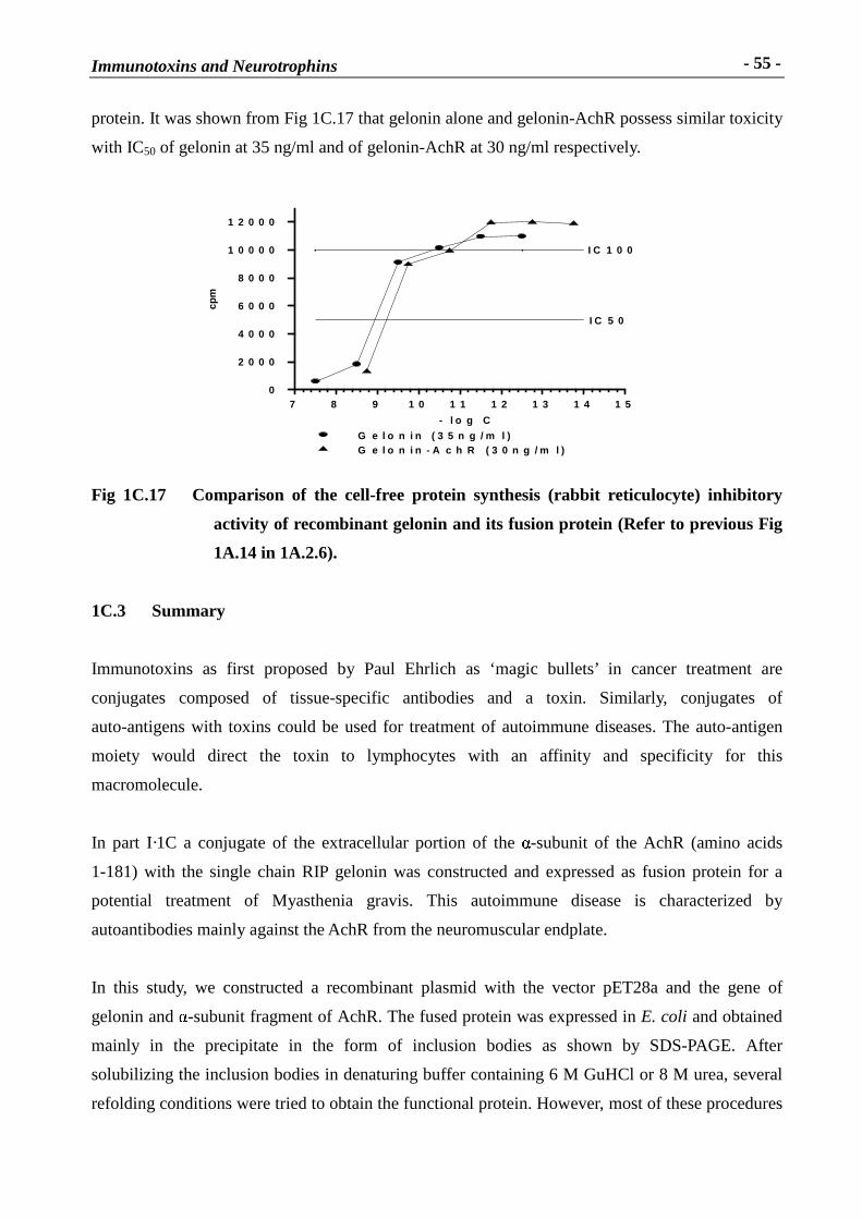
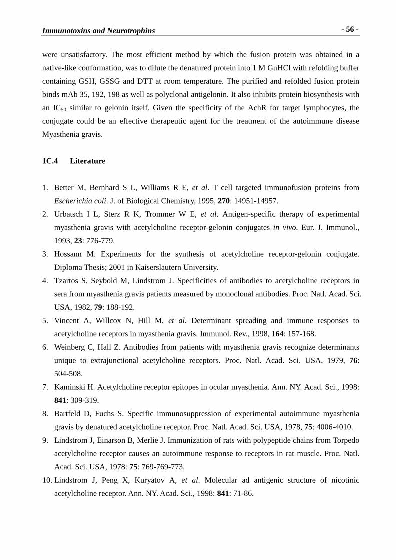
protein. It was shown from Fig 1C.17 that gelonin alone and gelonin-AchR possess similar toxicity
with IC50 of gelonin at 35 ng/ml and of gelonin-AchR at 30 ng/ml respectively.
Fig 1C.17 Compar ison of the cell-free protein synthesis (rabbit reticulocyte) inhibitory
activity of recombinant gelonin and its fusion protein (Refer to previous Fig
1A.14 in 1A.2.6).
1C.3 Summary
Immunotoxins as first proposed by Paul Ehrlich as ‘magic bullets’ in cancer treatment are
conjugates composed of tissue-specific antibodies and a toxin. Similarly, conjugates of
auto-antigens with toxins could be used for treatment of autoimmune diseases. The auto-antigen
moiety would direct the toxin to lymphocytes with an affinity and specificity for this
macromolecule.
In part I·1C a conjugate of the extracellular portion of the � -subunit of the AchR (amino acids
1-181) with the single chain RIP gelonin was constructed and expressed as fusion protein for a
potential treatment of Myasthenia gravis. This autoimmune disease is characterized by
autoantibodies mainly against the AchR from the neuromuscular endplate.
In this study, we constructed a recombinant plasmid with the vector pET28a and the gene of
gelonin and � -subunit fragment of AchR. The fused protein was expressed in E. coli and obtained
mainly in the precipitate in the form of inclusion bodies as shown by SDS-PAGE. After
solubilizing the inclusion bodies in denaturing buffer containing 6 M GuHCl or 8 M urea, several
refolding conditions were tried to obtain the functional protein. However, most of these procedures
- l o g C
7 8 9 1 0 1 1 1 2 1 3 1 4 1 5
cpm
0
2 0 0 0
4 0 0 0
6 0 0 0
8 0 0 0
1 0 0 0 0
1 2 0 0 0
I C 1 0 0
I C 5 0
G e l o n i n ( 3 5 n g / m l )G e l o n i n - A c h R ( 3 0 n g / m l )
Immunotoxins and Neurotrophins
- 56 -
were unsatisfactory. The most efficient method by which the fusion protein was obtained in a
native-like conformation, was to dilute the denatured protein into 1 M GuHCl with refolding buffer
containing GSH, GSSG and DTT at room temperature. The purified and refolded fusion protein
binds mAb 35, 192, 198 as well as polyclonal antigelonin. It also inhibits protein biosynthesis with
an IC50 similar to gelonin itself. Given the specificity of the AchR for target lymphocytes, the
conjugate could be an effective therapeutic agent for the treatment of the autoimmune disease
Myasthenia gravis.
1C.4 L iterature
1. Better M, Bernhard S L, Williams R E, et al. T cell targeted immunofusion proteins from
Escherichia coli. J. of Biological Chemistry, 1995, 270: 14951-14957.
2. Urbatsch I L, Sterz R K, Trommer W E, et al. Antigen-specific therapy of experimental
myasthenia gravis with acetylcholine receptor-gelonin conjugates in vivo. Eur. J. Immunol.,
1993, 23: 776-779.
3. Hossann M. Experiments for the synthesis of acetylcholine receptor-gelonin conjugate.
Diploma Thesis; 2001 in Kaiserslautern University.
4. Tzartos S, Seybold M, Lindstrom J. Specificities of antibodies to acetylcholine receptors in
sera from myasthenia gravis patients measured by monoclonal antibodies. Proc. Natl. Acad. Sci.
USA, 1982, 79: 188-192.
5. Vincent A, Willcox N, Hill M, et al. Determinant spreading and immune responses to
acetylcholine receptors in myasthenia gravis. Immunol. Rev., 1998, 164: 157-168.
6. Weinberg C, Hall Z. Antibodies from patients with myasthenia gravis recognize determinants
unique to extrajunctional acetylcholine receptors. Proc. Natl. Acad. Sci. USA, 1979, 76:
504-508.
7. Kaminski H. Acetylcholine receptor epitopes in ocular myasthenia. Ann. NY. Acad. Sci., 1998:
841: 309-319.
8. Bartfeld D, Fuchs S. Specific immunosuppression of experimental autoimmune myasthenia
gravis by denatured acetylcholine receptor. Proc. Natl. Acad. Sci. USA, 1978, 75: 4006-4010.
9. Lindstrom J, Einarson B, Merlie J. Immunization of rats with polypeptide chains from Torpedo
acetylcholine receptor causes an autoimmune response to receptors in rat muscle. Proc. Natl.
Acad. Sci. USA, 1978: 75: 769-769-773.
10. Lindstrom J, Peng X, Kuryatov A, et al. Molecular ad antigenic structure of nicotinic
acetylcholine receptor. Ann. NY. Acad. Sci., 1998: 841: 71-86.
Immunotoxins and Neurotrophins
- 57 -
11. Kao I, Drachman D. Thymic muscle cells bear acetylcholine receptors: possible relation to
myasthenia gravis. Science, 1977, 195: 74-75.
12. Nelson S, Conti-Troconi B. Adult thymuses express an embryonic nicotinic acetylcholine
receptor-like protein. J. Neuroimmunol, 1990, 29: 81-92.
13. Schluep M, Willcox N, Vincent A, et al. Acetylcholine receptors in human thymic myoid cells
in situ: an immunohistological study. Ann. Neurol, 1987, 22: 212-222.
14. Farrar J, Portolano S, Willcox N, et al. Diverse Fabs specific for receptor epitopes from a