Journal of Molecular Graphics and Modelling 38 (2012) 123–136
Contents lists available at SciVerse ScienceDirect
Journal of Molecular Graphics and Modelling
j ourna l h o me p age: www.elsev ier .com/ locate /JMGM
mpact of the CXCR4 structure on docking-based virtual screening ofIV entry inhibitors
esús M. Planesasa,1, Violeta I. Pérez-Nuenoa,b,∗, José I. Borrell a,1, Jordi Teixidóa,1
Grup d’Enginyeria Molecular, Institut Químic de Sarriá (IQS), Universitat Ramon Llull, Barcelona, SpainINRIA Nancy, LORIA, 615 rue du Jardin Botanique, 54600 Villers-lès-Nancy, France
r t i c l e i n f o
rticle history:ccepted 25 June 2012vailable online 3 July 2012
Herein we analyze in depth the receptor-based virtual screening (VS) performance of the five recent crys-tallized CXCR4 structures along with a CXCR4 rhodopsin-based homology model. All CXCR4 Protein DataBank (PDB) structures are co-crystallized with a small organic antagonist except structure 3OE0, which isco-crystallized with a cyclic peptide analog. Evaluation of the CXCR4 models was done by retrospectivedocking-based VS using a test set of 248 known CXCR4 inhibitors from 4 different chemotype familiesand 4696 different presumed inactives. The performance of the docking protocol using the five differentprotein structures was assessed in terms of pose prediction and hits detection using 12 different docking
caffold retrieval scoring functions and a scoring function with rescoring. Results show that 3OE6 structure achieves thehighest docking-based performance with an average area under the curve (aAUC) of 0.84 and an aver-age enrichment factor (aEF) of 11.7 at 1% of decoys screened. CXCR4 rhodopsin-like homology modelperforms comparable to the crystallized structures in the 1% of database screened. Moreover, a detailedanalysis of the retrospective docking results using the CXCR4 homology model in Discovery Studio allowsus to hypothesize the existence of multiple binding sub-sites in CXCR4 binding pocket.
HIV cell infection is a multi-stage and complex process. It startsith the virus entry [1] into the host membrane. The first stage
f the entry process is the virus attachment to the cell membranehrough receptor CD4. Secondly, viral proteins gp120 [2] and gp41ind to the co-receptors CCR5 and CXCR4 [3] to facilitate virusnchoring to the host cellular membrane. Finally, the viral enve-ope undergoes conformational changes that allow the insertion ofhe gp41 fusion peptide into the host membrane.
One of the strategies to block the HIV entry process is inhibitinghe binding of the viral protein gp120 to the co-receptors CXCR4 orCR5. The first interaction between gp120 and these co-receptors
s through their extracellular loops. CXCR4 and CCR5 are both
hemokine receptors which belong to the GPCR superfamily [4]nd are implicated in a wide range of human diseases [5]. This sub-amily of GPCR proteins is responsible for transmitting signals from
∗ Corresponding author at: INRIA Nancy, LORIA, 615 rue du Jardin Botanique,4600 Villers-lès-Nancy, France. Tel.: +33 3 83593045/+34 93 267 20 00;ax: +33 3 83413079/+34 93 205 62 66.
the extracellular environment as well as the recognition of extra-cellular ligands. All GPCRs have a common characteristic structurerelatively hydrophobic with 7 transmembrane domains. The N-terminus is located in the extracellular side and the transmembranehelices are connected by three extracellular loops whereas the C-terminus is located in the intracellular side facing the cytoplasmand three intracellular loops connect the interior domain of thetransmembrane helices (Fig. 1). The transmembrane helices areexpanded in the plasmatic membrane as anticlockwise bundle ofalpha-helices. Therefore extracellular loops fulfill important rolesin receptor activation and consequently in ligand binding [6].
An important achievement was accomplished at the end of 2010when Wu et al. [7] crystallized five independent CXCR4 structureswith a resolution of 2.5–3.2 A (Table 1). These crystal structurescontain a small organic molecule derived from isothiourea [8] (IT1t)in Protein Data Bank structures (PDBs) 3ODU, 3OE6, 3OE8 and 3OE9,and a 16-residue cyclic peptide [9] (CVX15) in PDB 3OE0, as antag-onists bound to the CXCR4 structure.
In all the PDBs we can observe that the ligand binding siteis located in the second extracellular loop, ECL2, close to theN-terminus (Fig. 2). Also, superposition of ligand binding pock-
ets shows similar overlapping volume. The residues that inducecommon interactions in both ligands are Tyr116, Asp187 andGlu288 (Fig. 3). Those residues are common in CXCR4 antago-nist binding as it has been widely discussed in previous literature
124 J.M. Planesas et al. / Journal of Molecular Graphics and Modelling 38 (2012) 123–136
Fig. 1. Backbone scheme of the co-receptor CXCR4 composed by the seven typicalGPCR transmembrane domains, the intracellular C-terminus and the extracellularN-terminus with their corresponding triad of extracellular and intracellular loops.
Fig. 2. CXCR4 ligand binding pocket complexed with the small organic antagonistIT1t, colored in green, superimposed with the structure of the peptide ligand CVX15,colored in purple. (For interpretation of the references to color in this figure legend,the reader is referred to the web version of the article.)
Table 1Resolution and characteristics of CXCR4 structures published by Wu et al.
[10,11]. Although the overall orientation differs between ligands,hydrophobic interactions highly contribute in the binding affinityof the majority of CXCR4 antagonists. The residues Ala95, Leu91,Val112, His113, Tyr116, Phe292, Tyr255 and Ile259 play importantroles in hydrophobic binding. Moreover, Tyr45 on TM1, Trp94 andAsp97 on TM2, Tyr116 on TM3, Asp171 on TM4, Asp262 on TM6and His281 and Glu288 on TM7, are critical residues involved instabilizing interactions with known CXCR4 antagonists [12–14]
Nevertheless, in the case of the peptide ligand it is observed a
ligand-induced conformational change in the extracellular side ofthe helix 5 [7]. Hence, the docking-based virtual screening (VS) ofsmall peptides might be more dependent on the CXCR4 structureused for the screening.
Phe 199 (F1995.38) Gln 200 (Q2005.39) Asp 262 (D2626.58)
Leu 266 (L266) Glu 277 (E277)
His 281 (H2817.32) Ile 284 (I2847.3 5)
Ser 285 (S2857.36) Glu 288 (E2887.39)
Fig. 3. Protein–ligand interaction map for each complex structure. Structure3OE0, whose co-crystallized ligand CVX15 has a large volume presents, manyprotein–ligand interactions in comparison with the rest of complexes whosecommon co-crystallized ligand IT1t is smaller. Some different interactions canbe appreciated between PDBs 3ODU, 3OE6, 3OE8, 3OE9. However, superposi-tion of crystallized IT1t binding modes is very similar in all cases. In bracketsBallesteros–Weinstein residues notation [65].
r Graphics and Modelling 38 (2012) 123–136 125
2
2
tcC
trsGCS(D
bmtooACwtr
ti[fp
Fig. 4. Superposition of CXCR4 structures PDB 3ODU (red) and the homology model
J.M. Planesas et al. / Journal of Molecula
. Methods
.1. Virtual screening data preparation
In the last years GPCR and also chemokine receptors have beguno be determined successfully by protein crystallography. In thease of the chemokine co-receptor CXCR4, Wu et al. obtained fiveXCR4 structures at the end of 2010.
We analyzed the five CXCR4 structures published in the Pro-ein Data Bank by retrospective docking-based (VS) using differentepresentative force-filed based, empirical, and knowledge-basedcoring functions [15]: AutoDock Vina [16] from AutoDock,oldScore [17], ChemScore [17] and GoldScore rescored withhemScore from Gold 4.1, Glide 5.5 [18], TotalScore provided inurflex [19] from Sybyl X 1.1 [20], Ligandfit scoring functionsLigScore1; LigScore2 [21]; PLP1; PLP2 [22,23]; Jain [23], PMF andock Score) implemented in Discovery Studio 2.5.
Moreover, we compared these results with the ones retrievedy our previously published CXCR4 rhodopsin-based homologyodel built when there were not available CXCR4 crystal struc-
ures [24]. This homology model agrees with the folding structuref the crystal PDBs now available. Fig. 4 shows the superpositionf our homology model and the crystal CXCR4 structure 3ODU.
detailed structural comparison between homology models andXCR4 crystal structures was done by Kufareva et al. [25]. Herein,e compare our homology model against the crystal structures in
erms of retrospective docking performance, especially the earlyecovery.
To perform the retrospective docking-based screening we usedhe test set of Pérez-Nueno et al. [24]. This test set contains 248
nhibitors representative of 4 CXCR4 antagonist scaffold families8,26] (Fig. 5) and 4696 different presumed inactives extracted [24]rom the Maybridge Screening Collection whose physicochemicalroperties (molecular weight, number of rotatable single bonds,
(yellow). Residues Asp171, Asp262 and Glu288 have been used to define superpo-sition. (For interpretation of the references to color in this figure legend, the readeris referred to the web version of the article.)
NH
NH
HN
N
NH
N
HN
HN
AMD3100
N
NH
N
HN
N
NH
N
HN
p-phenylenebis(methylene)LinkedBis-azamacrocycle
NH
HN
O
NH
O
NH
H2N NH
KRH-1636
N
N
N
N
N
H
H
N
imidazopyridine-tetrahydro-8-quinolinamine
Fig. 5. Representative compounds of the chemotype families used as actives in the test set 24 to guarantee diversity of true binders.
126 J.M. Planesas et al. / Journal of Molecular Graphics and Modelling 38 (2012) 123–136
Table 2Ligands and decoys molecular properties: summary of the 1D physicochemical properties of active and decoy molecules. Standard deviation values in parentheses.
umber of hydrogen-bond acceptor atoms, number of hydrogen-ond donor atoms, octanol–water partition coefficient, number ofydrophobic atoms, PSA, total positive partial charges and totalegative partial charges) match similar property values than theelected actives [27]. Table 2 shows that the average and standardeviations of these properties calculated by MOE 2009 [28] areuite similar for the active and inactive pools. Supplementary Fig.
shows the ROC curves obtained when ligands and decoys werelassified by their total positive partial charge (PEOE PC+), total neg-tive partial charge (PEOE PC−), total polar surface area (TPSA) andog octanol/water partition coefficient (log P) in order to evaluatehe relevance of the decoy set. It can be seen that TPSA, log P andEOE PC− discriminate randomly actives from decoys, as expectedn a proper selection of a decoy set. However, the PEOE PC+ ROCurve favors actives retrieval given that the total positive partialharge distribution for the decoy set is a bit lower than the activeigand set.
Both ligands and decoys were protonated at physiological pH,asteiger partial charges were assigned and geometric structureas minimized using MMFF94 forcefield implemented in MOE.
Protein structures were prepared prior to their docking, usinghe own protein preparation module implemented in each dock-ng tool: for AutoDock Vina we used ADT program to prepare theorresponding PDBQT-protein file, Protein Setup tool was used forold, Protein Preparation Wizard for Glide, Prepare Protein Structure
or Sybyl and Prepare Protein in the case of Discovery Studio. Therotein structure was protonated and minimized using forcefields:PLS2005, TRIPOS and CHARMm for Glide, Sybyl X and Discoverytudio protocols respectively. His281 and His113, allocated in theear boundaries outside the binding site definition, were proton-ted equally in all preparation programs. Redundant homodimericnd homotrimeric chains were suppressed. Only the larger chainsith less gaps in the binding pocket region were kept in order
o avoid gaps inserted between residues known to make impor-ant interactions with the bound ligand (for example, in 3ODUtructure, the second shortest protein chain lacks the Pro27, whichresents a direct interaction with the peptidic ligand in structureOE0 (Fig. 3)); a second example is the central monomeric unit
n structure 3OE8 which presents gaps corresponding to residueseu267, Glu268, Ile269, Ile270 and Lys271. These residues do notnteract with the small ligand IT1t, but in the case of ligand CVX15hey are allocated much closer to the peptidic ligand and takingnto account that CXCR4 has a deep binding pocket it seems con-enient not to underestimate. A third example is one of the chainsn structure 3OE9 which starts from the residue 35 and presentsaps corresponding to residues Cys28, Phe29, Arg30, Asn31, andaps as well corresponding to residues Gln66, Lys67, Lys68 andeu69. Again in this case, residues Cys28, Phe29, Arg30 and Asn31re placed very close to the ligand CVX15 and in a deep and biginding site these residues could interact with other CXCR4 lig-
nds. Additionally, water molecules present only in PDBs 3ODU andOE0, as well as chemical molecules used as crystallization aiders,ere eliminated. For structure 3ODU, binding site water moleculesere both kept and eliminated for comparison purposes.
CXCR4 binding pocket was defined setting the volume, wherethe ligands could interact with the protein, delimited by residuesAsp171, Asp262 and Glu288 (Fig. 6). Several mutagenic studies con-firm interactions between Asp171, Asp262 and Glu288 residueswith different ligands such as bicyclam AMD3100 [29–31], mono-cyclams AMD3465 and AMD3529 [32] or non cyclams like AMD070[10] in different ways. This triad of acidic residues also interactswith peptidic ligands like T-140 a 14-residue polypeptide [33,34]or cyclopentapeptides antagonists like FC131 and their derivates[35].
We docked Pérez-Nueno et al. test set of actives and decoysonto each CXCR4 PDB and the CXCR4 homology model using theaforementioned 12 scoring functions and the scoring functionwith rescoring. All the screened compounds were ranked by theirretrieved scores and the final ranking lists were represented ina ROC plot [36]. The area under the ROC plot (AUC) [36,37] andthe normalized partial area under the curve, or partial area index,were quantified to evaluate the VS performance. As high AUC val-ues do not guarantee that the top ranked poses will correspond toactive compounds [38], we calculated the partial area under thecurve [39,40] (pAUC) at 1%, 5% and 10% of false positives. The cor-responding values were normalized to partial area indexes [41].
Fig. 6. Definition of the CXCR4 binding site by residues Asp171, Asp262 and Glu288in the N-terminus region. The small co-crystallized ligand IT1t is shown in greencolor. It is allocated on one side of the pocket making interactions with Asp171residue. (For interpretation of the references to color in this figure legend, the readeris referred to the web version of the article.)
J.M. Planesas et al. / Journal of Molecular Graphics and Modelling 38 (2012) 123–136 127
F 4 tesm
ocf
asrm
ig. 7. ROC plots of the retrospective docking-based VS of Pérez-Nueno et al. CXCRodel.
In order to compare the overall AUC and the pAUC valuesbtained for each structure we calculate the average area under theurve (aAUC) as well as the average pAUC of all scoring functionsor each PDB.
Furthermore, evaluation [42] of early recovery of actives was
ssessed by the enrichment factor (EF) [43] at 1%, 5% and 10% ofcreened database and, additionally, by the percentage of activesetrieved at 1%, 5% and 10% of decoys. Moreover, the average enrich-ent factor (aEF) at 1%, 5% and 10% of database screened, and the
t set for each of the five experimental CXCR4 structures and the CXCR4 homology
average of the percentage of true positives retrieved at 1%, 5% and10% of decoys were also calculated based in all 13 scoring functionsused in the analysis.
2.3. Accuracy in pose prediction and docking reliability
The accuracy in pose prediction was evaluated by selfdock-ing the CXCR4 PDBs containing the same cognate ligand usingthe aforementioned scoring functions under the same docking
1 r Graphics and Modelling 38 (2012) 123–136
cs
btatrtbw
3
3
wGSRcn
tfot
c(Rs
3
ataPToar
outapcafrntmedsswai
Fig. 8. Definition of three sub-sites (green, blue, and red) in the CXCR4 homologymodel using Discovery Studio. LigandFit assays to dock every pose into the three sub-sites (green, blue, and red) and also into the combination of 2 sub-sites (green–blue,
28 J.M. Planesas et al. / Journal of Molecula
onditions [44]. This allows the analysis of possible geometric andhape differences between CXCR4 structures.
The docking reliability was evaluated for each CXCR4 complexy calculating the root-mean square-deviation (RMSD) betweenhe position of the co-crystallized ligand, taken as the reference,nd the binding mode predicted by the docking function, also byhe best docked pose (taken as the one with the lowest RMSD withespect to the crystal structure), the average RMSD (aRMSD) [45] ofhe 10 best poses returned for each docking function and the num-er of best docked poses with a RMSD value equal or below to 2 Aere calculated.
. Results
.1. Pose prediction
The two different CXCR4 co-crystallized ligands, IT1t and CVX15,ere selfdocked into their respective protein structures usingoldScore, ChemScore, GoldScore rescored with ChemScore, Glide,urflex and Ligandfit docking functions. Table 3 shows the bestMSD poses returned for each docking function and each PDB, thealculated average RMSD (aRMSD) of the 10 best poses, and theumber of docked poses with a RMSD value equal or below to 2 A.
The main weakness of using RMSD to evaluate the pose predic-ion is its dependency to the molecular size [46]. This is well notedor the cyclic peptide ligand, CVX15, in PDB 3OE0. The RMSD valuesbtained for 3OE0 are clearly higher than the results obtained forhe small organic ligand IT1t co-crystallized in the rest of PDBs.
Comparing the results for PDBs whose cognate ligand is IT1t, itan be observed that PDB 3ODU returns the lowest aRMSD value4.0 A), as well as the highest number of best docked poses with aMSD equal or below to 2 A. In addition, PDB 3ODU is the receptortructure with the highest crystallographic resolution (2.5 A).
.2. Docking-based virtual screening performance
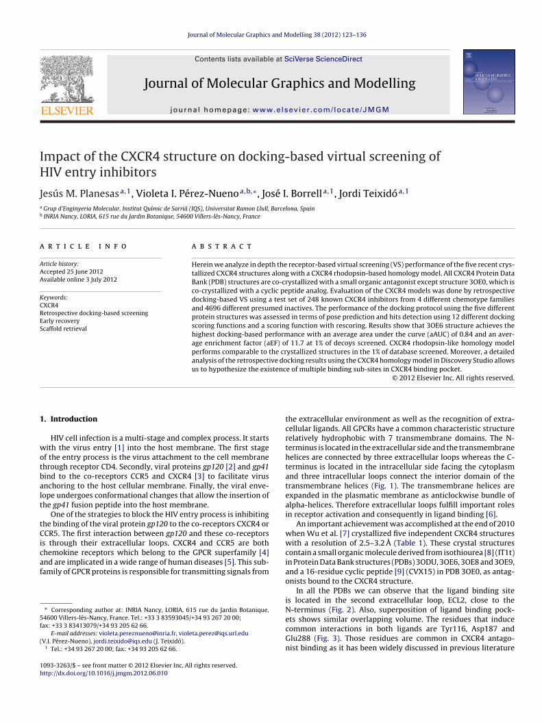
Pérez-Nueno et al. test set of actives and decoys was dockedgainst all five CXCR4 PDBs and the CXCR4 homology model usinghe aforementioned docking functions. Docked poses were rankedttending to their docking score. Fig. 7 shows the ROC plots for eachDB and each scoring function. AUC values are shown in Table 5a.ables 4a and 4b show the early recovery performance in termsf EF at 1%, 5% and 10% of the screened database and the percent-ge of true positives selected at 1%, 5% and 10% of false positives,espectively.
It can be observed that the ROC curves for the CXCR4 homol-gy model obtained using Ligandfit scoring functions do not arriventil 100% of screened database. Discovery Studio is very sensi-ive to the size of the ligand, the geometry of the binding pocket,nd the definition of the binding pocket sphere to retrieve accuraterotein–ligand interaction energies. Hence, if good interactionsannot be found with a given definition of the binding pocket for
determined ligand, the software will not return a docking scoreor that ligand. When defining the CXCR4 binding pocket as for theest of the docking software (see Section 2), Discovery Studio didot retrieve results for 200 out of the 248 actives. Hence, in ordero improve hits retrieval, the binding site of the CXCR4 homology
odel was defined first into two and after into three partition lev-ls using Discovery Studio (Fig. 8). Discovery Studio partition levelsivide randomly one binding site into a specified number of sub-ites. In this manner, the docking engine Ligandfit explores each
ubsite and all the different combinations of them. Consequentlyhen n subsites are defined, the test set is docked n factorial times
long the binding site. Docking-based VS was carried out explor-ng in a single run every sub-site, and also the combinations of
green–red and blue–red). (For interpretation of the references to color in this figurelegend, the reader is referred to the web version of the article.)
two different sub-sites when three sub-sites were defined. Hitsretrieval and VS performance improved significantly when definingthree sub-sites with respect to defining only one or two sub-sites.The Ligandfit ROC curves shown in Fig. 7 for the CXCR4 homologymodel represent the results when dividing the pocket in three sub-sites. The fact that dividing the CXCR4 pocket in sub-sites increasesthe VS performance agrees with recent published literature whichsupport the fact that at least two binding sub-sites exist [12–14]in the main CXCR4 binding pocket located between ECL2 and theN-terminus, and also with the fact that antagonists like the bicy-clam AMD3100, the monocyclam AMD3465, and the non-cyclamAMD11070 bind to CXCR4 in three different binding modes [47].The distribution of actives, when those were docked into the threesub-sites, was mainly between the pockets defined by sub-sites 1-2and 2-3. 100 actives were docked in sub-site 1-2 and 116 activeswere docked in sub-site 1-3. In pockets defined by sub-sites 1-3,1-1, and 2-2, only 9, 2, and 1 actives were docked, respectively. Inaddition, if we classify the compounds retrieved in each pocket sub-site by chemotype families, we retrieve 52% of AMD derivates, 37%of tetrahydro-quinolinamides, 21% of macrocycles and 35% of KRHderivates in the pocket defined by sub-sites 1-2, and 32% of AMDderivates, 58% of tetrahydro-quinolinamides, 58% of macrocyclesand 35% of KRH derivates in the pocket defined by sub-sites 2-3.
In general, ROC plot analysis shows that the docking-based VS
is selective in choosing ligands from decoys for the retrospectivetest set. Concretely, comparing early recovery values obtained forall PDBs, it can be observed that PDB 3OE6 obtains the highest top
J.M. Planesas et al. / Journal of Molecular Graphics and Modelling 38 (2012) 123–136 129
Table 3Pose prediction analysis when cognate ligands were selfdocked into their respective protein structures. Data in bold represents the best values obtained for each dockingfunction when comparing all five structures. PDB 3OE0 shows the highest RMSD values due to its higher molecular weight.
a Ligand CVX15, co-crystallized in PDB 3OE0, with more than 56 rotatable bonds
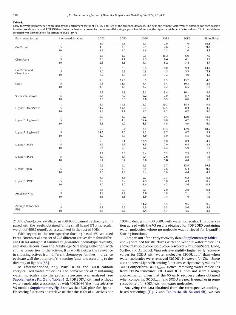
F values (values shown in bold in Table 4a) and also the highestEF (11.2 at 1% of screened database, 7.5 at 5% and 5.4 at 10% ofatabase screened) taking into account all scoring functions used,ven scoring functions with poorest results. Equally, the averageercentage of true positives (Table 4b) returned at 1%, 5% and 10%f false positives is favorable for PDB 3OE6 (19.3% of true positives at% of false positives, 44.1% at 5% and 57.2% at 10% of false positives)mong all scoring functions.
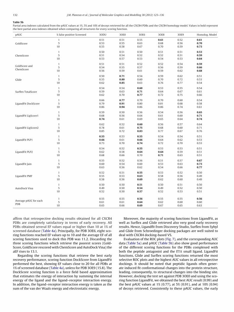
Comparing AUC and pAUC data values at 1%, 5% and 10% ofcreened decoys for all PDBs, it can be observed that PDB 3OE6btains also the highest top AUC and pAUC values (values shownn bold in Tables 5a and 5b), the highest aAUC value (0.82) and theighest average pAUC values (0.56 at 1%, 0.64 at 5% and 0.69 at 10%f decoys screened, respectively) among all scoring functions.
Enrichment factors, early recovery, AUC and pAUC resultsor the homology model (HomolMod) are also shown inables 4a, 4b, 5a and 5b, respectively.
.3. Scaffold retrieval analysis
Pérez-Nueno et al. test set contains 248 actives fromour chemotype families: AMD derivates [29,48], tetrahydro-uinolinamines [49,50], macrocycles [51,52] and KRH derivates53,54]. To evaluate the ability of the docking protocol to retrievehese diverse scaffold families, we analyzed the percentage ofctives returned by the scoring functions attending to its intrinsichemotype classification at 1%, 5%, and 10% of screened database.oreover, to assess how selective is the scaffold retrieval we chose
DB 3OE6 as CXCR4 working structure, being the most powerfuln terms of aAUC, pAUC, and early recovery performance. Fig. 9hows the scaffold retrieval rates. It can be observed that at 1%f screened database all four CXCR4 scaffolds are found. Only foretrahydro-quinolinamines and for KRH derivates, one and four
coring functions respectively, did not retrieve any compound at% of screened database and at 5% of screened database only onecoring function failed to recover any KRH derivates. It is alsoell noted that at 5% of screened database the scoring function
14.2 5.7 6.0 6.2
ds Glide and AutoDock Vina docking requirements.
LigScore2 recovered more than 40% of all chemotype CXCR4 fam-ilies and other scoring functions like Surflex-TotalScore, PLP1, andPMF recovered more than 30% of all CXCR4 scaffolds. Analysis at10% of screened database revealed that on average all scaffoldswere retrieved in more than 50% of their family compounds unlessfor tetrahydro-quinolinamines and KRH derivates whose averageretrieval rate was 47.4% and 46.2%, respectively.
4. Discussion
Docking evaluation of five CXCR4 crystal structures and aCXCR4 homology model was carried out in terms of poseprediction and retrospective docking-based VS. We used repre-sentative force-field based scoring functions like GoldScore andDockScore, empirical scoring functions like ChemScore, Surflex-TotalScore, Glide, LigScore1, LigScore2, PLP1, PLP2 and Jain, andalso knowledge-based scoring functions such as PMF or thehybrid knowledge-based and empirical scoring function AutodockVina.
With respect to pose prediction, RMSD results for the top 10binding poses retrieved for all PDBs using all aforementioned dock-ing functions are not totally satisfactory. Average RMSD values fromTable 3 are all higher than 2 A. Only four aRMSD results reachedvalues between 2 and 4 A, i.e. with the PDB 3ODU for the sco-ring functions GoldScore (3.9 A), ChemScore (2.7 A) and GoldScorerescored with ChemScore (3.7 A) and with the PDB 3OE8 using Glide(3.8 A). Moreover, Table 3 also shows only four binding modes withRMSD values below 2 A, i.e. for PDB 3ODU the best RMSD pose was1.0 A using GoldScore and GoldScore rescored with ChemScore andfor PDB 3OE6 and 3OE8 we obtained in both cases 1.5 A using Glide.
Comparing pose prediction between the PDBs it can be observedthat PDB 3ODU obtains on average the most accurate results (meanaRMSD of 4.5 A) using as scoring functions GoldScore, ChemScore
and GoldScore rescored with ChemScore. As we stated before, it isnecessary to take into account that RMSD metric depends on themolecular size of the ligand to dock, and therefore results obtainedfrom docking the peptidic ligand CVX15 (molecular weight of
130 J.M. Planesas et al. / Journal of Molecular Graphics and Modelling 38 (2012) 123–136
Table 4aEarly recovery performance expressed by the enrichment factor at 1%, 5%, and 10% of the screened database. The best enrichment factor values obtained for each scoringfunction are shown in bold. PDB 3OE6 retrieves the best enrichment factors across all docking approaches. Moreover, the highest enrichment factor value at 1% of the databasescreened was also obtained for structure 3OE6 (19.7).
138.6 g/mol), co-crystallized in PDB 3OE0, cannot be directly com-ared with the results obtained for the small ligand IT1t (moleculareight of 406.7 g/mol), co-crystallized in the rest of PDBs.
With regard to the retrospective docking-based VS, we usedérez-Nueno et al. test set of 248 different actives from four differ-nt CXCR4 antagonist families to guarantee chemotype diversity,nd 4696 decoys from the Maybridge Screening Collection withimilar properties to the actives. It is worth noting the relevancen choosing actives from different chemotype families in order tovaluate well the potency of the scoring functions according to theiversity of ligands [55].
As aforementioned, only PDBs 3ODU and 3OE0 containocrystallized water molecules. The convenience of maintainingater molecules into the protein structure was analyzed (see
upplementary Fig. 2 and Tables 1, 2. PDB 3ODU with and withoutaters molecules was compared with PDB 3OE6 (the most selectiveS model). Supplementary Fig. 2 shows that ROC plots for Ligand-it scoring functions do retrieve neither the 100% of all actives nor
100% of decoys for PDB 3ODU with water molecules. This observa-tion agreed with the VS results obtained for PDB 3OE0 containingwater molecules, where no molecule was retrieved for LigandFitScoring functions.
Comparison of the early recovery data (Supplementary Tables 1and 2) obtained for structures with and without water moleculesshows that GoldScore, GoldScore rescored with ChemScore, Glide,Surflex and Autodock Vina retrieve slightly higher early recoveryvalues for 3ODU with water molecules (3ODUwater) than whenwater molecules were removed (3ODU). However, for ChemScoreand the seven LigandFit scoring functions, early recovery values for3ODU outperform 3ODUwater. Hence, removing water moleculesfrom CXCR4 structures 3ODU and 3OE0 does not seem a roughapproximation given that the VS early recovery values obtained
when comparing 3ODUwater and 3ODU are nearly equal, or in somecases better, for 3ODU without water molecules.
Analyzing the data obtained from the retrospective docking-based screenings (Fig. 7 and Tables 4a, 4b, 5a and 5b), we can
J.M. Planesas et al. / Journal of Molecular Graphics and Modelling 38 (2012) 123–136 131
Table 4bEarly recovery performance expressed as percentage of actives recovered when 1%, 5%, and 10% of decoys were respectively selected. Values in bold represent the bestpercentages obtained when comparing all structures for each scoring function.
Table 5aAUC values calculated from the ROC plot for all the CXCR4 PDBs and the CXCR4 homology model. Values in bold represent the best AUC values obtained when comparing allstructures for each scoring function.
132 J.M. Planesas et al. / Journal of Molecular Graphics and Modelling 38 (2012) 123–136
Table 5bPartial area indexes calculated from the pAUC values at 1%, 5% and 10% of decoys retrieved for all the CXCR4 PDBs and the CXCR4 homology model. Values in bold representthe best partial area indexes obtained when comparing all structures for every scoring function.
pAUC % false positive Screened 3ODU 3OE0 3OE6 3OE8 3OE9 Homolog. Model
Average pAUC for each1 0.55 0.55 0.56 0.55 0.55 0.56
aPPsrstSa
rp1DteIs
PDB5 0.61
10 0.65
ffirm that retrospective docking results obtained for all CXCR4DBs are completely satisfactory in terms of early recovery. AllDBs obtained several EF values equal or higher than 10 at 1% ofcreened database (Table 4a). Principally, for PDB 3OE6, eight sco-ing functions reached EF values up to 10 and the average EF of allcoring functions used to dock this PDB was 11.2. Discarding thehree scoring functions which retrieve the poorest scores (Gold-core, GoldScore rescored with ChemScore and AutoDock Vina) theEF rises to 13.1.
Regarding the scoring functions that retrieve the best earlyecovery performance, scoring function DockScore from LigandFiterformed the best, showing EF values close to 20 for all PDBs at% of screened database (Table 4a), unless for PDB 3OE9 (15.8). TheockScore scoring function is a force field based approximation
hat estimates the energy of interaction by summing the internalnergy of the ligand and the ligand–receptor interaction energy.n addition, the ligand–receptor interaction energy is taken as theum of the van der Waals energy and electrostatic energy.
0.61 0.64 0.62 0.60 0.600.66 0.69 0.67 0.65 0.63
Moreover, the majority of scoring functions from LigandFit, aswell as Surflex and Glide retrieved also very good early recoveryresults. Hence, Ligandfit from Discovery Studio, Surflex from Sybyland Glide from Schroedinger docking packages are well suited todeal with CXCR4 docking-based VS.
Evaluation of the ROC plots (Fig. 7), and the corresponding AUCdata (Table 5a) and pAUC (Table 5b) also show good performanceof the different scoring functions for the PDBs complexed withboth the peptide antagonist and the IT1t small ligand. LigandFitfunctions, Glide and Surflex scoring functions returned the mostselective ROC plots and the highest AUC values in all retrospectivedockings. It should be noted that peptidic ligands often gener-ate induced fit conformational changes into the protein structure,leading, consequently, to structural changes into the binding site.
However, docking the test set against PDB 3OE0 and using the sco-ring function LigandFit, we obtained the best AUC result (0.99) andthe best pAUC values at 1% (0.77), at 5% (0.91), and at 10% (0.94)of decoys retrieved. Consistently to these pAUC values, the early
J.M. Planesas et al. / Journal of Molecular Graphics and Modelling 38 (2012) 123–136 133
Fig. 9. Scaffold diversity retrieval plots for PDB 3OE6. These graphics show the percentage of actives by chemotype family retrieved using all different scoring functions (barsi n of to
rd
ua(bPeaCAcfcorss
hcPfttCL3s
n colors) at 1%, 5% and 10% of the screened database, respectively. (For interpretatiof the article.)
ecovery was also the highest (77.4% of actives at 1% of recoveredecoys) along all retrospective dockings.
If we compare the ROC plots and their corresponding AUC val-es by PDB (Table 5a), all PDBs obtained average AUC values (aAUC)round 0.8, corresponding the highest aAUC value to PDB 3OE60.82). Also, the PDB 3OE6 obtained the best AUC values (Table 5a inold) when compared with the AUC values achieved for the rest ofDBs. Comparison of the early recovery and AUC values obtained forach scoring function and all the structures shows that PDB 3OE6gain returns the most selective results for GoldScore rescored withhemScore, LigandFit LigScore2, LigandFit PLP1, LigandFit PMF andutoDock Vina scoring functions. Attending to these results, wehose PDB 3OE6 for a scaffold retrieval analysis using the scoringunctions selected in this work. This analysis shows that all fourhemotype families were retrieved for all scoring functions at 10%f the screened database and only one scoring function did notetrieve KRH compounds at 5% of screened database. Therefore,caffold retrieval results obtained for PDB 3OE6 are also completelyatisfactory.
Comparing the docking-based VS results obtained for the CXCR4omology model with the ones obtained for all the CXCR4 PDBs, itan be observed that the homology model captures as well as theDB complexes the early recovery performance. Good enrichmentactors at 1% of the screened database and good percentage ofrue positives at 1% of decoys were retrieved for the scoring func-ions: GoldScore (15.3 and 34.7%) and GoldScore rescored with
hemScore (14.5 and 24.2%), LigandFit LigScore1 (18.1 and 38.7%),igandFit LigScore2 (18.5 and 35.5%) and LigandFit Jain (19.3 and7.1%). On average, early recovery results were similar and inome cases even better than those obtained for the crystallized
he references to color in this figure legend, the reader is referred to the web version
structures. Nevertheless, generally poor early recovery resultswere achieved using the scoring functions: Glide, LigandFit PLP1,LigandFit PLP2, LigandFit PMF and AutoDock Vina. Additionally, forthe homology model, it was observed that early recovery valuesbecame less selective when increasing the percentage of screeneddatabase.
In the same way when comparing the ROC plots for the homol-ogy model and the crystal structures it can be observed that thehighest average pAUC value at 1% of retrieved decoys (Table 5b)was achieved by the homology model as well as by the 3OE6 PDB(average pAUC of 0.56). Moreover, average pAUC values at 5% and10% of retrieved decoys for the homology model were very similarto the values obtained for the crystal structures.
Hence, the homology model performed as well as the crystalstructures in the first percentages of database screened, whichsuggests that using an homology model created from a templatecrystallized with a representative ligand may be a decent approachto separate members of a particular inhibitor class from inactives invirtual screening experiments. This observation is consistent withseveral papers that support the use of homology models for GPCR[56,57] predictions and protein kinases VS [58]. Vilar et al. sup-port the notion that homology models often provide a reasonableframework for discussing experimental data. They state that crystalstructures are only static representations of proteins and neglect-ing protein flexibility by using only one rigid representation of thetarget has been shown to adversely affect virtual screening per-
formance. They claim that receptor ensemble docking, a methodwhere docking is performed on several distinct structures and theresults are subsequently combined, has been successfully used toaccount for protein flexibility in virtual screening campaigns [56].
1 r Grap
Cerlbaa
ttiaimCitmnrlAia
ditwsts
iCrbtrtomkTitdcrcihfsduolfissoiHi
34 J.M. Planesas et al. / Journal of Molecula
Hence, combining docking results obtained using differentXCR4 crystal structures or even different CXCR4 homology mod-ls could help to account for protein flexibility and to improve VSesults. Furthermore, it is also worth mentioning that if a particu-ar protein family has a greater degree of induced fit upon ligandinding, then homology models created from a template with anppropriate ligand bound will produce better docking results than
crystal structure with no ligand or a dissimilar ligand bound [56].In summary, although the homology model performs as well as
he crystal structures in the first percentages of database screenedhe total VS performance shows a limited predictive power. CXCR4s generally a target difficult to dock. Retrospective docking resultsnd binding mode prediction are not completely satisfactory. Bind-ng mode analyses of the docked poses onto the CXCR4 homology
odel allowed us to hypothesize multiple binding sub-sites inXCR4 binding pocket. This hypothesis could explain the difficulty
n retrieving good receptor-based VS results. Recent studies showhat there are several notable differences in the binding mode of the
onocyclam (such as AMD3465), bicyclam (such as AMD3100) andon-cyclam (such as MX122) antagonists [10,12–14]. Moreover,ecent literature suggests that the CXCR4 conformation stabi-ized i.e. by AMD3100 differs slightly from the one stabilized byMD3465, mainly in the orientation of TM7. This may explain the
mproved efficacy of AMD3465 over AMD3100 in inhibiting thection of CXCL12 [13].
Furthermore, CXCR4 flexibility makes also difficult to get goodocking results. For example, by including receptor backbone flex-
bility, it is possible to consolidate all the mutation data pertainingo the binding of the cyclam compounds [14]. Techniques that dealith “difficult targets”, that is to say targets with multiple binding
ites or multiple binding modes, have recently appeared in ordero improve the screening performance, such as consensus shapecreening [59–61].
As expected, according to the experiment carried out (selfdock-ng of the two different CXCR4 co-crystallized ligands, IT1t andVX15, into their respective protein structures using different sco-ing functions), the highest resolution structures performed theest in the compound pose prediction analysis. However, regardinghe docking-based VS performance, it turned out that the lowestesolution structure, 3OE6, provided the best early enrichment ofhe actives in the screening dataset. This result leads to the debatef prioritizing conformational selection or induced fit concepts. Theajority of active ligands compiled from the literature are just
nown to bind CXCR4, without an exact idea of their binding mode.aking this into account, together with the fact that CXCR4 proteins known to be an allosteric transmembrane protein [62,63], leadso the hypothesis that the majority of active ligands in our compiledatabase bind in a more stable mode within the lowest resolutiononformation in the light of our early recovery docking enrichmentesults. CXCR4 structures can be crystallized in slightly differentonformations, regardless of the crystal resolution, which capturesn some way the induced fit binding capacity of the protein. We canave two CXCR4 crystal complexes at the same resolution with dif-
erent interactions in the binding pocket [64] (i.e. 3OE8 and 3OE9tructures, see Fig. 3) or two different resolution CXCR4 dimers withifferent interactions (i.e. 3ODU and 3OE9, see Fig. 3) which can gives an idea of the allosteric interactions. Therefore, given the lightf the results in this study, we hypothesize that the lowest reso-ution CXCR4 crystal structure describes better the CXCR4 inducedt binding effect, allowing the wide range of different active ligandcaffold families to be better accommodated in different pocketubsites in 3OE6. Our homology model also shares this behavior
f 3OE6, which is able to accommodate the wide range of activesn different pocket subsites retrieving good early recovery values.ence, it is necessary to take a compromise when performing dock-
ng based virtual screening on allosteric proteins between choosing
hics and Modelling 38 (2012) 123–136
a higher resolution crystal structure or a lower resolution crystalstructure but with a conformation describing better the inducedfit mechanism of the protein. Moreover, if the induced fit effectis significantly important, it might be even better to use a goodmodel (or good homology model if crystal structures are not avail-able) built to account for protein flexibility, i.e. by consensus shapescreening and receptor ensemble docking, using templates with anappropriate ligand bound. Supplementary Fig. 3 shows a compari-son between the binding pockets of the highest (3ODU) and lowest(3OE6) resolution CXCR4 crystal structures used in this study interms of H bonding, lipophilic potential, and electrostatic potentialsurface maps. Moreover, a detailed binding pocket interaction mapshowing the different interactions listed in Fig. 3 is shown for both3ODU and 3OE6 structures. The H bonding surface map is similar forboth structures, however the lowest resolution structure seems tohave a bigger and more defined hydrophobic binding pocket centralregion surrounded by high electronegativity areas (see hydrogenbonding surface map on the left), which corresponds to the brownhigh lipophilic region in the lipophilic potential surface map, anda bigger and central blue region in the electrostatic potential map.The different binding pocket interactions between both structuresare mainly between the residues Glu32, Lys38, Ala98, His113 andArg188.
5. Conclusions
In the present study we have compared and evaluated for thefirst time all CXCR4 structures published by Wu et al. together withour previously published CXCR4 rhodopsin-based homology modelunder the point of view of their receptor based virtual screeningperformance.
With the aim of doing an evaluation that guarantees diversityduring the global process we used Pérez-Nueno et al. test set of248 known actives comprising four different chemotypes [43] ofinhibitors. Additionally, we have used different kinds of scoringfunctions, attending to the nature of the computation algorithm,to dock our previously reported CXCR4 homology model and dif-ferent CXCR4 structures co-crystallized either with a small organicmolecule or a peptide antagonist.
With regard to pose prediction, results are not completely satis-factory. Binding modes obtained from the different dockings seemnot to be accurate enough.
Nevertheless, retrospective docking analysis shows a goodselection of known CXCR4 binders from decoys when defininga binding pocket delimited by residues Asp171, Asp262, andGlu288. In a similar manner, the homology model shows highselectivity in the classification of actives against decoys mainlyin the first percentages of database screened. A detailed analysisof the retrospective docking results using the CXCR4 homol-ogy model in Discovery Studio allows us to hypothesize theexistence of at least two binding sub-sites in CXCR4 bindingpocket.
Summarizing the docking-based VS performance, we found thatthe crystal structure (3OE6) retrieved the most accurate results,especially regarding the early recovery (aEF of 11.2 at 1% of decoysscreened) and the ROC plot analysis (aAUC of 0.82), despite beingthe lowest resolution structure (3.2 A). Moreover, scaffold retrievalanalysis of PDB 3OE6 confirmed that all chemotype families usedin this study were early recovered for all scoring functions. It isalso worth noting that enrichment factors and AUC data obtainedfor PDB 3OE0 (co-crystallized with the peptide antagonist CVX15)
are very similar to the rest of PDBs (co-crystallized with the smallligand IT1t). Hence, the fact that peptidic ligands often gener-ate induced fit conformational changes into the protein structureseems not to affect in this case the majority of VS results.
r Grap
fwmtgbfitdfitnCaCep
A
tasdf
A
f2
R
[
[
[
[
[
[
[
[
[
[
[
[
[
[
[
[
[
[
[
[
[
[
[
[
[
[
[
[
[
[
J.M. Planesas et al. / Journal of Molecula
Overall, herein we analyze in depth the receptor-based VS per-ormance of the five recent crystallized CXCR4 structures, alongith our previously published CXCR4 rhodopsin-based homologyodel. We propose PDB 3OE6 as the most suitable crystal structure
o carry on new prospective docking analyses with the maximumuarantees. We also show that our previous CXCR4 rhodopsin-ased homology model discriminates actives from decoys at therst percentage of database screened as well as the crystal struc-ures do. We hypothesize that these both structures perform wellue to their capacity to capture in some way a more general inducedt conformation to which a wide range of ligands can bind due tohe known CXCR4 allosteric behavior. These results support theotion that if no information is available, as it was the case ofXCR4 target years ago, homology models can be useful. Moreover,
detailed analysis of the retrospective docking results using theXCR4 homology model in Discovery Studio allows us to hypoth-size the existence of multiple binding sub-sites in CXCR4 bindingocket.
cknowledgements
The authors thank the Servei de Disseny de Farmacs (SDF) inhe Centre de Supercomputacio de Catalunya (CESCA) for providingccess to the computational software used through. This work isupported by the Programa Nacional de Biomedicina (Ministerioe Educación y Ciencia, SAF2010-21617-C02-02). V.I.P.N. is gratefulor a Marie Curie IEF Fellowship grant reference DOVSA 254128.
ppendix A. Supplementary data
Supplementary data associated with this article can beound, in the online version, at http://dx.doi.org/10.1016/j.jmgm.012.06.010.
eferences
[1] J.C. Tilton, R.W. Doms, Entry inhibitors in the treatment of HIV-1 infection,Antiviral Research 85 (2010) 91–100.
[2] S.A. Trushin, G.D. Bren, A.D. Badley, CXCR4 tropic HIV-1 gp120 inhibition ofSDF-1�-induced chemotaxis requires Lck and is associated with Cofilin phos-phorylation, Open Virology Journal 4 (2010) 157.
[3] P.R. Harrigan, L. Swenson, R.A. McGovern, G.J. Pollock, The determinants andconsequences of HIV coreceptor switching, HIV Medical Update 4 (2009).
[4] T. Haga, S. Takeda, G Protein-coupled Receptors: Structure, Function, andLigand Screening, CRC Press, Boca Raton, 2005.
[5] W.T. Choi, J. An, Biology and clinical relevance of chemokines and chemokinereceptors CXCR4 and CCR5 in human diseases, Experimental Biology andMedicine 236 (2011) 637–647.
[6] M. Peeters, G. van Westen, Q. Li, A. IJzerman, Importance of the extracellu-lar loops in G protein-coupled receptors for ligand recognition and receptoractivation, Trends in Pharmacological Sciences 32 (2011) 35–42.
[7] B. Wu, E.Y.T. Chien, C.D. Mol, G. Fenalti, W. Liu, V. Katritch, R. Abagyan, A. Brooun,P. Wells, F.C. Bi, Structures of the CXCR4 chemokine GPCR with small-moleculeand cyclic peptide antagonists, Science 330 (2010) 1066–1071.
[8] C.A. Mosley, L.J. Wilson, J.M. Wiseman, J.W. Skudlarek, D.C. Liotta, Recentpatents regarding the discovery of small molecule CXCR4 antagonists, ExpertOpinion on Therapeutic Patients 19 (2009) 23–38.
[9] G. Moncunill, M. Armand-Ugon, I. Clotet-Codina, E. Pauls, E. Ballana, A. Llano, B.Romagnoli, J.W. Vrijbloed, F.O. Gombert, B. Clotet, Anti-HIV activity and resis-tance profile of the CXC chemokine receptor 4 antagonist POL3026, MolecularPharmacology 73 (2008) 1264.
10] S. Pettersson, V.I. Pérez Nueno, M.P. Mena, B. Clotet, J.A. Esté, J.I. Borrell, J.Teixidó, Novel monocyclam derivatives as HIV entry inhibitors: design, syn-thesis anti HIV evaluation, and their interaction with the CXCR4 co receptor,ChemMedChem 5 (2010) 1272–1281.
11] V.I. Perez-Nueno, D.W. Ritchie, J.I. Borrell, J. Teixido, ChemInform abstract: clus-tering and classifying diverse HIV entry inhibitors using a novel consensusshape-based virtual screening approach: further evidence for multiple bind-ing sites within the CCR5 extracellular pocket, Journal of Chemical Information
and Modeling 48 (2008) 2146–2165.
12] S.P. Kawatkar, M. Yan, H. Gevariya, M.Y. Lim, S. Eisold, X. Zhu, Z. Huang, J. An,Computational analysis of the structural mechanism of inhibition of chemokinereceptor CXCR4 by small molecule antagonists, Experimental Biology andMedicine 236 (2011) 844–850.
[
[
hics and Modelling 38 (2012) 123–136 135
13] M.A.C. Neves, S. Simões, M.L. Sá e Melo, Ligand-guided optimization of CXCR4homology models for virtual screening using a multiple chemotype approach,Journal of Computer-Aided Molecular Design 24 (2010) 1023–1033.
14] A.R. Lam, S. Bhattacharya, K. Patel, S.E. Hall, A. Mao, N. Vaidehi, Importance ofreceptor flexibility in binding of cyclam compounds to the chemokine receptorCXCR4, Journal of Chemical Information and Modeling 51 (2010) 137–147.
15] R. Wang, Y. Lu, X. Fang, S. Wang, An extensive test of 14 scoring functions usingthe PDB bind refined set of 800 protein–ligand complexes, Journal of ChemicalInformation and Computer Science 44 (2004) 2114–2125.
16] O. Trott, A.J. Olson, AutoDock Vina: improving the speed and accuracy of dock-ing with a new scoring function, efficient optimization, and multithreading,Journal of Computational Chemistry 31 (2010) 455–461.
18] R.A. Friesner, J.L. Banks, R.B. Murphy, T.A. Halgren, J.J. Klicic, T. Daniel, M.P.Repasky, E.H. Knoll, M. Shelley, J.K. Perry, Glide: a new approach for rapid,accurate docking and scoring 1. Method and assessment of docking accuracy,Journal of Medicinal Chemistry 47 (2004) 1739–1749.
20] SYBYL-X 1.1, Tripos International, 1699 South Hanley Rd., St. Louis, MO 63144,USA.
21] A. Krammer, P.D. Kirchhoff, X. Jiang, C.M. Venkatachalam, M. Waldman,LigScore: a novel scoring function for predicting binding affinities, Journal ofMolecular Graphics and Modelling 23 (2005) 395–407.
22] D.K. Gehlhaar, G.M. Verkhivker, P.A. Rejto, C.J. Sherman, D.R. Fogel, L.J. Fogel,S.T. Freer, Molecular recognition of the inhibitor AG-1343 by HIV-1 protease:conformationally flexible docking by evolutionary programming, Chemistryand Biology 2 (1995) 317–324.
23] Y. Bustanji, M.O. Taha, A.M. Yousef, A.G. Al-Bakri, Berberine potently inhibitsprotein tyrosine phosphatase 1B: investigation by docking simulation andexperimental validation, Journal of Enzyme Inhibition and Medicinal Chemistry21 (2006) 163–171.
24] V.I. Pérez-Nueno, D.W. Ritchie, O. Rabal, R. Pascual, J.I. Borrell, J. Teixido, Com-parison of ligand-based and receptor-based virtual screening of HIV entryinhibitors for the CXCR4 and CCR5 receptors using 3D ligand shape matchingand ligand–receptor docking, Journal of Chemical Information and Modeling48 (2008) 509–533.
25] I. Kufareva, M. Rueda, V. Katritch, G. Dock, R.C. Stevens, R. Abagyan, Status ofGPCR modeling and docking as reflected by community-wide GPCR Dock 2010assessment, Structure 19 (2011) 1108–1126.
26] X. Liang, CXCR4, inhibitors and mechanisms of action, Chemical Biology andDrug Design 72 (2008) 97–110.
27] A.P. Graves, R. Brenk, B.K. Shoichet, Decoys for docking, Journal of MedicinalChemistry 48 (2005) 3714–3728.
28] MOE (Molecular Operating Environment), Chemical Computing Group Inc.,Montreal, Canada, 2009.
29] S. Hatse, K. Princen, K. Vermeire, L.O. Gerlach, M.M. Rosenkilde, T.W. Schwartz,G. Bridger, E. De Clercq, D. Schols, Mutations at the CXCR4 interaction sitesfor AMD3100 influence anti-CXCR4 antibody binding and HIV-1 entry, FEBSLetters 546 (2003) 300–306.
30] L.O. Gerlach, R.T. Skerlj, G.J. Bridger, T.W. Schwartz, Molecular interactionsof cyclam and bicyclam non-peptide antagonists with the CXCR4 chemokinereceptor, Journal of Biological Chemistry 276 (2001) 14153–14160.
31] M.M. Rosenkilde, L.O. Gerlach, J.S. Jakobsen, R.T. Skerlj, G.J. Bridger, T.W.Schwartz, Molecular mechanism of AMD3100 antagonism in the CXCR4 recep-tor, Journal of Biological Chemistry 279 (2004) 3033.
32] M.M. Rosenkilde, L.O. Gerlach, S. Hatse, R.T. Skerlj, D. Schols, G.J. Bridger, T.W.Schwartz, Molecular mechanism of action of monocyclam versus bicyclam non-peptide antagonists in the CXCR4 chemokine receptor, Journal of BiologicalChemistry 282 (2007) 27354–27365.
33] H. Tamamura, Y. Xu, T. Hattori, X. Zhang, R. Arakaki, K. Kanbara, A. Omagari,A. Otaka, T. Ibuka, N. Yamamoto, A low-molecular-weight inhibitor against thechemokine receptor CXCR4: a strong anti-HIV peptide T140, Biochemical andBiophysical Research Communications 253 (1998) 877–882.
34] J.O. Trent, Z. Wang, J.L. Murray, W. Shao, H. Tamamura, N. Fujii, S.C. Peiper, Lipidbilayer simulations of CXCR4 with inverse agonists and weak partial agonists,Journal of Biological Chemistry 278 (2003) 47136.
35] J. Våbenø, G.V. Nikiforovich, G.R. Marshall, Insight into the binding mode forcyclopentapeptide antagonists of the CXCR4 receptor, Chemical Biology andDrug Design 67 (2006) 346–354.
36] T. Fawcett, An introduction to ROC analysis, Pattern Recognition Letters 27(2006) 861–874.
37] Y. Jiang, C.E. Metz, R.M. Nishikawa, A receiver operating characteristic partialarea index for highly sensitive diagnostic tests, Radiology 201 (1996) 745.
38] P. Sonego, A. Kocsor, S. Pongor, ROC analysis: applications to the classificationof biological sequences and 3D structures, Briefings in Bioinformatics 9 (2008)198.
39] N.A. Obuchowski, ROC analysis, American Journal of Roentgenology 184 (2005)364.
40] T.A. Lasko, J.G. Bhagwat, K.H. Zou, L. Ohno-Machado, The use of receiver oper-ating characteristic curves in biomedical informatics, Journal of BiomedicalInformatics 38 (2005) 404–415.
41] D.K. McClish, Analyzing a portion of the ROC curve, Medical Decision Making 9(1989) 190.
42] A.N. Jain, Bias, reporting, and sharing: computational evaluations of dockingmethods, Journal of Computer-Aided Molecular Design 22 (2008) 201–212.
43] M.D. Mackey, J.L. Melville, Better than random? The chemotype enrich-ment problem, Journal of Chemical Information and Modeling 49 (2009)1154–1162.
44] A.N. Jain, A. Nicholls, Recommendations for evaluation of computa-tional methods, Journal of Computer-Aided Molecular Design 22 (2008)133–139.
45] J.B. Cross, D.C. Thompson, B.K. Rai, J.C. Baber, K.Y. Fan, Y. Hu, C. Humblet, Com-parison of several molecular docking programs: pose prediction and virtualscreening accuracy, Journal of Chemical Information and Modeling 49 (2009)1455–1474.
46] J. Kirchmair, P. Markt, S. Distinto, G. Wolber, T. Langer, Evaluation of the per-formance of 3D virtual screening protocols: RMSD comparisons, enrichmentassessments, and decoy selection––What can we learn from earlier mistakes?Journal of Computer-Aided Molecular Design 22 (2008) 213–228.
47] R.S.Y. Wong, V. Bodart, M. Metz, J. Labrecque, G. Bridger, S.P. Fricker, Com-parison of the potential multiple binding modes of bicyclam, monocylam, andnoncyclam small-molecule CXC chemokine receptor 4 inhibitors, MolecularPharmacology 74 (2008) 1485–1495.
48] E. De Clercq, Recent advances on the use of the CXCR4 antagonist plerixafor(AMD3100 MozobilTM) and potential of other CXCR4 antagonists as stem cellmobilizers, Pharmacology and Therapeutics 128 (2010) 509–518.
49] J.G. Catalano, K.S. Gudmundsson, A. Svolto, S.D. Boggs, J.F. Miller, A. Spaltenstein,M. Thomson, P. Wheelan, D.J. Minick, D.P. Phelps, Synthesis of a novel tri-cyclic 1, 2, 3, 4, 4a, 5, 6, 10b-octahydro-1, 10-phenanthroline ring system andCXCR4 antagonists with potent activity against HIV-1, Bioorganic and Medici-nal Chemistry Letters 20 (2010) 2186–2190.
50] G. Bridger, R. Skerlj, A. Kaller, C. Harwig, D. Bogucki, T.R. Wilson, J. Crawford,E.J. Mceachern, B. Atsma, S. Nan, Chemokine Receptor Binding HeterocyclicCompounds, 2004.
51] G.J. Bridger, R.T. Skerlj, S. Padmanabhan, S.A. Martellucci, G.W. Henson, S. Struyf,M. Witvrouw, D. Schols, E. De Clercq, Synthesis and structure–activity rela-tionships of phenylenebis (methylene)-linked bis-azamacrocycles that inhibitHIV-1 and HIV-2 replication by antagonism of the chemokine receptor CXCR4,Journal of Medicinal Chemistry 42 (1999) 3971–3981.
52] G.J. Bridger, R.T. Skerlj, D. Thornton, S. Padmanabhan, S.A. Martellucci,
G.W. Henson, M.J. Abrams, N. Yamamoto, K.D. Vreese, Synthesis andstructure–activity relationships of phenylenebis (methylene)-linked bis-tetraazamacrocycles that inhibit HIV replication Effects of macrocyclic ringsize and substituents on the aromatic linker, Journal of Medicinal Chemistry38 (1995) 366–378.
[
hics and Modelling 38 (2012) 123–136
53] K. Ichiyama, S. Yokoyama-Kumakura, Y. Tanaka, R. Tanaka, K. Hirose, K. Ban-nai, T. Edamatsu, M. Yanaka, Y. Niitani, N. Miyano-Kurosaki, A duodenallyabsorbable CXC chemokine receptor 4 antagonist KRH-1636, exhibits a potentand selective anti-HIV-1 activity, PNAS 100 (2003) 4185–4190.
54] T. Yamazaki, A. Saitou, M. Ono, S. Yokoyama, K. Bannai, K. Hirose, M. Yanaka,Novel Nitrogenous Compound and Use Thereof, 2003.
55] G.L. Warren, C.W. Andrews, A.M. Capelli, B. Clarke, J. LaLonde, M.H. Lambert,M. Lindvall, N. Nevins, S.F. Semus, S. Senger, A critical assessment of dock-ing programs and scoring functions, Journal of Medicinal Chemistry 49 (2006)5912–5931.
56] S. Vilar, G. Ferino, S.S. Phatak, B. Berk, C.N. Cavasotto, S. Costanzi, Docking-based virtual screening for ligands of G protein-coupled receptors: not onlycrystal structures but also in silico models, Journal of Molecular Graphics andModelling 29 (2011) 614–623.
57] L. Roumen, M. Sanders, B. Vroling, I.J.P. de Esch, J. de Vlieg, R. Leurs, J.P.G.Klomp, S.B. Nabuurs, C. de Graaf, In silico veritas: the pitfalls and challengesof predicting GPCR–ligand interactions, Pharmaceuticals 4 (2011) 1196–1215.
58] W.M. Rockey, A.H. Elcock, Structure selection for protein kinase docking andvirtual screening: homology models or crystal structures? Current Protein andPeptide Science 7 (2006) 437–457.
59] V.I. Perez-Nueno, D.W. Ritchie, Using consensus-shape clustering to identifypromiscuous ligands and protein targets and to choose the right query forshape-based virtual screening, Journal of Chemical Information and Modeling51 (2011) 1233–1248.
60] V.I. Pérez-Nueno, D.W. Ritchie, Applying in silico tools to the discovery of novelCXCR4 inhibitors, Drug Development Research 72 (2011) 95–111.
61] V.I. Pérez-Nueno, D.W. Ritchie, Identifying and characterizing promiscuous tar-gets: implications for virtual screening, Expert Opinion on Drug Discovery 7(2012) 1–17.
62] L.T. May, K. Leach, P.M. Sexton, A. Christopoulos, Allosteric modulation of gprotein-coupled receptors, Annual Review of Pharmacology and Toxicology 47(2007) 1–51.
63] J.M. Janz, Y. Ren, R.J. Looby, M.A. Kazmi, P. Sachdev, A. Grunbeck, L. Haggis, D.Chinnapen, A.Y. Lin, C. Seibert, Direct interaction between an allosteric agonistpepducin and the chemokine receptor CXCR4, Journal of the American ChemicalSociety 40 (2011) 15878–15881.
64] P. Maurice, M. Kamal, R. Jockers, Asymmetry of GPCR oligomers supports their
functional relevance, Trends in Pharmacological Sciences 32 (2011) 514–520.
65] J.A. Ballesteros, H. Weinstein, Integrated methods for the construction ofthree-dimensional models and computational probing of structure–functionrelations in G protein-coupled receptors, Methods in Neurosciences 25 (1995)366–428.