31
Implantation, Extraembryonic Membranes, Placental Structure and Classification
Implantation, Extraembryonic Membranes,
Placental StructureandClassification
A t t a c h m e n t a n d I m p l a n t a t i o n
Implantation is the first stage in development of the placenta. In most cases, implantation is preceded by a close interaction of embryonic trophoblast and endometrial epithelial cells that is known as adhesion or attachment.
Implantation also is known as the stage where the blastocyst embeds itself in the endometrium, the inner membrane of the uterus. This usually occurs near the top of the uterus and on the posterior wall.
Among other things, attachment involves a tight intertwining of microvilli on the maternal and embryonic cells. Following attachment, the blastocyst is no longer easily flushed from the lumen of the uterus. In species that carry multiple offspring, attachment is preceeded by a remarkably even spacing of embryos through the uterus. This process appears to result from uterine contractions and in some cases involves migration of embryos from one uterine horn to another (transuterinemigration).
The effect of implantation in all cases is to obtain very close apposition between embryonic and maternal tissues. There are, however, substantial differences among species in the process of implantation, particularly with regard to "invasiveness," or how much the embryo erodes into maternal tissue.
In species like horses and pigs, attachment and implantation are essentially equivalent. In contrast, implantation in humans involves the embryo eroding deeply into the substance of the uterus.
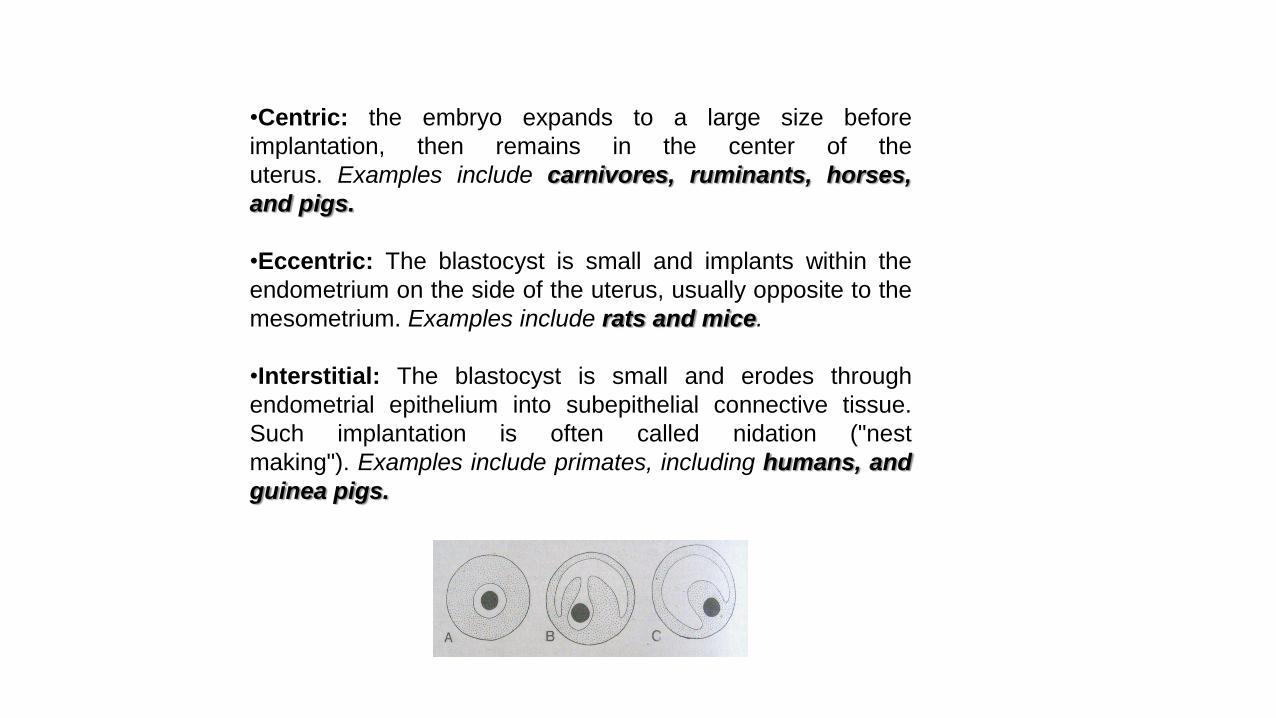
•Centric: the embryo expands to a large size before
implantation, then remains in the center of the
uterus. Examples include carnivores, ruminants, horses,
and pigs.
•Eccentric: The blastocyst is small and implants within the
endometrium on the side of the uterus, usually opposite to the
mesometrium. Examples include rats and mice.
•Interstitial: The blastocyst is small and erodes through
endometrial epithelium into subepithelial connective tissue.
Such implantation is often called nidation ("nest
making"). Examples include primates, including humans, and
guinea pigs.

It has been difficult to attribute any particular advantage to the degree of invasiveness seen during implantation. One possibleexception is that most species having highly invasive embryos have systems for prenatal transfer of antibodies from the mother tothe fetus.
For eccentric and interstitial implantations, what allows the embryo to invade the uterine substance? In some species it appearsthat the blastocyst is a passive participant, and the underlying endometrium degenerates. In other cases, including carnivores andprobably humans, the embryo seems to be the aggressor and trophoblast actively invades into the endometrium. It's likely thatboth tissues participate to some degree.
In species that undergo interstitial implantation, an interesting phenomenon called the decidual cell reaction occurs. This involvestransformation of uterine stromal and endothelial cells into a tissue called the decidua, which becomes a substantial portion of theplacenta and is expelled with the remainder of the placenta at the time of birth. The decidua is a prominent feature of the humanplacenta.
It is clear that steroid hormones from the ovary are necessary to prepare the endometrium for implantation and for the process ofimplantation itself. In some species, progesterone alone appears to be adequate, while in others, estrogen and progesterone arerequired for implantation.
In addition to the differences among species in the implantation process per se, there are also situations in which the timing ofimplantation varies. The usual case is for attachment and implantation to occur within a few days after the blastocyst reaches theuterus. In many animals, however, implantation can be delayed for substantial periods of time, during which the blastocyst enters aquiescent state called embryonic diapause. Delayed implantation seems to be a strategy used to regulate time of birth so that itoccurs when environmental conditions are favorable.
Read it !

Following hatching, the conceptus undergoes massive growth.
In the cow, the blastocyst is about 3 mm in diameter around day 13, which undergoes maximum growth to 250 mm in length within the next four days and appears as a filamentousthread. By day 18 of gestation, the blastocyst occupies space in both uterine horns.
In the sow, the development of blastocyst is even more dramatic, where it grows from 2mm spheres on day 10 to about 200 mm in length in the next 24-48 h reaching lengths of 800-1000 mm by day 16 (growth is at a rate of 4-8 mm/h).
The dramatic growth of the conceptus is due largely to development of a set of membranes called the extraembryonic membranes.
The pig, sheep and cow are characterized as having filamentous or threadlike blastocysts prior to attachment.
In the mare, however, blastocysts do not change into a thread like structure but remain spherical.
As the hatched blastocyst begins to grow, it develops an additional layer called primitive endoderm just beneath, but in contact with the inner cell mass which continues to growdownwards eventually lining the trophoblast.
At the same time, it also forms an evagination at the ventral portion of the inner cell mass to form the yolk sac, a transient extra embryonic membrane that regresses in size as theconceptus develops.
As the blastocyst continues to expand, the newly formed double membrane (the trophoblast and mesoderm) becomes the chorion. Further development of the blastocyst causes thechorion to push upward in the dorso lateral region of the conceptus and begins to surround it.
The chorion begins to send “wing-like” projections above the embryo, the amnion begins to form. Fusion of the chorion over the dorsal portion of the embryo results in formation of acomplete sac called amnion around the embryo.
The amnion is filled with fluid and serves to hydraulically protect the embryo from mechanical perturbations.
As an anti-adhesion material to prevent tissues in the rapidly developing embryo from adhering to each other.
The amnionic vesicle can be palpated in the cow between days 30 and 45 and feels like a small, turgid balloon inside the uterus. The embryo, however, is quite fragile during this earlyperiod and amnionic vesicle palpation should be performed with caution.
During the same time that the amnion is developing, a small evagination from the posterior region of the primitive gut begins to form. This sac-like evagination is referred to as theallantoic sac that collects liquid waste from the embryo.
As the embryo grows, the allantois continues to expand and eventually will make contact with the chorion.
When the allantois reaches a certain volume, it presses against the chorion and eventually fuses with it. When fusion takes place the two membranes are called the allantochorion. Theallantochorionic membrane is the fetal contribution to the placenta and will provide the surface for attachments to the endometrium.
Read it !
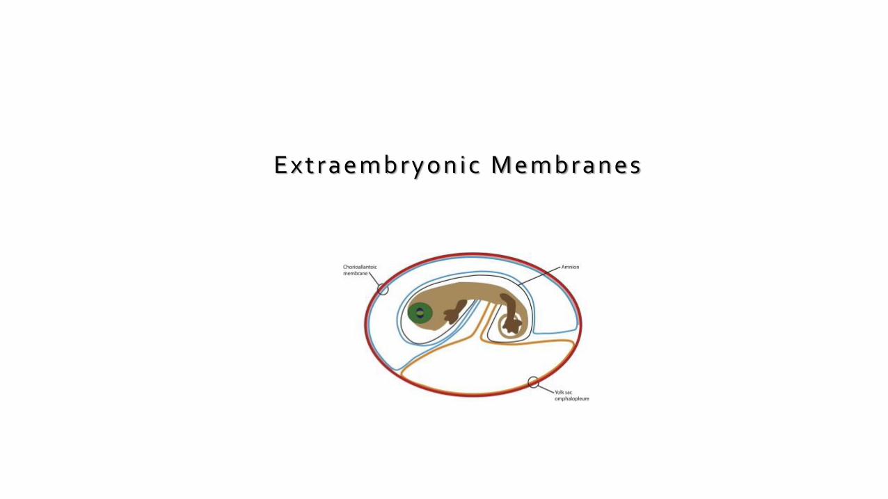
E x t r a e m b r y o n i c M e m b r a n e s

The embryos of reptiles, birds, and mammals produce 4 extraembryonic membranes,
AmnionYolk sacChorion, andAllantois
In birds and most reptiles, the embryo with its extraembryonic membranes develops within a shelled egg.The amnion protects the embryo in a sac filled with amniotic fluid.The yolk sac contains yolk — the sole source of food until hatching. Yolk is a mixture of proteins and lipoproteins.The chorion lines the inner surface of the shell (which is permeable to gases) and participates in the exchange of O2
and CO2 between the embryo and the outside air.The allantois stores metabolic wastes (chiefly uric acid) of the embryo and, as it grows larger, also participates in gas exchange

Amnionic membrane is two cell layers1) epiblast derived extraembryonic ectodermal layer2) thin non-vascular extraembryonic mesodermAs the amnion enlarges it encompasses the embryo on theventral side, merging around the umbilical cord.Amnion forms the epithelial layer of the umbilical cordWith embryo growth the amnion obliterates the chorioniccavityAmnionic sac is fluid filled called amnionic fluid: theembryo is bathed in the fluid
Amnion
Mechanical protection: hydrostatic pressureAllows free movement -which aids in neuromuscular developmentAntibacterialAllow for fetal growthProtection from adhesions
Amnion Function

Hypoblast -the primary yolk sac or Heuser'smembrane.Day 12 -Second wave of cell migration -forms definitive yolksacComposed of extrembryonic endodermEarly nutrition (2-3 weeks) for the embryo -later shrinking -nonfunctional –Meckels diverticulum (outpocketing of smallintestine)Connects to midgut via the yolk sac stalkDerivatives:Early blood cells forms from blood islandsPrimordial germ cellsThe early gut, epithelium of the respiratory and digestivetracts
Yolk Sac
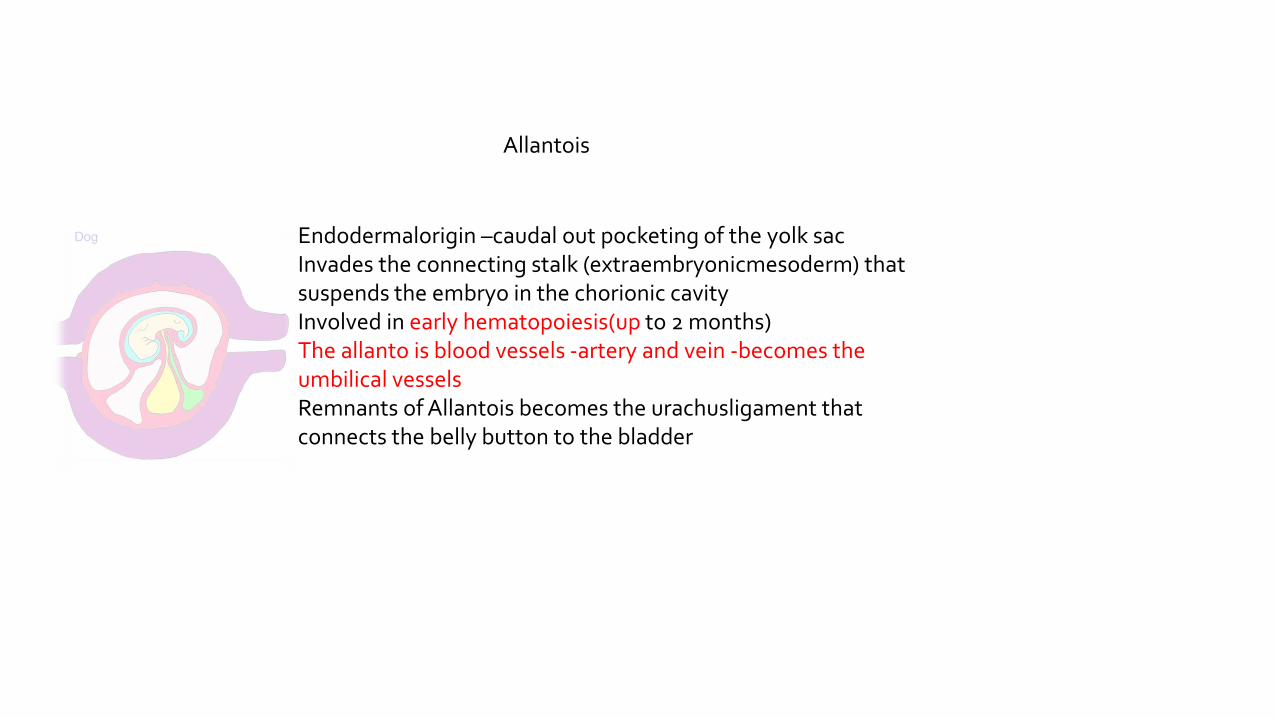
Endodermalorigin –caudal out pocketing of the yolk sacInvades the connecting stalk (extraembryonicmesoderm) thatsuspends the embryo in the chorionic cavityInvolved in early hematopoiesis(up to 2 months)The allanto is blood vessels -artery and vein -becomes theumbilical vesselsRemnants of Allantois becomes the urachusligament thatconnects the belly button to the bladder
Allantois

Chorioniccavity (extraembryonic coelom)-lined with extraembryonic mesodermChorionic cavity expands separating amnion from cytotrophoblastChorionicsac consist of:Cytotrophoblastic layerSyncytiotrophoblastic layerExtraembryonic somatic mesodermThe Chorion / maternal endometrium forms the placentaChorionforms stem villi
Chorion
Placental barrier decreases with gestation
Placental Barrier –syncytiotrophoblast+ basal lamina, basallamin+ fetal capillary endothelium
Syncytiotrophoblasts –many microvilli, no majorhistocompatibility antigens

Decidua is the functional layer of endometrium which is shed during parturition.Decidual Reaction–stromal cells –accumulate glycogen and lipid, called Decidual CellsDecidua basalis-forms maternal component of the placenta; associates with the chorion frondosomDecidua capsularis-superficallayer overlying the entire embryoblast-this layer eventually degenerates;associates with the chorion laeveDecidua parietalis-all remaining parts of the endometrium-not associated with the embryo
Decidua

What is the Placenta ?

The placenta is a:
“Vascular (supplied with blood vessels) organ in most mammals that unites the fetus to the uterus of the mother.
It mediates the metabolic exchanges of the developing individual through an intimate association of embryonic tissues and of certain uterine tissues, serving the functions of nutrition, respiration, and excretion.”

The placenta is also known as a hemochorical villous organ meaning that the maternal blood comes in contact
with the chorion and that villi protrude out of this same structure.
As the fetus is growing and developing, it requires a certain amount of gases and nutrients to help support its
needs throughout pregnancy. Because the fetus is unable to do so on its own, it is the placenta that carries out this
function.

What are the main roles/functions of the placenta?

The placenta provides the connection between fetus and mother in order to help carry outmany different functions that it is incapable to do alone. During pregnancy, the placenta has 6main roles to maintain good health and a good environment for the fetus:
•Respiration•Nutrition•Excretion•Protection•Endocrine•Immunity

I NO2
H2OFesaltscarbohydrates, amino acids, lipidsvitamins, hormones, antibodiesdrugs, alcoholviruses (rubella, varicella-zoster, HIV)
O U TCO2
H2Osaltsurea, uric acidcreatininebilirubin, hormones,RBC antigens
What passes across the placenta?
What stays behind?
How are wastes removed?
The blood supply of the developing fetus is continuous with that of the placenta. The placenta extracts food and oxygen from the uterus. Carbon dioxide and other wastes (e.g., urea) are transferred to the mother for disposal by her excretory organs.

Classification Based on Placental Shape and Contact Points

The placenta is composed of two different surfaces, thematernal surface, facing towards the outside, and the fetalsurface, facing towards the inside, or the fetus. On the fetalsurface, there is the umbilical cord, the link between theplacenta and the fetus.
The placentas of all eutherian (placental) mammals providecommon structural and functional features, but there arestriking differences among species in gross and microscopicstructure of the placenta. Two characteristics are particularlydivergent and form bases for classification of placental types:
1.The gross shape of the placenta and the distribution ofcontact sites between fetal membranes and endometrium.
2.The number of layers of tissue between maternal and fetalvascular systems.
Differences in these two properties allow classification ofplacentas into several fundamental types.

Examination of placentae from different species reveals striking differences in their shape and the area of contact between fetal and maternal tissue:
•Diffuse: Almost the entire surface of the allantochorion is involved in formation of the placenta. Seen in horses and pigs.
•Cotyledonary: Multiple, discrete areas of attachment called cotyledons are formed by interaction of patches of allantochorion with endometrium. The fetal portions of this type of placenta are called cotyledons, the maternal contact sites (caruncles), and the cotyledon-caruncle complex a placentome. This type of placentation is observed in ruminants.
•Zonary: The placenta takes the form of a complete or incomplete band of tissue surrounding the fetus. Seen in carnivores like dogs and cats, seals, bears, and elephants.
•Discoid: A single placenta is formed and is discoid in shape. Seen in primates and rodents.

Classification Based on Layers Between Fetal and Maternal Blood

Just prior to formation of the placenta, there are a total of sixlayers of tissue separating maternal and fetal blood. There arethree layers of fetal extraembryonic membranes in thechorioallantoic placenta of all mammals, all of which arecomponents of the mature placenta:
1.Endothelium lining allantoic capillaries2.Connective tissue in the form of chorioallantoic mesoderm3.Chorionic epithelium, the outermost layer of fetal membranes derived from trophoblast
There are also three layers on the maternal side, but the number of these layers which are retained - that is, not destroyed in the process of placentation - varies greatly among species. The three potential maternal layers in a placenta are:
1.Endothelium lining endometrial blood vessels2.Connective tissue of the endometrium3.Endometrial epithelial cells

One classification scheme for placentas is based on which maternal layers are r e t a i n e d in the placenta, which of course is the same as stating which maternal tissue is in contact with chorionic epithelium of the fetus.
Each of the possibilities is observed in some group of mammals.
Type of Placenta
Maternal Layers Retained
ExamplesEndometrialEpithelium
ConnectiveTissue
UterineEndothelium
Epitheliochorial + + + Horses, swine, ruminants
Endotheliochorial - - + Dogs, cats
Hemochorial - - - Humans, rodents

Summary of Species Differences in Placental Architecture
The placental mammals have evolved a variety of placental types which can be broadly classified using the nomenclature described above slide. Not all combinations of those classification schemes are seen or are
likely to ever be seen - for instance, no mammal is known to have a diffuse, endotheliochorial, or a hemoendothelial placenta. Placental types for "familiar" mammals are summarized below, with
supplemental information provided for a variety of "non-familiar" species.
Type of Placenta Common Examples
Diffuse, epitheliochorial Horses and pigs
Cotyledonary, epitheliochorial Ruminants (cattle, sheep, goats, deer)
Zonary, endotheliochorial Carnivores (dog, cat, ferret)
Discoid, hemochorial Humans, apes, monkeys and rodents

The difference between marsupials & placentals
Although marsupials and placental animals are both mammals, there are several distinguishing features thatdifferentiate the two groups.
The biggest difference between marsupials and placentals liesin the possession a placenta, the oxygen- and nutrient-richorgan that attaches growing embryos of placental mammals totheir mothers. Marsupials, on the other hand, have no internalplacenta and must therefore absorb nutrients from the yolk oftheir ovum; however, once the young are born, they spend amuch longer time suckling than do placental young. Essentially,marsupials spend far more time nurturing and nursing theiryoung after they are born than placentals, mammals that investmore time and energy in pregnancy.

Placenta syndesmochorialis, adeciduata, villosa cotyledonataRuminants (cattle, sheep, goats, deer)
Placenta epithelio-chorialis, adeciduata, villosa diffusa incompleta
Pig
Placenta epithelio-chorialis, adeciduata, villosa diffusa completa
Horse and donkey
Placenta haemo-chroialis, deciduata, villosa discoidea
Humans, apes, monkeys and rodents
Placenta endothelio-chorialis, deciduata, villosa zonaria
Carnivores (dog, cat, ferret)
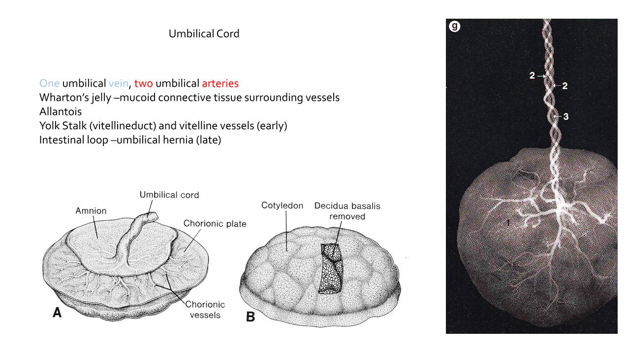
Umbilical Cord
One umbilical vein, two umbilical arteriesWharton’s jelly –mucoid connective tissue surrounding vesselsAllantoisYolk Stalk (vitellineduct) and vitelline vessels (early)Intestinal loop –umbilical hernia (late)
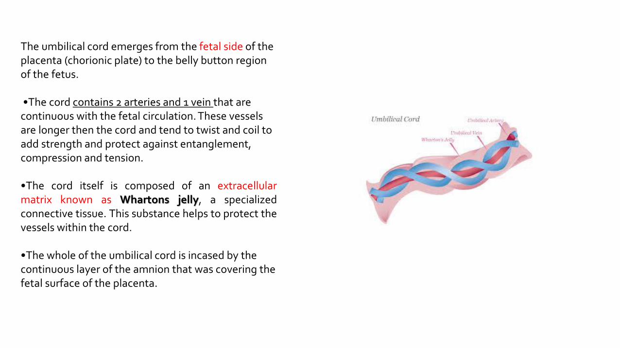
The umbilical cord emerges from the fetal side of the placenta (chorionic plate) to the belly button region of the fetus.
•The cord contains 2 arteries and 1 vein that are continuous with the fetal circulation. These vessels are longer then the cord and tend to twist and coil to add strength and protect against entanglement, compression and tension.
•The cord itself is composed of an extracellularmatrix known as Whartons jelly, a specializedconnective tissue. This substance helps to protect thevessels within the cord.
•The whole of the umbilical cord is incased by the continuous layer of the amnion that was covering the fetal surface of the placenta.

Fetal –Contained within vesselsUmbilical Arteries –chorionic plate –branches to stem villi –capillariesin terminal villi –return via umbilical veinMaternal –Free-flowing lakeSpiral arteries open into intervillous space and bath the villi150 ml of maternal bloodExchanged -3-4 times/minuteReduced blood pressure in intervillous spaceOxygenated blood to the chorionic plate, return baths the villi
Placental Circulation

references
• https://www.encyclopedia.com/
• Langman's Medical Embryology
• BRS Embryology (Board Review Series)
• Essentials of Domestic Animal Embryology


