14
Impression Cytology: Shipping Dry Eye Assessment and Management Penny Asbell, MD, MBA Brendan Barry, BA Seth Epstein,MD Yi Wei, PhD
| Date post: | 25-Dec-2015 |
| Category: |
Documents |
| Upload: | winifred-marian-davis |
| View: | 214 times |
| Download: | 0 times |
Impression Cytology:Shipping
Dry Eye Assessment and Management Penny Asbell, MD, MBA
Brendan Barry, BASeth Epstein,MD
Yi Wei, PhD
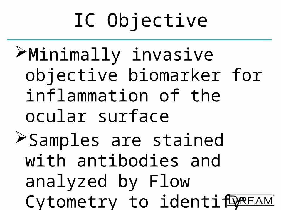
IC Objective
Minimally invasive objective biomarker for inflammation of the ocular surface
Samples are stained with antibodies and analyzed by Flow Cytometry to identify effector cells
Evaluation of Treatment Efficacy
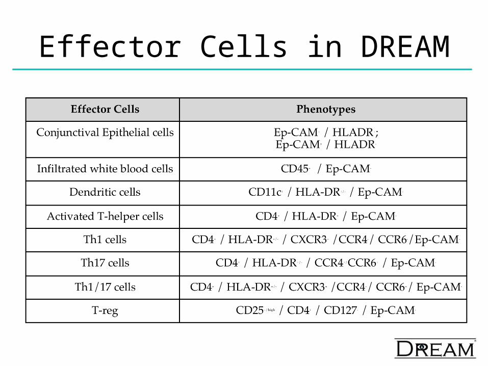
Effector Cells in DREAM
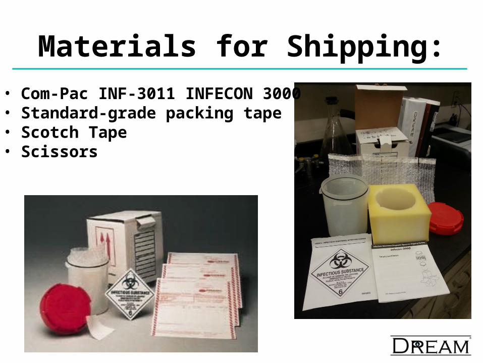
Materials for Shipping:• Com-Pac INF-3011 INFECON 3000 • Standard-grade packing tape• Scotch Tape• Scissors
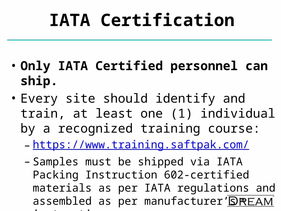
IATA Certification
• Only IATA Certified personnel can ship.• Every site should identify and train, at least one (1)
individual by a recognized training course: – https://www.training.saftpak.com/– Samples must be shipped via IATA Packing Instruction
602-certified materials as per IATA regulations and assembled as per manufacturer’s instructions.
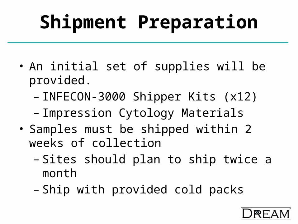
Shipment Preparation
• An initial set of supplies will be provided.– INFECON-3000 Shipper Kits (x12)– Impression Cytology Materials
• Samples must be shipped within 2 weeks of collection– Sites should plan to ship twice a month– Ship with provided cold packs
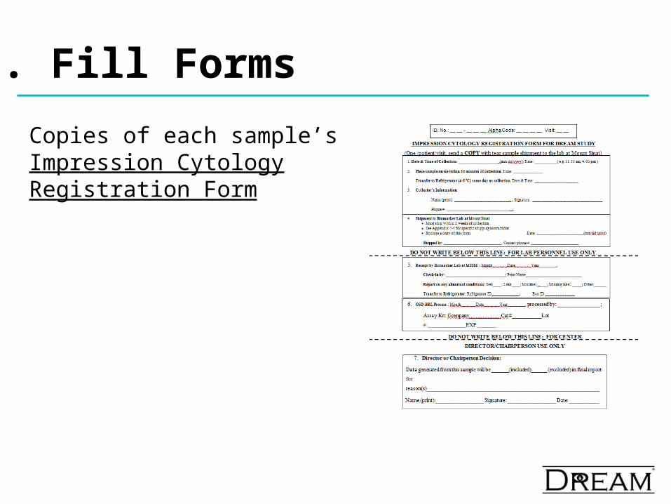
1. Fill Forms
Copies of each sample’s Impression Cytology Registration Form
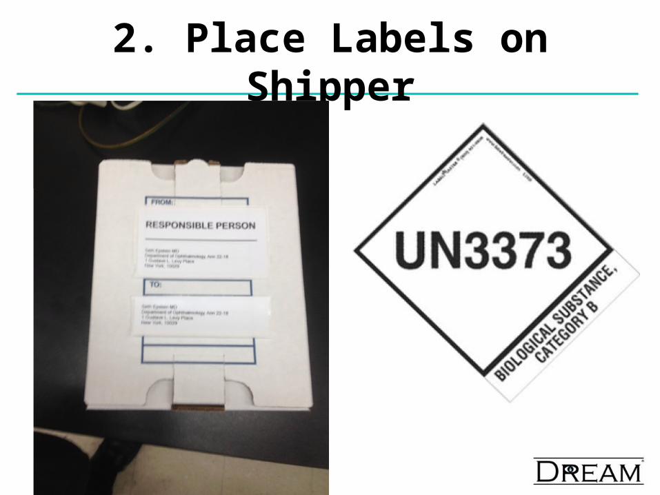
2. Place Labels on Shipper
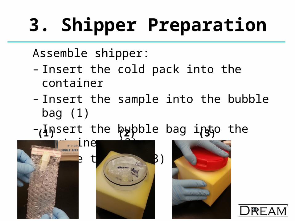
3. Shipper Preparation
Assemble shipper:– Insert the cold pack into the container – Insert the sample into the bubble bag (1)– Insert the bubble bag into the container (2)– Secure the lid (3)(1) (3)(2)
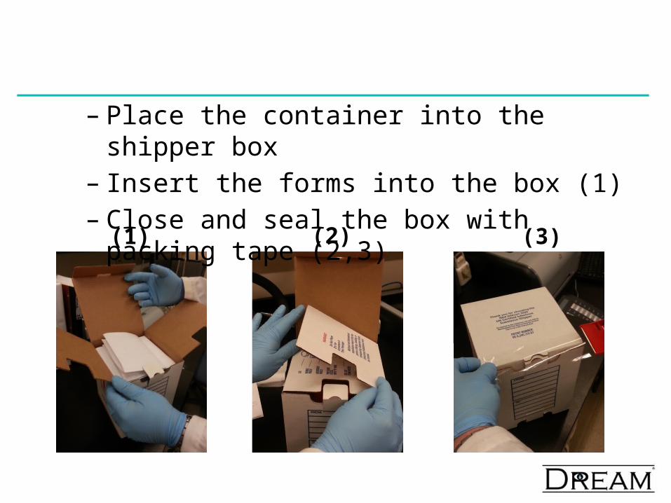
– Place the container into the shipper box– Insert the forms into the box (1)– Close and seal the box with packing tape (2,3)
(1) (2) (3)
X
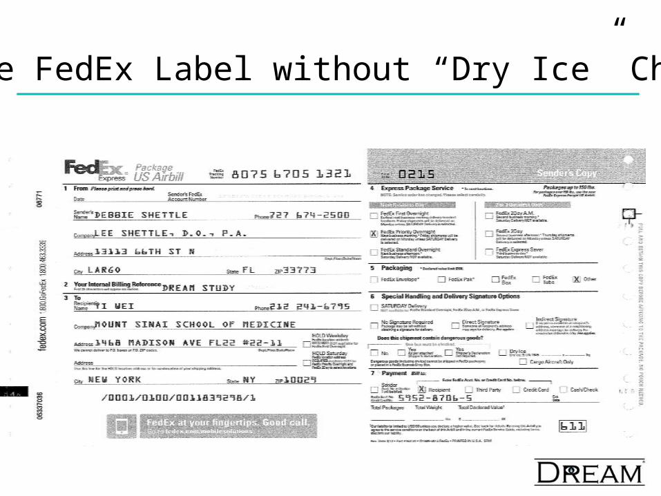
4. Use FedEx Label without “Dry Ice” Checked
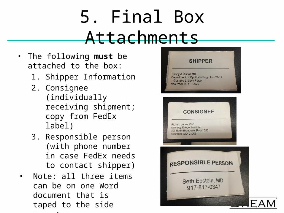
5. Final Box Attachments• The following must be attached
to the box:1. Shipper Information2. Consignee (individually
receiving shipment; copy from FedEx label)
3. Responsible person (with phone number in case FedEx needs to contact shipper)
• Note: all three items can be on one Word document that is taped to the side
• Remember your Category B label!!
Impression Cytology : Summary
• Flow Cytometry provides an objective minimally invasive metric of ocular inflammation
• IC provides Biomarkers of Inflammation of the Ocular Surface
• May provide a method to better classify severity of DED
• Will add to our understanding of DED and the mechanism of action that leads to inflammation of the ocular surface