184
Improving Umbilical Cord Blood Stem Cell Engraftment by Ex Vivo Expansion of Hematopoietic Stem and Progenitor Cells Lucia Elisabeth Duinhouwer

Improving Umbilical Cord Blood Stem Cell Engraftment by Ex Vivo Expansion of Hematopoietic Stem and Progenitor Cells
Lucia Elisabeth Duinhouwer

ISBN: 978-94-6169-958-9
Layout and printed by: Optima Grafische Communicatie, Rotterdam, the Netherlands
Improving Umbilical Cord Blood Stem Cell Engraftment by Ex Vivo Expansion of Hematopoietic Stem and Progenitor Cells
Het verbeteren van klinische uitkomsten na stamceltransplantatie met navelstreng-bloed door het vermeerderen van hematologische stam- en voorlopercellen
Proefschrift
ter verkrijging van de graad van doctor aan de Erasmus Universiteit Rotterdam
op gezag van de rector magnificus
Prof.dr. H.A.P. Pols
en volgens besluit van het College voor Promoties.
De openbare verdediging zal plaatsvinden opdinsdag 15 november 2016 om 15.30 uur
door
Lucia Elisabeth Duinhouwergeboren te Rotterdam

PromotIECommISSIE
Promotor: Prof.dr. J.J. Cornelissen
Overige leden: Prof.dr. J.P. van Leeuwen Prof.dr. B. Löwenberg Prof.dr. J.J. Zwaginga
Co-promotor: Dr. E. Braakman
taBLE of ContEnt
Chapter 1 General introduction and outline of the thesis 7
Chapter 2 Impaired thymopoiesis predicts for a high risk of severe infections after double umbilical cord blood transplantation
35
Chapter 3 Wnt3a protein reduces the number of mouse hematopoietic stem cells in serum-free cultures in an apoptosis-independent manner
59
Chapter 4 Wnt3a protein reduces growth factor-driven expansion of human hematopoietic stem and progenitor cells in serum-free cultures
77
Chapter 5 In vitro and in vivo evaluation of StemRegenin1-expanded umbilical cord blood-derived hematopoietic stem and progenitor cells
95
Chapter 6 Magnetic resonance detection of CD34+ cells from umbilical cord blood using a 19F label
117
Chapter 7 Towards clinical application of StemRegenin1-expanded umbilical cord blood-derived hematopoietic stem and progenitor cells: manufacture and clinical study design
133
Chapter 8 General discussion 149
appendices Summary 163Nederlandse samenvatting 169Publication list 173PhD Portfolio 175Curriculum vitae 178Dankwoord 179


A
Chapter 1General introduction and outline of the thesis

General introduction and outline of the thesis 9
11. HEmatoPoIESIS anD HEmatoPoIEtIC StEm CELLS
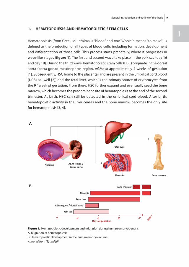
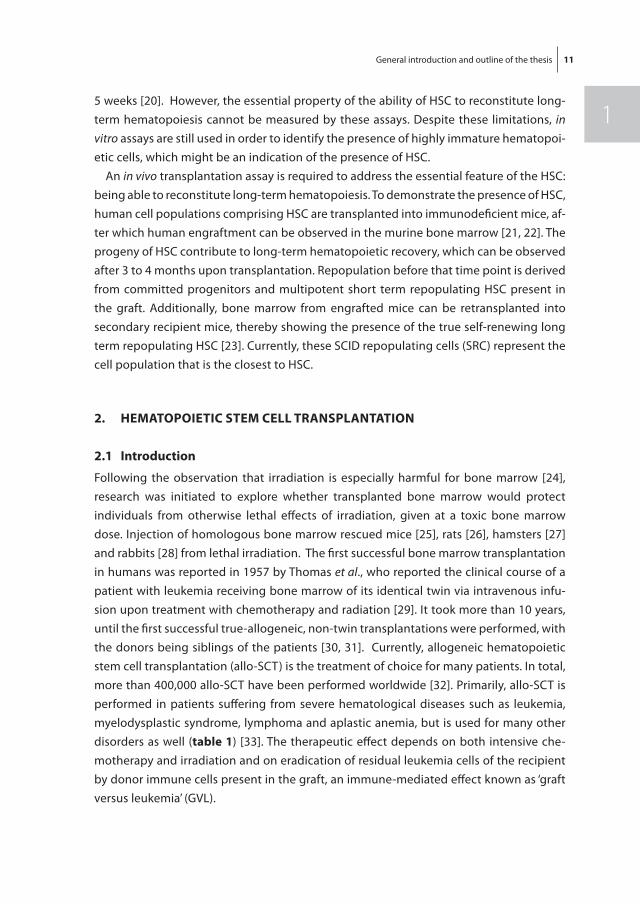
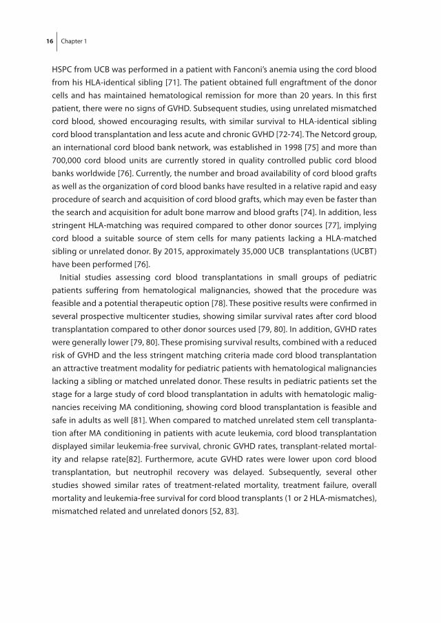
Hematopoiesis (from Greek: αἷμα/aíma is “blood” and ποιεῖν/poieín means “to make”) is defi ned as the production of all types of blood cells, including formation, development and diff erentiation of those cells. This process starts prenatally, where it progresses in wave-like stages (fi gure 1). The fi rst and second wave take place in the yolk sac (day 16 and day 19). During the third wave, hematopoietic stem cells (HSC) originate in the dorsal aorta (aorta-gonad-mesonephros region, AGM) at approximately 4 weeks of gestation [1]. Subsequently, HSC home to the placenta (and are present in the umbilical cord blood (UCB) as well [2]) and the fetal liver, which is the primary source of erythrocytes from the 9th week of gestation. From there, HSC further expand and eventually seed the bone marrow, which becomes the predominant site of hematopoiesis at the end of the second trimester. At birth, HSC can still be detected in the umbilical cord blood. After birth, hematopoietic activity in the liver ceases and the bone marrow becomes the only site for hematopoiesis [3, 4].
Yolk sac AGM region /dorsal aorta
Fetal liver
Placenta Bone marrow
0 20 40 60 80Days of gestation
Bone marrow
Placenta
Fetal liver
AGM region / dorsal aorta
Yolk sac
A
B
figure 1. Hematopoietic development and migration during human embryogenesisA. Migration of hematopoiesis B. Hematopoietic development in the human embryo in time. Adapted from [5] and [6]
10 Chapter 1
In adults, all mature blood cells are derived from a small population of HSC that reside in the bone marrow. HSC are self-renewing and have the ability to differentiate into pro-genitors of all different hematopoietic lineages and eventually generate mature blood cells, including erythrocytes, platelets, and leukocytes. Within the bone marrow, HSC reside in anatomically defined regions, the so-called niche. The bone marrow niche con-sists of niche cells (e.g. osteoblasts, adipocytes and mesenchymal stromal cells), extracel-lular matrix and soluble factors such as cytokines, chemokines and signaling molecules derived from the extracellular matrix [7]. Fate decisions of HSC, balancing self-renewal, proliferation and differentiation, are controlled in the bone marrow niche by signaling molecules, such as bone morphogenetic proteins and hematopoietic cytokines, direct cell-cell contact of HSC with the niche cells and interaction with the extracellular matrix [8, 9]. In addition, other factors like the circadian rhythm and oxygen level regulate HSC maintenance by influencing the niche cells and the production of cytokines [10, 11]. It is hypothesized that the bone marrow comprises two distinct niches: an endosteal niche housing the quiescent HSC with self-renewing capacity and a perivascular niche where more actively proliferating HSC reside [12-14].
In contrast to more mature blood cells, HSC are morphologically indistinguishable from immature hematopoietic progenitor cells. In order to identify HSC flow cytometry, gene expression profiling, in vitro functional assays and (re)transplantation into immunodefi-cient mice have been used. HSC are characterized by the expression of a number of cell surface markers which can be used to purify and quantitate HSC. In humans, the marker profile that leads to the highest enrichment of HSC is: CD34+, CD38-, CD45RA-, CD90+ and CD49f+ [15]. In mice, HSC are highly purified within the LSK (Lin-, c-Kit+, Sca-1+), CD34-, CD150+, CD48-, CD41-, flt3- and CD49blo population [16].
Transcription factors are able to modulate expression levels of their target genes and are thereby able to influence cell fate decisions. Several genes have been identified that appear to be active in HSC, including Scl1, Gata1, Gata2 and Runx1 [17] as well as associ-ated transcription factors forming the gene regulatory network of HSC [18]. However, the ability of HSC to both self-renew and differentiate complicates capturing HSC function in one gene regulatory network. It is beyond the scope of this thesis to describe these proposed gene regulatory networks in further detail.
Multipotency, a key feature of HSC, can be demonstrated by colony forming unit (CFU) assays. Upon culture of human bone marrow in semisolid methylcellulose medium, colonies containing granulocytes, erythrocytes, monocytes and megakaryocytes (CFU-GEMM) are formed, indicating the presence of pluripotent hematopoietic progenitors [19]. Another technique identifying cells with HSC features is the long-term culture initi-ating cell (LTC-IC) assay. This assay, where cells are cultured for 5 to 8 weeks on a suitable feeder cell layer followed by replating in methylcellulose to score colonies, identifies primitive hematopoietic cells, based on their capacity to produce progenitors for at least
General introduction and outline of the thesis 11
15 weeks [20]. However, the essential property of the ability of HSC to reconstitute long-term hematopoiesis cannot be measured by these assays. Despite these limitations, in vitro assays are still used in order to identify the presence of highly immature hematopoi-etic cells, which might be an indication of the presence of HSC.
An in vivo transplantation assay is required to address the essential feature of the HSC: being able to reconstitute long-term hematopoiesis. To demonstrate the presence of HSC, human cell populations comprising HSC are transplanted into immunodeficient mice, af-ter which human engraftment can be observed in the murine bone marrow [21, 22]. The progeny of HSC contribute to long-term hematopoietic recovery, which can be observed after 3 to 4 months upon transplantation. Repopulation before that time point is derived from committed progenitors and multipotent short term repopulating HSC present in the graft. Additionally, bone marrow from engrafted mice can be retransplanted into secondary recipient mice, thereby showing the presence of the true self-renewing long term repopulating HSC [23]. Currently, these SCID repopulating cells (SRC) represent the cell population that is the closest to HSC.
2. HEmatoPoIEtIC StEm CELL tranSPLantatIon
2.1 Introduction
Following the observation that irradiation is especially harmful for bone marrow [24], research was initiated to explore whether transplanted bone marrow would protect individuals from otherwise lethal effects of irradiation, given at a toxic bone marrow dose. Injection of homologous bone marrow rescued mice [25], rats [26], hamsters [27] and rabbits [28] from lethal irradiation. The first successful bone marrow transplantation in humans was reported in 1957 by Thomas et al., who reported the clinical course of a patient with leukemia receiving bone marrow of its identical twin via intravenous infu-sion upon treatment with chemotherapy and radiation [29]. It took more than 10 years, until the first successful true-allogeneic, non-twin transplantations were performed, with the donors being siblings of the patients [30, 31]. Currently, allogeneic hematopoietic stem cell transplantation (allo-SCT) is the treatment of choice for many patients. In total, more than 400,000 allo-SCT have been performed worldwide [32]. Primarily, allo-SCT is performed in patients suffering from severe hematological diseases such as leukemia, myelodysplastic syndrome, lymphoma and aplastic anemia, but is used for many other disorders as well (table 1) [33]. The therapeutic effect depends on both intensive che-motherapy and irradiation and on eradication of residual leukemia cells of the recipient by donor immune cells present in the graft, an immune-mediated effect known as ‘graft versus leukemia’ (GVL).
12 Chapter 1
2.2 Immunological principles of allogeneic hematopoietic stem cell transplantation
Two types of allogeneic immune reactivity should be distinguished. First, the so-called host versus graft (HVG) reaction, mediated by recipient T cells, which may result in rejection of the graft and thereby in graft failure. Rejection is primarily determined by differences in highly polymorphic human leukocyte antigens (HLA) between donor and recipient [34]. HLA antigens are essentially involved in the afferent and efferent phases of a T cell dependent immune response and therefore abundantly expressed on hema-topoietic cells (both class I and II), but also on every nucleated cell (class I) [35, 36]. Major HLA-antigens continuously present foreign and autologous peptides to CD4+ T cells (class II) or to CD8+ T cells (class I) in order to direct an immune response towards infectious micro-organisms, but not to recipient tissues [37]. In addition to major HLA antigens, mi-nor antigens have been identified, which refer to peptides derived from human proteins and tissues, that may differ between donor and recipient and may also be involved in alloreactivity after transplantation [38]. Furthermore, the degree of immune suppression given to the recipient also strongly affects the probability of rejection and whether or not the donor graft is depleted from T cells [39]. Apart from host versus graft reactions, a reverse type of alloreactivity can occur, exerted by donor T cells present in the donor graft recognizing recipient tissues (graft versus host), again due to differences in HLA major and HLA minor antigens. Clinically, such alloreactivity is termed graft versus host disease (GVHD) with acute and chronic variants based upon the time of onset and clinical manifestations [40]. Acute GVHD may present itself as a maculopapular rash, persistent nausea and/or emesis, abdominal cramps with diarrhea and liver function abnormalities consistent with hepatitis [41, 42]. In contrast, patients with chronic GVHD demonstrate skin involvement resembling lichen planus or the cutaneous manifestations of sclero-derma, dry oral mucosa with ulcerations and sclerosis of the gastrointestinal tract and liver function abnormalities consistent with chronic hepatitis [43]. Chronic GVHD can be
table 1. Indications for allo-SCT [33]
malignancies other diseases
Acute myeloid leukemia Aplastic anemia
Acute lymphoblastic leukemia Paroxysmal nocturnal hemoglobinuria
Chronic myeloid leukemia Fanconi’s anemia
Myelodysplastic syndrome Blackfan-Diamond anemia
Non-Hodgkin’s lymphoma Thalassemia major
Hodgkin’s disease Sickle cell anemia
Chronic lymphocytic leukemia Severe combined immunodeficiency
Multiple myeloma Wiskott-Aldrich syndrome
Juvenile chronic myeloid leukemia Inborn errors of metabolism
General introduction and outline of the thesis 13
1preceded by acute GVHD, either direct or after a disease-free interval, or it may occur de novo without clinically evident acute GVHD preceding. Although GVHD is a severe complication of allo-HSCT, that needs immunosuppressive treatment, the alloreactive T cells may also recognize and eradicate the underlying leukemia, an effect known as the GVL effect. This GVL effect plays a major role in reducing the risk of relapse following allo-HSCT [44]. Both GVHD and GVL are the result of graft cells recognizing recipient cells, based on differences in HLA antigens. However, it is still unclear whether the specificity of T cells responsible for GVHD differ from that of T cells mediating the GVL effect. There are several strategies to prevent GVH alloreactivity, including T cell depletion of the graft and the use of immunosuppressive agents such as cyclosporine, tacrolimus, sirolimus, myco-phenolate and corticosteroids. However, these strategies also suppress GVL effects and may therefore be associated with an enhanced frequency of relapse [45]. Prevention of host versus graft alloreactivity is primarily based on matching for HLA-antigens between donor and recipient in conjunction with immunosuppressive therapy prior to transplan-tation, aimed at inhibition of the recipients immune response. In contrast to solid organ transplantation, allo-HSCT is generally followed by tolerance for donor hematopoietic cells by the recipient, resulting in a durable engrafted and functional hematopoietic system [46]. Tolerance results from both central and peripheral mechanisms, including central (thymic) deletion of newly developed, donor derived auto-reactive T cells and peripheral tolerance by the functional inhibition of auto(recipient)reactive donor T cells by regulatory T cells and inhibitory cytokines such as IL-10 [47]. In general, tolerance is established by 6 months after transplantation allowing then to stop further immunosup-pressive therapy, provided that no graft versus host disease is present.
2.3 Conditioning regimens prior to hematopoietic stem cell transplantation
The aim of condition therapy prior to allo-SCT is threefold: reducing the disease burden, deterging the bone marrow and thereby making room for the donor cells and suppress-ing the recipients immune system and thereby allowing engraftment of the HSC [48]. However, the latter aim is most probably the most important, as the high rates of engraft-ment following non-myeloablative conditioning has shown that intensive immunosup-pressive therapy is sufficient[49]. Several regimens are defined based on their intensity and toxicity. Three types of conditioning were defined by Bacigalupo et al. [50], namely, myeloablative (MA) conditioning, reduced intensity conditioning (RIC) and nonmyeloab-lative (NMA) conditioning. MA conditioning is defined as a combination of agents ex-pected to produce profound pancytopenia and myeloablation within 1 to 3 weeks from administration; pancytopenia is long lasting, usually irreversible, and in most instances fatal, unless hematopoiesis is restored by hematopoietic stem cell infusion. MA regimens that are frequently applied include total body irradiation (TBI, 10-12 Gy) in combination with cyclophosphamide and the combination of busulfan and cyclophosphamide. NMA
14 Chapter 1
regimens are those that will cause minimal cytopenia, do not require stem cell support, and mainly consist of immunosuppressive agents. A well-known example of the latter is the fludarabine plus TBI (2 Gy) schedule, as was developed by Storb et al. in Seattle [51], which results in engraftment and appears to be associated with a low rate of early mortality. RIC regimens are an intermediate category of regimens that fit neither the MA nor NMA definition. RIC regimens cause cytopenia, which may be prolonged and may require stem cell support, but it is possible that autologous recovery would occur eventually. An array of RIC regimen were developed the last decades, including disease specific regimens and more generally applicable regimens, but randomized comparative studies are lacking, leaving the question which regimen is to be preferred unanswered.
2.4 Hematologic and immunologic reconstitution following hematopoietic stem cell transplantation
Hematopoietic recovery following allo-SCT depends on many variables, but the quality of the donor graft in terms of stem cell numbers and whether the graft was manipu-lated prior to transplantation are the main denominators of hematopoietic recovery. In general, following allo-SCT from an HLA-identical sibling donor, neutrophil recovery and platelet recovery usually take, respectively, approximately 14 and 25 days [52]. Alterna-tive donor stem cell grafts usually are associated with a more protracted recovery, which is most pronounced following umbilical cord blood stem cell transplantation (UCBT), which may take 3 to 4 weeks for neutrophil recovery and up to 3 months for platelet recovery, which is due to the low number of stem cells and the multiple HLA-mismatches between donor and recipient [53, 54]. Lymphoid recovery may take much longer than myeloid recovery, which is mainly due to insufficient thymopoiesis after allo-SCT in adult patients. While NK cells may rapidly recover, B cells usually take up to several months and T cell recovery may even be extended up to 12 months [55]. Full immune reconstitution requires the restoration of both non-hematopoietic and hematopoietic components. The non-hematopoietic components of the immune system (the skin and mucosa surfaces) are part of the innate (natural) immunity, which is present from birth and which is not adapted or developed during infections such as the adaptive part of the immune system, including B cells and T cells. Especially, CD4+ T cell counts may be insufficient beyond one year post-transplantation, indicating the slow and retarded recovery of adaptive immune function [56]. For T cell recovery, both peripheral expansion of transplanted T cells and formation of newly developed T cell in the thymus (thymopoiesis) are pivotal. Impaired recovery of thymopoiesis is a major risk factor for retarded T cell recovery and subsequent susceptibility for opportunistic infections and treatment related mortality [57]. Thymopoiesis can be monitored by measuring the frequency of signal joint T cell receptor rearrangement excision circles (sjTRECs). SjTREC positive CD3+ cells are T cells
General introduction and outline of the thesis 15
1newly formed by thymopoiesis. Measuring sjTREC positive T cells is however not routinely applied in clinical care.
Many variables may affect the timing of reconstitution, including the occurrence of GVHD, conditioning regimen, type of donor, source of hematopoietic progenitor cells, number of HLA-mismatches and immune suppressive therapy applied post-transplan-tation[55]. Several therapeutic strategies have been proposed to enhance immune reconstitution following allo-SCT, however, most of these strategies have only been tested in preclinical settings [58, 59]. One of these approaches is the administration of the cytokine interleukine-7 (Il-7). In animal models and in humans, administration of Il-7 resulted in strong expansion of newly developed naïve T cells [60, 61]. In a phase I trial, administration of recombinant Il-7 enhanced T cell recovery, without increasing the incidence of GVHD [62]. Other approaches include adoptive T cell therapy by using either unselected donor lymphoid cells or donor T cells selected on the basis of antigen specificity, such as anti-cytomegalovirus (CMV) specificity [63, 64].
2.5 Donors and stem cell sources
Outcome after allo-SCT depends on many variables, including type and stage of underly-ing disease, patient co-morbidities, timing of the transplant and choice of donor. The potential benefit of allo-SCT and potential risks of the procedure are usually taken into account in the decision making whether or not allo-SCT is the preferred treatment in a particular patient or whether chemotherapy or autologous transplantation is to be preferred [65]. As mentioned earlier, matching of HLA-haplotypes between donor and patient is a key part of successful allo-SCT [66, 67]. An HLA-matched sibling of the patient is the preferred donor of choice, but 75% of the patients lack such a matched sibling donor. As a second option, HLA-matched unrelated donors can be identified using a worldwide donor database containing more than 20 million HLA-typed volunteers. For up to 50-60% of Caucasian patients a suitable matched unrelated donor may be identified, but for patients without such a donor, alternative donors can be considered, including haplo-identical donors and umbilical cord blood [68].
3. UmBILICaL CorD BLooD StEm CELL tranSPLantatIon
3.1 Umbilical cord blood as a source for hematopoietic stem cells
During fetal development, Hematopoiesis takes place in the yolk sac, the fetal liver and finally, in the bone marrow upon settling of HSCin their bone marrow niches. Hemato-poietic stem and progenitor cells (HSPC) can be found in fetal blood [69] and especially in umbilical cord blood (UCB) around birth [2]. These UCB-derived HSPCappeared to be an alternative source of HSPC for allo-SCT [70], In 1988, the first transplantation with
16 Chapter 1
HSPC from UCB was performed in a patient with Fanconi’s anemia using the cord blood from his HLA-identical sibling [71]. The patient obtained full engraftment of the donor cells and has maintained hematological remission for more than 20 years. In this first patient, there were no signs of GVHD. Subsequent studies, using unrelated mismatched cord blood, showed encouraging results, with similar survival to HLA-identical sibling cord blood transplantation and less acute and chronic GVHD [72-74]. The Netcord group, an international cord blood bank network, was established in 1998 [75] and more than 700,000 cord blood units are currently stored in quality controlled public cord blood banks worldwide [76]. Currently, the number and broad availability of cord blood grafts as well as the organization of cord blood banks have resulted in a relative rapid and easy procedure of search and acquisition of cord blood grafts, which may even be faster than the search and acquisition for adult bone marrow and blood grafts [74]. In addition, less stringent HLA-matching was required compared to other donor sources [77], implying cord blood a suitable source of stem cells for many patients lacking a HLA-matched sibling or unrelated donor. By 2015, approximately 35,000 UCB transplantations (UCBT) have been performed [76].
Initial studies assessing cord blood transplantations in small groups of pediatric patients suffering from hematological malignancies, showed that the procedure was feasible and a potential therapeutic option [78]. These positive results were confirmed in several prospective multicenter studies, showing similar survival rates after cord blood transplantation compared to other donor sources used [79, 80]. In addition, GVHD rates were generally lower [79, 80]. These promising survival results, combined with a reduced risk of GVHD and the less stringent matching criteria made cord blood transplantation an attractive treatment modality for pediatric patients with hematological malignancies lacking a sibling or matched unrelated donor. These results in pediatric patients set the stage for a large study of cord blood transplantation in adults with hematologic malig-nancies receiving MA conditioning, showing cord blood transplantation is feasible and safe in adults as well [81]. When compared to matched unrelated stem cell transplanta-tion after MA conditioning in patients with acute leukemia, cord blood transplantation displayed similar leukemia-free survival, chronic GVHD rates, transplant-related mortal-ity and relapse rate[82]. Furthermore, acute GVHD rates were lower upon cord blood transplantation, but neutrophil recovery was delayed. Subsequently, several other studies showed similar rates of treatment-related mortality, treatment failure, overall mortality and leukemia-free survival for cord blood transplants (1 or 2 HLA-mismatches), mismatched related and unrelated donors [52, 83].
General introduction and outline of the thesis 17
13.2 Umbilical cord blood stem cell transplantation: challenges and
recommendations
Prolonged hematopoietic recovery and a higher incidence of graft failure were observed after cord blood transplantation, which appeared to be associated with the number of HSPC present in the graft and the degree of HLA mismatch] [54, 84, 85]. As a result, a higher risk for non-relapse mortality became apparent [79]. To avoid his, guidelines advocate the use of UCB grafts with at least 3.0 x 10⁷/kg nucleated cells (NC) for single unit transplantations [79], and even higher cell numbers in the case of HLA-mismatches. However, these thresholds, combined with the criteria for HLA matching, severely limit the number of grafts suitable for transplantation. To improve the outcome of UCBT, the field has come up with various alternative strategies. We will discuss the most promising strategies, some of which are currently used in clinical practice.
4. DoUBLE UmBILICaL CorD BLooD StEm CELL tranSPLantatIonS
4.1 rationale and history of double umbilical cord blood stem cell transplantation
Co-infusing two cord blood units, the so-called double UCBT (dUCBT), was used as a strategy to increase the cell dose in order to improve engraftment and recovery, while using the same criteria for HLA histocompatibility. The first dUCBTs were performed in 1999 in two adult patients suffering from acute lymphoid and chronic myeloid leukemia, respectively. Donor engraftment was observed in both patients, who however both died three months post-transplant (due to relapse and hemorrhage, respectively) [86]. Barker et al. reported the first case of dual donor chimerism after dUCBT using two partially HLA-matched unrelated umbilical cord blood grafts [87]. In the years thereafter, it was shown that dUCBT was safe and feasible after both MA [88] and RIC regimens [89]. Following these studies, Eurocord advised to perform dUCBT if cell dose criteria could not be met using a single cord blood unit and since 2005, according to Eurocord reports, the number of dUCBT performed in adult patients has surpassed the number of single UCBT [86].
4.2 Graft predominance after double umbilical cord blood stem cell transplantation
In the majority of patients receiving a dUCBT, initial combined engraftment of both units is rapidly followed by single-unit-dominance in favor of one of the two grafts [88-91]. By day 21 post-transplantation, single-unit-dominance can be detected in more than 80% of the patients [92-94]. Although the exact mechanisms determining predominance are still largely unknown, an important hypothesis is that predominance is based on an immune interaction between the cord blood units [95, 96]. According to a study in
18 Chapter 1
mice, mixed chimerism was established upon infusing of two CD34+-selected units, whereas addition of the corresponding CD34- cells restored single-unit-dominance [96]. Furthermore, some studies showed that CD3+ cell dose might be a predictor for cord blood predominance following both MA and RIC dUCBT [53, 95]. Early T cell chimerism of the predominating unit (day 7 and day 11 post-transplantation, respectively) predicts the long-term engrafting unit [93, 97] and in addition, administration of anti-thymocyte globulin shortly before infusion of the grafts, resulted in a higher incidence of mixed double chimerism [98], suggesting that an immunological mechanism involving T cells accounts for single-unit-dominance. Detailed engraftment kinetics studies showed that CD4+ lymphocytes expand early after SCT, suggesting that CD4+ T cells may play a pivotal role in the underlying graft-versus-graft response [93]. In addition, CD4+ T cells of the pre-dominating graft reactive against a non-matched HLA-class II antigen expressed by the disappearing unit were detectable in blood of most patients after dUCBT [99], suggest-ing a pivotal role for alloreactive CD4+ T cells in immediate rejection. However, prediction of the winning unit prior to transplantation is still not feasible. Another factor may be the CD34+ cell viability, which presumably reflects the quality of the overall graft, with grafts with a CD34+ viability exceeding 75% showing a higher rate of engraftment compared to grafts with a lower CD34+ viability [53, 100]. In addition, the number of ALDH-bright cells, as a reflection of viable progenitor cells, might influence donor chimerism, engraftment and recovery [101]. Order of UCB infusion or level of HLA matching with the patient did not appear to influence engraftment kinetics [95].
4.3 Clinical outcome following double umbilical cord blood stem cell transplantation
Sustained engraftment is usually observed in 85% to 100% of patients after dUCBT, both after MA and NMA conditioning [88, 91, 102, 103]. Median time to neutrophil recovery upon dUCBT varies from 12 days [91] to 36 days [94], depending on the conditioning regi-men used and administration of granulocyte colony-stimulating factor (G-CSF) following transplantation. Conflicting results are reported on relapse and progression after dUCBT compared to other graft sources. Several studies report a lower incidence of relapse or progression upon dUCBT compared to single UCBT [104] and transplantation with grafts obtained from either matched-related or –unrelated donors [105]. Strikingly, there was no association with an increased risk of severe GVHD. However, others have reported no advantage in progression-free-survival following dUCBT [106-108]. It is important to emphasize that transplantation outcome is affected by many variables, including donor source, conditioning regimen used, characteristics of the underlying disease, post-transplant treatment and overall patient health. Therefore, conflicting results might be due to different treatment protocols and selected patient populations. In current clinical
General introduction and outline of the thesis 19
1practice, dUCBT is considered an appropriate alternative for patients lacking a single adequately dosed cord blood unit.
5. In vItro ExPanSIon of UCB-DErIVED HEmatoPoIEtIC StEm CELLS
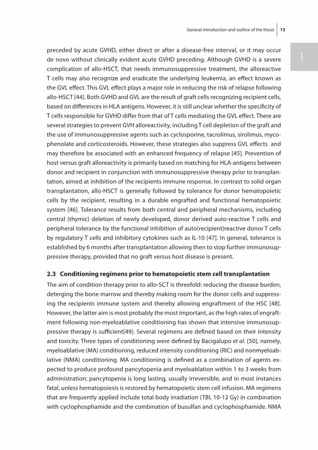
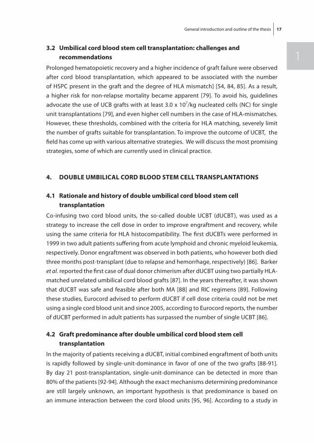
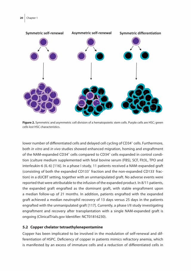
Because UCB grafts contain a relatively low number of HSPC and the dose of HSPC infused is related to patient survival and time required for engraftment [84, 105], it was hypoth-esized that expansion of HSPC in vitro prior to transplantation may result in improved engraftment and recovery compared to transplantation with non-expanded cord blood HSPC. Robust expansion of long-term repopulating HSC ex vivo, however, remains a chal-lenge. Culturing HSPC with different combinations of hematopoietic cytokines such as stem cell factor (SCF), Fms-related tyrosine kinase 3 ligand (Flt3L), thrombopoietin (TPO) and granulocyte-macrophage colony-stimulating factor (GM –CSF) resulted in massive expansion of committed HPC which is accompanied by a loss or at best maintenance of primitive HSC with long-term repopulation ability [109-112]. In addition to these prolif-eration- and differentiation-inducing cytokines, ex vivo expansion of HSC may require additional signals that inhibit differentiation of HSC during culture. Expansion of HSC requires symmetric self-renewal cell divisions in which both daughter cells retain all HSC characteristics (figure 2). Asymmetric cell divisions in which one daughter cell is still a HSC and the other daughter cell is a more differentiated progenitor cell leads to main-tenance of HSC number in culture, whereas symmetric differentiation cell divisions in which both daughter cells lost HSC characteristics results in a loss of HSC during culture. Decades of research has resulted in the identification of many developmental regulators and chemical modulators that have been implicated to be involved in the regulation of self-renewal, proliferation, differentiation and survival of HSC and that may increase the frequency of HSC symmetrical self-renewal cell divisions in culture [113]. Several of these factors have been evaluated for their potential to expand HSC in culture. Ex vivo HSC expansion protocols using some of these factors are currently evaluated in clinical trials. Several of these early phase clinical trials have reported promising results [7]. However, the search for the most optimal expansion protocol continues.
5.1 nicotinamide
Nicotinamide (NAM) is a form of vitamin-B3, which inhibits several classes of ribosylase enzymes and sirtuin1 (SIRT1), a class III NAD+-dependent-histone-deacetylase, and is implicated to play an role in the regulation of cell adhesion, polarity, migration, prolifera-tion and differentiation [114]. In addition, NAM modulates the fate of embryonic stem cells [115]. In primary growth factor-driven expansion cultures with UCB-derived CD34+ cells, addition of NAM resulted in a higher number of immature CD34+CD38- cells, a
20 Chapter 1
lower number of diff erentiated cells and delayed cell cycling of CD34+ cells. Furthermore, both in vitro and in vivo studies showed enhanced migration, homing and engraftment of the NAM-expanded CD34+ cells compared to CD34+ cells expanded in control condi-tion (culture medium supplemented with fetal bovine serum (FBS), SCF, Flt3L, TPO and interleukin-6 (IL-6) [116]. In a phase I study, 11 patients received a NAM-expanded graft (consisting of both the expanded CD133+ fraction and the non-expanded CD133- frac-tion) in a dUCBT setting, together with an unmanipulated graft. No adverse events were reported that were attributable to the infusion of the expanded product. In 8/11 patients, the expanded graft engrafted as the dominant graft, with stable engraftment upon a median follow-up of 21 months. In addition, patients engrafted with the expanded graft achieved a median neutrophil recovery of 13 days versus 25 days in the patients engrafted with the unmanipulated graft [117]. Currently, a phase I/II study investigating engraftment and recovery after transplantation with a single NAM-expanded graft is ongoing (ClinicalTrials.gov Identifi er: NCT01816230).
5.2 Copper chelator tetraethylenepentamine
Copper has been implicated to be involved in the modulation of self-renewal and dif-ferentiation of HSPC. Defi ciency of copper in patients mimics refractory anemia, which is manifested by an excess of immature cells and a reduction of diff erentiated cells in
Symmetric self-renewal Asymmetric self-renewal Symmetric differentiation
figure 2. Symmetric and asymmetric cell division of a hematopoietic stem cells. Purple cells are HSC; green cells lost HSC characteristics.
General introduction and outline of the thesis 21
1the bone marrow [118]. In vitro, copper defi ciency results in delayed diff erentiation and prolonged proliferation of HSPC, while increased cellular copper levels result in acceler-ated diff erentiation [119]. It appeared that reduction of the cellular chelatable copper content, rather than the overall copper content, resulted in the observed eff ects of cop-per defi ciency [120]. Copper chelator tetraethylenepentamine (TEPA) can be used to modulate the level of cellular chelatable copper. Addition of TEPA to expansion cultures (containing FBS, SCF, Flt3L, TPO and IL-6) results in robust expansion of HSPC. These HSPC show both high levels of engraftment and multi-lineage diff erentiation potential upon transplantation in NOD/SCID mice [121]. In a phase I/II study, co-transplantation of TEPA-expanded CD133+ CB-derived HSPC and unmanipulated HSPC from the same graft appeared to be safe and feasible [122]. Nine out of 10 patients engrafted with a median time to neutrophil and platelet recovery of 30 and 48 days respectively which is similar compared to recipients of unmanipulated single UCB grafts.
5.3 notch ligands
Notch and its ligands (Jagged-1, Jagged-2, Delta-1) are expressed in the bone marrow micro-environment and by HSC, which may suggest that Notch is an important regulator in hematopoiesis [123]. The immobilized form of Delta-1 has been shown to promote expansion of murine and human HSPC in culture and to increase the number of SRC with secondary transplantation ability. The eff ects of Notch ligand appeared to be dose-dependent and HSPC expansion was only observed using lower Delta-1 concentrations [124, 125]. In a clinical phase I study, upon co-transplantation of a Notch ligand-expanded graft and an unmanipulated graft, patients engrafted with an accelerated time to neutro-phil recovery of 16 days. In the fi rst week after transplantation, hematopoietic recovery was mainly derived from the expanded graft, but upon day 80 all hematopoietic cells in blood and marrow were derived from the unmanipulated graft [126]. These data suggest that either long term HSC were lost during expansion or that the expanded graft, lacking T cells, was rejected by the unmanipulated graft.
5.4 Wingless-related integration site proteins
Wingless-related integration site (Wnt) signaling has been implicated to play a role in the regulation HSPC fate decisions in vivo and might promote HSPC self-renewal by inhibit-ing diff erentiation. However, the eff ects of Wnt on the ex vivo expansion of HSPC remain controversial. In primary cultures of Bcl2-mouse Lin-Sca-c-Kit+ cells (LSK cells, enriched for HSPC in mice) supplemented with cytokines and serum, Willert et al. and Reya et al. showed induction of proliferation and inhibition of diff erentiation upon the addition of purifi ed Wnt3a [127, 128]. However, other studies observed a decrease in mouse LSK cells upon addition of Wnt3a to the culture medium [129], which is also described in this thesis. In addition, we describe the eff ect of the addition of Wnt3a to expansion cultures
22 Chapter 1
with human UCB-derived HSPC. To date, no clinical trials with Wnt protein expanded HSPC have been initiated.
5.5 aryl hydrocarbon receptor antagonist Stemregenin1
The Aryl hydrocarbon Receptor (AhR) pathway is involved in HSC self-renewal and inhibi-tion of differentiation. AhR-KO mice have an increased number of hyperproliferative LSK cells [130], while treatment of donor mice with AhR agonist TCDD results in impaired competitive engraftment [131]. Using high throughput screening of a library of small molecules, purine derivative StemRegenin1 (SR1), an AhR antagonist, was identified pro-moting the ex vivo expansion of CD34+ cells [132]. Addition of SR1 to expansion cultures supplemented with SCF, Flt3L, TPO and IL-6 resulted in a 50-fold increase in the number of CD34+ cells and a 17-fold increase in SRC, the cells that are capable of hematopoi-etic reconstitution in sublethally irradiated mice [132]. Transplantation of SR1-expanded CD34+ cells in a double cord blood setting appeared safe and feasible and resulted in engraftment in all 17 transplanted patients. The median time to neutrophil and platelet recovery was 15 and 49 days, respectively, which was faster compared to an historical control group receiving unmanipulated dUCBT. In 11/17 patients, hematopoiesis was primarily derived from the expanded unit and in those patients, neutrophil recovery was even more rapid with a median of 11 days versus 23 days in the 6/17 patients engrafting with the unmanipulated unit [133]. These results set the stage for clinical studies in a single UCBT setting (ClinicalTrials.gov Identifier: NCT01930162).
5.6 Um729 and Um171
In a recent study, a library-screen of more than 5000 low-molecular-weight compounds identified UM729 as an AhR-independent compound capable of expansion of human CD34+CD45RA- cells [134]. Further characterization and optimization resulted in the synthesis of the analog UM171, which appeared to be 10 to 20 times more potent than UM729. When compared to SR1, UM171 resulted in similar CD34+ and higher CD34+CD45RA- cell frequencies and absolute numbers after 12 days of in vitro culture in the presence of SCF, Flt3L and TPO. Transplantation of UM171-expanded cells into NSG mice resulted in higher levels of human chimerism compared to transplantation of SR1-expanded cells. In addition, transplantation of the input equivalent of only 50-100 UM171-expanded cells resulted in long-term multilineage engraftment, which was not the case upon transplantation of the same amount of SR1-expanded cells. These data indicate that UM171 enables robust ex vivo expansion of human CB cells with long-term in vivo repopulating capacity. Currently, a phase I/II trial exploring the safety and feasibil-ity of transplantation of UM171-expanded CD34+ cells in a single graft setting is ongoing (ClinicalTrials.gov Identifier: NCT02668315).
General introduction and outline of the thesis 23
15.7 Co-culture with mesenchymal stem cells
Most expansion protocols use selected CD34+ or CD133+ HSPC and subsequent culture with the addition of exogenous growth factors and differentiation-inhibiting factors. An-other approach is to co-culture cord blood cells with cells that are present in the micro-environment of HSC in bone marrow, such as mesenchymal stem cells (MSC). Mimicking the in vivo ‘niche’ of the HSC is thought to provide the complex molecular signals that are important for HSC self-renewal, proliferation and differentiation. Co-culturing CB cells for 14 days on primary MSC cells in the presence of FBS, SCF, TPO and G-CSF resulted in a significant increase of CD34+ cells and CFU [135]. Transplantation of MSC-expanded cells in a dUCBT setting together with a unmanipulated graft appeared to be safe and feasible [136]. Compared to historical controls that had received an unmanipulated dUCBT, the cumulative incidence of neutrophil engraftment at day 26 and platelet engraftment at day 60 was significantly increased. Although about half of the patients had hematopoi-esis derived from both units in the first month after transplantation, long-term engraft-ment was produced primarily by the unit of the unmanipulated cord blood in all patients, suggesting either loss of self-renewing HSC in the expanded graft or rejection of the manipulated graft by T cells present in the unmanipulated graft.
The quest to the most optimal expansion protocol is still ongoing. An optimized pro-tocol might involve a combination of several factors that act synergistically in order to induce cell proliferation while maintaining self-renewal capacities and inhibit differentia-tion [137].
6. aImS anD oUtLInE of tHE tHESIS
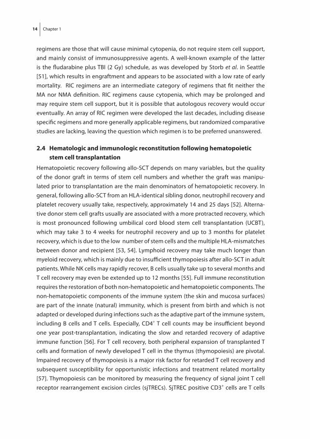
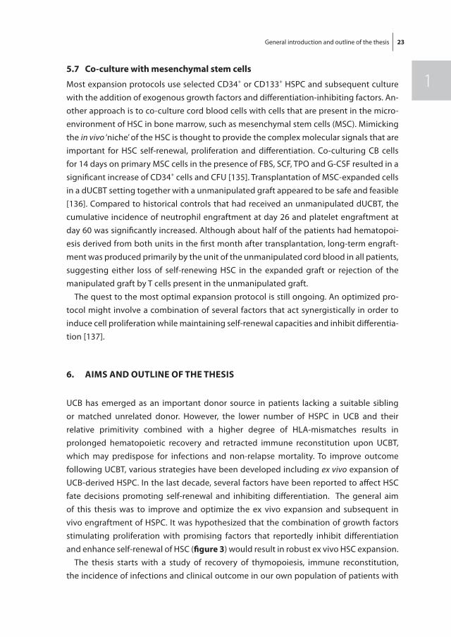
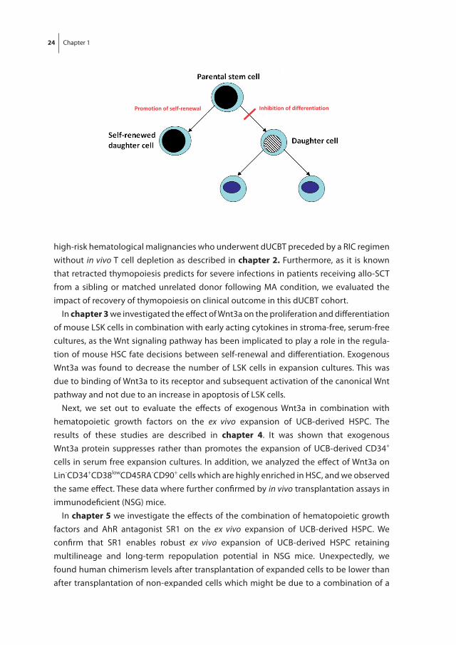
UCB has emerged as an important donor source in patients lacking a suitable sibling or matched unrelated donor. However, the lower number of HSPC in UCB and their relative primitivity combined with a higher degree of HLA-mismatches results in prolonged hematopoietic recovery and retracted immune reconstitution upon UCBT, which may predispose for infections and non-relapse mortality. To improve outcome following UCBT, various strategies have been developed including ex vivo expansion of UCB-derived HSPC. In the last decade, several factors have been reported to affect HSC fate decisions promoting self-renewal and inhibiting differentiation. The general aim of this thesis was to improve and optimize the ex vivo expansion and subsequent in vivo engraftment of HSPC. It was hypothesized that the combination of growth factors stimulating proliferation with promising factors that reportedly inhibit differentiation and enhance self-renewal of HSC (figure 3) would result in robust ex vivo HSC expansion.
The thesis starts with a study of recovery of thymopoiesis, immune reconstitution, the incidence of infections and clinical outcome in our own population of patients with
24 Chapter 1
high-risk hematological malignancies who underwent dUCBT preceded by a RIC regimen without in vivo T cell depletion as described in chapter 2. Furthermore, as it is known that retracted thymopoiesis predicts for severe infections in patients receiving allo-SCT from a sibling or matched unrelated donor following MA condition, we evaluated the impact of recovery of thymopoiesis on clinical outcome in this dUCBT cohort.
In chapter 3 we investigated the effect of Wnt3a on the proliferation and differentiation of mouse LSK cells in combination with early acting cytokines in stroma-free, serum-free cultures, as the Wnt signaling pathway has been implicated to play a role in the regula-tion of mouse HSC fate decisions between self-renewal and differentiation. Exogenous Wnt3a was found to decrease the number of LSK cells in expansion cultures. This was due to binding of Wnt3a to its receptor and subsequent activation of the canonical Wnt pathway and not due to an increase in apoptosis of LSK cells.
Next, we set out to evaluate the effects of exogenous Wnt3a in combination with hematopoietic growth factors on the ex vivo expansion of UCB-derived HSPC. The results of these studies are described in chapter 4. It was shown that exogenous Wnt3a protein suppresses rather than promotes the expansion of UCB-derived CD34+ cells in serum free expansion cultures. In addition, we analyzed the effect of Wnt3a on Lin-CD34+CD38lowCD45RA-CD90+ cells which are highly enriched in HSC, and we observed the same effect. These data where further confirmed by in vivo transplantation assays in immunodeficient (NSG) mice.
In chapter 5 we investigate the effects of the combination of hematopoietic growth factors and AhR antagonist SR1 on the ex vivo expansion of UCB-derived HSPC. We confirm that SR1 enables robust ex vivo expansion of UCB-derived HSPC retaining multilineage and long-term repopulation potential in NSG mice. Unexpectedly, we found human chimerism levels after transplantation of expanded cells to be lower than after transplantation of non-expanded cells which might be due to a combination of a
Promotion of self-renewal Inhibition of differentiation
General introduction and outline of the thesis 25
1B cell-prone NSG mouse model and a reduced number of lymphoid progenitors in the expanded cell population.
To non-invasively monitor and quantify homing of transplanted HSPC in the bone mar-row, we developed a 19F-labeling protocol for HSPC as described in chapter 6. Our results show sufficient intracellular labeling of UCB-derived CD34+ cells using 19F-containing Poly(Lactic-co-Glycolic Acid) (PLGA) nanoparticles. These labeled cells were detectable with both flow cytometry and magnetic resonance spectroscopy (MRS), while maintain-ing their ability to proliferate and differentiate.
In chapter 7, the development of a Good Manufacturing Practice (GMP)-compliant expansion protocol for clinical application, based on the results presented in chapter 5, is described. In addition, the protocol for a clinical study addressing the safety and feasibility of the transplantation of SR1-expanded HSPC in a single UCB unit setting is presented.
Finally, in chapter 8, the results, implications and future perspectives of the experi-mental chapters are discussed in a broader perspective.
26 Chapter 1
rEfErEnCES
1. Baron MH, Isern J, Fraser ST. The embryonic origins of erythropoiesis in mammals. Blood. 2012 May 24;119(21):4828-37.
2. Nakahata T, Ogawa M. Hemopoietic colony-forming cells in umbilical cord blood with extensive capability to generate mono- and multipotential hemopoietic progenitors. J Clin Invest. 1982 Dec;70(6):1324-8.
3. Brauner P, Nibbelink M, Flachs P, Vitkova I, Kopecky P, Mertelikova I, et al. Fast decline of hema-topoiesis and uncoupling protein 2 content in human liver after birth: location of the protein in Kupffer cells. Pediatr Res. 2001 Mar;49(3):440-7.
4. Dzierzak E, Robin C. Placenta as a source of hematopoietic stem cells. Trends Mol Med. 2010 Aug;16(8):361-7.
5. Magnon C, Frenette PS. Hematopoietic stem cell trafficking. 2008. 6. Mikkola HK, Orkin SH. The journey of developing hematopoietic stem cells. Development. 2006
Oct;133(19):3733-44. 7. Lund TC, Boitano AE, Delaney CS, Shpall EJ, Wagner JE. Advances in umbilical cord blood manipu-
lation-from niche to bedside. Nat Rev Clin Oncol. 2015 Mar;12(3):163-74. 8. Yoshihara H, Arai F, Hosokawa K, Hagiwara T, Takubo K, Nakamura Y, et al. Thrombopoietin/MPL
signaling regulates hematopoietic stem cell quiescence and interaction with the osteoblastic niche. Cell Stem Cell. 2007 Dec 13;1(6):685-97.
9. Zhang J, Niu C, Ye L, Huang H, He X, Tong WG, et al. Identification of the haematopoietic stem cell niche and control of the niche size. Nature. 2003 Oct 23;425(6960):836-41.
10. Ceradini DJ, Kulkarni AR, Callaghan MJ, Tepper OM, Bastidas N, Kleinman ME, et al. Progenitor cell trafficking is regulated by hypoxic gradients through HIF-1 induction of SDF-1. Nat Med. 2004 Aug;10(8):858-64.
11. Mendez-Ferrer S, Lucas D, Battista M, Frenette PS. Haematopoietic stem cell release is regulated by circadian oscillations. Nature. 2008 Mar 27;452(7186):442-7.
12. Mendez-Ferrer S, Michurina TV, Ferraro F, Mazloom AR, Macarthur BD, Lira SA, et al. Mesen-chymal and haematopoietic stem cells form a unique bone marrow niche. Nature. 2010 Aug 12;466(7308):829-34.
13. Naveiras O, Nardi V, Wenzel PL, Hauschka PV, Fahey F, Daley GQ. Bone-marrow adipocytes as nega-tive regulators of the haematopoietic microenvironment. Nature. 2009 Jul 9;460(7252):259-63.
14. Ding L, Saunders TL, Enikolopov G, Morrison SJ. Endothelial and perivascular cells maintain hae-matopoietic stem cells. Nature. 2012 Jan 26;481(7382):457-62.
15. Notta F, Doulatov S, Laurenti E, Poeppl A, Jurisica I, Dick JE. Isolation of single human hematopoietic stem cells capable of long-term multilineage engraftment. Science. 2011 Jul 8;333(6039):218-21.
16. Kiel MJ, Yilmaz OH, Iwashita T, Yilmaz OH, Terhorst C, Morrison SJ. SLAM family receptors distin-guish hematopoietic stem and progenitor cells and reveal endothelial niches for stem cells. Cell. 2005 Jul 1;121(7):1109-21.
17. Bonzanni N, Garg A, Feenstra KA, Schutte J, Kinston S, Miranda-Saavedra D, et al. Hard-wired heterogeneity in blood stem cells revealed using a dynamic regulatory network model. Bioinfor-matics. 2013 Jul 1;29(13):i80-8.
18. Gottgens B. Regulatory network control of blood stem cells. Blood. 2015 Apr 23;125(17):2614-20. 19. Fauser AA, Messner HA. Identification of megakaryocytes, macrophages, and eosinophils in colo-
nies of human bone marrow containing neurtophilic granulocytes and erythroblasts. Blood. 1979 May;53(5):1023-7.
General introduction and outline of the thesis 27
1 20. Sutherland HJ, Lansdorp PM, Henkelman DH, Eaves AC, Eaves CJ. Functional characterization of
individual human hematopoietic stem cells cultured at limiting dilution on supportive marrow stromal layers. Proc Natl Acad Sci U S A. 1990 May;87(9):3584-8.
21. Kamel-Reid S, Dick JE. Engraftment of immune-deficient mice with human hematopoietic stem cells. Science. 1988 Dec 23;242(4886):1706-9.
22. Theocharides AP, Rongvaux A, Fritsch K, Flavell RA, Manz MG. Humanized hemato-lymphoid system mice. Haematologica. 2016 Jan;101(1):5-19.
23. Morita Y, Ema H, Nakauchi H. Heterogeneity and hierarchy within the most primitive hematopoi-etic stem cell compartment. J Exp Med. 2010 Jun 7;207(6):1173-82.
24. Jacobson LO, Marks EK, Lorenz E. The hematological effects of ionizing radiations. Radiology. 1949 Mar;52(3):371-95.
25. Jacobson LO, Simmons EL, Marks EK, Eldredge JH. Recovery from radiation injury. Science. 1951 May 4;113(2940):510-11.
26. Fishler MC, Cole LJ, Bond VP, Milne WL. Therapeutic effect of rat bone marrow injection in rats exposed to lethal whole body x-radiation. Am J Physiol. 1954 May;177(2):236-42.
27. Smith WW, Marston RQ, Gonshery L, Alderman IM, Ruth HJ. X-irradiation in hamsters, and effects of streptomycin and marrow-spleen homogenate treatment. Am J Physiol. 1955 Oct;183(1):98-110.
28. Porter KA, Murray JE. Long-term study of x-irradiated rabbits with bone-marrow homotransplants. J Natl Cancer Inst. 1958 Jan;20(1):189-205.
29. Thomas ED, Lochte HL, Jr., Lu WC, Ferrebee JW. Intravenous infusion of bone marrow in patients receiving radiation and chemotherapy. N Engl J Med. 1957 Sep 12;257(11):491-6.
30. Gatti RA, Meuwissen HJ, Allen HD, Hong R, Good RA. Immunological reconstitution of sex-linked lymphopenic immunological deficiency. Lancet. 1968 Dec 28;2(7583):1366-9.
31. Bach FH, Albertini RJ, Joo P, Anderson JL, Bortin MM. Bone-marrow transplantation in a patient with the Wiskott-Aldrich syndrome. Lancet. 1968 Dec 28;2(7583):1364-6.
32. Gratwohl A, Pasquini MC, Aljurf M, Atsuta Y, Baldomero H, Foeken L, et al. One million haemopoietic stem-cell transplants: a retrospective observational study. Lancet Haematol. 2015 Mar;2(3):e91-e100.
33. Copelan EA. Hematopoietic stem-cell transplantation. N Engl J Med. 2006 Apr 27;354(17):1813-26. 34. Nowak J. Role of HLA in hematopoietic SCT. Bone Marrow Transplant. 2008 Oct;42 Suppl 2:S71-6. 35. Goldberg AC, Rizzo LV. MHC structure and function - antigen presentation. Part 1. Einstein (Sao
Paulo). 2015 Jan-Mar;13(1):153-6. 36. Goldberg AC, Rizzo LV. MHC structure and function - antigen presentation. Part 2. Einstein (Sao
Paulo). 2015 Jan-Mar;13(1):157-62. 37. Geneugelijk K, Thus KA, Spierings E. Predicting alloreactivity in transplantation. J Immunol Res.
2014;2014:159479. 38. Goulmy E, Voogt P, van Els C, de Bueger M, van Rood J. The role of minor histocompatibility anti-
gens in GVHD and rejection: a mini-review. Bone Marrow Transplant. 1991;7 Suppl 1:49-51. 39. Olsson R, Remberger M, Schaffer M, Berggren DM, Svahn BM, Mattsson J, et al. Graft failure in the
modern era of allogeneic hematopoietic SCT. Bone Marrow Transplant. 2013 Apr;48(4):537-43. 40. Filipovich AH, Weisdorf D, Pavletic S, Socie G, Wingard JR, Lee SJ, et al. National Institutes of Health
consensus development project on criteria for clinical trials in chronic graft-versus-host disease: I. Diagnosis and staging working group report. Biol Blood Marrow Transplant. 2005 Dec;11(12):945-56.
41. Flowers ME, Kansu E, Sullivan KM. Pathophysiology and treatment of graft-versus-host disease. Hematol Oncol Clin North Am. 1999 Oct;13(5):1091-112, viii-ix.
28 Chapter 1
42. Goyal RK, Goyal M, Sankaranarayan K. Grading acute graft-versus-host disease: time to reconsider. Pediatr Transplant. 2015 May;19(3):252-4.
43. Flowers ME, Martin PJ. How we treat chronic graft-versus-host disease. Blood. 2015 Jan 22;125(4):606-15.
44. Horowitz MM, Gale RP, Sondel PM, Goldman JM, Kersey J, Kolb HJ, et al. Graft-versus-leukemia reactions after bone marrow transplantation. Blood. 1990 Feb 1;75(3):555-62.
45. Cornelissen JJ, Lowenberg B. Developments in T-cell depletion of allogeneic stem cell grafts. Curr Opin Hematol. 2000 Nov;7(6):348-52.
46. Mosaad YM. Immunology of hematopoietic stem cell transplant. Immunol Invest. 2014;43(8):858-87.
47. Chiffoleau E, Walsh PT, Turka L. Apoptosis and transplantation tolerance. Immunol Rev. 2003 Jun;193:124-45.
48. Vriesendorp HM. Aims of conditioning. Exp Hematol. 2003 Oct;31(10):844-54. 49. Storb R, Gyurkocza B, Storer BE, Sorror ML, Blume K, Niederwieser D, et al. Graft-versus-host
disease and graft-versus-tumor effects after allogeneic hematopoietic cell transplantation. J Clin Oncol. 2013 Apr 20;31(12):1530-8.
50. Bacigalupo A, Ballen K, Rizzo D, Giralt S, Lazarus H, Ho V, et al. Defining the intensity of condition-ing regimens: working definitions. Biol Blood Marrow Transplant. 2009 Dec;15(12):1628-33.
51. Storb RF, Champlin R, Riddell SR, Murata M, Bryant S, Warren EH. Non-myeloablative transplants for malignant disease. Hematology Am Soc Hematol Educ Program. 2001:375-91.
52. Eapen M, Rocha V, Sanz G, Scaradavou A, Zhang MJ, Arcese W, et al. Effect of graft source on unre-lated donor haemopoietic stem-cell transplantation in adults with acute leukaemia: a retrospec-tive analysis. Lancet Oncol. 2010 Jul;11(7):653-60.
53. Avery S, Shi W, Lubin M, Gonzales AM, Heller G, Castro-Malaspina H, et al. Influence of infused cell dose and HLA match on engraftment after double-unit cord blood allografts. Blood. 2011 Mar 24;117(12):3277-85; quiz 478.
54. Barker JN, Scaradavou A, Stevens CE. Combined effect of total nucleated cell dose and HLA match on transplantation outcome in 1061 cord blood recipients with hematologic malignancies. Blood. 2010 Mar 4;115(9):1843-9.
55. Storek J, Geddes M, Khan F, Huard B, Helg C, Chalandon Y, et al. Reconstitution of the immune system after hematopoietic stem cell transplantation in humans. Semin Immunopathol. 2008 Dec;30(4):425-37.
56. Tomblyn M, Chiller T, Einsele H, Gress R, Sepkowitz K, Storek J, et al. Guidelines for preventing infec-tious complications among hematopoietic cell transplantation recipients: a global perspective. Biol Blood Marrow Transplant. 2009 Oct;15(10):1143-238.
57. Wils EJ, van der Holt B, Broers AE, Posthumus-van Sluijs SJ, Gratama JW, Braakman E, et al. Insuf-ficient recovery of thymopoiesis predicts for opportunistic infections in allogeneic hematopoietic stem cell transplant recipients. Haematologica. 2011 Dec;96(12):1846-54.
58. Min D, Taylor PA, Panoskaltsis-Mortari A, Chung B, Danilenko DM, Farrell C, et al. Protection from thymic epithelial cell injury by keratinocyte growth factor: a new approach to improve thymic and peripheral T-cell reconstitution after bone marrow transplantation. Blood. 2002 Jun 15;99(12):4592-600.
59. Wils EJ, Cornelissen JJ. Thymopoiesis following allogeneic stem cell transplantation: new possibili-ties for improvement. Blood Rev. 2005 Mar;19(2):89-98.
60. Broers AE, Posthumus-van Sluijs SJ, Spits H, van der Holt B, Lowenberg B, Braakman E, et al. Inter-leukin-7 improves T-cell recovery after experimental T-cell-depleted bone marrow transplanta-
General introduction and outline of the thesis 29
1tion in T-cell-deficient mice by strong expansion of recent thymic emigrants. Blood. 2003 Aug 15;102(4):1534-40.
61. Mackall CL, Fry TJ, Gress RE. Harnessing the biology of IL-7 for therapeutic application. Nat Rev Immunol. 2011 May;11(5):330-42.
62. Perales MA, Goldberg JD, Yuan J, Koehne G, Lechner L, Papadopoulos EB, et al. Recombinant hu-man interleukin-7 (CYT107) promotes T-cell recovery after allogeneic stem cell transplantation. Blood. 2012 Dec 6;120(24):4882-91.
63. Mackinnon S, Thomson K, Verfuerth S, Peggs K, Lowdell M. Adoptive cellular therapy for cyto-megalovirus infection following allogeneic stem cell transplantation using virus-specific T cells. Blood Cells Mol Dis. 2008 Jan-Feb;40(1):63-7.
64. Andre-Schmutz I, Dal Cortivo L, Fischer A, Cavazzana-Calvo M. Improving immune reconstitution while preventing GvHD in allogeneic stem cell transplantation. Cytotherapy. 2005;7(2):102-8.
65. Cornelissen JJ, Gratwohl A, Schlenk RF, Sierra J, Bornhauser M, Juliusson G, et al. The European LeukemiaNet AML Working Party consensus statement on allogeneic HSCT for patients with AML in remission: an integrated-risk adapted approach. Nat Rev Clin Oncol. 2012 Oct;9(10):579-90.
66. Lee SJ, Klein J, Haagenson M, Baxter-Lowe LA, Confer DL, Eapen M, et al. High-resolution donor-recipient HLA matching contributes to the success of unrelated donor marrow transplantation. Blood. 2007 Dec 15;110(13):4576-83.
67. Loiseau P, Busson M, Balere ML, Dormoy A, Bignon JD, Gagne K, et al. HLA Association with hematopoietic stem cell transplantation outcome: the number of mismatches at HLA-A, -B, -C, -DRB1, or -DQB1 is strongly associated with overall survival. Biol Blood Marrow Transplant. 2007 Aug;13(8):965-74.
68. Cornelissen JJ, Blaise D. Hematopoietic stem cell transplantation for patients with AML in first complete remission. Blood. 2016 Jan 7;127(1):62-70.
69. Migliaccio G, Migliaccio AR, Petti S, Mavilio F, Russo G, Lazzaro D, et al. Human embryonic hemo-poiesis. Kinetics of progenitors and precursors underlying the yolk sac----liver transition. J Clin Invest. 1986 Jul;78(1):51-60.
70. Broxmeyer HE, Douglas GW, Hangoc G, Cooper S, Bard J, English D, et al. Human umbilical cord blood as a potential source of transplantable hematopoietic stem/progenitor cells. Proc Natl Acad Sci U S A. 1989 May;86(10):3828-32.
71. Gluckman E, Broxmeyer HA, Auerbach AD, Friedman HS, Douglas GW, Devergie A, et al. Hemato-poietic reconstitution in a patient with Fanconi’s anemia by means of umbilical-cord blood from an HLA-identical sibling. N Engl J Med. 1989 Oct 26;321(17):1174-8.
72. Wagner JE, Rosenthal J, Sweetman R, Shu XO, Davies SM, Ramsay NK, et al. Successful transplanta-tion of HLA-matched and HLA-mismatched umbilical cord blood from unrelated donors: analysis of engraftment and acute graft-versus-host disease. Blood. 1996 Aug 1;88(3):795-802.
73. Kurtzberg J, Laughlin M, Graham ML, Smith C, Olson JF, Halperin EC, et al. Placental blood as a source of hematopoietic stem cells for transplantation into unrelated recipients. N Engl J Med. 1996 Jul 18;335(3):157-66.
74. Barker JN, Krepski TP, DeFor TE, Davies SM, Wagner JE, Weisdorf DJ. Searching for unrelated donor hematopoietic stem cells: availability and speed of umbilical cord blood versus bone marrow. Biol Blood Marrow Transplant. 2002;8(5):257-60.
75. Hakenberg P, Kogler G, Wernet P. NETCORD: a cord blood allocation network. Bone Marrow Trans-plant. 1998 Jul;22 Suppl 1:S17-8.
76. Ballen KK, Verter F, Kurtzberg J. Umbilical cord blood donation: public or private? Bone Marrow Transplant. 2015 Oct;50(10):1271-8.
30 Chapter 1
77. Petersdorf EW, Gooley TA, Anasetti C, Martin PJ, Smith AG, Mickelson EM, et al. Optimizing outcome after unrelated marrow transplantation by comprehensive matching of HLA class I and II alleles in the donor and recipient. Blood. 1998 Nov 15;92(10):3515-20.
78. Locatelli F, Rocha V, Chastang C, Arcese W, Michel G, Abecasis M, et al. Factors associated with outcome after cord blood transplantation in children with acute leukemia. Eurocord-Cord Blood Transplant Group. Blood. 1999 Jun 1;93(11):3662-71.
79. Eapen M, Rubinstein P, Zhang MJ, Stevens C, Kurtzberg J, Scaradavou A, et al. Outcomes of transplantation of unrelated donor umbilical cord blood and bone marrow in children with acute leukaemia: a comparison study. Lancet. 2007 Jun 9;369(9577):1947-54.
80. Kurtzberg J, Prasad VK, Carter SL, Wagner JE, Baxter-Lowe LA, Wall D, et al. Results of the Cord Blood Transplantation Study (COBLT): clinical outcomes of unrelated donor umbilical cord blood transplantation in pediatric patients with hematologic malignancies. Blood. 2008 Nov 15;112(10):4318-27.
81. Laughlin MJ, Barker J, Bambach B, Koc ON, Rizzieri DA, Wagner JE, et al. Hematopoietic engraft-ment and survival in adult recipients of umbilical-cord blood from unrelated donors. N Engl J Med. 2001 Jun 14;344(24):1815-22.
82. Rocha V, Labopin M, Sanz G, Arcese W, Schwerdtfeger R, Bosi A, et al. Transplants of umbilical-cord blood or bone marrow from unrelated donors in adults with acute leukemia. N Engl J Med. 2004 Nov 25;351(22):2276-85.
83. Laughlin MJ, Eapen M, Rubinstein P, Wagner JE, Zhang MJ, Champlin RE, et al. Outcomes after transplantation of cord blood or bone marrow from unrelated donors in adults with leukemia. N Engl J Med. 2004 Nov 25;351(22):2265-75.
84. Wagner JE, Barker JN, DeFor TE, Baker KS, Blazar BR, Eide C, et al. Transplantation of unrelated donor umbilical cord blood in 102 patients with malignant and nonmalignant diseases: influence of CD34 cell dose and HLA disparity on treatment-related mortality and survival. Blood. 2002 Sep 1;100(5):1611-8.
85. Gluckman E, Rocha V, Boyer-Chammard A, Locatelli F, Arcese W, Pasquini R, et al. Outcome of cord-blood transplantation from related and unrelated donors. Eurocord Transplant Group and the European Blood and Marrow Transplantation Group. N Engl J Med. 1997 Aug 7;337(6):373-81.
86. Rocha V, Crotta A, Ruggeri A, Purtill D, Boudjedir K, Herr AL, et al. Double cord blood transplanta-tion: extending the use of unrelated umbilical cord blood cells for patients with hematological diseases. Best Pract Res Clin Haematol. 2010 Jun;23(2):223-9.
87. Barker JN, Weisdorf DJ, Wagner JE. Creation of a double chimera after the transplantation of umbilical-cord blood from two partially matched unrelated donors. N Engl J Med. 2001 Jun 14;344(24):1870-1.
88. Barker JN, Weisdorf DJ, DeFor TE, Blazar BR, McGlave PB, Miller JS, et al. Transplantation of 2 par-tially HLA-matched umbilical cord blood units to enhance engraftment in adults with hematologic malignancy. Blood. 2005 Feb 1;105(3):1343-7.
89. Barker JN, Weisdorf DJ, DeFor TE, Blazar BR, Miller JS, Wagner JE. Rapid and complete donor chi-merism in adult recipients of unrelated donor umbilical cord blood transplantation after reduced-intensity conditioning. Blood. 2003 Sep 1;102(5):1915-9.
90. Ruggeri A, de Latour RP, Rocha V, Larghero J, Robin M, Rodrigues CA, et al. Double cord blood transplantation in patients with high risk bone marrow failure syndromes. Br J Haematol. 2008 Nov;143(3):404-8.
General introduction and outline of the thesis 31
1 91. Brunstein CG, Barker JN, Weisdorf DJ, DeFor TE, Miller JS, Blazar BR, et al. Umbilical cord blood
transplantation after nonmyeloablative conditioning: impact on transplantation outcomes in 110 adults with hematologic disease. Blood. 2007 Oct 15;110(8):3064-70.
92. Delaney C, Gutman JA, Appelbaum FR. Cord blood transplantation for haematological malignan-cies: conditioning regimens, double cord transplant and infectious complications. Br J Haematol. 2009 Oct;147(2):207-16.
93. Somers JA, Brand A, van Hensbergen Y, Mulder A, Oudshoorn M, Sintnicolaas K, et al. Double umbilical cord blood transplantation: a study of early engraftment kinetics in leukocyte subsets using HLA-specific monoclonal antibodies. Biol Blood Marrow Transplant. 2013 Feb;19(2):266-73.
94. Somers JA, Braakman E, van der Holt B, Petersen EJ, Marijt EW, Huisman C, et al. Rapid induc-tion of single donor chimerism after double umbilical cord blood transplantation preceded by reduced intensity conditioning: results of the HOVON 106 phase II study. Haematologica. 2014 Nov;99(11):1753-61.
95. Ramirez P, Wagner JE, DeFor TE, Blazar BR, Verneris MR, Miller JS, et al. Factors predicting single-unit predominance after double umbilical cord blood transplantation. Bone Marrow Transplant. 2012 Jun;47(6):799-803.
96. Eldjerou LK, Chaudhury S, Baisre-de Leon A, He M, Arcila ME, Heller G, et al. An in vivo model of double-unit cord blood transplantation that correlates with clinical engraftment. Blood. 2010 Nov 11;116(19):3999-4006.
97. Newell LF, Milano F, Nicoud IB, Pereira S, Gooley TA, Heimfeld S, et al. Early CD3 peripheral blood chimerism predicts the long-term engrafting unit following myeloablative double-cord blood transplantation. Biol Blood Marrow Transplant. 2012 Aug;18(8):1243-9.
98. Berglund S, Okas M, Gertow J, Uhlin M, Mattsson J. Stable mixed donor-donor chimerism after double cord blood transplantation. Int J Hematol. 2009 Nov;90(4):526-31.
99. Cornelissen JJ, Wijers R, van Bergen CAM, Somers JA, Braakman E, Gratama JW, et al. Early CD4+ T-Cell Effector Alloreactivity Towards Multiple Mismatched HLA Class II Alleles Is Associated with Graft Predominance after Double Umbilical Cord Blood Transplantation (dUCBT). 57th ASH An-nual Meeting & Exposition; December 03, 2015; Orlando, United States of America: Blood; 2015. p. 387.
100. Scaradavou A, Smith KM, Hawke R, Schaible A, Abboud M, Kernan NA, et al. Cord blood units with low CD34+ cell viability have a low probability of engraftment after double unit transplantation. Biol Blood Marrow Transplant. 2010 Apr;16(4):500-8.
101. Page KM, Gentry T, Shoulars K, Horwitz M, Herrmann N, Meadows N, et al. Use of Aldehyde De-hydrogenase Content to Predict Dominating Cord in Double Cord Blood Transplant. ASH Annual Meeting Abstracts. 2009 November 20, 2009;114(22):2155-.
102. Becker AJ, Mc CE, Till JE. Cytological demonstration of the clonal nature of spleen colonies derived from transplanted mouse marrow cells. Nature. 1963 Feb 2;197:452-4.
103. Ballen KK, Spitzer TR, Yeap BY, McAfee S, Dey BR, Attar E, et al. Double unrelated reduced-intensity umbilical cord blood transplantation in adults. Biol Blood Marrow Transplant. 2007 Jan;13(1):82-9.
104. Kindwall-Keller TL, Hegerfeldt Y, Meyerson HJ, Margevicius S, Fu P, van Heeckeren W, et al. Prospective study of one- vs two-unit umbilical cord blood transplantation following reduced intensity conditioning in adults with hematological malignancies. Bone Marrow Transplant. 2012 Jul;47(7):924-33.
105. Brunstein CG, Gutman JA, Weisdorf DJ, Woolfrey AE, Defor TE, Gooley TA, et al. Allogeneic hema-topoietic cell transplantation for hematologic malignancy: relative risks and benefits of double umbilical cord blood. Blood. 2010 Nov 25;116(22):4693-9.
32 Chapter 1
106. Wagner JE, Jr., Eapen M, Carter S, Wang Y, Schultz KR, Wall DA, et al. One-unit versus two-unit cord-blood transplantation for hematologic cancers. N Engl J Med. 2014 Oct 30;371(18):1685-94.
107. Chen YB, Aldridge J, Kim HT, Ballen KK, Cutler C, Kao G, et al. Reduced-intensity conditioning stem cell transplantation: comparison of double umbilical cord blood and unrelated donor grafts. Biol Blood Marrow Transplant. 2012 May;18(5):805-12.
108. Ruggeri A, Sanz G, Bittencourt H, Sanz J, Rambaldi A, Volt F, et al. Comparison of outcomes after single or double cord blood transplantation in adults with acute leukemia using different types of myeloablative conditioning regimen, a retrospective study on behalf of Eurocord and the Acute Leukemia Working Party of EBMT. Leukemia. 2014 Apr;28(4):779-86.
109. Sauvageau G, Iscove NN, Humphries RK. In vitro and in vivo expansion of hematopoietic stem cells. Oncogene. 2004 Sep 20;23(43):7223-32.
110. Goff JP, Shields DS, Greenberger JS. Influence of cytokines on the growth kinetics and immuno-phenotype of daughter cells resulting from the first division of single CD34(+)Thy-1(+)lin- cells. Blood. 1998 Dec 1;92(11):4098-107.
111. Glimm H, Eaves CJ. Direct evidence for multiple self-renewal divisions of human in vivo repopulat-ing hematopoietic cells in short-term culture. Blood. 1999 Oct 1;94(7):2161-8.
112. Ivanovic Z, Duchez P, Chevaleyre J, Vlaski M, Lafarge X, Dazey B, et al. Clinical-scale cultures of cord blood CD34(+) cells to amplify committed progenitors and maintain stem cell activity. Cell Transplant. 2011;20(9):1453-63.
113. Walasek MA, van Os R, de Haan G. Hematopoietic stem cell expansion: challenges and opportuni-ties. Ann N Y Acad Sci. 2012 Aug;1266:138-50.
114. Glowacki G, Braren R, Firner K, Nissen M, Kuhl M, Reche P, et al. The family of toxin-related ecto-ADP-ribosyltransferases in humans and the mouse. Protein Sci. 2002 Jul;11(7):1657-70.
115. Vaca P, Berna G, Martin F, Soria B. Nicotinamide induces both proliferation and differentiation of embryonic stem cells into insulin-producing cells. Transplant Proc. 2003 Aug;35(5):2021-3.
116. Peled T, Shoham H, Aschengrau D, Yackoubov D, Frei G, Rosenheimer GN, et al. Nicotinamide, a SIRT1 inhibitor, inhibits differentiation and facilitates expansion of hematopoietic progenitor cells with enhanced bone marrow homing and engraftment. Exp Hematol. 2012 Apr;40(4):342-55 e1.
117. Horwitz ME, Chao NJ, Rizzieri DA, Long GD, Sullivan KM, Gasparetto C, et al. Umbilical cord blood expansion with nicotinamide provides long-term multilineage engraftment. J Clin Invest. 2014 Jul;124(7):3121-8.
118. Koca E, Buyukasik Y, Cetiner D, Yilmaz R, Sayinalp N, Yasavul U, et al. Copper deficiency with increased hematogones mimicking refractory anemia with excess blasts. Leuk Res. 2008 Mar;32(3):495-9.
119. Peled T, Landau E, Prus E, Treves AJ, Nagler A, Fibach E. Cellular copper content modulates dif-ferentiation and self-renewal in cultures of cord blood-derived CD34+ cells. Br J Haematol. 2002 Mar;116(3):655-61.
120. Peled T, Glukhman E, Hasson N, Adi S, Assor H, Yudin D, et al. Chelatable cellular copper modulates differentiation and self-renewal of cord blood-derived hematopoietic progenitor cells. Exp Hema-tol. 2005 Oct;33(10):1092-100.
121. Peled T, Landau E, Mandel J, Glukhman E, Goudsmid NR, Nagler A, et al. Linear polyamine copper chelator tetraethylenepentamine augments long-term ex vivo expansion of cord blood-derived CD34+ cells and increases their engraftment potential in NOD/SCID mice. Exp Hematol. 2004 Jun;32(6):547-55.
122. de Lima M, McMannis J, Gee A, Komanduri K, Couriel D, Andersson BS, et al. Transplantation of ex vivo expanded cord blood cells using the copper chelator tetraethylenepentamine: a phase I/II clinical trial. Bone Marrow Transplant. 2008 May;41(9):771-8.
123. Varnum-Finney B, Purton LE, Yu M, Brashem-Stein C, Flowers D, Staats S, et al. The Notch ligand, Jagged-1, influences the development of primitive hematopoietic precursor cells. Blood. 1998 Jun 1;91(11):4084-91.
124. Varnum-Finney B, Brashem-Stein C, Bernstein ID. Combined effects of Notch signaling and cy-tokines induce a multiple log increase in precursors with lymphoid and myeloid reconstituting ability. Blood. 2003 Mar 1;101(5):1784-9.
125. Delaney C, Varnum-Finney B, Aoyama K, Brashem-Stein C, Bernstein ID. Dose-dependent effects of the Notch ligand Delta1 on ex vivo differentiation and in vivo marrow repopulating ability of cord blood cells. Blood. 2005 Oct 15;106(8):2693-9.
126. Delaney C, Heimfeld S, Brashem-Stein C, Voorhies H, Manger RL, Bernstein ID. Notch-mediated expansion of human cord blood progenitor cells capable of rapid myeloid reconstitution. Nat Med. 2010 Feb;16(2):232-6.
127. Willert K, Brown JD, Danenberg E, Duncan AW, Weissman IL, Reya T, et al. Wnt proteins are lipid-modified and can act as stem cell growth factors. Nature. 2003 May 22;423(6938):448-52.
128. Reya T, Duncan AW, Ailles L, Domen J, Scherer DC, Willert K, et al. A role for Wnt signalling in self-renewal of haematopoietic stem cells. Nature. 2003 May 22;423(6938):409-14.
129. Nemeth MJ, Topol L, Anderson SM, Yang Y, Bodine DM. Wnt5a inhibits canonical Wnt signaling in hematopoietic stem cells and enhances repopulation. Proc Natl Acad Sci U S A. 2007 Sep 25;104(39):15436-41.
130. Gasiewicz TA, Singh KP, Casado FL. The aryl hydrocarbon receptor has an important role in the regulation of hematopoiesis: implications for benzene-induced hematopoietic toxicity. Chem Biol Interact. 2010 Mar 19;184(1-2):246-51.
131. Singh KP, Wyman A, Casado FL, Garrett RW, Gasiewicz TA. Treatment of mice with the Ah receptor agonist and human carcinogen dioxin results in altered numbers and function of hematopoietic stem cells. Carcinogenesis. 2009 Jan;30(1):11-9.
132. Boitano AE, Wang J, Romeo R, Bouchez LC, Parker AE, Sutton SE, et al. Aryl hydrocarbon recep-tor antagonists promote the expansion of human hematopoietic stem cells. Science. 2010 Sep 10;329(5997):1345-8.
133. Wagner JE, Jr., Brunstein CG, Boitano AE, DeFor TE, McKenna D, Sumstad D, et al. Phase I/II Trial of StemRegenin-1 Expanded Umbilical Cord Blood Hematopoietic Stem Cells Supports Testing as a Stand-Alone Graft. Cell Stem Cell. 2016 Jan 7;18(1):144-55.
134. Fares I, Chagraoui J, Gareau Y, Gingras S, Ruel R, Mayotte N, et al. Cord blood expansion. Pyrimi-doindole derivatives are agonists of human hematopoietic stem cell self-renewal. Science. 2014 Sep 19;345(6203):1509-12.
135. Robinson SN, Ng J, Niu T, Yang H, McMannis JD, Karandish S, et al. Superior ex vivo cord blood expansion following co-culture with bone marrow-derived mesenchymal stem cells. Bone Marrow Transplant. 2006 Feb;37(4):359-66.
136. de Lima M, McNiece I, Robinson SN, Munsell M, Eapen M, Horowitz M, et al. Cord-blood engraft-ment with ex vivo mesenchymal-cell coculture. N Engl J Med. 2012 Dec 13;367(24):2305-15.
137. Dahlberg A, Brashem-Stein C, Delaney C, Bernstein ID. Enhanced generation of cord blood hema-topoietic stem and progenitor cells by culture with StemRegenin1 and Delta1(Ext-IgG.). Leukemia. 2014 Oct;28(10):2097-101.

A
Chapter 2Impaired thymopoiesis predicts for a high risk of severe infections after double umbilical cord blood transplantation
Lucia E. Duinhouwer1*, Nick Beije2*, Bronno van der Holt3, Anita Rijken-Schelen1, Cor H. Lamers2, Judith Somers1,4, Eric Braakman1 and Jan J. Cornelissen1
1 Department of Hematology, Erasmus MC Cancer Institute, Rotterdam, the Netherlands2 Department of Medical Oncology, Erasmus MC Cancer Institute, Rotterdam, the
Netherlands 3 HOVON Data Center, Department of Hematology, Erasmus MC Cancer Institute, Rotterdam,
the Netherlands 4 Sanquin Blood Supply, Rotterdam, the Netherlands
* These authors contributed equally to this manuscript
Submitted
36 Chapter 2
aBStraCt
Background
Double umbilical cord blood transplantation is an important treatment option in patients in need of an allogeneic hematopoietic stem cell transplantation but lacking a matched related or unrelated adult donor. Hematopoietic recovery and immune reconstitution, in particular recovery of thymopoiesis, however, are retarded, which may predispose for infections and non-relapse mortality. Earlier, we reported that retarded thymic recovery strongly predicts for subsequent opportunistic infections in recipients of matched re-lated and unrelated transplants. Here, we set out to assess whether impaired recovery of thymopoiesis also predicts for opportunistic infections in recipients of double umbilical cord blood transplantation and whether it is associated with infection-related mortality, non-relapse mortality, relapse-free survival and overall survival.
Patients and methods
Fifty-five patients with high-risk hematological malignancies receiving a double um-bilical cord blood transplantation preceded by a reduced intensity conditioning regimen without in vivo T cell depletion were included. Recovery of B cells, NK cells, and T cells (including CD4+ and CD8+ subsets) was assessed at baseline and at 1, 2, 3, 6, 12 and 24 months post-transplantation by flow cytometry. In addition, recovery of thymopoiesis was assessed using signal joint T cell receptor rearrangement excision circle (sjTREC) analysis. The impact of recovery of lymphocyte subsets and thymopoiesis at 3 months post-transplantation on the incidence of subsequent infections, infection-related mor-tality, non-relapse mortality, relapse-free survival and overall survival was assessed by Cox regression analysis.
results
T cell recovery was severely protracted in the majority of patients: the lower reference limit of absolute cell counts of CD4+ and CD8+ cells was not reached 24 months post-transplant in 64% and 73% of patients, respectively. SjTREC+ T cells were undetectable in 38% and 27% of patients at 3 and 12 months post-transplantation, respectively. A total of 128 CTC grade 3-4 infections were observed in the first year post-transplantation, of which most infections occurred in the first 30 days post-transplant. The cumulative incidence of grade 2-4 and grade 3-4 infections was 89% and 83% at 6 months post-transplant and 95% and 87% at 12 months post-transplant, respectively. Non-relapse mortality at 12 months post-transplant was 16%, of which 78% infection-related mortal-ity. One-year overall survival was 73%. Patients who failed to recover thymopoiesis at 3 months post-transplant were at a 3.3-fold higher risk of subsequent severe grade 3-4 infections. (HR 3.25, 95% CI 1.26-8.43, p=0.015).
Impaired thymopoiesis predicts for a high risk of severe infections 37
2
overall conclusion
Immune reconstitution, and especially T cell recovery and thymopoiesis, is impaired after double umbilical cord blood transplantation. Patients who fail to recover thymopoiesis at 3 months post-transplantation are at high risk for subsequent severe infections.
IntroDUCtIon
Umbilical cord blood (UCB) is an important alternative hematopoietic stem cell (HSC) source for patients needing an allogeneic stem cell transplantation (allo-SCT) who lack a suitable matched related or unrelated donor (1). However, UCB transplantation (UCBT) is associated with retarded hematopoietic recovery and immune reconstitution, resulting in higher rates of infection compared to conventional HSC sources (2-4). Especially, the relatively low number of progenitor cells present in the graft is associated with a higher rate of graft failure and retarded recovery (5-7). Alternatively, two UCB grafts (double, dUCBT) instead of one were applied to increase the total nucleated cell (TNC) and progenitor cell dose (8-10). That approach appeared associated with less graft failure, however hematopoietic recovery is still slow and results most time from one predomi-nating unit (8, 11).
Immune reconstitution upon allo-SCT is a complex process that consists of both the innate and the adaptive immune system. Recovery of innate immunity, characterized by restoration of mucosal barriers, neutrophil recovery and natural killer (NK) cell recovery occurs within the first weeks post- transplant (12). T cell reconstitution, which is part of the adaptive immune system, can take years to fully recover (13, 14). T cell recovery after allo-SCT occurs via two independent routes. Initially, lymphopenia-induced homeostatic proliferation of mature T cells (either present in the graft or residual host T cells) results in the expansion of a T cell population with a limited T cell receptor (TCR) repertoire (4). Long-term effective immune reconstitution with a broad T cell repertoire depends on thymic production of new naïve T cells (thymopoiesis) (15). The presence of these newly formed naïve T cells can be detected by the measurement of signal joint T-cell receptor rearrangement excision DNA circles (sjTREC), that are formed during TCR rearrangement. Previously, we showed that retarded thymopoiesis strongly predicts for opportunistic infections in recipients of matched related and unrelated transplants (16). Several stud-ies showed higher rates of infectious morbidity and mortality upon single unit UCBT as compared to related or unrelated donor allo-SCT, which appeared to be due to delayed functional T cell recovery (17-19). Delayed immune recovery and a high incidence of in-fectious complications were observed in patients with high-risk haematological diseases after dUCBT preceded by either myeloablative (MA) or reduced intensity conditioning
38 Chapter 2
(RIC) (20). However, it is still unclear to what extend impaired T cell recovery and thymo-poiesis predicts for subsequent severe infections in dUCBT patients.
In the current study, hematopoietic recovery and immune recovery, including recovery of thymopoiesis, were evaluated in 55 poor-risk hematological patients receiving dUCBT upon conditioning using a RIC regimen, without T cell depletion. Additionally, it was ex-plored whether impaired recovery of thymopoiesis predicted for subsequent infections or was associated with infection-related mortality (IRM), non-relapse mortality (NRM), relapse-free survival (RFS) and overall survival (OS).
mEtHoDS
Patients and cord blood units
Patients receiving a dUCBT in the Erasmus MC Cancer Institute between September 2007 and December 2013 were included in this study. Patients were eligible for inclusion if they met the following criteria: (i) a diagnosis of a high-risk hematologic malignancy (as defined in (21)) in need for an allogeneic stem cell transplantation but lacking a matched related or unrelated donor and therefore eligible for dUCBT; (ii) receiving a RIC regimen without in vivo T cell depletion and (iii) presence of a peripheral blood sample to determine immune reconstitution and thymopoiesis available at 3 months post-transplantation. The institutional review board approved the protocols and all patients provided written informed consent. All patients were treated according to the HOVON 106 (11) and 115 study protocols (EudraCTnr.: 2008-000053-35 and 2012-001188-55 / Netherlands Trial Registry: NTR1573 and NTR3535).
Cord blood unit (CBU) selection was based on TNC dose and human leukocyte antigen (HLA) match. The minimum TNC dose required was set at 1.5 x 10⁷/kg recipient body weight for each individual CBU and at 4.0 x 10⁷/kg for both CBUs combined. HLA match-ing of CBUs and recipient was performed for HLA-A and -B antigens at the serological split resolution level and for HLA-DRB1 at the 4-digit resolution level. A minimal match grade of 4/6 between both individual CBUs and recipient and between CBUs was required.
Conditioning regimen, supportive care and transplantation procedures
Conditioning, supportive care and graft-versus-host disease (GVHD) prophylaxis were as previously described (11). In short, all patients received a RIC regimen, consisting of cyclophosphamide at day -7 (60 mg/kg), fludarabine at days -6, -5, -4 and -3 (total dose of 160 mg/m2) and total-body irradiation (TBI) at days -2 and -1 (total dose 4 Gy). CBU were routinely infused on two consecutive days (day 0 and day +1). No hematopoietic growth factors were prescribed post-transplant per institution policy. GVHD prophylaxis consist-ed of cyclosporine A and mycophenolate mofetil. Patients received infection prophylaxis
Impaired thymopoiesis predicts for a high risk of severe infections 39
2
by ciprofloxacin and fluconazole at least until neutrophil counts measured ≥ 0.2 x 10⁹/L on two consecutive days. In addition, patients received valaciclovir for prevention of herpes infections starting at day 0. Cotrimoxazol for prevention of pneumocystis jirovecii was started after neutrophil recovery for at least one year following transplantation.
Infections
All infections diagnosed following transplantation between day +1 and day +365 post-transplant were evaluated and scored by grade, localization and causative micro-organism according to the NCI common toxicity criteria (CTC) version 3.0, as described before (16, 22, 23). CTC grade 3-4 infections were defined as severe (CTC grade 3) or life-threatening (CTC grade 4) with the need for admission and intravenous treatment. An infection was defined and scored as recurrent if an asymptomatic period of at least 4 weeks was documented between two infectious episodes. Patients were excluded from evaluation of infections when cytogenetic or morphological relapse was diagnosed, starting at the day the relapse was diagnosed. Death was considered associated with infection if findings consistent with an infection were present at the time of death or if the pathogen was detected in an autopsy specimen. In addition, death caused by an infection could be either direct (e.g. pneumonia) or indirect (e.g. sepsis leading to respi-ratory distress). Cytomegalovirus (CMV) and Epstein-Barr virus (EBV) reactivations were diagnosed and treated as described previously (22). Both CMV and EBV reactivations were scored as infections.
Immune reconstitution analysis
Peripheral blood samples were acquired at baseline (after conditioning, but prior to dUCBT) and at 1, 2, 3, 6 and 12 months post-transplantation to determine post-transplant recovery of lymphocyte subsets i.e. CD3+, CD4+, CD8+, CD19+ and CD3-CD16/56+ cells, using four-color single platform flow cytometry as described before (16). In addition, the 3, 6 and 12 month samples were used to determine the frequency and absolute number of sjTREC+ T cells.
Quantification of signal joint t cell receptor rearrangement excision circles
The frequency of sjTREC+ cells per 105 CD3+ T cell was determined at 3, 6 and 12 months post-transplant. CD3+ cells were isolated from whole blood using immunomagnetic posi-tive purification (RoboSep™, Stemcell Technologies, Grenoble, France) according to the instructions of the manufacturer. This resulted in a median purity of 99.0% (range 81.0% - 100%). DNA was purified from the CD3+ T cells using the Illustra blood genomicPrep Mini Spin Kit (GE Healthcare Life Sciences, Eindhoven, the Netherlands) according to manufacturer’s instructions. SjTRECs were quantified by a previously described 5’nucle-ase based real time quantitative polymerase chain reaction (RT-qPCR) assay using the
40 Chapter 2
ABI PRISM 7700 sequence detector (Applied Biosystems, Foster City, CA, USA) (24). To compensate for variations in input DNA we used the constant gene segment of the TCRA gene (C-alpha) as an endogenous reference gene. The sequences for detection of both the C-alpha gene and of sjTREC+ T cells are described earlier (16). The sjTREC frequency in CD3+ lymphocytes was calculated by normalizing the sjTREC RT-qPCR data to the C-alpha RT-qPCR data. SjTREC frequency per 105 T cells was calculated by the following formula: [(2x10⁵/2ΔCT)/percentage of purity of T cells]. Since the frequency of sjTREC+ T cells in peripheral blood is not only influenced by thymic output, but by peripheral expansion as well (25), we calculated the content of sjTREC+ T cells per milliliter blood, which is not influenced by post-rearrangement expansion and may therefore be a better estimate of thymic output (26). SjTREC+ T cell content (sjTREC+ T cells/ml blood) was calculated by multiplying the absolute number of CD3+ T cells (in 105/ml) by the sjTREC+ frequency.
Statistical considerations
Endpoints of the study were neutrophil and platelet recovery, chimerism, acute GVHD (aGVHD), chronic GVHD (cGVHD), CTC grade 2-4 infections, IRM, NRM, relapse-related mortality (RM) and OS.
Time to neutrophil recovery was defined as the first two consecutive days on which neutrophils exceeded 0.5 x 10⁹/L. Time to platelet recovery was defined as the first of three consecutive days on which platelets exceeded 50 x 109/L without platelet support for 7 days. Successful engraftment was defined as neutrophil recovery in association with do-nor hematopoiesis of > 10% in the bone marrow. Complete donor chimerism was defined as > 95% donor hematopoiesis (single or both donors) and < 5% recipient hematopoiesis in bone marrow; mixed chimerism was defined as 10% to 95% donor hematopoiesis and > 5% recipient hematopoiesis in the bone marrow. Chimerism analysis was performed as previously described (11). aGVHD was graded according to the Glucksberg criteria updated according to Przepiorka et al. (27, 28). cGVHD was scored according to the Seattle classification for limited and extensive chronic GVHD (29). Relapse before the occurrence of aGVHD or cGVHD was considered a competing risk at the day of one of these events for calculating the cumulative incidence of aGVHD and cGVHD. The time to occurrence of CTC grade 2-4, 3-4 and 4 infections was calculated from the day of transplantation (day 0) to the day of occurrence. Three time intervals were constructed, the first one starting at the day of transplantation and ending 30 days post-transplant, the second interval ranging from day +31 to +180 and the third interval from day +181 and day +365. Infections were expressed as ‘infection rate’ defined by the total number of infections per 100 patient days. OS was calculated from the date of transplantation until death. Patients still alive at the date of last contact were then censored. Causes of death were classified as RM or NRM and these were considered competing risks. IRM was defined as mortality in which any infection was involved, with relapse as a competing risk.
Impaired thymopoiesis predicts for a high risk of severe infections 41
2
Risk scores used for the prediction of NRM included the European Group for Blood and Marrow Transplantation (EBMT) risk score (30) and the hematopoietic cell transplanta-tion comorbidity index (HCT-CI), developed by the Seattle group (31). Univariate logistic regression was performed to evaluate factors associated with sjTREC+ T cell presence (as a dichotomous outcome present versus absent). Parameters analyzed were age (continuous), gender, infused CD34+ cells (continuous), infused CD3+ cells (continuous), number of infused CD34+ cells winning cord (continuous), number of infused CD3+ cells winning cord (continuous), EBMT risk score (2-3 vs 4-5 vs 6-7), HCT-CI score (0 vs 1-2 vs >=3), aGVHD (grade 2-4) and HLA match of recipient versus winning cord (4/6 vs 5/6-6/6). Recovery of thymopoiesis was defined as the presence of detectable sjTREC+ T cells. Univariate Cox regression analyses was performed to evaluate the impact of the recovery of thymopoiesis at 3 months (dichotomous; present versus absent), age (continuous), gender, infused CD34+ cells (continuous), infused CD3+ cells (continuous), infused CD34+ cells winning cord (continuous), infused CD3+ cells winning cord (continuous), total number of CD3+ T cells, CD4+ T cells, CD8+ T cells, B cells and NK cells in PB at 3 months (all continuous), EBMT risk score (2-3 vs 4-5 vs 6-7), HCT-CI score(0 vs 1-2 vs >=3), CMV status of the patients (positive versus negative), HLA matching of the winning cord blood graft (4/6 versus 5/6-6/6) and preceding grade 2-4 aGVHD (present versus absent) on clinical outcome beyond that time point. Multivariate analysis of logistic regression and Cox regression was performed for parameters with a p-value <0.05 in univariate analysis. For parameters significant in Cox multivariate regression analysis hazard ratios (HR), cor-responding 95% confidence intervals (95% CI), and p-values were calculated accordingly. Differences in Kaplan-Meier curves were calculated using the log-rank test, or the log-rank test for trend if appropriate. All reported p-values are two-sided, and a significance level α = 0.05 was used. All data analyses were performed with Stata/SE version 12 (StataCorp LP, College Station, TX, USA).
rESULtS
Patient characteristics
Fifty-five patients were included. Patients’ characteristics are shown in table 1. The median age at inclusion was 51 years (range 23-68 years). Diagnoses included acute leu-kemia (53%), chronic leukemia (9%), lymphoma (11%), and myelodysplastic syndromes, myeloproliferative diseases or bone marrow failure (MDS/MPL/BMF; 27%). Seventeen percent of the patients had a high (6 or 7) EBMT risk score, while 34% of patients were at higher risk for NRM according to the HCT-CI. The majority of patients received either two 4/6 matched units (36%) or one 4/6 matched unit combined with a 5/6 matched unit (42%).
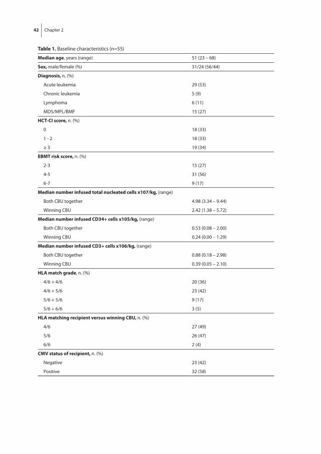
42 Chapter 2
table 1. Baseline characteristics (n=55)
median age, years (range) 51 (23 – 68)
Sex, male/female (%) 31/24 (56/44)
Diagnosis, n. (%)
Acute leukemia 29 (53)
Chronic leukemia 5 (9)
Lymphoma 6 (11)
MDS/MPL/BMF 15 (27)
HCt-CI score, n. (%)
0 18 (33)
1 - 2 18 (33)
≥ 3 19 (34)
EBmt risk score, n. (%)
2-3 15 (27)
4-5 31 (56)
6-7 9 (17)
median number infused total nucleated cells x107/kg, (range)
Both CBU together 4.98 (3.34 – 9.44)
Winning CBU 2.42 (1.38 – 5.72)
median number infused CD34+ cells x105/kg, (range)
Both CBU together 0.53 (0.08 – 2.00)
Winning CBU 0.24 (0.00 – 1.29)
median number infused CD3+ cells x106/kg, (range)
Both CBU together 0.88 (0.18 – 2.98)
Winning CBU 0.39 (0.05 – 2.10)
HLa match grade, n. (%)
4/6 + 4/6 20 (36)
4/6 + 5/6 23 (42)
5/6 + 5/6 9 (17)
5/6 + 6/6 3 (5)
HLa matching recipient versus winning CBU, n. (%)
4/6 27 (49)
5/6 26 (47)
6/6 2 (4)
CmV status of recipient, n. (%)
Negative 23 (42)
Positive 32 (58)
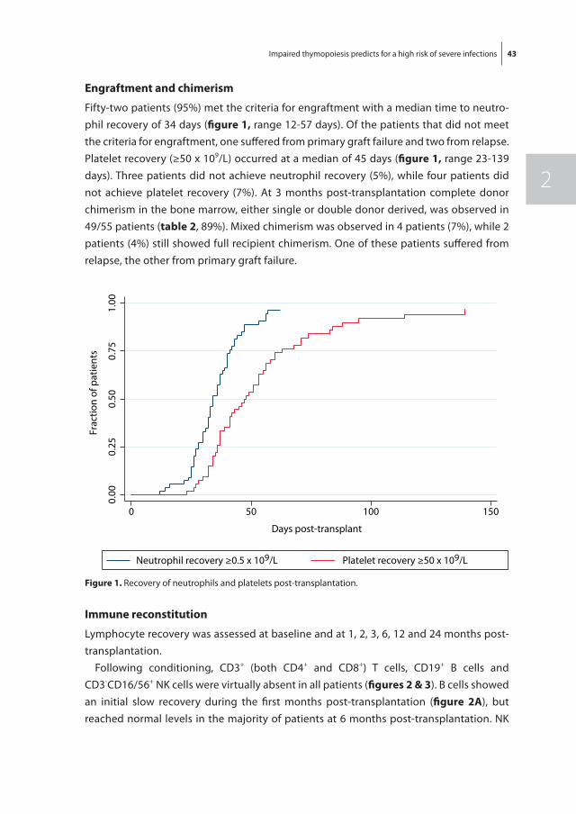
Impaired thymopoiesis predicts for a high risk of severe infections 43
2
Engraftment and chimerism
Fifty-two patients (95%) met the criteria for engraftment with a median time to neutro-phil recovery of 34 days (figure 1, range 12-57 days). Of the patients that did not meet the criteria for engraftment, one suffered from primary graft failure and two from relapse. Platelet recovery (≥50 x 109/L) occurred at a median of 45 days (figure 1, range 23-139 days). Three patients did not achieve neutrophil recovery (5%), while four patients did not achieve platelet recovery (7%). At 3 months post-transplantation complete donor chimerism in the bone marrow, either single or double donor derived, was observed in 49/55 patients (table 2, 89%). Mixed chimerism was observed in 4 patients (7%), while 2 patients (4%) still showed full recipient chimerism. One of these patients suffered from relapse, the other from primary graft failure.
Immune reconstitution
Lymphocyte recovery was assessed at baseline and at 1, 2, 3, 6, 12 and 24 months post-transplantation.
Following conditioning, CD3+ (both CD4+ and CD8+) T cells, CD19+ B cells and CD3-CD16/56+ NK cells were virtually absent in all patients (figures 2 & 3). B cells showed an initial slow recovery during the first months post-transplantation (figure 2a), but reached normal levels in the majority of patients at 6 months post-transplantation. NK
0.00
0.25
0.50
0.75
1.00
0 50 100 150
Days post-transplant
Neutrophil recovery ≥0.5 x 10⁹/L Platelet recovery ≥50 x 109/L
Frac
tion
of p
atie
nts
figure 1. Recovery of neutrophils and platelets post-transplantation.
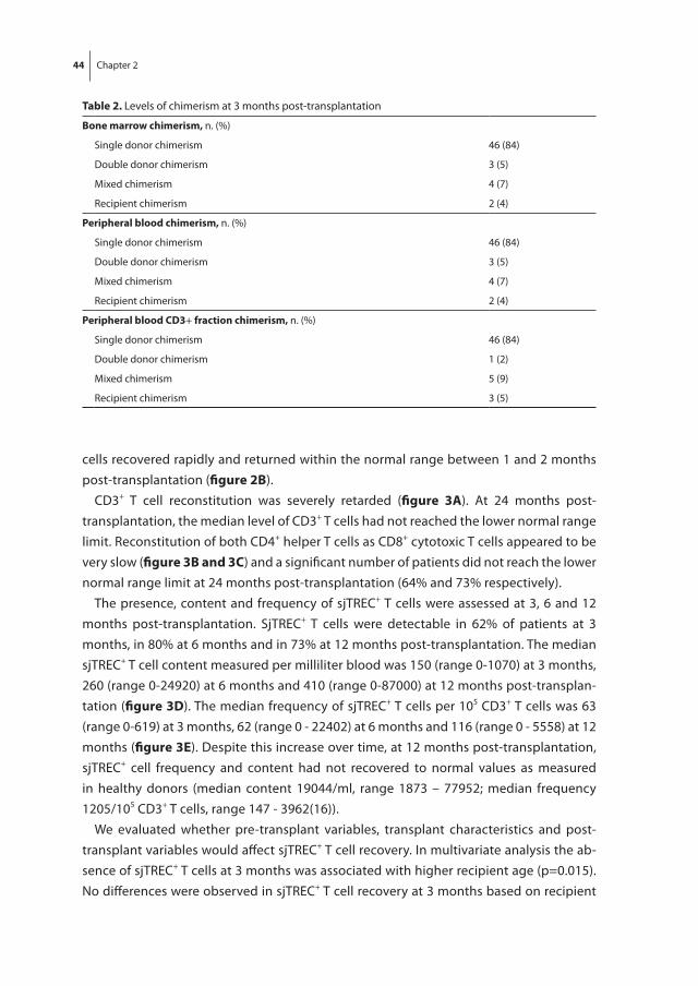
44 Chapter 2
cells recovered rapidly and returned within the normal range between 1 and 2 months post-transplantation (figure 2B).
CD3+ T cell reconstitution was severely retarded (figure 3a). At 24 months post-transplantation, the median level of CD3+ T cells had not reached the lower normal range limit. Reconstitution of both CD4+ helper T cells as CD8+ cytotoxic T cells appeared to be very slow (figure 3B and 3C) and a significant number of patients did not reach the lower normal range limit at 24 months post-transplantation (64% and 73% respectively).
The presence, content and frequency of sjTREC+ T cells were assessed at 3, 6 and 12 months post-transplantation. SjTREC+ T cells were detectable in 62% of patients at 3 months, in 80% at 6 months and in 73% at 12 months post-transplantation. The median sjTREC+ T cell content measured per milliliter blood was 150 (range 0-1070) at 3 months, 260 (range 0-24920) at 6 months and 410 (range 0-87000) at 12 months post-transplan-tation (figure 3D). The median frequency of sjTREC+ T cells per 105 CD3+ T cells was 63 (range 0-619) at 3 months, 62 (range 0 - 22402) at 6 months and 116 (range 0 - 5558) at 12 months (figure 3E). Despite this increase over time, at 12 months post-transplantation, sjTREC+ cell frequency and content had not recovered to normal values as measured in healthy donors (median content 19044/ml, range 1873 – 77952; median frequency 1205/105 CD3+ T cells, range 147 - 3962(16)).
We evaluated whether pre-transplant variables, transplant characteristics and post-transplant variables would affect sjTREC+ T cell recovery. In multivariate analysis the ab-sence of sjTREC+ T cells at 3 months was associated with higher recipient age (p=0.015). No differences were observed in sjTREC+ T cell recovery at 3 months based on recipient
table 2. Levels of chimerism at 3 months post-transplantation
Bone marrow chimerism, n. (%)
Single donor chimerism 46 (84)
Double donor chimerism 3 (5)
Mixed chimerism 4 (7)
Recipient chimerism 2 (4)
Peripheral blood chimerism, n. (%)
Single donor chimerism 46 (84)
Double donor chimerism 3 (5)
Mixed chimerism 4 (7)
Recipient chimerism 2 (4)
Peripheral blood CD3+ fraction chimerism, n. (%)
Single donor chimerism 46 (84)
Double donor chimerism 1 (2)
Mixed chimerism 5 (9)
Recipient chimerism 3 (5)
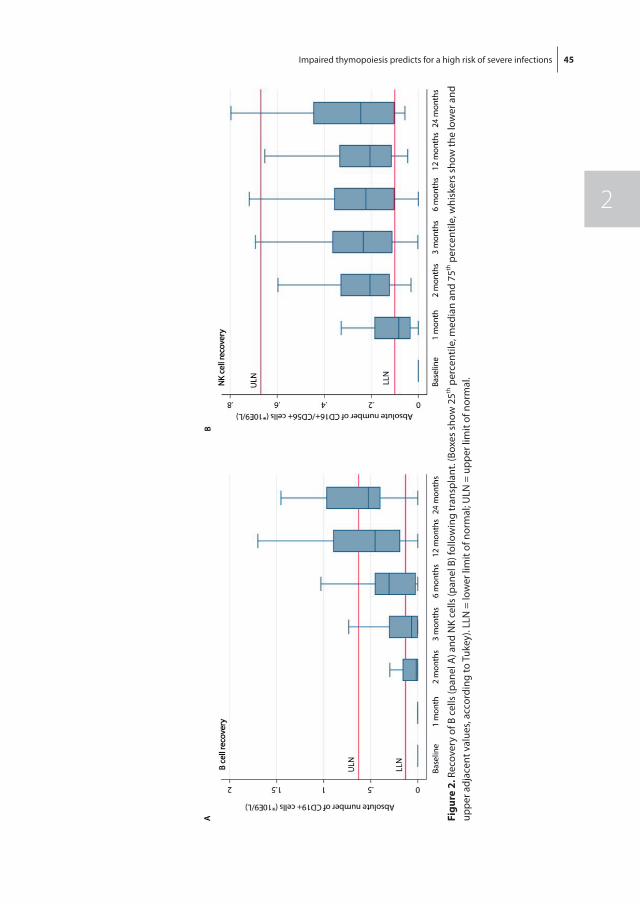
Impaired thymopoiesis predicts for a high risk of severe infections 45
2
figu
re 2
. Rec
over
y of
B c
ells
(pan
el A
) and
NK
cells
(pan
el B
) fol
low
ing
tran
spla
nt. (
Boxe
s sh
ow 2
5th p
erce
ntile
, med
ian
and
75th
per
cent
ile, w
hisk
ers
show
the
low
er a
nd
uppe
r adj
acen
t val
ues,
acco
rdin
g to
Tuk
ey).
LLN
= lo
wer
lim
it of
nor
mal
; ULN
= u
pper
lim
it of
nor
mal
.
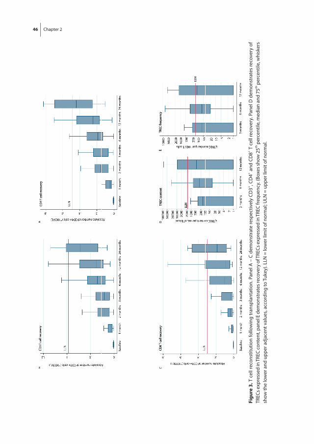
46 Chapter 2
figu
re 3
. T c
ell r
econ
stitu
tion
follo
win
g tr
ansp
lant
atio
n. P
anel
A –
C d
emon
stra
te re
spec
tivel
y CD
3+ , CD
4+ and
CD
8+ T c
ell r
ecov
ery.
Pan
el D
dem
onst
rate
s re
cove
ry o
f TR
ECs e
xpre
ssed
in T
REC
cont
ent,
pane
l E d
emon
stra
tes r
ecov
ery
of T
RECs
exp
ress
ed in
TRE
C fr
eque
ncy.
(Box
es sh
ow 2
5th p
erce
ntile
, med
ian
and
75th
per
cent
ile, w
hisk
ers
show
the
low
er a
nd u
pper
adj
acen
t val
ues,
acco
rdin
g to
Tuk
ey).
LLN
= lo
wer
lim
it of
nor
mal
; ULN
= u
pper
lim
it of
nor
mal
.
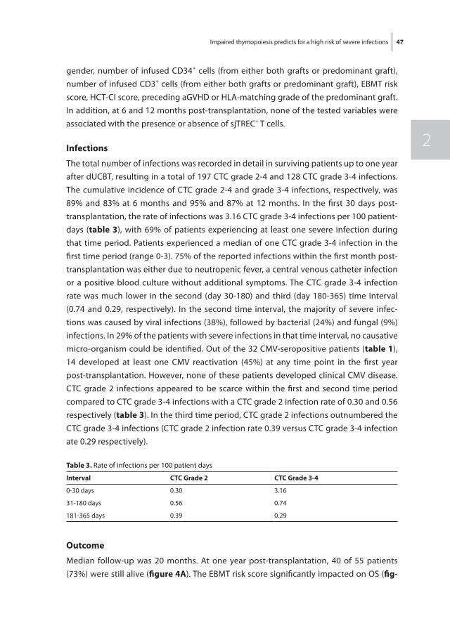
Impaired thymopoiesis predicts for a high risk of severe infections 47
2
gender, number of infused CD34+ cells (from either both grafts or predominant graft), number of infused CD3+ cells (from either both grafts or predominant graft), EBMT risk score, HCT-CI score, preceding aGVHD or HLA-matching grade of the predominant graft. In addition, at 6 and 12 months post-transplantation, none of the tested variables were associated with the presence or absence of sjTREC+ T cells.
Infections
The total number of infections was recorded in detail in surviving patients up to one year after dUCBT, resulting in a total of 197 CTC grade 2-4 and 128 CTC grade 3-4 infections. The cumulative incidence of CTC grade 2-4 and grade 3-4 infections, respectively, was 89% and 83% at 6 months and 95% and 87% at 12 months. In the first 30 days post-transplantation, the rate of infections was 3.16 CTC grade 3-4 infections per 100 patient-days (table 3), with 69% of patients experiencing at least one severe infection during that time period. Patients experienced a median of one CTC grade 3-4 infection in the first time period (range 0-3). 75% of the reported infections within the first month post-transplantation was either due to neutropenic fever, a central venous catheter infection or a positive blood culture without additional symptoms. The CTC grade 3-4 infection rate was much lower in the second (day 30-180) and third (day 180-365) time interval (0.74 and 0.29, respectively). In the second time interval, the majority of severe infec-tions was caused by viral infections (38%), followed by bacterial (24%) and fungal (9%) infections. In 29% of the patients with severe infections in that time interval, no causative micro-organism could be identified. Out of the 32 CMV-seropositive patients (table 1), 14 developed at least one CMV reactivation (45%) at any time point in the first year post-transplantation. However, none of these patients developed clinical CMV disease. CTC grade 2 infections appeared to be scarce within the first and second time period compared to CTC grade 3-4 infections with a CTC grade 2 infection rate of 0.30 and 0.56 respectively (table 3). In the third time period, CTC grade 2 infections outnumbered the CTC grade 3-4 infections (CTC grade 2 infection rate 0.39 versus CTC grade 3-4 infection ate 0.29 respectively).
table 3. Rate of infections per 100 patient days
Interval CtC Grade 2 CtC Grade 3-4
0-30 days 0.30 3.16
31-180 days 0.56 0.74
181-365 days 0.39 0.29
outcome
Median follow-up was 20 months. At one year post-transplantation, 40 of 55 patients (73%) were still alive (figure 4a). The EBMT risk score significantly impacted on OS (fig-
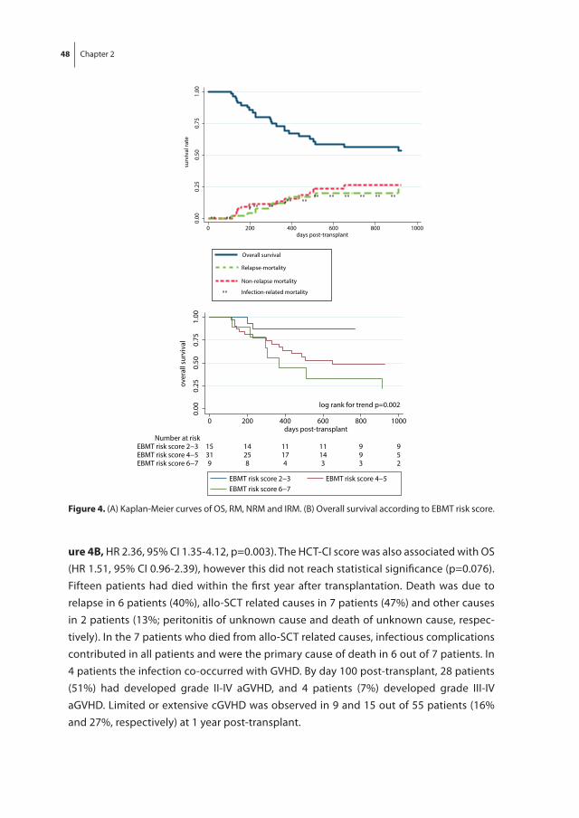
48 Chapter 2
ure 4B, HR 2.36, 95% CI 1.35-4.12, p=0.003). The HCT-CI score was also associated with OS (HR 1.51, 95% CI 0.96-2.39), however this did not reach statistical significance (p=0.076). Fifteen patients had died within the first year after transplantation. Death was due to relapse in 6 patients (40%), allo-SCT related causes in 7 patients (47%) and other causes in 2 patients (13%; peritonitis of unknown cause and death of unknown cause, respec-tively). In the 7 patients who died from allo-SCT related causes, infectious complications contributed in all patients and were the primary cause of death in 6 out of 7 patients. In 4 patients the infection co-occurred with GVHD. By day 100 post-transplant, 28 patients (51%) had developed grade II-IV aGVHD, and 4 patients (7%) developed grade III-IV aGVHD. Limited or extensive cGVHD was observed in 9 and 15 out of 55 patients (16% and 27%, respectively) at 1 year post-transplant.
0.00
0.25
0.50
0.75
1.00
0 200 400 600 800 1000days post-transplant
Overall survival
Non-relapse mortality
Relapse-mortality
Infection-related mortality
surv
ival
rate
0.00
0.25
0.50
0.75
1.00
9 8 4 3 3 2EBMT risk score 6−731 25 17 14 9 5EBMT risk score 4−515 14 11 11 9 9EBMT risk score 2−3
Number at risk
0 200 400 600 800 1000days post-transplant
EBMT risk score 2−3 EBMT risk score 4−5EBMT risk score 6−7
over
all s
urvi
val
log rank for trend p=0.002
figure 4. (A) Kaplan-Meier curves of OS, RM, NRM and IRM. (B) Overall survival according to EBMT risk score.
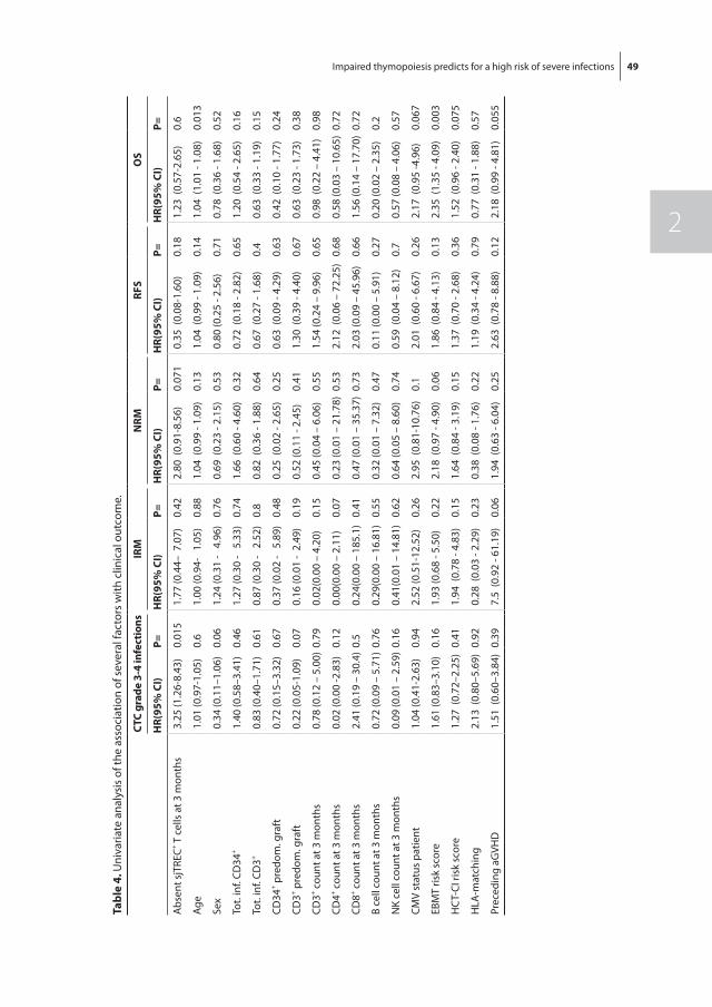
Impaired thymopoiesis predicts for a high risk of severe infections 49
2
tabl
e 4.
Uni
varia
te a
naly
sis
of th
e as
soci
atio
n of
sev
eral
fact
ors
with
clin
ical
out
com
e.
CtC
grad
e 3-
4 in
fect
ions
Irm
nrm
rfS
oS
Hr(
95%
CI)
P=H
r(95
% C
I)P=
Hr(
95%
CI)
P=H
r(95
% C
I)P=
Hr(
95%
CI)
P=
Abs
ent s
jTRE
C+ T c
ells
at 3
mon
ths
3.25
(1.2
6-8.
43)
0.01
51.
77 (0
.44–
7.0
7)0.
422.
80 (
0.91
-8.5
6)0.
071
0.35
(0.
08-1
.60)
0.18
1.23
(0.
57-2
.65)
0.6
Age
1.01
(0.9
7-1.
05)
0.6
1.00
(0.9
4-
1.05
)0.
881.
04 (
0.99
- 1.
09)
0.13
1.04
(0.
99 -
1.09
)0.
141.
04 (
1.01
- 1.
08)
0.01
3
Sex
0.34
(0.1
1–1.
06)
0.06
1.24
(0.3
1 -
4.9
6)0.
760.
69 (
0.23
- 2.
15)
0.53
0.80
(0.2
5 - 2
.56)
0.71
0.78
(0.
36 -
1.68
)0.
52
Tot.
inf.
CD34
+1.
40 (0
.58–
3.41
)0.
461.
27 (0
.30
- 5
.33)
0.74
1.66
(0.
60 -
4.60
)0.
320.
72 (
0.18
- 2.
82)
0.65
1.20
(0.
54 -
2.65
)0.
16
Tot.
inf.
CD3+
0.83
(0.4
0–1.
71)
0.61
0.87
(0.3
0 -
2.5
2)0.
80.
82 (
0.36
- 1.
88)
0.64
0.67
(0.
27 -
1.68
)0.
40.
63 (
0.33
- 1.
19)
0.15
CD34
+ pre
dom
. gra
ft0.
72 (0
.15–
3.32
)0.
670.
37 (0
.02
- 5
.89)
0.48
0.25
(0.
02 -
2.65
)0.
250.
63 (
0.09
- 4.
29)
0.63
0.42
(0.
10 -
1.77
)0.
24
CD3+ p
redo
m. g
raft
0.22
(0.0
5-1.
09)
0.07
0.16
(0.0
1 -
2.49
)0.
190.
52 (0
.11
- 2.4
5)0.
411.
30 (
0.39
- 4.
40)
0.67
0.63
(0.
23 -
1.73
)0.
38
CD3+ c
ount
at 3
mon
ths
0.78
(0.1
2 –
5.00
)0.
790.
02(0
.00
– 4.
20)
0.15
0.45
(0.0
4 –
6.06
)0.
551.
54 (0
.24
– 9.
96)
0.65
0.98
(0.
22 –
4.4
1)0.
98
CD4+ c
ount
at 3
mon
ths
0.02
(0.0
0 -2
.83)
0.12
0.00
(0.0
0 –
2.11
)0.
070.
23 (0
.01
– 21
.78)
0.53
2.12
(0.
06 –
72.
25)
0.68
0.58
(0.0
3 –
10.6
5)0.
72
CD8+ c
ount
at 3
mon
ths
2.41
(0.1
9 –
30.4
)0.
50.
24(0
.00
– 18
5.1)
0.41
0.47
(0.0
1 –
35.3
7)0.
732.
03 (0
.09
– 45
.96)
0.66
1.56
(0.1
4 –
17.7
0)0.
72
B ce
ll co
unt a
t 3 m
onth
s0.
72 (0
.09
– 5.
71)
0.76
0.29
(0.0
0 –
16.8
1)0.
550.
32 (0
.01
– 7.
32)
0.47
0.11
(0.0
0 –
5.91
)0.
270.
20 (0
.02
– 2.
35)
0.2
NK
cell
coun
t at 3
mon
ths
0.09
(0.0
1 –
2.59
)0.
160.
41(0
.01
– 14
.81)
0.62
0.64
(0.0
5 –
8.60
)0.
740.
59 (
0.04
– 8
.12)
0.7
0.57
(0.0
8 –
4.06
)0.
57
CMV
stat
us p
atie
nt1.
04 (0
.41-
2.63
)0.
942.
52 (0
.51-
12.5
2)0.
262.
95 (
0.81
-10.
76)
0.1
2.01
(0.
60 -
6.67
)0.
262.
17 (
0.95
-4.9
6)0.
067
EBM
T ris
k sc
ore
1.61
(0.8
3–3.
10)
0.16
1.93
(0.6
8 - 5
.50)
0.22
2.18
(0.
97 -
4.90
)0.
061.
86 (
0.84
- 4.
13)
0.13
2.35
(1.
35 -
4.09
)0.
003
HC
T-CI
risk
sco
re1.
27 (
0.72
–2.2
5)0.
411.
94 (
0.78
- 4.
83)
0.15
1.64
(0.
84 -
3.19
)0.
151.
37 (
0.70
- 2.
68)
0.36
1.52
(0.
96 -
2.40
)0.
075
HLA
-mat
chin
g2.
13 (
0.80
–5.6
9)0.
920.
28 (
0.03
- 2.
29)
0.23
0.38
(0.
08 -
1.76
)0.
221.
19 (
0.34
- 4.
24)
0.79
0.77
(0.
31 -
1.88
)0.
57
Prec
edin
g aG
VHD
1.51
(0.
60–3
.84)
0.39
7.5
(0.9
2 - 6
1.19
)0.
061.
94 (
0.63
- 6.
04)
0.25
2.63
(0.
78 -
8.88
)0.
122.
18 (
0.99
- 4.
81)
0.05
5
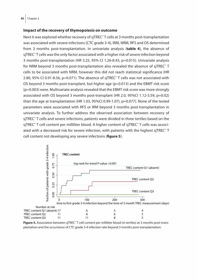
50 Chapter 2
Impact of the recovery of thymopoiesis on outcome
Next it was explored whether recovery of sjTREC+ T cells at 3 months post-transplantation was associated with severe infections (CTC grade 3-4), IRM, NRM, RFS and OS determined from 3 months post-transplantation. In univariate analysis (table 4), the absence of sjTREC+ T cells was the only factor associated with a higher risk of severe infection beyond 3 months post-transplantation (HR 3.25, 95% CI 1.26-8.43, p=0.015). Univariate analysis for NRM beyond 3 months post-transplantation also revealed the absence of sjTREC+ T cells to be associated with NRM, however this did not reach statistical significance (HR 2.80, 95% CI 0.91-8.56, p=0.071). The absence of sjTREC+ T cells was not associated with OS beyond 3 months post-transplant, but higher age (p=0.013) and the EBMT risk score (p=0.003) were. Multivariate analysis revealed that the EBMT risk score was more strongly associated with OS beyond 3 months post-transplant (HR 2.0, 95%CI 1.12-3.59, p=0.02) than the age at transplantation (HR 1.03, 95%CI 0.99-1.07), p=0.077). None of the tested parameters were associated with RFS or IRM beyond 3 months post-transplantation in univariate analysis. To further address the observed association between recovery of sjTREC+ T cells and severe infections, patients were divided in three tertiles based on the sjTREC+ T cell content per milliliter blood. A higher content of sjTREC+ T cells was associ-ated with a decreased risk for severe infection, with patients with the highest sjTREC+ T cell content not developing any severe infections (figure 5).
0.00
0.25
0.50
0.75
1.00
11 11 8 5TREC content Q311 8 6 5TREC content Q217 6 5 4TREC content Q1 (absent)
Number at risk
0 100 200 300time to �rst grade 3-4 infection beyond the time of 3 month TREC measurement (days)
TREC content
TREC content Q1 (absent)
TREC content Q2
TREC content Q3
frac
tion
of p
atie
nts
with
gra
de 3
-4 in
fect
ion
log-rank for trend P-value <0.001
figure 5. Association between sjTREC+ T cell content per milliliter blood (in tertiles) at 3 months post-trans-plantation and the occurrence of CTC grade 3-4 infection rate beyond 3 months post-transplantation.
Impaired thymopoiesis predicts for a high risk of severe infections 51
2
DISCUSSIon
Immune reconstitution upon UCBT is severely retarded and as a direct consequence, UCBT is associated with a significant risk of opportunistic infections (32). Kinetics of immune reconstitution upon UCBT have been widely studied, although most of these studies were performed in a single graft setting and are based on mixed populations of patients, receiving various conditioning and immunosuppressive regimens (15, 17-19, 33-35). Few studies reported on recovery of thymopoiesis upon dUCBT (20, 36, 37). This study, to our knowledge, is the first to describe immune reconstitution including thymopoiesis in a uniform population of 55 adult patients, receiving dUCBT upon a RIC regimen omitting in vivo T cell depletion. In addition, the present study is the first to correlate recovery of thymopoiesis with clinical outcome upon dUCBT and shows the predictive impact of impaired sjTREC+ T cell recovery at 3 months on the risk of severe infections beyond that time point.
Although CD3+ T cell recovery was severely protracted and the majority of patients had CD3+ T cell levels below the normal range up to 24 months post-transplantation, sjTREC+ T cells were detected at low levels in 62% of patients at 3 months post-transplantation. Our results are in contrast to several other studies, were sjTREC+ T cells were detected in 20% (20), 11% (37) and 0% (38) of patients around the same time point upon dUCBT. However, in these studies, patients received either MA conditioning or a RIC regimen combined with antithymocyte globulin (ATG), a polyclonal antibody raised against thymocyte antigens resulting in profound in vivo T cell depletion (39). Cytotoxic che-motherapy and radiation negatively affect thymic reconstitution (40, 41). RIC regimens may therefore cause less damage to the thymus compared to MA conditioning, resulting in faster recovery of thymopoiesis. In addition, several studies showed an association between the use of a RIC regimen and enhanced immune reconstitution (42-44). The use of ATG results in a lower incidence of GVHD, but however, might result in delayed immune reconstitution as well. Enhanced B and T cell recovery is reported upon the use of ATG-free conditioning regimens in UCBT, both MA or RIC, in both pediatric and adult cohorts (45-47). Therefore, the high incidence of patients with detectable sjTREC+ T cells at 3 months post-transplantation could be well explained by the relatively mild conditioning regimen used in our study. In a comparable cohort of 46 adult patients receiving a matched related (n=33) or unrelated (n=13) allo-SCT upon an ATG-free RIC regimen (Fludarabine 90 mg/m2 and TBI 2 Gy) in our center, detectable sjTREC+ T cells were observed in 67% of patients at 3 months and 86% at 12 months, which is similar to the above described proportions of sjTREC+ T cell positivity in patients receiving dUCBT. These results indicate, that upon an ATG-free RIC regimen, dUCBT and adult donor allo-SCT show comparable reconstitution of thymopoiesis. Previously, both age and GVHD have been implicated to impact the reconstitution of thymopoiesis (44, 48-50). While an
52 Chapter 2
association between higher age and undetectable sjTREC+ T cells was observed in the current study, this association was not observed for the presence of GVHD.
In an earlier study, we showed that insufficient thymopoiesis predicted for severe infections and NRM in patients receiving a matched related or unrelated donor allo-SCT upon MA conditioning (16). In the current study, patients who failed to recover thymo-poiesis at 3 months post-transplantation had a 3.3-fold higher risk for subsequent severe infections. This association was not observed between CD3+, CD4+ and CD8+ T cell counts at 3 months post-transplantation and subsequent severe infections, indicating that the newly formed naïve T cells are pivotal for adequate infectious immunity. In addition, the content of sjTREC+ T cells was associated with the risk for severe infection at that time point. These results compare well with Brown et al. (37) who demonstrated that clear-ance of CMV viremia after dUCBT depended on the recovery of thymopoiesis. Although several other studies assessed thymopoiesis upon dUCBT(20, 36-38) and infectious complications are a known problem upon UCBT(32, 46), severe infections after dUCBT were not yet linked to recovery of thymopoiesis.
In line with Sauter et al. (46) the highest infection rate was observed in the first 30 days post-transplantation. They described 61% of patients experiencing at least one infection in this time period, which compares well to the cumulative incidence of 69% of severe infections in our cohort. Of note is that most of the infections scored in the present study in the first month post-transplantation were neutropenic fevers, central venous catheter infections or positive blood cultures without an evident focus. These infections may have occurred and been reported more frequently because of longer hospitaliza-tion and therefore increased surveillance, leading to an overestimation of infections in the first 30 days post-transplantation. In a comparable adult donor recipient cohort, as mentioned earlier, the highest rate of CTC grade 2-4 infections was observed in the first 30 days post-transplantation as well, although this rate was markedly lower compared to the observed infection rate upon dUCBT (adult donor 1.09 CTC grade 2-4 infections per 100 patient-days versus 3.46 in the dUCBT group). In the second time interval of 30 days to 180 days post-transplantation, viral infections were the most common cause of infectious morbidity, which is in line with literature (4, 20, 32). In addition, 45% of the CMV-seropositive patients developed one or more CMV reactivation during the first year post-transplant. This is in line with the study performed by Sauter et al. (46), where a CMV reactivation rate of 51% was observed in 180 CMV-seropositive patients that received dUCBT upon conditioning omitting in vivo T cell depletion. However, clinical CMV disease was reported in 25 patients, whereas no cases of clinical CMV disease were observed in the current study. This difference might be due to the prophylactic use of valaciclovir for the prevention of herpes infections in our center. Although primarily used for the prevention of herpes infections, valaciclovir is a more potent CMV suppressor in patients receiving allo-SCT than acyclovir (51), which was used by Sauter and colleagues.
Impaired thymopoiesis predicts for a high risk of severe infections 53
2
As mentioned earlier, this study is the first to assess immune reconstitution including thymopoiesis in a homogeneous cohort of 55 adult patients, receiving a RIC regimen omitting in vivo T cell depletion and to correlate recovery of thymopoiesis to clinical out-come. Although this population of 55 patients is larger than any other cohort described in this context, the limited cohort size was a limitation for the present study, especially for associations with clinical outcomes in which there were only a few events.
In conclusion, failure to recover thymopoiesis after RIC-dUCBT without in vivo T cell depletion resulted in a higher risk of developing subsequent severe infections. This may lead to increased NRM and adverse outcome. These results highlight the need to improve thymic function post-transplantation, for example by the administration of the cytokines interleukin-7 or keratinocyte growth factor (52-54). Furthermore, risk of infections might be reduced by adoptive transfer of virus or fungal specific T cells (55, 56). In addition, patients lacking thymic recovery at 3 months post-transplantation might benefit from intensified supportive measures in order to prevent infections and associated mortality. Therefore, we propose to consider monitoring sjTREC+ T cell recovery in dUCBT patients, to identify patients in need for intensified support.
54 Chapter 2
rEfErEnCES
1. Ballen KK, Gluckman E, Broxmeyer HE. Umbilical cord blood transplantation: the first 25 years and beyond. Blood. 2013 Jul 25;122(4):491-8.
2. Rocha V, Labopin M, Sanz G, Arcese W, Schwerdtfeger R, Bosi A, et al. Transplants of umbilical-cord blood or bone marrow from unrelated donors in adults with acute leukemia. N Engl J Med. 2004 Nov 25;351(22):2276-85.
3. Laughlin MJ, Eapen M, Rubinstein P, Wagner JE, Zhang MJ, Champlin RE, et al. Outcomes after transplantation of cord blood or bone marrow from unrelated donors in adults with leukemia. N Engl J Med. 2004 Nov 25;351(22):2265-75.
4. Szabolcs P, Niedzwiecki D. Immune reconstitution after unrelated cord blood transplantation. Cytotherapy. 2007;9(2):111-22.
5. Barker JN, Scaradavou A, Stevens CE. Combined effect of total nucleated cell dose and HLA match on transplantation outcome in 1061 cord blood recipients with hematologic malignancies. Blood. 2010 Mar 4;115(9):1843-9.
6. Wagner JE, Barker JN, DeFor TE, Baker KS, Blazar BR, Eide C, et al. Transplantation of unrelated donor umbilical cord blood in 102 patients with malignant and nonmalignant diseases: influence of CD34 cell dose and HLA disparity on treatment-related mortality and survival. Blood. 2002 Sep 1;100(5):1611-8.
7. Gluckman E, Rocha V, Boyer-Chammard A, Locatelli F, Arcese W, Pasquini R, et al. Outcome of cord-blood transplantation from related and unrelated donors. Eurocord Transplant Group and the European Blood and Marrow Transplantation Group. N Engl J Med. 1997 Aug 7;337(6):373-81.
8. Barker JN, Weisdorf DJ, DeFor TE, Blazar BR, McGlave PB, Miller JS, et al. Transplantation of 2 par-tially HLA-matched umbilical cord blood units to enhance engraftment in adults with hematologic malignancy. Blood. 2005 Feb 1;105(3):1343-7.
9. Barker JN, Weisdorf DJ, DeFor TE, Blazar BR, Miller JS, Wagner JE. Rapid and complete donor chi-merism in adult recipients of unrelated donor umbilical cord blood transplantation after reduced-intensity conditioning. Blood. 2003 Sep 1;102(5):1915-9.
10. Barker JN, Weisdorf DJ, Wagner JE. Creation of a double chimera after the transplantation of umbilical-cord blood from two partially matched unrelated donors. N Engl J Med. 2001 Jun 14;344(24):1870-1.
11. Somers JA, Braakman E, van der Holt B, Petersen EJ, Marijt EW, Huisman C, et al. Rapid induc-tion of single donor chimerism after double umbilical cord blood transplantation preceded by reduced intensity conditioning: results of the HOVON 106 phase II study. Haematologica. 2014 Nov;99(11):1753-61.
12. Storek J, Geddes M, Khan F, Huard B, Helg C, Chalandon Y, et al. Reconstitution of the immune system after hematopoietic stem cell transplantation in humans. Semin Immunopathol. 2008 Dec;30(4):425-37.
13. Rufer N, Helg C, Chapuis B, Roosnek E. Human memory T cells: lessons from stem cell transplanta-tion. Trends Immunol. 2001 Mar;22(3):136-41.
14. Mackall CL, Granger L, Sheard MA, Cepeda R, Gress RE. T-cell regeneration after bone marrow trans-plantation: differential CD45 isoform expression on thymic-derived versus thymic-independent progeny. Blood. 1993 Oct 15;82(8):2585-94.
15. Clave E, Lisini D, Douay C, Giorgiani G, Busson M, Zecca M, et al. Thymic function recovery after unrelated donor cord blood or T-cell depleted HLA-haploidentical stem cell transplantation cor-relates with leukemia relapse. Front Immunol. 2013;4:54.
Impaired thymopoiesis predicts for a high risk of severe infections 55
2
16. Wils EJ, van der Holt B, Broers AE, Posthumus-van Sluijs SJ, Gratama JW, Braakman E, et al. Insuf-ficient recovery of thymopoiesis predicts for opportunistic infections in allogeneic hematopoietic stem cell transplant recipients. Haematologica. 2011 Dec;96(12):1846-54.
17. Parody R, Martino R, Rovira M, Vazquez L, Vazquez MJ, de la Camara R, et al. Severe infections after unrelated donor allogeneic hematopoietic stem cell transplantation in adults: comparison of cord blood transplantation with peripheral blood and bone marrow transplantation. Biol Blood Marrow Transplant. 2006 Jul;12(7):734-48.
18. Safdar A, Rodriguez GH, De Lima MJ, Petropoulos D, Chemaly RF, Worth LL, et al. Infections in 100 cord blood transplantations: spectrum of early and late posttransplant infections in adult and pediatric patients 1996-2005. Medicine (Baltimore). 2007 Nov;86(6):324-33.
19. Komanduri KV, St John LS, de Lima M, McMannis J, Rosinski S, McNiece I, et al. Delayed immune reconstitution after cord blood transplantation is characterized by impaired thymopoiesis and late memory T-cell skewing. Blood. 2007 Dec 15;110(13):4543-51.
20. Ruggeri A, Peffault de Latour R, Carmagnat M, Clave E, Douay C, Larghero J, et al. Outcomes, infections, and immune reconstitution after double cord blood transplantation in patients with high-risk hematological diseases. Transpl Infect Dis. 2011 Oct;13(5):456-65.
21. Somers JA, Brand A, van Hensbergen Y, Mulder A, Oudshoorn M, Sintnicolaas K, et al. Double umbilical cord blood transplantation: a study of early engraftment kinetics in leukocyte subsets using HLA-specific monoclonal antibodies. Biol Blood Marrow Transplant. 2013 Feb;19(2):266-73.
22. Broers AE, van der Holt B, Haze S, Braakman E, Gratama JW, Lowenberg B, et al. A comparison of postengraftment infectious morbidity and mortality after allogeneic partially T cell-depleted peripheral blood progenitor cell transplantation versus T cell-depleted bone marrow transplanta-tion. Exp Hematol. 2005 Aug;33(8):912-9.
23. Beije N, Versluis J, Kraan J, Gratama JW, Sleijfer S, Cornelissen JJ. Circulating endothelial cell enu-meration demonstrates prolonged endothelial damage in recipients of myeloablative allogeneic stem cell transplantation. Haematologica. 2015 Jun;100(6):e246-9.
24. Broers AE, Meijerink JP, van Dongen JJ, Posthumus SJ, Lowenberg B, Braakman E, et al. Quantifica-tion of newly developed T cells in mice by real-time quantitative PCR of T-cell receptor rearrange-ment excision circles. Exp Hematol. 2002 Jul;30(7):745-50.
25. Hazenberg MD, Otto SA, de Pauw ES, Roelofs H, Fibbe WE, Hamann D, et al. T-cell receptor excision circle and T-cell dynamics after allogeneic stem cell transplantation are related to clinical events. Blood. 2002 May 1;99(9):3449-53.
26. Ribeiro RM, Perelson AS. Determining thymic output quantitatively: using models to interpret experimental T-cell receptor excision circle (TREC) data. Immunol Rev. 2007 Apr;216:21-34.
27. Glucksberg H, Storb R, Fefer A, Buckner CD, Neiman PE, Clift RA, et al. Clinical manifestations of graft-versus-host disease in human recipients of marrow from HL-A-matched sibling donors. Transplantation. 1974 Oct;18(4):295-304.
28. Przepiorka D, Weisdorf D, Martin P, Klingemann HG, Beatty P, Hows J, et al. 1994 Consensus Confer-ence on Acute GVHD Grading. Bone Marrow Transplant. 1995 Jun;15(6):825-8.
29. Shulman HM, Sullivan KM, Weiden PL, McDonald GB, Striker GE, Sale GE, et al. Chronic graft-versus-host syndrome in man. A long-term clinicopathologic study of 20 Seattle patients. Am J Med. 1980 Aug;69(2):204-17.
30. Gratwohl A, Hermans J, Goldman JM, Arcese W, Carreras E, Devergie A, et al. Risk assessment for patients with chronic myeloid leukaemia before allogeneic blood or marrow transplantation. Chronic Leukemia Working Party of the European Group for Blood and Marrow Transplantation. Lancet. 1998 Oct 3;352(9134):1087-92.
56 Chapter 2
31. Sorror ML, Maris MB, Storb R, Baron F, Sandmaier BM, Maloney DG, et al. Hematopoietic cell trans-plantation (HCT)-specific comorbidity index: a new tool for risk assessment before allogeneic HCT. Blood. 2005 Oct 15;106(8):2912-9.
32. Danby R, Rocha V. Improving engraftment and immune reconstitution in umbilical cord blood transplantation. Front Immunol. 2014;5:68.
33. Niehues T, Rocha V, Filipovich AH, Chan KW, Porcher R, Michel G, et al. Factors affecting lymphocyte subset reconstitution after either related or unrelated cord blood transplantation in children -- a Eurocord analysis. Br J Haematol. 2001 Jul;114(1):42-8.
34. Tanaka J, Sugita J, Asanuma S, Arita K, Shono Y, Kikutchi M, et al. Increased number of CD16(+)CD56(dim) NK cells in peripheral blood mononuclear cells after allogeneic cord blood transplanta-tion. Hum Immunol. 2009 Sep;70(9):701-5.
35. Talvensaari K, Clave E, Douay C, Rabian C, Garderet L, Busson M, et al. A broad T-cell repertoire diversity and an efficient thymic function indicate a favorable long-term immune reconstitution after cord blood stem cell transplantation. Blood. 2002 Feb 15;99(4):1458-64.
36. Kanda J, Chiou LW, Szabolcs P, Sempowski GD, Rizzieri DA, Long GD, et al. Immune recovery in adult patients after myeloablative dual umbilical cord blood, matched sibling, and matched unrelated donor hematopoietic cell transplantation. Biol Blood Marrow Transplant. 2012 Nov;18(11):1664-76 e1.
37. Brown JA, Stevenson K, Kim HT, Cutler C, Ballen K, McDonough S, et al. Clearance of CMV viremia and survival after double umbilical cord blood transplantation in adults depends on reconstitu-tion of thymopoiesis. Blood. 2010 May 20;115(20):4111-9.
38. Ballen K, Mendizabal AM, Cutler C, Politikos I, Jamieson K, Shpall EJ, et al. Phase II trial of parathy-roid hormone after double umbilical cord blood transplantation. Biol Blood Marrow Transplant. 2012 Dec;18(12):1851-8.
39. Simpson D. T-cell depleting antibodies: new hope for induction of allograft tolerance in bone marrow transplantation? BioDrugs. 2003;17(3):147-54.
40. Chung B, Barbara-Burnham L, Barsky L, Weinberg K. Radiosensitivity of thymic interleukin-7 pro-duction and thymopoiesis after bone marrow transplantation. Blood. 2001 Sep 1;98(5):1601-6.
41. Mackall CL, Fleisher TA, Brown MR, Andrich MP, Chen CC, Feuerstein IM, et al. Age, thymopoiesis, and CD4+ T-lymphocyte regeneration after intensive chemotherapy. N Engl J Med. 1995 Jan 19;332(3):143-9.
42. Jimenez M, Martinez C, Ercilla G, Carreras E, Urbano-Ispizua A, Aymerich M, et al. Reduced-intensity conditioning regimen preserves thymic function in the early period after hematopoietic stem cell transplantation. Exp Hematol. 2005 Oct;33(10):1240-8.
43. Chao NJ, Liu CX, Rooney B, Chen BJ, Long GD, Vredenburgh JJ, et al. Nonmyeloablative regimen preserves “niches” allowing for peripheral expansion of donor T-cells. Biol Blood Marrow Trans-plant. 2002;8(5):249-56.
44. Uhlin M, Sairafi D, Berglund S, Thunberg S, Gertow J, Ringden O, et al. Mesenchymal stem cells inhibit thymic reconstitution after allogeneic cord blood transplantation. Stem Cells Dev. 2012 Jun 10;21(9):1409-17.
45. Chiesa R, Gilmour K, Qasim W, Adams S, Worth AJ, Zhan H, et al. Omission of in vivo T-cell depletion promotes rapid expansion of naive CD4+ cord blood lymphocytes and restores adaptive immunity within 2 months after unrelated cord blood transplant. Br J Haematol. 2012 Mar;156(5):656-66.
46. Sauter C, Abboud M, Jia X, Heller G, Gonzales AM, Lubin M, et al. Serious infection risk and immune recovery after double-unit cord blood transplantation without antithymocyte globulin. Biol Blood Marrow Transplant. 2011 Oct;17(10):1460-71.
47. Lindemans CA, Chiesa R, Amrolia PJ, Rao K, Nikolajeva O, de Wildt A, et al. Impact of thymoglobulin prior to pediatric unrelated umbilical cord blood transplantation on immune reconstitution and clinical outcome. Blood. 2014 Jan 2;123(1):126-32.
48. Clave E, Busson M, Douay C, Peffault de Latour R, Berrou J, Rabian C, et al. Acute graft-versus-host disease transiently impairs thymic output in young patients after allogeneic hematopoietic stem cell transplantation. Blood. 2009 Jun 18;113(25):6477-84.
49. Sairafi D, Mattsson J, Uhlin M, Uzunel M. Thymic function after allogeneic stem cell transplan-tation is dependent on graft source and predictive of long term survival. Clin Immunol. 2012 Mar;142(3):343-50.
50. Weinberg K, Blazar BR, Wagner JE, Agura E, Hill BJ, Smogorzewska M, et al. Factors affecting thymic function after allogeneic hematopoietic stem cell transplantation. Blood. 2001 Mar 1;97(5):1458-66.
51. Zaia JA. Prevention of cytomegalovirus disease in hematopoietic stem cell transplantation. Clin Infect Dis. 2002 Oct 15;35(8):999-1004.
52. Broers AE, Posthumus-van Sluijs SJ, Spits H, van der Holt B, Lowenberg B, Braakman E, et al. Inter-leukin-7 improves T-cell recovery after experimental T-cell-depleted bone marrow transplanta-tion in T-cell-deficient mice by strong expansion of recent thymic emigrants. Blood. 2003 Aug 15;102(4):1534-40.
53. Mackall CL, Fry TJ, Gress RE. Harnessing the biology of IL-7 for therapeutic application. Nat Rev Immunol. 2011 May;11(5):330-42.
54. Perales MA, Goldberg JD, Yuan J, Koehne G, Lechner L, Papadopoulos EB, et al. Recombinant hu-man interleukin-7 (CYT107) promotes T-cell recovery after allogeneic stem cell transplantation. Blood. 2012 Dec 6;120(24):4882-91.
55. Mackinnon S, Thomson K, Verfuerth S, Peggs K, Lowdell M. Adoptive cellular therapy for cyto-megalovirus infection following allogeneic stem cell transplantation using virus-specific T cells. Blood Cells Mol Dis. 2008 Jan-Feb;40(1):63-7.
56. Andre-Schmutz I, Dal Cortivo L, Fischer A, Cavazzana-Calvo M. Improving immune reconstitution while preventing GvHD in allogeneic stem cell transplantation. Cytotherapy. 2005;7(2):102-8.

A
Chapter 3Wnt3a protein reduces the number of mouse hematopoietic stem cells in serum-free cultures in an apoptosis-independent manner
Nesrin Tüysüz1, Lucia E. Duinhouwer2, Elwin W. J. C. Rombouts2, Dorota Kurek1, Louis van Bloois3, Reinier van der Linden4, Enrico Mastrobattista3, Jan J. Cornelissen2, Eric Braakman2, Derk ten Berge1*
1Department of Cell Biology, Erasmus University Medical Center, Rotterdam, The Netherlands 2Department of Hematology, Erasmus University Medical Center, Rotterdam, The
Netherlands3Department of Pharmaceutics, Utrecht Institute for Pharmaceutical Sciences, Utrecht
University, Utrecht, The Netherlands. 4Hubrecht Institute, University Medical Center Utrecht, Utrecht, The Netherlands
Submitted
60 Chapter 3
aBStraCt
The Wnt signaling pathway has been implicated to play a role in the regulation of mouse hematopoietic stem cell fate decisions between self-renewal and differentiation. How-ever, the role of Wnt proteins in ex vivo cultures of hematopoietic stem cells remains controversial. Here, it is shown that exogenous Wnt3a combined with hematopoietic growth factors leads to a reduction in the number of Lin-, Sca1-, and c-Kit+ (LSK) cells in a stroma-free, serum-free culture system. The same negative effect on the number of CD48-, CD150+, CD34low primitive LSK cells was observed when a more stable Wnt signal, provided in the form of a lipid-stabilized Wnt3a protein or the GSK3β inhibitor CHIR99021, was used. The decrease in mouse LSK number occurred at later stages of the culture. Inducible overexpression of the anti-apoptotic BCL2 protein did not prevent the Wnt-induced loss of primitive LSK cells, suggesting that the loss was not due to apopto-sis. These data suggest that Wnt signals impede the maintenance of hematopoietic stem cells in serum-free cultures.
Wnt3a reduces the number of mouse HSPC in culture 61
3
IntroDUCtIon
Wingless-related integration site (Wnt) signals have been shown to act as self-renewal factors for both embryonic [1] and adult stem cells in a diversity of mammalian tissues, including intestines, skin and mammary gland [2]. They also provide critical cues for maintenance or expansion of stem cells in cultures of a variety of adult stem cells both from mouse and man [3–8]. Several signal transduction cascades may be induced upon binding of Wnt ligands to their receptors [9], the best-characterized one being the ca-nonical or β-catenin pathway. In this pathway, β-catenin is phosphorylated by glycogen synthase kinase-3β (GSK-3β) to be targeted for proteasomal degradation. Upon binding of a Wnt protein to the Frizzled (Fz) family of receptors and to low-density lipoprotein receptor-related protein 5 (LRP5) or LRP6 co-receptors, the phosphorylation of β-catenin is inhibited. Stabilized β-catenin translocates into the nucleus where it interacts with TCF/LEF transcription factors to activate targeted gene expression [10].
Over the past decades, a variety of studies have implicated a possible role for canoni-cal Wnt signaling in the control of mammalian hematopoietic stem cell (HSC) function. Mice carrying a null allele of Wnt3a [11] or overexpressing the Wnt inhibitor Dickkopf1 in the bone marrow niche [12] display impaired HSC self-renewal. When stimulated by a constitutively active form of β-catenin or by purified Wnt3a protein, Wnt signaling increased self-renewal capacity of apoptosis-resistant, transgenic HSC ex vivo [13,14]. However, mouse models with constitutively activated Wnt signaling displayed a multi-lineage differentiation block and loss of HSCs [15,16], and later studies using purified Wnt proteins in ex vivo cultures of normal mouse HSC reported controversial outcomes. In one study, Wnt5a was suggested to improve repopulation capacity of mouse HSC by inhibiting canonical Wnt pathway induced by Wnt3a protein [17], while in another it was shown to impede maintenance of HSC [18]. Several other studies showed that active β-catenin induces apoptosis in HSPC [19,20]. Taken together, these studies underscore the controversies on the effect of Wnt activation on HSC proliferation, differentiation and apoptosis.
It has been known for some time that Wnt proteins have a very short half-life in serum-free cell culture media, losing its activity within several hours [21]. This complicates the interpretation of the results obtained for the role of exogenous Wnt proteins on ex vivo HSC proliferation. Thus, addition of Wnt3a protein to cell cultures would result in inter-mittent rather than continuous activation of the pathway, which may be insufficient to inhibit HSC differentiation. In this study, the effect of Wnt signals in combination with hematopoietic growth factors on ex vivo cultures of mouse HSC is studied and the role of apoptosis and of Wnt3a protein stability on the response of HSC to Wnt signals is investigated.
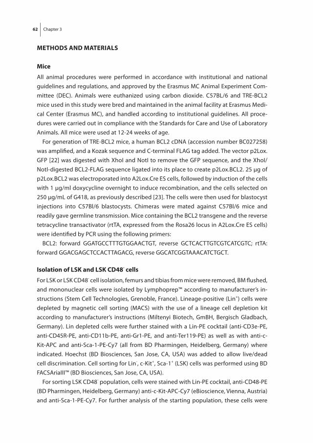
62 Chapter 3
mEtHoDS anD matErIaLS
mice
All animal procedures were performed in accordance with institutional and national guidelines and regulations, and approved by the Erasmus MC Animal Experiment Com-mittee (DEC). Animals were euthanized using carbon dioxide. C57BL/6 and TRE-BCL2 mice used in this study were bred and maintained in the animal facility at Erasmus Medi-cal Center (Erasmus MC), and handled according to institutional guidelines. All proce-dures were carried out in compliance with the Standards for Care and Use of Laboratory Animals. All mice were used at 12-24 weeks of age.
For generation of TRE-BCL2 mice, a human BCL2 cDNA (accession number BC027258) was amplified, and a Kozak sequence and C-terminal FLAG tag added. The vector p2Lox.GFP [22] was digested with XhoI and NotI to remove the GFP sequence, and the XhoI/NotI-digested BCL2-FLAG sequence ligated into its place to create p2Lox.BCL2. 25 µg of p2Lox.BCL2 was electroporated into A2Lox.Cre ES cells, followed by induction of the cells with 1 µg/ml doxycycline overnight to induce recombination, and the cells selected on 250 μg/mL of G418, as previously described [23]. The cells were then used for blastocyst injections into C57Bl/6 blastocysts. Chimeras were mated against C57Bl/6 mice and readily gave germline transmission. Mice containing the BCL2 transgene and the reverse tetracycline transactivator (rtTA, expressed from the Rosa26 locus in A2Lox.Cre ES cells) were identified by PCR using the following primers:
BCL2: forward GGATGCCTTTGTGGAACTGT, reverse GCTCACTTGTCGTCATCGTC; rtTA: forward GGACGAGCTCCACTTAGACG, reverse GGCATCGGTAAACATCTGCT.
Isolation of LSK and LSK CD48- cells
For LSK or LSK CD48- cell isolation, femurs and tibias from mice were removed, BM flushed, and mononuclear cells were isolated by Lymphoprep™ according to manufacturer’s in-structions (Stem Cell Technologies, Grenoble, France). Lineage-positive (Lin+) cells were depleted by magnetic cell sorting (MACS) with the use of a lineage cell depletion kit according to manufacturer’s instructions (Miltenyi Biotech, GmBH, Bergisch Gladbach, Germany). Lin depleted cells were further stained with a Lin-PE cocktail (anti-CD3e-PE, anti-CD45R-PE, anti-CD11b-PE, anti-Gr1-PE, and anti-Ter119-PE) as well as with anti-c-Kit-APC and anti-Sca-1-PE-Cy7 (all from BD Pharmingen, Heidelberg, Germany) where indicated. Hoechst (BD Biosciences, San Jose, CA, USA) was added to allow live/dead cell discrimination. Cell sorting for Lin-, c-Kit+, Sca-1+ (LSK) cells was performed using BD FACSAriaIII™ (BD Biosciences, San Jose, CA, USA).
For sorting LSK CD48- population, cells were stained with Lin-PE cocktail, anti-CD48-PE (BD Pharmingen, Heidelberg, Germany) anti-c-Kit-APC-Cy7 (eBioscience, Vienna, Austria) and anti-Sca-1-PE-Cy7. For further analysis of the starting population, these cells were
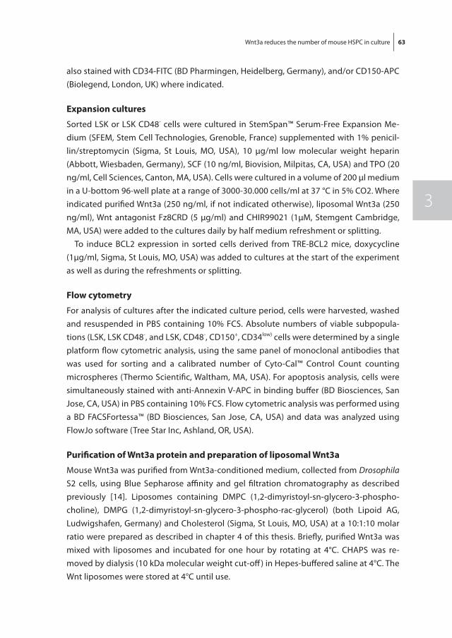
Wnt3a reduces the number of mouse HSPC in culture 63
3
also stained with CD34-FITC (BD Pharmingen, Heidelberg, Germany), and/or CD150-APC (Biolegend, London, UK) where indicated.
Expansion cultures
Sorted LSK or LSK CD48- cells were cultured in StemSpan™ Serum-Free Expansion Me-dium (SFEM, Stem Cell Technologies, Grenoble, France) supplemented with 1% penicil-lin/streptomycin (Sigma, St Louis, MO, USA), 10 μg/ml low molecular weight heparin (Abbott, Wiesbaden, Germany), SCF (10 ng/ml, Biovision, Milpitas, CA, USA) and TPO (20 ng/ml, Cell Sciences, Canton, MA, USA). Cells were cultured in a volume of 200 µl medium in a U-bottom 96-well plate at a range of 3000-30.000 cells/ml at 37 °C in 5% CO2. Where indicated purified Wnt3a (250 ng/ml, if not indicated otherwise), liposomal Wnt3a (250 ng/ml), Wnt antagonist Fz8CRD (5 µg/ml) and CHIR99021 (1µM, Stemgent Cambridge, MA, USA) were added to the cultures daily by half medium refreshment or splitting.
To induce BCL2 expression in sorted cells derived from TRE-BCL2 mice, doxycycline (1µg/ml, Sigma, St Louis, MO, USA) was added to cultures at the start of the experiment as well as during the refreshments or splitting.
flow cytometry
For analysis of cultures after the indicated culture period, cells were harvested, washed and resuspended in PBS containing 10% FCS. Absolute numbers of viable subpopula-tions (LSK, LSK CD48-, and LSK, CD48-, CD150+, CD34low) cells were determined by a single platform flow cytometric analysis, using the same panel of monoclonal antibodies that was used for sorting and a calibrated number of Cyto-Cal™ Control Count counting microspheres (Thermo Scientific, Waltham, MA, USA). For apoptosis analysis, cells were simultaneously stained with anti-Annexin V-APC in binding buffer (BD Biosciences, San Jose, CA, USA) in PBS containing 10% FCS. Flow cytometric analysis was performed using a BD FACSFortessa™ (BD Biosciences, San Jose, CA, USA) and data was analyzed using FlowJo software (Tree Star Inc, Ashland, OR, USA).
Purification of Wnt3a protein and preparation of liposomal Wnt3a
Mouse Wnt3a was purified from Wnt3a-conditioned medium, collected from Drosophila S2 cells, using Blue Sepharose affinity and gel filtration chromatography as described previously [14]. Liposomes containing DMPC (1,2-dimyristoyl-sn-glycero-3-phospho-choline), DMPG (1,2-dimyristoyl-sn-glycero-3-phospho-rac-glycerol) (both Lipoid AG, Ludwigshafen, Germany) and Cholesterol (Sigma, St Louis, MO, USA) at a 10:1:10 molar ratio were prepared as described in chapter 4 of this thesis. Briefly, purified Wnt3a was mixed with liposomes and incubated for one hour by rotating at 4°C. CHAPS was re-moved by dialysis (10 kDa molecular weight cut-off ) in Hepes-buffered saline at 4°C. The Wnt liposomes were stored at 4°C until use.
64 Chapter 3
Luciferase reporter assay
Mouse LSL cells, which express luciferase in response to TCF promoter binding [24], were routinely cultured at 37oC and 5% CO2 in culture medium containing DMEM (Invitrogen, Life Technologies, Bleiswijk, The Netherlands), 10% FCS (HyClone, Thermo Scientific, Waltham, MA, USA), 100 U/ml Penicillin and 100 µg/ml Streptomycin (both from Invitro-gen, Life Technologies, Bleiswijk, The Netherlands). Prior to the activity assays, 25,000 LSL cells were plated in each well of a 96-well plate and grown for 24 hours. Wnt3a reagents were separately incubated in culture medium with or without serum for various periods of time at 37oC in 96-well plates. After the incubation intervals, media with Wnt3a reagents were added to LSL cells. Upon an additional overnight incubation with the indicated reagents, relative luciferase units were measured with a Glomax multiplate reader.
Western blotting
Lineage-negative (Lin-) cells from TRE-BCL2 mice were isolated by FACS sorting as de-scribed above. 100,000 cells were seeded for each condition and were cultured for 24h with and without doxycycline (1µg/ml, Sigma, St Louis, MO, USA) treatment. Whole-cell extracts were then isolated from total cells. Cells were washed twice in PBS and lysed in RIPA buffer (Thermo Scientific, Waltham, MA, USA) including protease and phosphatase inhibitors. Protein concentrations were measured by BCA protein assay kit (Thermo Sci-entific, Waltham, MA, USA) and equal amounts of protein were subjected to SDS-PAGE, followed by western blot analysis using anti-Flag antibody (1:1500, Sigma, St Louis, MO, USA).
Statistical analysis
Data are presented as mean ± SD. Statistical significance was determined by a Student’s two-tail t-test (unpaired and ratio-paired), performed using GraphPad Prism software (version 6.0; GraphPad), and p<0.05 was defined as statistically significant.
rESULtS
Wnt/beta-catenin signals reduce the number of mouse LSK cells in stroma- and serum-free cultures
The effect of the combination of hematopoietic growth factors and exogenous Wnt3a on HSPC in stroma- and serum-free cultures was assessed. For this, FACS-sorted LSK cells were cultured for 10 days in serum-free medium containing stem cell factor (SCF) and Thrombopoietin (TPO). Addition of Wnt3a protein did not significantly affect the expan-sion of total nucleated cells (figure 1a). However, treatment with purified Wnt3a protein resulted in a 2-fold decrease in the number of Lin- cells relative to control condition
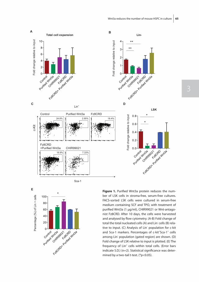
Wnt3a reduces the number of mouse HSPC in culture 65
3
Figure 2
C
AFo
ld c
hang
e re
lativ
e to
inpu
tTotal cell expansion
B
D
Fold
cha
nge
rela
tive
to in
put
Contro
l
Purifie
d Wnt3
a
CHIR99
021
Fz8CRD
Fz8CRD+ P
urifie
d Wnt3
a0.0
0.2
0.4
0.6
0.8
LSK
*
*
E
Fz8CRD2.95% 18.4%24.8%
Purified Wnt3aControl
7.33%18.9%
Fz8CRD+Purified Wnt3a CHIR99021
c-K
it
Sca-1
Lin-
Contro
l
Purifie
d Wnt3
a
CHIR99
021
Fz8CRD
Fz8CRD+ P
urifie
d Wnt3
a0
2
4
6
8
10
Fold
cha
nge
rela
tive
to in
put
**
**
Contro
l
Purifie
d Wnt3
a
CHIR99
021
Fz8CRD
Fz8CRD+ P
urifie
d Wnt3
a0
1
2
3
4
Lin-
Contro
l
Purifie
d Wnt3
a
CHIR99
021
Fz8CRD
Fz8CRD+ P
urifie
d Wnt3
a0
20
40
60
80
100
Per
cent
age
(%) o
f Lin
+ c
ells *
figure 1. Purifi ed Wnt3a protein reduces the num-ber of LSK cells in stroma-free, serum-free cultures. FACS-sorted LSK cells were cultured in serum-free medium containing SCF and TPO, with treatment of purifi ed Wnt3a (1 µg/ml), CHIR99021 or Wnt-antago-nist Fz8CRD. After 10 days, the cells were harvested and analysed by fl ow cytometry. (A-B) Fold change of total the total nucleated cells (A) and Lin- cells (B) rela-tive to input. (C) Analysis of Lin- population for c-kit and Sca-1 markers. Percentages of c-kit+Sca-1+ cells among Lin- population (gated region) are shown. (D) Fold change of LSK relative to input is plotted. (E) The frequency of Lin+ cells within total cells. (Error bars indicate S.D.) (n=2). Statistical signifi cance was deter-mined by a two-tail t-test. (*p<0.05).
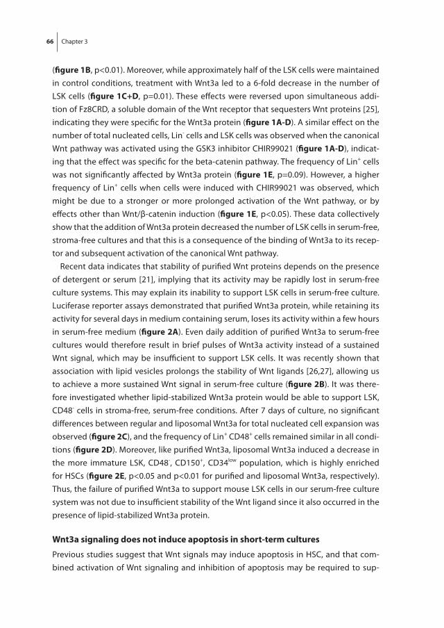
66 Chapter 3
(figure 1B, p<0.01). Moreover, while approximately half of the LSK cells were maintained in control conditions, treatment with Wnt3a led to a 6-fold decrease in the number of LSK cells (figure 1C+D, p=0.01). These effects were reversed upon simultaneous addi-tion of Fz8CRD, a soluble domain of the Wnt receptor that sequesters Wnt proteins [25], indicating they were specific for the Wnt3a protein (figure 1a-D). A similar effect on the number of total nucleated cells, Lin- cells and LSK cells was observed when the canonical Wnt pathway was activated using the GSK3 inhibitor CHIR99021 (figure 1a-D), indicat-ing that the effect was specific for the beta-catenin pathway. The frequency of Lin+ cells was not significantly affected by Wnt3a protein (figure 1E, p=0.09). However, a higher frequency of Lin+ cells when cells were induced with CHIR99021 was observed, which might be due to a stronger or more prolonged activation of the Wnt pathway, or by effects other than Wnt/β-catenin induction (figure 1E, p<0.05). These data collectively show that the addition of Wnt3a protein decreased the number of LSK cells in serum-free, stroma-free cultures and that this is a consequence of the binding of Wnt3a to its recep-tor and subsequent activation of the canonical Wnt pathway.
Recent data indicates that stability of purified Wnt proteins depends on the presence of detergent or serum [21], implying that its activity may be rapidly lost in serum-free culture systems. This may explain its inability to support LSK cells in serum-free culture. Luciferase reporter assays demonstrated that purified Wnt3a protein, while retaining its activity for several days in medium containing serum, loses its activity within a few hours in serum-free medium (figure 2a). Even daily addition of purified Wnt3a to serum-free cultures would therefore result in brief pulses of Wnt3a activity instead of a sustained Wnt signal, which may be insufficient to support LSK cells. It was recently shown that association with lipid vesicles prolongs the stability of Wnt ligands [26,27], allowing us to achieve a more sustained Wnt signal in serum-free culture (figure 2B). It was there-fore investigated whether lipid-stabilized Wnt3a protein would be able to support LSK, CD48- cells in stroma-free, serum-free conditions. After 7 days of culture, no significant differences between regular and liposomal Wnt3a for total nucleated cell expansion was observed (figure 2C), and the frequency of Lin+ CD48+ cells remained similar in all condi-tions (figure 2D). Moreover, like purified Wnt3a, liposomal Wnt3a induced a decrease in the more immature LSK, CD48-, CD150+, CD34low population, which is highly enriched for HSCs (figure 2E, p<0.05 and p<0.01 for purified and liposomal Wnt3a, respectively). Thus, the failure of purified Wnt3a to support mouse LSK cells in our serum-free culture system was not due to insufficient stability of the Wnt ligand since it also occurred in the presence of lipid-stabilized Wnt3a protein.
Wnt3a signaling does not induce apoptosis in short-term cultures
Previous studies suggest that Wnt signals may induce apoptosis in HSC, and that com-bined activation of Wnt signaling and inhibition of apoptosis may be required to sup-
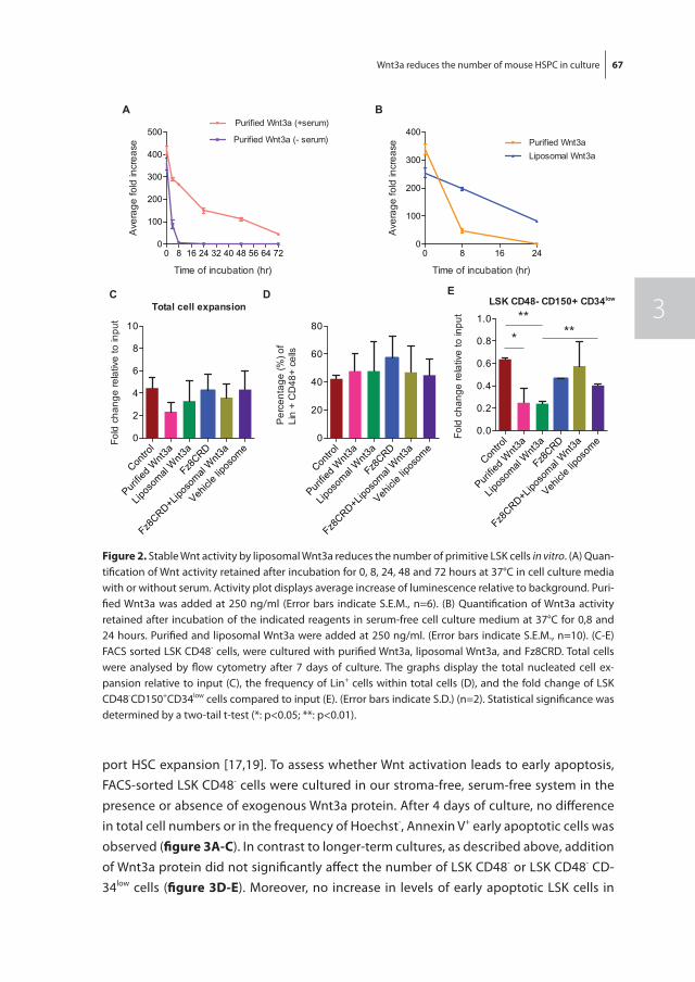
Wnt3a reduces the number of mouse HSPC in culture 67
3
port HSC expansion [17,19]. To assess whether Wnt activation leads to early apoptosis, FACS-sorted LSK CD48- cells were cultured in our stroma-free, serum-free system in the presence or absence of exogenous Wnt3a protein. After 4 days of culture, no difference in total cell numbers or in the frequency of Hoechst-, Annexin V+ early apoptotic cells was observed (figure 3a-C). In contrast to longer-term cultures, as described above, addition of Wnt3a protein did not significantly affect the number of LSK CD48- or LSK CD48- CD-34low cells (figure 3D-E). Moreover, no increase in levels of early apoptotic LSK cells in
Figure 3
D
LSK CD48- CD150+ CD34low
***
E
C
A B
0 8 16 24 32 40 48 56 64 720
100
200
300
400
500
Time of incubation (hr)
Ave
rage
fold
incr
ease
Purified Wnt3a (+serum)
Purified Wnt3a (- serum)
0 8 16 240
100
200
300
400
Time of incubation (hr)
Ave
rage
fold
incr
ease Purified Wnt3a
Liposomal Wnt3a
Contro
l
Purifie
d Wnt3
a
Lipos
omal
Wnt3
a
Fz8CRD
Fz8CRD+Lip
osom
al W
nt3a
Vehicl
e lipo
some
0
2
4
6
8
10
Fold
cha
nge
rela
tive
to in
put
Total cell expansion
Fold
cha
nge
rela
tive
to in
put
Per
cent
age
(%) o
f Li
n +
CD
48+
cells
Contro
l
Purifie
d Wnt3
a
Lipos
omal
Wnt3
a
Fz8CRD
Fz8CRD+Lip
osom
al W
nt3a
Vehicl
e lipo
some
0
20
40
60
80
Contro
l
Purifie
d Wnt3
a
Lipos
omal
Wnt3
a
Fz8CRD
Fz8CRD+Lip
osom
al W
nt3a
Vehicl
e lipo
some
0.0
0.2
0.4
0.6
0.8
1.0**
figure 2. Stable Wnt activity by liposomal Wnt3a reduces the number of primitive LSK cells in vitro. (A) Quan-tification of Wnt activity retained after incubation for 0, 8, 24, 48 and 72 hours at 37°C in cell culture media with or without serum. Activity plot displays average increase of luminescence relative to background. Puri-fied Wnt3a was added at 250 ng/ml (Error bars indicate S.E.M., n=6). (B) Quantification of Wnt3a activity retained after incubation of the indicated reagents in serum-free cell culture medium at 37°C for 0,8 and 24 hours. Purified and liposomal Wnt3a were added at 250 ng/ml. (Error bars indicate S.E.M., n=10). (C-E) FACS sorted LSK CD48- cells, were cultured with purified Wnt3a, liposomal Wnt3a, and Fz8CRD. Total cells were analysed by flow cytometry after 7 days of culture. The graphs display the total nucleated cell ex-pansion relative to input (C), the frequency of Lin+ cells within total cells (D), and the fold change of LSK CD48-CD150+CD34low cells compared to input (E). (Error bars indicate S.D.) (n=2). Statistical significance was determined by a two-tail t-test (*: p<0.05; **: p<0.01).
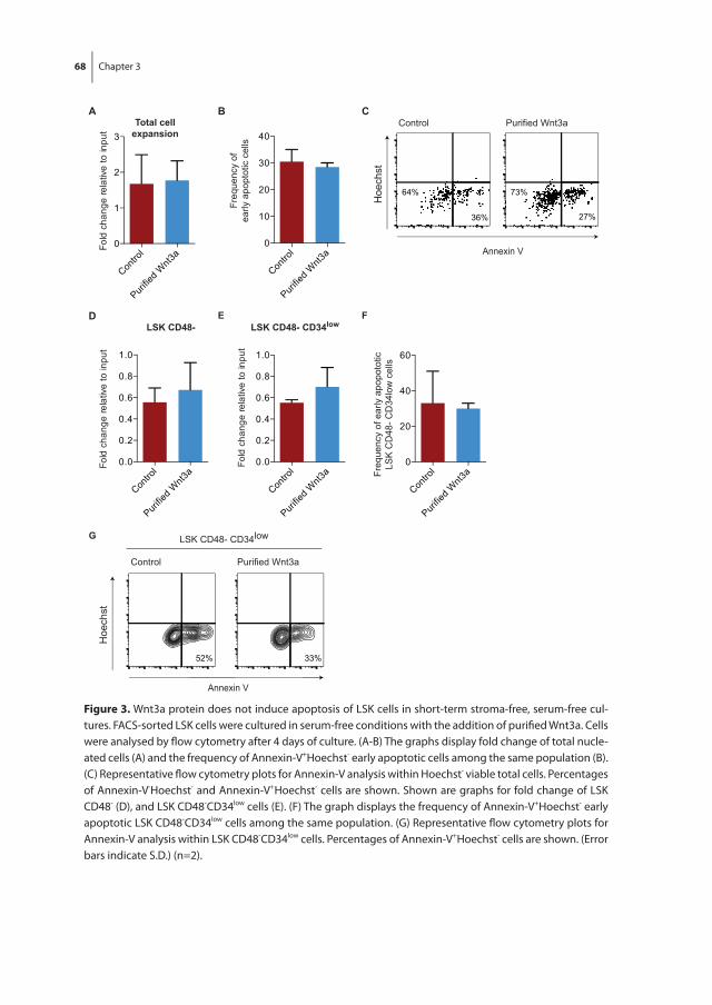
68 Chapter 3
Figure 4
A B
Contro
l
Purifie
d Wnt3
a0
1
2
3Fo
ld c
hang
e re
lativ
e to
inpu
t
Total cellexpansion
LSK CD48- CD34low
Contro
l
Purifie
d Wnt3
a0.0
0.2
0.4
0.6
0.8
1.0
Fold
cha
nge
rela
tive
to in
put
C
LSK CD48-
Contro
l
Purifie
d Wnt3
a0.0
0.2
0.4
0.6
0.8
1.0
Fold
cha
nge
rela
tive
to in
put
D
Contro
l
Purifie
d Wnt3
a0
10
20
30
40
Freq
uenc
y of
early
apo
ptot
ic c
ells
E F
64%
36%
73%
Hoe
chst
Annexin V
Purified Wnt3aControl
27%
Hoe
chst
Annexin V
33%52%
Purified Wnt3aControl
LSK CD48- CD34lowG
Contro
l
Purifie
d Wnt3
a0
20
40
60
Freq
uenc
y of
ear
ly a
popo
totic
LSK
CD
48- C
D34
low
cel
ls
Figure 4
A B
Contro
l
Purifie
d Wnt3
a0
1
2
3
Fold
cha
nge
rela
tive
to in
put
Total cellexpansion
LSK CD48- CD34low
Contro
l
Purifie
d Wnt3
a0.0
0.2
0.4
0.6
0.8
1.0
Fold
cha
nge
rela
tive
to in
put
C
LSK CD48-
Contro
l
Purifie
d Wnt3
a0.0
0.2
0.4
0.6
0.8
1.0
Fold
cha
nge
rela
tive
to in
put
D
Contro
l
Purifie
d Wnt3
a0
10
20
30
40
Freq
uenc
y of
early
apo
ptot
ic c
ells
E F
64%
36%
73%
Hoe
chst
Annexin V
Purified Wnt3aControl
27%
Hoe
chst
Annexin V
33%52%
Purified Wnt3aControl
LSK CD48- CD34lowG
Contro
l
Purifie
d Wnt3
a0
20
40
60
Freq
uenc
y of
ear
ly a
popo
totic
LSK
CD
48- C
D34
low
cel
ls
figure 3. Wnt3a protein does not induce apoptosis of LSK cells in short-term stroma-free, serum-free cul-tures. FACS-sorted LSK cells were cultured in serum-free conditions with the addition of purifi ed Wnt3a. Cells were analysed by fl ow cytometry after 4 days of culture. (A-B) The graphs display fold change of total nucle-ated cells (A) and the frequency of Annexin-V+Hoechst- early apoptotic cells among the same population (B). (C) Representative fl ow cytometry plots for Annexin-V analysis within Hoechst- viable total cells. Percentages of Annexin-V-Hoechst- and Annexin-V+Hoechst- cells are shown. Shown are graphs for fold change of LSK CD48- (D), and LSK CD48-CD34low cells (E). (F) The graph displays the frequency of Annexin-V+Hoechst- early apoptotic LSK CD48-CD34low cells among the same population. (G) Representative fl ow cytometry plots for Annexin-V analysis within LSK CD48-CD34low cells. Percentages of Annexin-V+Hoechst- cells are shown. (Error bars indicate S.D.) (n=2).
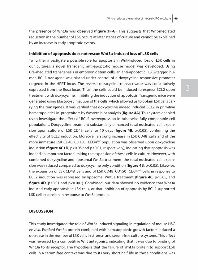
Wnt3a reduces the number of mouse HSPC in culture 69
3
the presence of Wnt3a was observed (figure 3f-G). This suggests that Wnt-mediated reduction in the number of LSK occurs at later stages of culture and cannot be explained by an increase in early apoptotic events.
Inhibition of apoptosis does not rescue Wnt3a-induced loss of LSK cells
To further investigate a possible role for apoptosis in Wnt-induced loss of LSK cells in our cultures, a novel transgenic anti-apoptotic mouse model was developed. Using Cre-mediated transgenesis in embryonic stem cells, an anti-apoptotic FLAG-tagged hu-man BCL2 transgene was placed under control of a doxycycline-responsive promoter targeted in the HPRT locus. The reverse tetracycline transactivator was constitutively expressed from the Rosa locus. Thus, the cells could be induced to express BCL2 upon treatment with doxycycline, inhibiting the induction of apoptosis. Transgenic mice were generated using blastocyst injection of the cells, which allowed us to obtain LSK cells car-rying the transgenes. It was verified that doxycycline indeed induced BCL2 in primitive hematopoietic Lin- progenitors by Western blot analysis (figure 4a). This system enabled us to investigate the effect of BCL2 overexpression in otherwise fully comparable cell populations. Doxycycline treatment substantially enhanced total nucleated cell expan-sion upon culture of LSK CD48- cells for 10 days (figure 4B, p<0.05), confirming the effectivity of BCL2 induction. Moreover, a strong increase in LSK CD48- cells and of the more immature LSK CD48- CD150+ CD34low population was observed upon doxycycline induction (figure 4C+D, p<0.05 and p<0.01, respectively), indicating that apoptosis was indeed an important factor limiting the expansion of these cells in culture. However, with combined doxycycline and liposomal Wnt3a treatment, the total nucleated cell expan-sion was reduced compared to doxycycline only condition (figure 4B, p<0.05). Likewise, the expansion of LSK CD48- cells and of LSK CD48- CD150+ CD34low cells in response to BCL2 induction was repressed by liposomal Wnt3a treatment (figure 4C, p<0.05, and figure 4D, p<0.01 and p<0.001). Combined, our data showed no evidence that Wnt3a induced early apoptosis in LSK cells, or that inhibition of apoptosis by BCL2 supported LSK cell expansion in response to Wnt3a protein.
DISCUSSIon
This study investigated the role of Wnt3a-induced signaling in regulation of mouse HSC ex vivo. Purified Wnt3a protein combined with hematopoietic growth factors induced a decrease in the number of LSK cells in stroma- and serum-free culture systems. This effect was reversed by a competitive Wnt antagonist, indicating that it was due to binding of Wnt3a to its receptor. The hypothesis that the failure of Wnt3a protein to support LSK cells in a serum-free context was due to its very short half-life in these conditions was
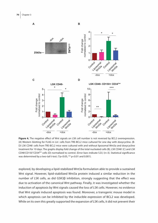
70 Chapter 3
explored, by developing a lipid-stabilized Wnt3a formulation able to provide a sustained Wnt signal. However, lipid-stabilized Wnt3a protein induced a similar reduction in the number of LSK cells, as did GSK3β inhibition, strongly suggesting that the effect was due to activation of the canonical Wnt pathway. Finally, it was investigated whether the induction of apoptosis by Wnt signals caused the loss of LSK cells. However, no evidence that Wnt signals induced apoptosis was found. Moreover, a transgenic mouse model in which apoptosis can be inhibited by the inducible expression of BCL2 was developed. While on its own this greatly supported the expansion of LSK cells, it did not prevent their
Figure 5
A B
C
-dox+dox
α-FLAG
25kDa Fold
cha
nge
norm
alize
d to
con
trol
Contro
l
Lipos
omal
Wnt3
a Contro
l
Lipos
omal
Wnt3
a
0
1
2
3
4 * *
-dox +dox
Fold
cha
nge
norm
alíz
ed to
con
trol
0
1
2
3
4
5
Contro
l
Lipos
omal
Wnt3
a Contro
l
Lipos
omal
Wnt3
a
-dox +dox
* *LSK CD48-
D
0
2
4
6
8
10
Fold
cha
nge
norm
alize
d to
con
trol
Contro
l
Lipos
omal
Wnt3
a Contro
l
Lipos
omal
Wnt3
a
-dox +dox
**
***LSK CD48- CD150+ CD34low
figure 4. The negative effect of Wnt signals on LSK cell number is not reversed by BCL2 overexpression. (A) Western blotting for FLAG in Lin- cells from TRE-BCL2 mice cultured for one day with doxycycline. (B-D) LSK CD48- cells from TRE-BCL2 mice were cultured with and without liposomal Wnt3a and doxycycline treatment for 10 days. The graphs display fold change of the total nucleated cells (B), LSK CD48- (C) and LSK CD48-CD150+CD34low cells (D) normalized to control. (Error bars indicate S.D.) (n=3). Statistical significance was determined by a two-tail t-test. (*p<0.05, ** p<0.01 and 0.001).
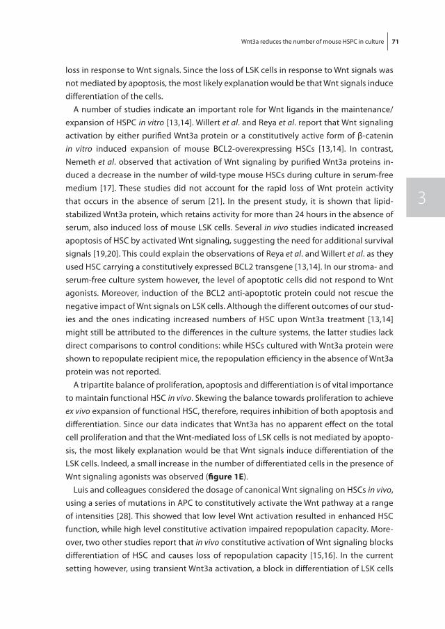
Wnt3a reduces the number of mouse HSPC in culture 71
3
loss in response to Wnt signals. Since the loss of LSK cells in response to Wnt signals was not mediated by apoptosis, the most likely explanation would be that Wnt signals induce differentiation of the cells.
A number of studies indicate an important role for Wnt ligands in the maintenance/expansion of HSPC in vitro [13,14]. Willert et al. and Reya et al. report that Wnt signaling activation by either purified Wnt3a protein or a constitutively active form of β-catenin in vitro induced expansion of mouse BCL2-overexpressing HSCs [13,14]. In contrast, Nemeth et al. observed that activation of Wnt signaling by purified Wnt3a proteins in-duced a decrease in the number of wild-type mouse HSCs during culture in serum-free medium [17]. These studies did not account for the rapid loss of Wnt protein activity that occurs in the absence of serum [21]. In the present study, it is shown that lipid-stabilized Wnt3a protein, which retains activity for more than 24 hours in the absence of serum, also induced loss of mouse LSK cells. Several in vivo studies indicated increased apoptosis of HSC by activated Wnt signaling, suggesting the need for additional survival signals [19,20]. This could explain the observations of Reya et al. and Willert et al. as they used HSC carrying a constitutively expressed BCL2 transgene [13,14]. In our stroma- and serum-free culture system however, the level of apoptotic cells did not respond to Wnt agonists. Moreover, induction of the BCL2 anti-apoptotic protein could not rescue the negative impact of Wnt signals on LSK cells. Although the different outcomes of our stud-ies and the ones indicating increased numbers of HSC upon Wnt3a treatment [13,14] might still be attributed to the differences in the culture systems, the latter studies lack direct comparisons to control conditions: while HSCs cultured with Wnt3a protein were shown to repopulate recipient mice, the repopulation efficiency in the absence of Wnt3a protein was not reported.
A tripartite balance of proliferation, apoptosis and differentiation is of vital importance to maintain functional HSC in vivo. Skewing the balance towards proliferation to achieve ex vivo expansion of functional HSC, therefore, requires inhibition of both apoptosis and differentiation. Since our data indicates that Wnt3a has no apparent effect on the total cell proliferation and that the Wnt-mediated loss of LSK cells is not mediated by apopto-sis, the most likely explanation would be that Wnt signals induce differentiation of the LSK cells. Indeed, a small increase in the number of differentiated cells in the presence of Wnt signaling agonists was observed (figure 1E).
Luis and colleagues considered the dosage of canonical Wnt signaling on HSCs in vivo, using a series of mutations in APC to constitutively activate the Wnt pathway at a range of intensities [28]. This showed that low level Wnt activation resulted in enhanced HSC function, while high level constitutive activation impaired repopulation capacity. More-over, two other studies report that in vivo constitutive activation of Wnt signaling blocks differentiation of HSC and causes loss of repopulation capacity [15,16]. In the current setting however, using transient Wnt3a activation, a block in differentiation of LSK cells
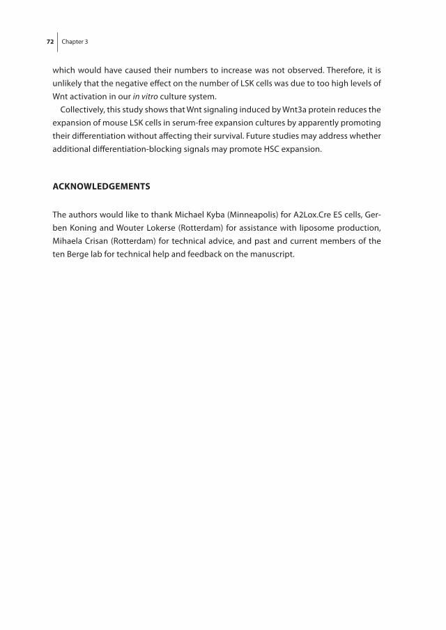
72 Chapter 3
which would have caused their numbers to increase was not observed. Therefore, it is unlikely that the negative effect on the number of LSK cells was due to too high levels of Wnt activation in our in vitro culture system.
Collectively, this study shows that Wnt signaling induced by Wnt3a protein reduces the expansion of mouse LSK cells in serum-free expansion cultures by apparently promoting their differentiation without affecting their survival. Future studies may address whether additional differentiation-blocking signals may promote HSC expansion.
aCKnoWLEDGEmEntS
The authors would like to thank Michael Kyba (Minneapolis) for A2Lox.Cre ES cells, Ger-ben Koning and Wouter Lokerse (Rotterdam) for assistance with liposome production, Mihaela Crisan (Rotterdam) for technical advice, and past and current members of the ten Berge lab for technical help and feedback on the manuscript.
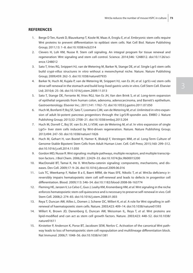
Wnt3a reduces the number of mouse HSPC in culture 73
3
rEfErEnCES
1. Berge D Ten, Kurek D, Blauwkamp T, Koole W, Maas A, Eroglu E, et al. Embryonic stem cells require Wnt proteins to prevent differentiation to epiblast stem cells. Nat Cell Biol. Nature Publishing Group; 2011;13: 1–8. doi:10.1038/ncb2314
2. Clevers H, Loh KM, Nusse R. Stem cell signaling. An integral program for tissue renewal and regeneration: Wnt signaling and stem cell control. Science. 2014;346: 1248012. doi:10.1126/sci-ence.1248012
3. Sato T, Vries RG, Snippert HJ, van de Wetering M, Barker N, Stange DE, et al. Single Lgr5 stem cells build crypt-villus structures in vitro without a mesenchymal niche. Nature. Nature Publishing Group; 2009;459: 262–5. doi:10.1038/nature07935
4. Barker N, Huch M, Kujala P, van de Wetering M, Snippert HJ, van Es JH, et al. Lgr5(+ve) stem cells drive self-renewal in the stomach and build long-lived gastric units in vitro. Cell Stem Cell. Elsevier Ltd; 2010;6: 25–36. doi:10.1016/j.stem.2009.11.013
5. Sato T, Stange DE, Ferrante M, Vries RGJ, Van Es JH, Van den Brink S, et al. Long-term expansion of epithelial organoids from human colon, adenoma, adenocarcinoma, and Barrett’s epithelium. Gastroenterology. Elsevier Inc.; 2011;141: 1762–72. doi:10.1053/j.gastro.2011.07.050
6. Huch M, Bonfanti P, Boj SF, Sato T, Loomans CJM, van de Wetering M, et al. Unlimited in vitro expan-sion of adult bi-potent pancreas progenitors through the Lgr5/R-spondin axis. EMBO J. Nature Publishing Group; 2013;32: 2708–21. doi:10.1038/emboj.2013.204
7. Huch M, Dorrell C, Boj SF, van Es JH, Li VSW, van de Wetering M, et al. In vitro expansion of single Lgr5+ liver stem cells induced by Wnt-driven regeneration. Nature. Nature Publishing Group; 2013;494: 247–50. doi:10.1038/nature11826
8. Huch M, Gehart H, van Boxtel R, Hamer K, Blokzijl F, Verstegen MM, et al. Long-Term Culture of Genome-Stable Bipotent Stem Cells from Adult Human Liver. Cell. Cell Press; 2015;160: 299–312. doi:10.1016/j.cell.2014.11.050
9. Gordon MD, Nusse R. Wnt signaling: multiple pathways, multiple receptors, and multiple transcrip-tion factors. J Biol Chem. 2006;281: 22429–33. doi:10.1074/jbc.R600015200
10. MacDonald BT, Tamai K, He X. Wnt/beta-catenin signaling: components, mechanisms, and dis-eases. Dev Cell. 2009;17: 9–26. doi:10.1016/j.devcel.2009.06.016
11. Luis TC, Weerkamp F, Naber B a E, Baert MRM, de Haas EFE, Nikolic T, et al. Wnt3a deficiency ir-reversibly impairs hematopoietic stem cell self-renewal and leads to defects in progenitor cell differentiation. Blood. 2009;113: 546–54. doi:10.1182/blood-2008-06-163774
12. Fleming HE, Janzen V, Lo Celso C, Guo J, Leahy KM, Kronenberg HM, et al. Wnt signaling in the niche enforces hematopoietic stem cell quiescence and is necessary to preserve self-renewal in vivo. Cell Stem Cell. 2008;2: 274–83. doi:10.1016/j.stem.2008.01.003
13. Reya T, Duncan AW, Ailles L, Domen J, Scherer DC, Willert K, et al. A role for Wnt signalling in self-renewal of haematopoietic stem cells. Nature. 2003;423: 409–14. doi:10.1038/nature01593
14. Willert K, Brown JD, Danenberg E, Duncan AW, Weissman IL, Reya T, et al. Wnt proteins are lipid-modified and can act as stem cell growth factors. Nature. 2003;423: 448–52. doi:10.1038/nature01611
15. Kirstetter P, Anderson K, Porse BT, Jacobsen SEW, Nerlov C. Activation of the canonical Wnt path-way leads to loss of hematopoietic stem cell repopulation and multilineage differentiation block. Nat Immunol. 2006;7: 1048–56. doi:10.1038/ni1381
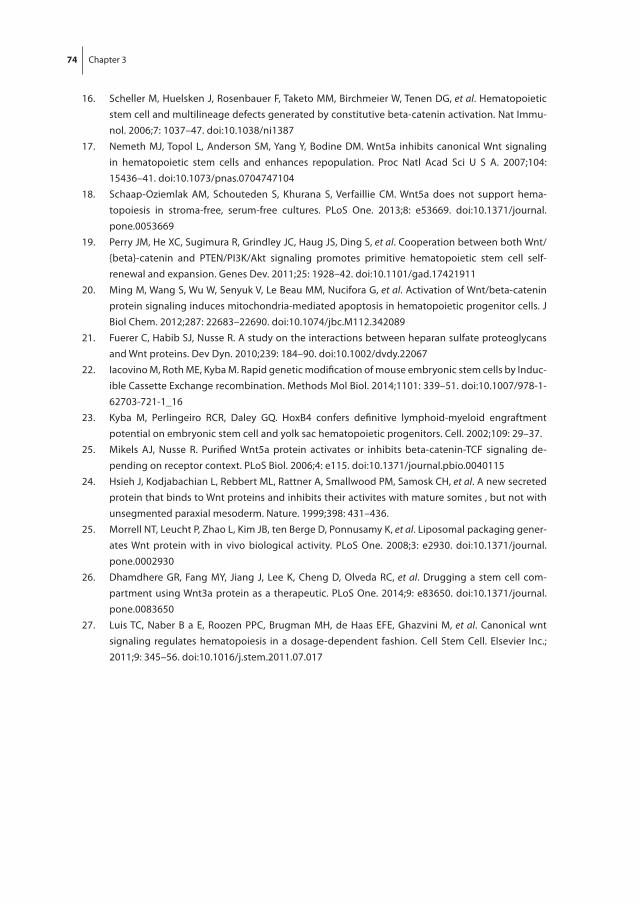
74 Chapter 3
16. Scheller M, Huelsken J, Rosenbauer F, Taketo MM, Birchmeier W, Tenen DG, et al. Hematopoietic stem cell and multilineage defects generated by constitutive beta-catenin activation. Nat Immu-nol. 2006;7: 1037–47. doi:10.1038/ni1387
17. Nemeth MJ, Topol L, Anderson SM, Yang Y, Bodine DM. Wnt5a inhibits canonical Wnt signaling in hematopoietic stem cells and enhances repopulation. Proc Natl Acad Sci U S A. 2007;104: 15436–41. doi:10.1073/pnas.0704747104
18. Schaap-Oziemlak AM, Schouteden S, Khurana S, Verfaillie CM. Wnt5a does not support hema-topoiesis in stroma-free, serum-free cultures. PLoS One. 2013;8: e53669. doi:10.1371/journal.pone.0053669
19. Perry JM, He XC, Sugimura R, Grindley JC, Haug JS, Ding S, et al. Cooperation between both Wnt/{beta}-catenin and PTEN/PI3K/Akt signaling promotes primitive hematopoietic stem cell self-renewal and expansion. Genes Dev. 2011;25: 1928–42. doi:10.1101/gad.17421911
20. Ming M, Wang S, Wu W, Senyuk V, Le Beau MM, Nucifora G, et al. Activation of Wnt/beta-catenin protein signaling induces mitochondria-mediated apoptosis in hematopoietic progenitor cells. J Biol Chem. 2012;287: 22683–22690. doi:10.1074/jbc.M112.342089
21. Fuerer C, Habib SJ, Nusse R. A study on the interactions between heparan sulfate proteoglycans and Wnt proteins. Dev Dyn. 2010;239: 184–90. doi:10.1002/dvdy.22067
22. Iacovino M, Roth ME, Kyba M. Rapid genetic modification of mouse embryonic stem cells by Induc-ible Cassette Exchange recombination. Methods Mol Biol. 2014;1101: 339–51. doi:10.1007/978-1-62703-721-1_16
23. Kyba M, Perlingeiro RCR, Daley GQ. HoxB4 confers definitive lymphoid-myeloid engraftment potential on embryonic stem cell and yolk sac hematopoietic progenitors. Cell. 2002;109: 29–37.
25. Mikels AJ, Nusse R. Purified Wnt5a protein activates or inhibits beta-catenin-TCF signaling de-pending on receptor context. PLoS Biol. 2006;4: e115. doi:10.1371/journal.pbio.0040115
24. Hsieh J, Kodjabachian L, Rebbert ML, Rattner A, Smallwood PM, Samosk CH, et al. A new secreted protein that binds to Wnt proteins and inhibits their activites with mature somites , but not with unsegmented paraxial mesoderm. Nature. 1999;398: 431–436.
25. Morrell NT, Leucht P, Zhao L, Kim JB, ten Berge D, Ponnusamy K, et al. Liposomal packaging gener-ates Wnt protein with in vivo biological activity. PLoS One. 2008;3: e2930. doi:10.1371/journal.pone.0002930
26. Dhamdhere GR, Fang MY, Jiang J, Lee K, Cheng D, Olveda RC, et al. Drugging a stem cell com-partment using Wnt3a protein as a therapeutic. PLoS One. 2014;9: e83650. doi:10.1371/journal.pone.0083650
27. Luis TC, Naber B a E, Roozen PPC, Brugman MH, de Haas EFE, Ghazvini M, et al. Canonical wnt signaling regulates hematopoiesis in a dosage-dependent fashion. Cell Stem Cell. Elsevier Inc.; 2011;9: 345–56. doi:10.1016/j.stem.2011.07.017


A
Chapter 4Wnt3a protein reduces growth factor-driven expansion of human hematopoietic stem and progenitor cells in serum-free cultures
Lucia E. Duinhouwer1, Nesrin Tüysüz2, Elwin W.J.C. Rombouts1, Mariette N.D. ter Borg1, Enrico Mastrobattista3, Jan Spanholtz4, Jan J. Cornelissen1, Derk ten Berge2, Eric Braakman1.
1Department of Hematology, Erasmus University Medical Centre, Rotterdam, The
Netherlands,2Erasmus MC Stem Cell Institute, Erasmus University Medical Centre, Rotterdam, The
Netherlands,3Institute for Pharmaceutical Sciences, Utrecht University, Utrecht, The Netherlands,4Glycostem, Oss, The Netherlands
PLoS ONE 2015 Mar 25;10(3):e0119086
78 Chapter 4
aBStraCt
Ex vivo expansion of hematopoietic stem and progenitor cells (HSPC) is a promising approach to improve insufficient engraftment after umbilical cord blood stem cell transplantation. Although culturing HSPC with hematopoietic cytokines results in robust proliferation, it is accompanied with extensive differentiation and loss of self-renewal capacity. Wingless-related integration site (Wnt) signaling has been implicated in regu-lating HSPC fate decisions in vivo and in promoting HSPC self-renewal by inhibition of differentiation, but the effects of Wnt on the ex vivo expansion of HSPC are controversial. Here, we demonstrate that exogenous Wnt3a protein suppresses rather than promotes the expansion of UCB-derived CD34+ cells in serum free expansion cultures. The reduced expansion was also observed in cultures initiated with Lin-CD34+CD38lowCD45RA-CD90+ cells which are highly enriched in HSC and was also observed in response to activation of beta-catenin signaling by GSK3beta inhibition. The presence of Wnt3a protein during the culture reduced the frequency of multilineage CFU-GEMM and the long-term re-population ability of the expanded HSPC. These data suggest that Wnt signaling reduces expansion of human HSPC in growth factor-driven expansion cultures by promoting differentiation of HSPC.
Wnt3a reduces growth-factor driven expansion of human HSPC 79
4
IntroDUCtIon
Allogeneic hematopoietic stem cell transplantation (allo-SCT) is an important part of treatment for patients suffering from hematological disorders, including leukemia, my-elodysplastic syndromes, and aplastic anemia. However, many patients lack a suitable sibling or human leucocyte antigen (HLA) matched unrelated donor (MUD). Because of its rapid availability and less stringent matching criteria[1], umbilical cord blood (UCB) is an important alternative source for hematopoietic stem and progenitor cells (HSPC). However, UCB-derived HSPC significantly differ from bone marrow- and peripheral blood-derived HSPC quantitatively and qualitatively. UCB grafts contain a relatively low number of HSPC which are relatively more primitive, resulting in impaired engraftment and a delayed hematopoietic recovery [1-5], during which patients are at increased risk for severe complications, including infections and bleeding. Several approaches have been pursued to improve engraftment after UCB transplantation, including the ex vivo expansion of HSPC.
HSC are defined by their self-renewal capacity and the ability to generate all different hematopoietic lineages. Although in vivo studies demonstrated that HSPC expand after transplantation [6], robust ex vivo expansion of long-term repopulating HSC remains a challenge. Culturing HSPC with different combinations of hematopoietic cytokines such as stem cell factor (SCF), Fms-related tyrosine kinase 3 ligand (Flt3L), Thrombopoietin (TPO) and granulocyte-macrophage colony-stimulating factor (GM –CSF) resulted in mas-sive expansion of committed HPC which is accompanied by a loss or at best maintenance of primitive HSC with long-term repopulation ability [7-11]. Additional signals are needed to support the expansion of primitive HSC in ex vivo culture systems. Several novel factors, such as the immobilized Notch-ligand Delta1, copper chelator tetra-ethylenepentamine (TEPA) and signals derived from mesenchymal stromal cells, were identified that may affect self-renewal of HSC and inhibit differentiation, thereby having the potential to improve ex vivo expansion protocols [12-14]. In addition, numerous promising factors have been tested in a pre-clinical setting, including developmental regulators such as fibroblast growth factor signaling, insulin-like growth factor, Angiopoietin-like proteins and Pleiotrophin and chemical modulators like all-trans retinoic acid, stemregenin1 and prostaglandin E2 (reviewed by Walasek et al. [15]).
The Wingless-related integration site (Wnt)/beta-catenin signaling pathway regulates cell fate decisions in many developmental processes in embryo and adult. Stimulation of cells with Wnt signaling proteins induces the stabilization and accumulation of the signal transducer protein beta-catenin, which then localizes into the nucleus where it regulates target gene expression (reviewed by Clevers et al. [16]). When combined with other growth factors, Wnt proteins can promote self-renewal in several types of stem cells, such as mammary, intestinal and embryonic stem cells [17-20]. Several studies, using different
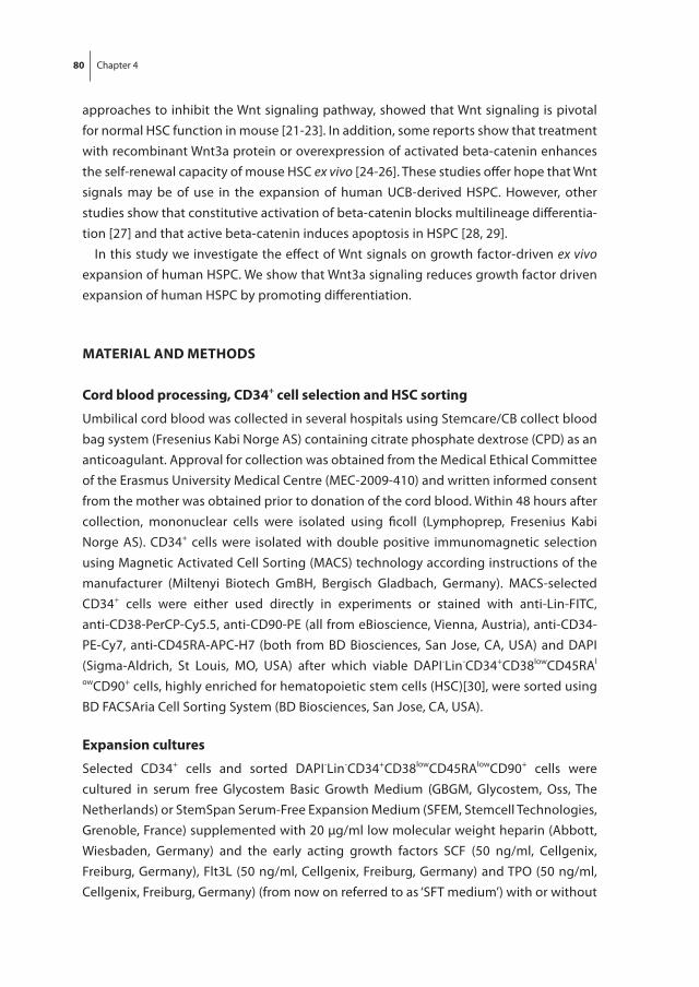
80 Chapter 4
approaches to inhibit the Wnt signaling pathway, showed that Wnt signaling is pivotal for normal HSC function in mouse [21-23]. In addition, some reports show that treatment with recombinant Wnt3a protein or overexpression of activated beta-catenin enhances the self-renewal capacity of mouse HSC ex vivo [24-26]. These studies offer hope that Wnt signals may be of use in the expansion of human UCB-derived HSPC. However, other studies show that constitutive activation of beta-catenin blocks multilineage differentia-tion [27] and that active beta-catenin induces apoptosis in HSPC [28, 29].
In this study we investigate the effect of Wnt signals on growth factor-driven ex vivo expansion of human HSPC. We show that Wnt3a signaling reduces growth factor driven expansion of human HSPC by promoting differentiation.
matErIaL anD mEtHoDS
Cord blood processing, CD34+ cell selection and HSC sorting
Umbilical cord blood was collected in several hospitals using Stemcare/CB collect blood bag system (Fresenius Kabi Norge AS) containing citrate phosphate dextrose (CPD) as an anticoagulant. Approval for collection was obtained from the Medical Ethical Committee of the Erasmus University Medical Centre (MEC-2009-410) and written informed consent from the mother was obtained prior to donation of the cord blood. Within 48 hours after collection, mononuclear cells were isolated using ficoll (Lymphoprep, Fresenius Kabi Norge AS). CD34+ cells were isolated with double positive immunomagnetic selection using Magnetic Activated Cell Sorting (MACS) technology according instructions of the manufacturer (Miltenyi Biotech GmBH, Bergisch Gladbach, Germany). MACS-selected CD34+ cells were either used directly in experiments or stained with anti-Lin-FITC, anti-CD38-PerCP-Cy5.5, anti-CD90-PE (all from eBioscience, Vienna, Austria), anti-CD34-PE-Cy7, anti-CD45RA-APC-H7 (both from BD Biosciences, San Jose, CA, USA) and DAPI (Sigma-Aldrich, St Louis, MO, USA) after which viable DAPI-Lin-CD34+CD38lowCD45RAl
owCD90+ cells, highly enriched for hematopoietic stem cells (HSC)[30], were sorted using BD FACSAria Cell Sorting System (BD Biosciences, San Jose, CA, USA).
Expansion cultures
Selected CD34+ cells and sorted DAPI-Lin-CD34+CD38lowCD45RAlowCD90+ cells were cultured in serum free Glycostem Basic Growth Medium (GBGM, Glycostem, Oss, The Netherlands) or StemSpan Serum-Free Expansion Medium (SFEM, Stemcell Technologies, Grenoble, France) supplemented with 20 μg/ml low molecular weight heparin (Abbott, Wiesbaden, Germany) and the early acting growth factors SCF (50 ng/ml, Cellgenix, Freiburg, Germany), Flt3L (50 ng/ml, Cellgenix, Freiburg, Germany) and TPO (50 ng/ml, Cellgenix, Freiburg, Germany) (from now on referred to as ‘SFT medium’) with or without
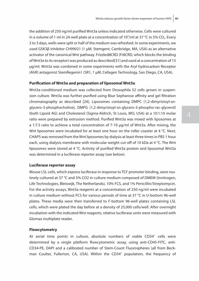
Wnt3a reduces growth-factor driven expansion of human HSPC 81
4
the addition of 250 ng/ml purified Wnt3a unless indicated otherwise. Cells were cultured in a volume of 1 ml in 24-well plate at a concentration of 105/ml at 37 °C in 5% CO2. Every 2 to 3 days, wells were split or half of the medium was refreshed. In some experiments, we used GSK3β inhibitor CH99021 (1 µM, Stemgent, Cambridge, MA, USA) as an alternative activator of the canonical Wnt pathway. Frizzled8CRD (Fr8CRD, which blocks the binding of Wnt3a to its receptor) was produced as described[31] and used at a concentration of 15 µg/ml. Wnt3a was combined in some experiments with the Aryl hydrocarbon Receptor (AhR) antagonist StemRegenin1 (SR1, 1 µM, Cellagen Technology, San Diego, CA, USA).
Purification of Wnt3a and preparation of liposomal Wnt3a
Wnt3a-conditioned medium was collected from Drosophila S2 cells grown in suspen-sion culture. Wnt3a was further purified using Blue Sepharose affinity and gel filtration chromatography as described [24]. Liposomes containing DMPC (1,2-dimyristoyl-sn-glycero-3-phosphocholine), DMPG (1,2-dimyristoyl-sn-glycero-3-phospho-rac-glycerol) (both Lipoid AG) and Cholesterol (Sigma-Aldrich, St Louis, MO, USA) at a 10:1:10 molar ratio were prepared by extrusion method. Purified Wnt3a was mixed with liposomes at a 1:7.5 ratio to achieve a total concentration of 7-10 µg/ml of Wnt3a. After mixing, the Wnt liposomes were incubated for at least one hour on the roller coaster at 4 °C. Next, CHAPS was removed from the Wnt liposomes by dialysis at least three times in PBS 1 hour each, using dialysis membrane with molecular weight cut-off of 10 kDa at 4 °C. The Wnt liposomes were stored at 4 °C. Activity of purified Wnt3a protein and liposomal Wnt3a was determined in a luciferase reporter assay (see below).
Luciferase reporter assay
Mouse LSL cells, which express luciferase in response to TCF promoter binding, were rou-tinely cultured at 37 °C and 5% CO2 in culture medium composed of DMEM (Invitrogen, Life Technologies, Bleiswijk, The Netherlands), 10% FCS, and 1% Penicillin/Streptomycin. For the activity assays, Wnt3a reagents at a concentration of 250 ng/ml were incubated in culture medium without FCS for various periods of time at 37 oC in U-bottom 96-well plates. These media were then transferred to F-bottom 96-well plates containing LSL cells, which were plated the day before at a density of 25,000 cells/well. After overnight incubation with the indicated Wnt reagents, relative luciferase units were measured with Glomax multiplate reader.
flowcytometry
At serial time points in culture, absolute numbers of viable CD34+ cells were determined by a single platform flowcytometric assay, using anti-CD45-FITC, anti-CD34-PE, DAPI and a calibrated number of Stem-Count Fluorospheres (all from Beck-man Coulter, Fullerton, CA, USA). Within the CD34+ population, the frequency of
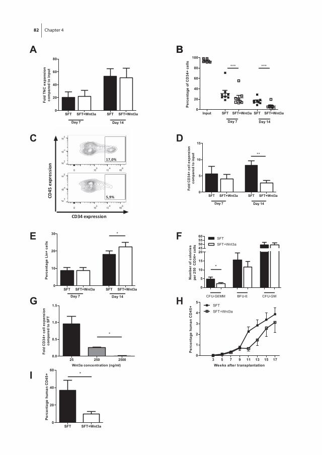
82 Chapter 4
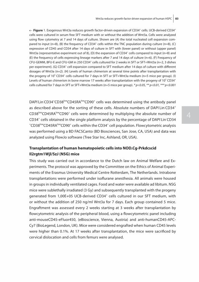
Wnt3a reduces growth-factor driven expansion of human HSPC 83
4
DAPI-Lin-CD34+CD38lowCD45RAlowCD90+ cells was determined using the antibody panel as described above for the sorting of these cells. Absolute numbers of DAPI-Lin-CD34+
CD38lowCD45RAlowCD90+ cells were determined by multiplying the absolute number of CD34+ cells obtained in the single platform analysis by the percentage of DAPI-Lin-CD34+CD38lowCD45RAlowCD90+ cells within the CD34+ cell population. Flowcytometric analysis was performed using a BD FACSCanto (BD Biosciences, San Jose, CA, USA) and data was analyzed using FlowJo software (Tree Star Inc, Ashland, OR, USA).
transplantation of human hematopoietic cells into noD.Cg-Prkdcscid Il2rgtm1Wjl/SzJ (nSG) mice
This study was carried out in accordance to the Dutch law on Animal Welfare and Ex-periments. The protocol was approved by the Committee on the Ethics of Animal Experi-ments of the Erasmus University Medical Centre Rotterdam, The Netherlands. Intrabone transplantations were performed under isoflurane anesthesia. All animals were housed in groups in individually ventilated cages. Food and water were available ad libitum. NSG mice were sublethally irradiated (3 Gy) and subsequently transplanted with the progeny generated from 1,00E+05 UCB-derived CD34+ cells cultured in our SFT medium, with or without the addition of 250 ng/ml Wnt3a for 7 days. Each group contained 5 mice. Engraftment was assessed every 2 weeks starting at 3 weeks after transplantation by flowcytometric analysis of the peripheral blood, using a flowcytometric panel including anti-mouseCD45-eFluor450, (eBioscience, Vienna, Austria) and anti-humanCD45-APC-Cy7 (BioLegend, London, UK). Mice were considered engrafted when human CD45 levels were higher than 0.1%. At 17 weeks after transplantation, the mice were sacrificed by cervical dislocation and cells from femurs were analysed.
← figure 1. Exogenous Wnt3a reduces growth factor-driven expansion of CD34+ cells. UCB-derived CD34+ cells were cultured in serum free SFT medium with or without the addition of Wnt3a. Cells were analyzed using flow cytometry at 7 and 14 days of culture. Shown are (A) the total nucleated cell expansion com-pared to input (n=8), (B) the frequency of CD34+ cells within the TNC population during culture (n=8), (C) expression of CD45 and CD34 after 14 days of culture in SFT with (lower panel) or without (upper panel) Wnt3a (representative experiment out of 8), (D) the expansion of CD34+ cells compared to input (n=8) and (E) the frequency of cells expressing lineage markers after 7 and 14 days of culture (n=6). (F) Frequency of CFU-GEMM, BFU-E and CFU-GM in 250 CD34+ cells cultured for 2 weeks in SFT or SFT+Wnt3a (n=2, 3 dishes per experiment). (G) CD34+ cell expansion compared to SFT medium after 14 days of culture with different dosages of Wnt3a (n=2). (H) Levels of human chimerism at several time points after transplantation with the progeny of 105 CD34+ cells cultured for 7 days in SFT or SFT+Wnt3a medium (n=5 mice per group). (I) Levels of human chimerism in bone marrow 17 weeks after transplantation with the progeny of 105 CD34+ cells cultured for 7 days in SFT or SFT+Wnt3a medium (n=5 mice per group). * p<0.05, ** p<0.01, *** p<0.001
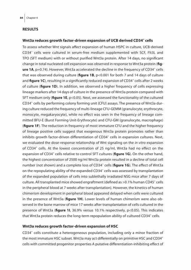
84 Chapter 4
rESULtS
Wnt3a reduces growth factor-driven expansion of UCB derived CD34+ cells
To assess whether Wnt signals affect expansion of human HSPC in culture, UCB-derived CD34+ cells were cultured in serum-free medium supplemented with SCF, Flt3L and TPO (SFT medium) with or without purified Wnt3a protein. After 14 days, no significant change in total nucleated cell expansion was observed in response to Wnt3a protein (fig-ure 1a, p=0.74). However, Wnt3a accelerated the decline in the frequency of CD34+ cells that was observed during culture (figure 1B, p<0.001 for both 7 and 14 days of culture and figure 1C), resulting in a significantly reduced expansion of CD34+ cells after 2 weeks of culture (figure 1D). In addition, we observed a higher frequency of cells expressing lineage markers after 14 days of culture in the presence of Wnt3a protein compared with SFT medium only (figure 1E, p<0.05). Next, we assessed the functionality of the cultured CD34+ cells by performing colony forming unit (CFU) assays. The presence of Wnt3a dur-ing culture reduced the frequency of multi-lineage CFU-GEMM (granulocyte, erythrocyte, monocyte, megakaryocyte), while no effect was seen in the frequency of lineage com-mitted BFU-E (Burst Forming Unit-Erythrocyte) and CFU-GM (granulocyte, macrophage) (figure 1f). The reduction in frequency of most immature CFU and the higher frequency of lineage positive cells suggest that exogenous Wnt3a protein promotes rather than inhibits growth factor-driven differentiation of CD34+ cells in expansion cultures. Next, we evaluated the dose-response relationship of Wnt signaling on the in vitro expansion of CD34+ cells. At the lowest concentration of 25 ng/ml, Wnt3a had no effect on the expansion of CD34+ cells relative to control SFT cultures (figure 1G). On the other hand, the highest concentration of 2500 ng/ml Wnt3a protein resulted in a decline of total cell number (not shown) and a complete loss of CD34+ cells (figure 1G). The effect of Wnt3a on the repopulating ability of the expanded CD34+ cells was assessed by transplantation of the expanded population of cells into sublethally irradiated NSG mice after 7 days of culture. All transplanted mice showed engraftment (defined as >0.1% human CD45+ cells in the peripheral blood at 7 weeks after transplantation). However, the kinetics of human chimerism development in peripheral blood appeared delayed when cells were cultured in the presence of Wnt3a (figure 1H). Lower levels of human chimerism were also ob-served in the bone marrow of mice 17 weeks after transplantation of cells cultured in the presence of Wnt3a (figure 1I, 36.9% versus 10.1% respectively, p<0.05). This indicates that Wnt3a protein reduces the long-term repopulation ability of cultured CD34+ cells.
Wnt3a reduces growth factor-driven expansion of HSC
CD34+ cells constitute a heterogeneous population, including only a minor fraction of the most immature HSC subset. Wnt3a may act differentially on primitive HSC and CD34+ cells with committed progenitor properties A putative differentiation-inhibiting effect of
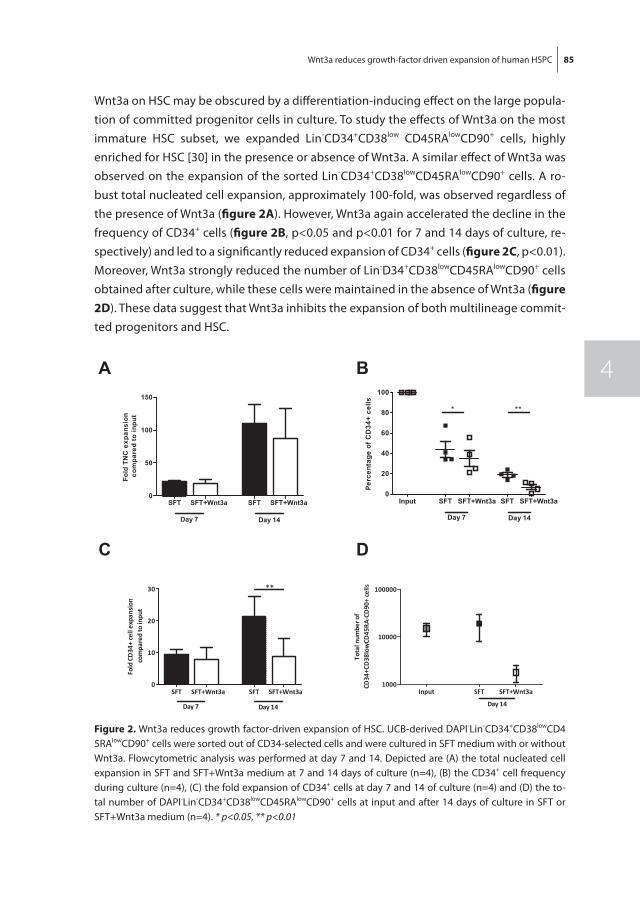
Wnt3a reduces growth-factor driven expansion of human HSPC 85
4
Wnt3a on HSC may be obscured by a differentiation-inducing effect on the large popula-tion of committed progenitor cells in culture. To study the effects of Wnt3a on the most immature HSC subset, we expanded Lin-CD34+CD38low CD45RAlowCD90+ cells, highly enriched for HSC [30] in the presence or absence of Wnt3a. A similar effect of Wnt3a was observed on the expansion of the sorted Lin-CD34+CD38lowCD45RAlowCD90+ cells. A ro-bust total nucleated cell expansion, approximately 100-fold, was observed regardless of the presence of Wnt3a (figure 2a). However, Wnt3a again accelerated the decline in the frequency of CD34+ cells (figure 2B, p<0.05 and p<0.01 for 7 and 14 days of culture, re-spectively) and led to a significantly reduced expansion of CD34+ cells (figure 2C, p<0.01). Moreover, Wnt3a strongly reduced the number of Lin-D34+CD38lowCD45RAlowCD90+ cells obtained after culture, while these cells were maintained in the absence of Wnt3a (figure 2D). These data suggest that Wnt3a inhibits the expansion of both multilineage commit-ted progenitors and HSC.
figure 2. Wnt3a reduces growth factor-driven expansion of HSC. UCB-derived DAPI-Lin-CD34+CD38lowCD45RAlowCD90+ cells were sorted out of CD34-selected cells and were cultured in SFT medium with or without Wnt3a. Flowcytometric analysis was performed at day 7 and 14. Depicted are (A) the total nucleated cell expansion in SFT and SFT+Wnt3a medium at 7 and 14 days of culture (n=4), (B) the CD34+ cell frequency during culture (n=4), (C) the fold expansion of CD34+ cells at day 7 and 14 of culture (n=4) and (D) the to-tal number of DAPI-Lin-CD34+CD38lowCD45RAlowCD90+ cells at input and after 14 days of culture in SFT or SFT+Wnt3a medium (n=4). * p<0.05, ** p<0.01
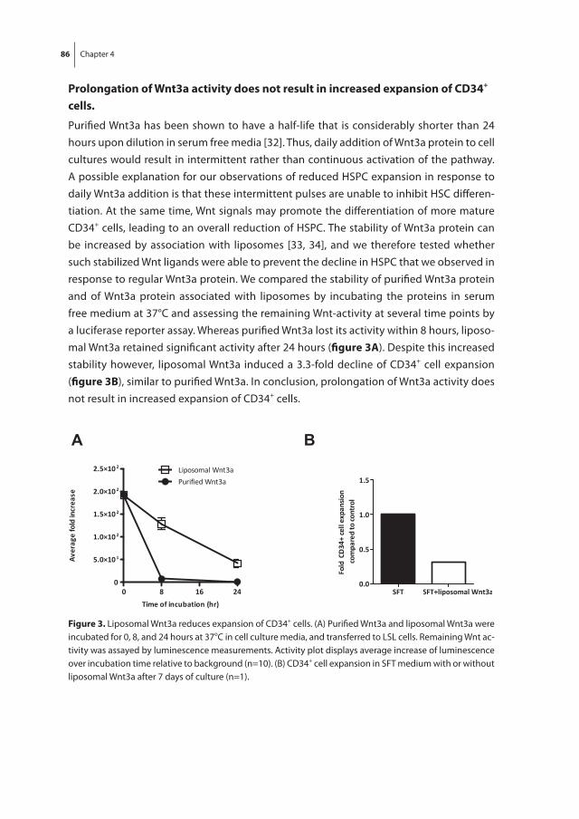
86 Chapter 4
Prolongation of Wnt3a activity does not result in increased expansion of CD34+ cells.
Purified Wnt3a has been shown to have a half-life that is considerably shorter than 24 hours upon dilution in serum free media [32]. Thus, daily addition of Wnt3a protein to cell cultures would result in intermittent rather than continuous activation of the pathway. A possible explanation for our observations of reduced HSPC expansion in response to daily Wnt3a addition is that these intermittent pulses are unable to inhibit HSC differen-tiation. At the same time, Wnt signals may promote the differentiation of more mature CD34+ cells, leading to an overall reduction of HSPC. The stability of Wnt3a protein can be increased by association with liposomes [33, 34], and we therefore tested whether such stabilized Wnt ligands were able to prevent the decline in HSPC that we observed in response to regular Wnt3a protein. We compared the stability of purified Wnt3a protein and of Wnt3a protein associated with liposomes by incubating the proteins in serum free medium at 37°C and assessing the remaining Wnt-activity at several time points by a luciferase reporter assay. Whereas purified Wnt3a lost its activity within 8 hours, liposo-mal Wnt3a retained significant activity after 24 hours (figure 3a). Despite this increased stability however, liposomal Wnt3a induced a 3.3-fold decline of CD34+ cell expansion (figure 3B), similar to purified Wnt3a. In conclusion, prolongation of Wnt3a activity does not result in increased expansion of CD34+ cells.
figure 3. Liposomal Wnt3a reduces expansion of CD34+ cells. (A) Purified Wnt3a and liposomal Wnt3a were incubated for 0, 8, and 24 hours at 37oC in cell culture media, and transferred to LSL cells. Remaining Wnt ac-tivity was assayed by luminescence measurements. Activity plot displays average increase of luminescence over incubation time relative to background (n=10). (B) CD34+ cell expansion in SFT medium with or without liposomal Wnt3a after 7 days of culture (n=1).
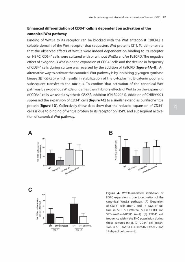
Wnt3a reduces growth-factor driven expansion of human HSPC 87
4
Enhanced differentiation of CD34+ cells is dependent on activation of the canonical Wnt pathway
Binding of Wnt3a to its receptor can be blocked with the Wnt antagonist Fz8CRD, a soluble domain of the Wnt receptor that sequesters Wnt proteins [31]. To demonstrate that the observed effects of Wnt3a were indeed dependent on binding to its receptor on HSPC, CD34+ cells were cultured with or without Wnt3a and/or Fz8CRD. The negative effect of exogenous Wnt3a on the expansion of CD34+ cells and the decline in frequency of CD34+ cells during culture was reversed by the addition of Fz8CRD (figure 4a+B). An alternative way to activate the canonical Wnt pathway is by inhibiting glycogen synthase kinase 3β (GSK3β) which results in stabilization of the cytoplasmic β-catenin pool and subsequent transfer to the nucleus. To confirm that activation of the canonical Wnt pathway by exogenous Wnt3a underlies the inhibitory effects of Wnt3a on the expansion of CD34+ cells we used a synthetic GSK3β-inhibitor (CHIR99021). Addition of CHIR99021 supressed the expansion of CD34+ cells (figure 4C) to a similar extend as purified Wnt3a protein (figure 1D). Collectively these data show that the reduced expansion of CD34+ cells is due to binding of Wnt3a protein to its receptor on HSPC and subsequent activa-tion of canonical Wnt pathway.
figure 4. Wnt3a-mediated inhibition of HSPC expansion is due to activation of the canonical Wnt3a pathway. (A) Expansion of CD34+ cells after 7 and 14 days of cul-ture in SFT, SFT+Wnt3a, SFT+Fr8CRD and SFT+Wnt3a+Fr8CRD (n=2). (B) CD34+ cell frequency within the TNC population during these cultures (n=2). (C) CD34+ cell expan-sion in SFT and SFT+CHIR99021 after 7 and 14 days of culture (n=2).
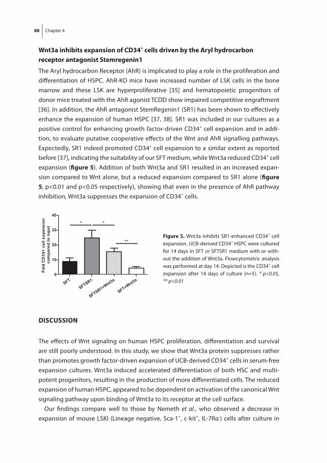
88 Chapter 4
Wnt3a inhibits expansion of CD34+ cells driven by the aryl hydrocarbon receptor antagonist Stemregenin1
The Aryl hydrocarbon Receptor (AhR) is implicated to play a role in the proliferation and differentiation of HSPC. AhR-KO mice have increased number of LSK cells in the bone marrow and these LSK are hyperproliferative [35] and hematopoietic progenitors of donor mice treated with the AhR agonist TCDD show impaired competitive engraftment [36]. In addition, the AhR antagonist StemRegenin1 (SR1) has been shown to effectively enhance the expansion of human HSPC [37, 38]. SR1 was included in our cultures as a positive control for enhancing growth factor-driven CD34+ cell expansion and in addi-tion, to evaluate putative cooperative effects of the Wnt and AhR signalling pathways. Expectedly, SR1 indeed promoted CD34+ cell expansion to a similar extent as reported before [37], indicating the suitability of our SFT medium, while Wnt3a reduced CD34+ cell expansion (figure 5). Addition of both Wnt3a and SR1 resulted in an increased expan-sion compared to Wnt alone, but a reduced expansion compared to SR1 alone (figure 5, p<0.01 and p<0.05 respectively), showing that even in the presence of AhR pathway inhibition, Wnt3a suppresses the expansion of CD34+ cells.
DISCUSSIon
The effects of Wnt signaling on human HSPC proliferation, differentiation and survival are still poorly understood. In this study, we show that Wnt3a protein suppresses rather than promotes growth factor-driven expansion of UCB-derived CD34+ cells in serum-free expansion cultures. Wnt3a induced accelerated differentiation of both HSC and multi-potent progenitors, resulting in the production of more differentiated cells. The reduced expansion of human HSPC, appeared to be dependent on activation of the canonical Wnt signaling pathway upon binding of Wnt3a to its receptor at the cell surface.
Our findings compare well to those by Nemeth et al., who observed a decrease in expansion of mouse LSKI (Lineage negative, Sca-1+, c-kit+, IL-7Rα-) cells after culture in
figure 5. Wnt3a inhibits SR1-enhanced CD34+ cell expansion. UCB-derived CD34+ HSPC were cultured for 14 days in SFT or SFTSR1 medium with or with-out the addition of Wnt3a. Flowcytometric analysis was performed at day 14. Depicted is the CD34+ cell expansion after 14 days of culture (n=5). * p<0.05, ** p<0.01
Wnt3a reduces growth-factor driven expansion of human HSPC 89
4
serum-free medium containing SCF, Flt3L and Wnt3a compared to culture conditions with growth factors only. In addition, they showed no enhanced repopulation capacity of Wnt3a-cultured LSKI cells compared to control-cultured cells[25]. Earlier, several studies showed that overexpression of beta-catenin in HSC resulted in a functional defect of hematopoiesis and loss of repopulating activity [27, 39]. These results are in line with our observations of enhanced differentiation of HSPC upon stimulation of the canonical Wnt pathway. However, Malhotra et al. reported that overexpression of activated beta-catenin may expand the pool of HSC, both phenotypically and functionally in long term cultures [40] . In addition, overexpression of the Wnt inhibitor Dickkopf-related protein 1 (Dkk1) was shown to significantly impair the self-renewal capacity of adult HSC[21, 22]. How to reconcile these contradictory results? Although at first sight contradictory, the many differences in types of cultures, cells growth factors added may explain some of the observed differences.
Willert et al. and Reya et al. earlier showed that purified Wnt3a induced proliferation, while inhibiting differentiation of HSC in growth-factor based cultures of mouse LSKT (Lineage negative, Sca-1+, c-kit+, Thy-1.1lo) [24, 26]. However, they used cells derived from BCL2 transgenic mice. The BCL2 anti-apoptotic signal in HSPC may counteract possible apoptotic signals induced by Wnt3a signaling. Moreover, they used serum, which may supply additional signals that allow canonical Wnt signaling to exert a differentiation inhibitory effect or a more pronounced effect on self-renewal. It would compare well to several other studies [28, 41], which show that the presence of additional factors is required to balance the activated Wnt pathway. Trowbridge et al. showed enhanced long-term repopulation after treating mice with a GSK3beta inhibitor, which regulates the canonical Wnt pathway and several other pathways as well[41]. Perry et al. showed that the combination of a PTEN deletion and activation of beta-catenin results in en-hanced self-renewal and expansion of HSC [28]. Another important pathway involved in HSC self-renewal and inhibition of differentiation is the pathway initiated by the aryl hydrocarbon Receptor (AhR). It is now well established that AhR-KO mice have increased number of LSK cells in the bone marrow and these LSK are hyperproliferative [35], while hematopoietic progenitors of donor mice treated with AhR agonist TCDD show impaired competitive engraftment[36]. In addition, the AhR antagonist StemRegenin1 (SR1) promotes expansion of human hematopoietic stem cells [37, 38]. We evaluated whether combined inhibition of the AhR pathway and activation of the Wnt pathway would result in enhanced expansion of HSPC. However, Wnt signaling also reduced the expansion of CD34+ HSPC in the presence of the aryl hydrocarbon receptor antagonist SR1. Another important factor that may affect the response of HSPC to Wnt signaling is the level of oxygenation. Expansion cultures are usually performed at normoxic levels. However, it is well established that the response of HSPC to hematopoietic cytokines is different under normoxic and hypoxic conditions, which mimics the in situ bone marrow environment
90 Chapter 4
[42]. The effect of stimulation of the canonical Wnt pathway on HSC expansion may be modulated by the level of oxygenation, which was already shown for the effect of the Wnt4-dependent pathway on the functional capacities of mesenchymal stem cells [43].
Apart from other cytokines and pathways involved, the dose and timing of Wnt activa-tion might play an important role. Our data show that both intermittent pulses of Wnt3a and more continuous exposure to Wnt3a have a similar negative effect on the expansion of CD34+ cells in serum-free expansion cultures. Luis et al reported that Wnts are tightly regulated in a dose-dependent fashion [44], which may affect their biological activity. Thereby, varying results (exerted via the canonical Wnt pathway) may result from differ-ent levels of Wnt signaling, such as can be achieved in different experimental conditions. The optimal level of Wnt signaling for HSC was suggested to be only slightly increased over normal physiological values, while higher levels resulted in impaired engraftment potential of HSC fashion [44].
Collectively, we show that exogenous Wnt3a proteins reduces the expansion of human HSPC in serum-free growth factor-driven HSPC expansion cultures by promoting their differentiation without apparently affecting their proliferation or survival. It cannot be excluded that additional signals, such as the induction of a hypoxic cellular response, are needed for a possible positive effect of canonical Wnt signaling on HSPC expansion. Future studies may address the fragile balance between canonical Wnt signaling and other pathways to determine whether combined activation of Wnt signaling and other signaling pathways may promote human HSPC expansion.
aCKnoWLEDGEmEntS
The authors would like to acknowledge Louis van Blooij of the Institute for Pharmaceuti-cal Sciences, Utrecht University for his help in the preparation of liposomal Wnt3a, and Régine Steegers-Theunissen of the department of Obstetrics and Gynaecology of the Erasmus Medical Centre Rotterdam and Gretel van Hoecke of the department of Obstet-rics and Gynaecology of the Albert Schweitzer Hospital in Dordrecht for their help in the collection of umbilical cord blood.
Wnt3a reduces growth-factor driven expansion of human HSPC 91
4
rEfErEnCES
1. Wagner JE, Barker JN, DeFor TE, Baker KS, Blazar BR, Eide C, et al. Transplantation of unrelated donor umbilical cord blood in 102 patients with malignant and nonmalignant diseases: influence of CD34 cell dose and HLA disparity on treatment-related mortality and survival. Blood. 2002 Sep 1;100(5):1611-8.
2. Laughlin MJ, Eapen M, Rubinstein P, Wagner JE, Zhang MJ, Champlin RE, et al. Outcomes after transplantation of cord blood or bone marrow from unrelated donors in adults with leukemia. N Engl J Med. 2004 Nov 25;351(22):2265-75.
3. Rocha V, Labopin M, Sanz G, Arcese W, Schwerdtfeger R, Bosi A, et al. Transplants of umbilical-cord blood or bone marrow from unrelated donors in adults with acute leukemia. N Engl J Med. 2004 Nov 25;351(22):2276-85.
4. Brunstein CG, Gutman JA, Weisdorf DJ, Woolfrey AE, Defor TE, Gooley TA, et al. Allogeneic hema-topoietic cell transplantation for hematologic malignancy: relative risks and benefits of double umbilical cord blood. Blood. 2010 Nov 25;116(22):4693-9.
5. Ng YY, van Kessel B, Lokhorst HM, Baert MR, van den Burg CM, Bloem AC, et al. Gene-expression profiling of CD34+ cells from various hematopoietic stem-cell sources reveals functional differ-ences in stem-cell activity. J Leukoc Biol. 2004 Feb;75(2):314-23.
6. Iscove NN, Nawa K. Hematopoietic stem cells expand during serial transplantation in vivo without apparent exhaustion. Curr Biol. 1997 Oct 1;7(10):805-8.
7. Sauvageau G, Iscove NN, Humphries RK. In vitro and in vivo expansion of hematopoietic stem cells. Oncogene. 2004 Sep 20;23(43):7223-32.
8. Goff JP, Shields DS, Greenberger JS. Influence of cytokines on the growth kinetics and immuno-phenotype of daughter cells resulting from the first division of single CD34(+)Thy-1(+)lin- cells. Blood. 1998 Dec 1;92(11):4098-107.
9. Glimm H, Eaves CJ. Direct evidence for multiple self-renewal divisions of human in vivo repopulat-ing hematopoietic cells in short-term culture. Blood. 1999 Oct 1;94(7):2161-8.
10. Ivanovic Z, Duchez P, Chevaleyre J, Vlaski M, Lafarge X, Dazey B, et al. Clinical-scale cultures of cord blood CD34(+) cells to amplify committed progenitors and maintain stem cell activity. Cell Transplant. 2011;20(9):1453-63.
11. Duchez P, Chevaleyre J, Vlaski M, Dazey B, Milpied N, Boiron JM, et al. Definitive setup of clinical scale procedure for ex vivo expansion of cord blood hematopoietic cells for transplantation. Cell Transplant. 2012;21(11):2517-21.
12. Delaney C, Heimfeld S, Brashem-Stein C, Voorhies H, Manger RL, Bernstein ID. Notch-mediated expansion of human cord blood progenitor cells capable of rapid myeloid reconstitution. Nat Med. 2010 Feb;16(2):232-6.
13. de Lima M, McMannis J, Gee A, Komanduri K, Couriel D, Andersson BS, et al. Transplantation of ex vivo expanded cord blood cells using the copper chelator tetraethylenepentamine: a phase I/II clinical trial. Bone Marrow Transplant. 2008 May;41(9):771-8.
14. de Lima M, McNiece I, Robinson SN, Munsell M, Eapen M, Horowitz M, et al. Cord-blood engraft-ment with ex vivo mesenchymal-cell coculture. N Engl J Med. 2012 Dec 13;367(24):2305-15.
15. Walasek MA, van Os R, de Haan G. Hematopoietic stem cell expansion: challenges and opportuni-ties. Ann N Y Acad Sci. 2012 Aug;1266:138-50.
16. Clevers H, Nusse R. Wnt/beta-catenin signaling and disease. Cell. 2012 Jun 8;149(6):1192-205. 17. Nusse R. Wnt signaling and stem cell control. Cell Res. 2008 May;18(5):523-7.
92 Chapter 4
18. Zeng YA, Nusse R. Wnt proteins are self-renewal factors for mammary stem cells and promote their long-term expansion in culture. Cell Stem Cell. 2010 Jun 4;6(6):568-77.
19. Sato MM, Nakashima A, Nashimoto M, Yawaka Y, Tamura M. Bone morphogenetic protein-2 enhances Wnt/beta-catenin signaling-induced osteoprotegerin expression. Genes Cells. 2009 Feb;14(2):141-53.
20. Lien WH, Polak L, Lin M, Lay K, Zheng D, Fuchs E. In vivo transcriptional governance of hair follicle stem cells by canonical Wnt regulators. Nat Cell Biol. 2014 Feb;16(2):179-90.
21. Luis TC, Weerkamp F, Naber BA, Baert MR, de Haas EF, Nikolic T, et al. Wnt3a deficiency irreversibly impairs hematopoietic stem cell self-renewal and leads to defects in progenitor cell differentia-tion. Blood. 2009 Jan 15;113(3):546-54.
22. Fleming HE, Janzen V, Lo Celso C, Guo J, Leahy KM, Kronenberg HM, et al. Wnt signaling in the niche enforces hematopoietic stem cell quiescence and is necessary to preserve self-renewal in vivo. Cell Stem Cell. 2008 Mar 6;2(3):274-83.
23. Zhao C, Blum J, Chen A, Kwon HY, Jung SH, Cook JM, et al. Loss of beta-catenin impairs the renewal of normal and CML stem cells in vivo. Cancer Cell. 2007 Dec;12(6):528-41.
24. Willert K, Brown JD, Danenberg E, Duncan AW, Weissman IL, Reya T, et al. Wnt proteins are lipid-modified and can act as stem cell growth factors. Nature. 2003 May 22;423(6938):448-52.
25. Nemeth MJ, Topol L, Anderson SM, Yang Y, Bodine DM. Wnt5a inhibits canonical Wnt signaling in hematopoietic stem cells and enhances repopulation. Proc Natl Acad Sci U S A. 2007 Sep 25;104(39):15436-41.
26. Reya T, Duncan AW, Ailles L, Domen J, Scherer DC, Willert K, et al. A role for Wnt signalling in self-renewal of haematopoietic stem cells. Nature. 2003 May 22;423(6938):409-14.
27. Scheller M, Huelsken J, Rosenbauer F, Taketo MM, Birchmeier W, Tenen DG, et al. Hematopoietic stem cell and multilineage defects generated by constitutive beta-catenin activation. Nat Immu-nol. 2006 Oct;7(10):1037-47.
28. Perry JM, He XC, Sugimura R, Grindley JC, Haug JS, Ding S, et al. Cooperation between both Wnt/{beta}-catenin and PTEN/PI3K/Akt signaling promotes primitive hematopoietic stem cell self-renewal and expansion. Genes Dev. 2011 Sep 15;25(18):1928-42.
29. Ming M, Wang S, Wu W, Senyuk V, Le Beau MM, Nucifora G, et al. Activation of Wnt/beta-catenin protein signaling induces mitochondria-mediated apoptosis in hematopoietic progenitor cells. J Biol Chem. 2012 Jun 29;287(27):22683-90.
30. Majeti R, Park CY, Weissman IL. Identification of a hierarchy of multipotent hematopoietic progeni-tors in human cord blood. Cell Stem Cell. 2007 Dec 13;1(6):635-45.
31. Hsieh JC, Rattner A, Smallwood PM, Nathans J. Biochemical characterization of Wnt-frizzled inter-actions using a soluble, biologically active vertebrate Wnt protein. Proc Natl Acad Sci U S A. 1999 Mar 30;96(7):3546-51.
32. Fuerer C, Habib SJ, Nusse R. A study on the interactions between heparan sulfate proteoglycans and Wnt proteins. Dev Dyn. 2010 Jan;239(1):184-90.
33. Morrell NT, Leucht P, Zhao L, Kim JB, ten Berge D, Ponnusamy K, et al. Liposomal packaging gener-ates Wnt protein with in vivo biological activity. PLoS One. 2008;3(8):e2930.
34. Zhao L, Rooker SM, Morrell N, Leucht P, Simanovskii D, Helms JA. Controlling the in vivo activity of Wnt liposomes. Methods Enzymol. 2009;465:331-47.
35. Gasiewicz TA, Singh KP, Casado FL. The aryl hydrocarbon receptor has an important role in the regulation of hematopoiesis: implications for benzene-induced hematopoietic toxicity. Chem Biol Interact. 2010 Mar 19;184(1-2):246-51.
Wnt3a reduces growth-factor driven expansion of human HSPC 93
4
36. Singh KP, Wyman A, Casado FL, Garrett RW, Gasiewicz TA. Treatment of mice with the Ah receptor agonist and human carcinogen dioxin results in altered numbers and function of hematopoietic stem cells. Carcinogenesis. 2009 Jan;30(1):11-9.
37. Boitano AE, Wang J, Romeo R, Bouchez LC, Parker AE, Sutton SE, et al. Aryl hydrocarbon recep-tor antagonists promote the expansion of human hematopoietic stem cells. Science. 2010 Sep 10;329(5997):1345-8.
38. Wagner JE, Brunstein CG, McKenna D, Sumstad D, Maahs S, Boitano AE, et al., editors. Safety and Exploratory Efficacy of Ex Vivo Expanded Umbilical Cord Blood (UCB) Hematopoietic Stem and Progenitor Cells (HSPC) Using Cytokines and Stem-Regenin 1 (SR1): Interim Results Of a Phase 1/2 Dose Escalation Clinical Study. 55th ASH Annual Meeting and Exposition; 2013 December 9, 2013; New Orleans, LA.
39. Kirstetter P, Anderson K, Porse BT, Jacobsen SE, Nerlov C. Activation of the canonical Wnt pathway leads to loss of hematopoietic stem cell repopulation and multilineage differentiation block. Nat Immunol. 2006 Oct;7(10):1048-56.
40. Malhotra S, Baba Y, Garrett KP, Staal FJ, Gerstein R, Kincade PW. Contrasting responses of lymphoid progenitors to canonical and noncanonical Wnt signals. J Immunol. 2008 Sep 15;181(6):3955-64.
41. Trowbridge JJ, Xenocostas A, Moon RT, Bhatia M. Glycogen synthase kinase-3 is an in vivo regula-tor of hematopoietic stem cell repopulation. Nat Med. 2006 Jan;12(1):89-98.
42. Ivanovic Z. Hypoxia or in situ normoxia: The stem cell paradigm. J Cell Physiol. 2009 May;219(2):271-5.
43. Leroux L, Descamps B, Tojais NF, Seguy B, Oses P, Moreau C, et al. Hypoxia preconditioned mesen-chymal stem cells improve vascular and skeletal muscle fiber regeneration after ischemia through a Wnt4-dependent pathway. Mol Ther. 2010 Aug;18(8):1545-52.
44. Luis TC, Naber BA, Roozen PP, Brugman MH, de Haas EF, Ghazvini M, et al. Canonical wnt signaling regulates hematopoiesis in a dosage-dependent fashion. Cell Stem Cell. 2011 Oct 4;9(4):345-56.

A
Chapter 5In vitro and in vivo evaluation of StemRegenin1-expanded umbilical cord blood-derived hematopoietic stem and progenitor cells
Lucia E. Duinhouwer1, Mariëtte N.D. Ter Borg1, Elwin W.J.C. Rombouts1, Jan J. Cornelissen1, Eric Braakman1
1Department of Hematology, Erasmus Medical Center, Rotterdam, The Netherlands
Submitted
96 Chapter 5
aBStraCt
The slow hematopoietic recovery following umbilical cord blood stem cell transplanta-tion (UCB-SCT) is considered to be primarily due to the relatively low number of hema-topoietic stem and progenitor cells (HSPC) in the graft. Aryl hydrocarbon receptor (AhR) antagonists, such as StemRegenin1 (SR1), have been shown to promote ex vivo expansion of UCB-derived CD34+ cells. Here, the effects of SR1 on the expansion of the most primi-tive subset of UCB-derived CD34+ cells were evaluated. It is shown that the combination of SR1 and the hematopoietic growth factors Stem Cell Factor (SCF), FMS-like tyrosine kinase 3 ligand (Flt3L) and Thrombopoietin (TPO) resulted in a 27-fold expansion of the phenotypically most primitive CD34+ cell subset (i.e. Lin-CD34+CD38lowCD45RAlowCD90+ cells) following serum-free culture of 2 weeks. That combination also resulted in a 105-fold expansion of the functionally most primitive subset as assessed by the long-term culture initiating cell (LTC-IC) assay. Transplantation of SR1-expanded cells into sublethally irradi-ated NSG mice resulted in robust long-term multilineage human engraftment in blood and bone marrow, although mice transplanted with expanded cells, with or without SR1, developed lower levels of human chimerism as compared to mice transplanted with non-expanded cells. The number of CD34+C10+ lymphoid progenitors was strongly reduced in the expanded population of cells, cultured with or without SR1, which might account for the lower level of human B cell chimerism, being the primary endpoint in the B cell-prone NSG mouse model. In conclusion, the combination of proliferation induc-ing cytokines combined with the differentiation inhibiting factor SR1 resulted in strong augmentation of phenotypic HSC with long-term repopulating capacity in vivo.
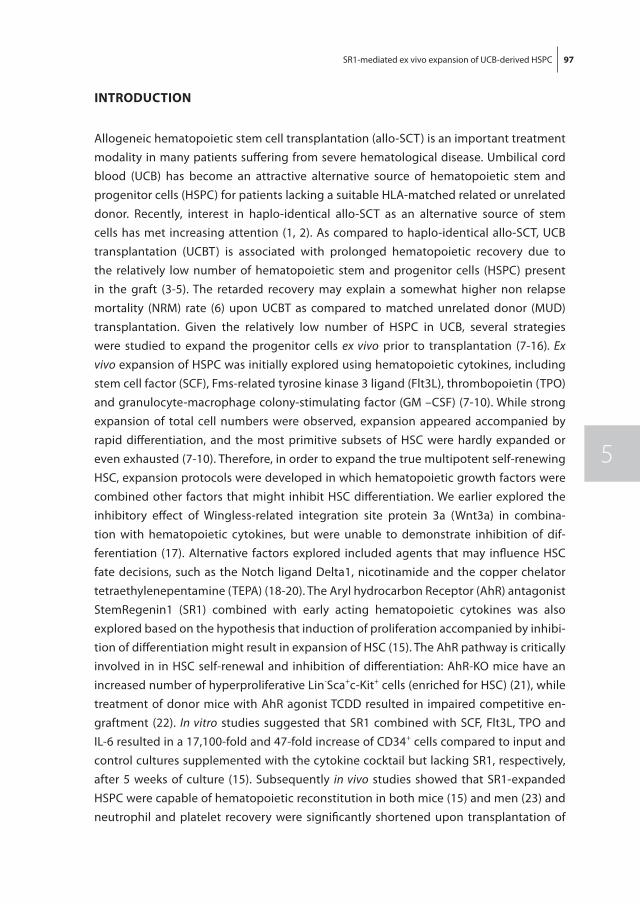
SR1-mediated ex vivo expansion of UCB-derived HSPC 97
5
IntroDUCtIon
Allogeneic hematopoietic stem cell transplantation (allo-SCT) is an important treatment modality in many patients suffering from severe hematological disease. Umbilical cord blood (UCB) has become an attractive alternative source of hematopoietic stem and progenitor cells (HSPC) for patients lacking a suitable HLA-matched related or unrelated donor. Recently, interest in haplo-identical allo-SCT as an alternative source of stem cells has met increasing attention (1, 2). As compared to haplo-identical allo-SCT, UCB transplantation (UCBT) is associated with prolonged hematopoietic recovery due to the relatively low number of hematopoietic stem and progenitor cells (HSPC) present in the graft (3-5). The retarded recovery may explain a somewhat higher non relapse mortality (NRM) rate (6) upon UCBT as compared to matched unrelated donor (MUD) transplantation. Given the relatively low number of HSPC in UCB, several strategies were studied to expand the progenitor cells ex vivo prior to transplantation (7-16). Ex vivo expansion of HSPC was initially explored using hematopoietic cytokines, including stem cell factor (SCF), Fms-related tyrosine kinase 3 ligand (Flt3L), thrombopoietin (TPO) and granulocyte-macrophage colony-stimulating factor (GM –CSF) (7-10). While strong expansion of total cell numbers were observed, expansion appeared accompanied by rapid differentiation, and the most primitive subsets of HSC were hardly expanded or even exhausted (7-10). Therefore, in order to expand the true multipotent self-renewing HSC, expansion protocols were developed in which hematopoietic growth factors were combined other factors that might inhibit HSC differentiation. We earlier explored the inhibitory effect of Wingless-related integration site protein 3a (Wnt3a) in combina-tion with hematopoietic cytokines, but were unable to demonstrate inhibition of dif-ferentiation (17). Alternative factors explored included agents that may influence HSC fate decisions, such as the Notch ligand Delta1, nicotinamide and the copper chelator tetraethylenepentamine (TEPA) (18-20). The Aryl hydrocarbon Receptor (AhR) antagonist StemRegenin1 (SR1) combined with early acting hematopoietic cytokines was also explored based on the hypothesis that induction of proliferation accompanied by inhibi-tion of differentiation might result in expansion of HSC (15). The AhR pathway is critically involved in in HSC self-renewal and inhibition of differentiation: AhR-KO mice have an increased number of hyperproliferative Lin-Sca+c-Kit+ cells (enriched for HSC) (21), while treatment of donor mice with AhR agonist TCDD resulted in impaired competitive en-graftment (22). In vitro studies suggested that SR1 combined with SCF, Flt3L, TPO and IL-6 resulted in a 17,100-fold and 47-fold increase of CD34+ cells compared to input and control cultures supplemented with the cytokine cocktail but lacking SR1, respectively, after 5 weeks of culture (15). Subsequently in vivo studies showed that SR1-expanded HSPC were capable of hematopoietic reconstitution in both mice (15) and men (23) and neutrophil and platelet recovery were significantly shortened upon transplantation of
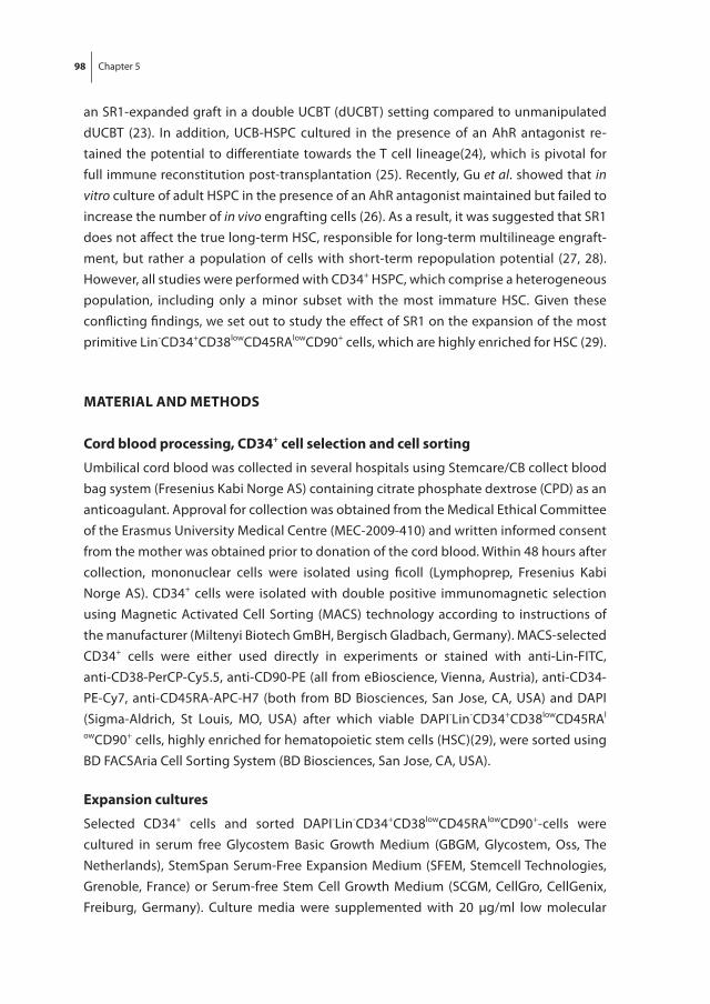
98 Chapter 5
an SR1-expanded graft in a double UCBT (dUCBT) setting compared to unmanipulated dUCBT (23). In addition, UCB-HSPC cultured in the presence of an AhR antagonist re-tained the potential to differentiate towards the T cell lineage(24), which is pivotal for full immune reconstitution post-transplantation (25). Recently, Gu et al. showed that in vitro culture of adult HSPC in the presence of an AhR antagonist maintained but failed to increase the number of in vivo engrafting cells (26). As a result, it was suggested that SR1 does not affect the true long-term HSC, responsible for long-term multilineage engraft-ment, but rather a population of cells with short-term repopulation potential (27, 28). However, all studies were performed with CD34+ HSPC, which comprise a heterogeneous population, including only a minor subset with the most immature HSC. Given these conflicting findings, we set out to study the effect of SR1 on the expansion of the most primitive Lin-CD34+CD38lowCD45RAlowCD90+ cells, which are highly enriched for HSC (29).
matErIaL anD mEtHoDS
Cord blood processing, CD34+ cell selection and cell sorting
Umbilical cord blood was collected in several hospitals using Stemcare/CB collect blood bag system (Fresenius Kabi Norge AS) containing citrate phosphate dextrose (CPD) as an anticoagulant. Approval for collection was obtained from the Medical Ethical Committee of the Erasmus University Medical Centre (MEC-2009-410) and written informed consent from the mother was obtained prior to donation of the cord blood. Within 48 hours after collection, mononuclear cells were isolated using ficoll (Lymphoprep, Fresenius Kabi Norge AS). CD34+ cells were isolated with double positive immunomagnetic selection using Magnetic Activated Cell Sorting (MACS) technology according to instructions of the manufacturer (Miltenyi Biotech GmBH, Bergisch Gladbach, Germany). MACS-selected CD34+ cells were either used directly in experiments or stained with anti-Lin-FITC, anti-CD38-PerCP-Cy5.5, anti-CD90-PE (all from eBioscience, Vienna, Austria), anti-CD34-PE-Cy7, anti-CD45RA-APC-H7 (both from BD Biosciences, San Jose, CA, USA) and DAPI (Sigma-Aldrich, St Louis, MO, USA) after which viable DAPI-Lin-CD34+CD38lowCD45RAl
owCD90+ cells, highly enriched for hematopoietic stem cells (HSC)(29), were sorted using BD FACSAria Cell Sorting System (BD Biosciences, San Jose, CA, USA).
Expansion cultures
Selected CD34+ cells and sorted DAPI-Lin-CD34+CD38lowCD45RAlowCD90+-cells were cultured in serum free Glycostem Basic Growth Medium (GBGM, Glycostem, Oss, The Netherlands), StemSpan Serum-Free Expansion Medium (SFEM, Stemcell Technologies, Grenoble, France) or Serum-free Stem Cell Growth Medium (SCGM, CellGro, CellGenix, Freiburg, Germany). Culture media were supplemented with 20 μg/ml low molecular
SR1-mediated ex vivo expansion of UCB-derived HSPC 99
5
weight heparin (Abbott, Wiesbaden, Germany) and the early acting growth factors SCF, Flt3L and TPO (50 ng/ml each, all from Cellgenix, Freiburg, Germany) (from now on re-ferred to as ‘SFT medium’) with or without the addition of 1 µM StemRegenin1 (SR1, Cel-lagen Technology, San Diego, CA, USA or Axon Medchem, Groningen, The Netherlands) unless indicated otherwise. Cells were cultured in a volume of 1 ml in a 24 well plate at a concentration of 105/ml at 37 °C in 5% CO2. Every 2 to 3 days, wells were split with addition of an equal volume of culture medium.
flow cytometry
At serial time points in culture, absolute numbers of viable CD34+ cells were determined by a single platform flow cytometric assay, using anti-CD45-FITC, anti-CD34-PE, DAPI and a calibrated number of Stem-Count Fluorospheres (all from Beckman Coulter, Fullerton, CA, USA). Within the CD34+ population, the frequency of DAPI-Lin-CD34+CD38lowCD45RAlowCD90+-cells was determined using the antibody panel as described above for the sorting of these cells. Absolute numbers of DAPI-Lin-CD34+CD38lowCD45RAlowCD90+-cells were determined by multiplying the absolute number of CD34+ cells obtained in the single platform analysis by the percentage of DAPI-Lin-CD34+CD38lowCD45RAlowCD90+-cells within the CD34+ cell population.
For cell division tracking during culture, the CellTrace™ Violet Cell Proliferation Kit (Life Technologies, Eugene, OR, USA) was used. Prior to culture, selected and sorted cells were labeled using CellTrace™ Violet, according to instructions of the manufacturer. Flow cytometry was performed at day 5 of culture. Flow cytometric analysis was performed using a BD FACSCanto (BD Biosciences, San Jose, CA, USA) and data was analyzed using FlowJo software (Tree Star Inc, Ashland, OR, USA).
Long term culture-initiating cell (LtC-IC) assays
The LTC-IC assay was performed as described(30). In short, confluent MS-5 stromal layers in 96-well plates were overlaid with sorted subsets of (expanded) CD34+ cells in a limiting dilution set-up, with 3000 cells per well as the highest concentration and titrating down by 1/3, using 20 wells per condition. Cells were cultured for 5 weeks at 37 °C in 5% CO2 in IMDM medium, containing 12% horse serum, 12% fetal calf serum, 2mM L-glutamine, 100 U/ml penicillin and 100 U/ml streptomycin, with medium change every week. After 5 weeks, the medium was removed and methylcellulose containing medium (Methocult GF H84434, Stemcell Technologies, Vancouver, BC, Canada) was added to each well. Upon another 14 days of culture, the percentage of wells containing a colony (=positive wells) was determined. The frequency of LTC-IC within each cell population was calculated us-ing the number of negative wells per dilution and Poisson statistics.
100 Chapter 5
transplantation of human hematopoietic cells into noD.Cg-Prkdcscid Il2rgtm1Wjl/SzJ (nSG) mice
NSG mice were purchased from The Jackson Laboratory (Bar Harbor, ME, USA) and housed under specific pathogen-free conditions in the Erasmus MC animal facility. The mice experiments were carried out in accordance to the Dutch law on Animal Welfare and Experiments. The protocol was approved by the Committee on the Ethics of Animal Experiments of the Erasmus University Medical Centre Rotterdam, The Netherlands. All animals were housed in groups in individually ventilated cages. Food and water were available ad libitum. All NSG mice were sublethally irradiated (2.5 – 3.5 Gy) and trans-planted intravenously with the indicated number of CD34+ cells, the progeny thereof or sorted subsets of CD34+ cells. Engraftment was assessed by flow cytometric analysis of the peripheral blood, using a panel including anti-mouseCD45-eFluor450, (eBioscience, Vienna, Austria) and anti-humanCD45-APC-Cy7 (BioLegend, London, UK). For the assess-ment of multilineage engraftment, peripheral blood and bone marrow cells were stained with anti-humanCD56-PE-Cy7, anti-humanCD33-PerCP-eFluor710, anti-humanCD3-APC and anti-humanCD19-PE (all from eBioscience, Vienna, Austria). Mice were considered engrafted when human CD45+ cell levels were higher than 0.1% in the bone marrow. The mice were sacrificed by cervical dislocation and cells from femurs were analyzed at the time points described in the results section.
Statistics
Paired or unpaired two-tailed t-tests were performed to test the differences between the different culture conditions, as appropriate. Differences were considered to be sta-tistically significant is p < 0.05. Statistical tests were performed in GraphPad Prism 5.0 (GraphdPad software, San Diego, CA, USA).
rESULtS
Serum-free cultures combining hematopoietic growth factors and StemRegenin1 result in expansion of the phenotypically most immature CD34+ cells that are enriched for LTC-IC
To confirm the effect of Stemregenin1 (SR1) on the expansion of HSPC in culture, puri-fied UCB-derived CD34+ cells were cultured in serum-free medium supplemented with SCF, Flt3L and TPO (SFT medium), with or without the addition of SR1. No significant difference was observed in expansion of CD45+ cells after 14 days of culture in either SFT or SFT+SR1 medium (table 1, figure 1a, p=0.23). During expansion, the frequency of CD34+ cells gradually declined due to concomitant differentiation of the expanding cells. Addition of SR1 delayed the decline in frequency of CD34+ cells, resulting in a higher frequency of CD34+ cells at both 7 and 14 days compared to SFT medium alone (table
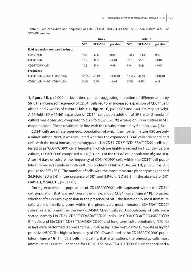
SR1-mediated ex vivo expansion of UCB-derived HSPC 101
5
1, figure 1B, p<0.001 for both time points), suggesting inhibition of differentiation by SR1. The increased frequency of CD34+ cells led to an increased expansion of CD34+ cells after 1 and 2 weeks of culture (table 1, figure 1C, p=0.0083 and p=0.006 respectively). A 55-fold (SD ±44.46) expansion of CD34+ cells upon addition of SR1 after 2 weeks of culture was observed, compared to a 23-fold (SD ±20.78) expansion upon culture in SFT medium alone. These results are in line with the results reported by Boitano et al. (15).
CD34+ cells are a heterogeneous population, of which the most immature HSC are only a minor subset. Next, it was evaluated whether the expanded CD34+ cells still contained cells with the most immature phenotype, i.e. Lin-CD34+CD38lowCD45RAlowCD90+ cells (re-ferred to as “CD34+CD90+ cells” hereafter), which are highly enriched for HSC (29). Before culture, CD34+CD90+ comprised 4.0% (SD ±2.1) of the CD34+ cell population (figure 1D). After 14 days of culture, the frequency of CD34+CD90+ cells within the CD34+ cell popu-lation remained stable in both culture conditions (table 1, figure 1D, p=0.38 for SFT; p=0.18 for SFT+SR1). The number of cells with the most immature phenotype expanded 26.9-fold (SD ±9.6) in the presence of SR1 and 8.9-fold (SD ±9.5) in the absence of SR1 (table 1, figure 1E, p=0.0003).
During expansion, a population of CD45RA+CD90+ cells appeared within the CD34+ cell population that was not present in unexpanded CD34+ cells (figure 1f). To assess whether after ex vivo expansion in the presence of SR1, the functionally most immature cells were primarily present within the phenotypic most immature CD45RAlowCD90+ subset or also present in the new CD45RA+CD90+ subset, 3 populations of cells were sorted, namely Lin-CD34+CD38lowCD45RAlowCD90+ cells, Lin-CD34+CD38lowCD45RAlowCD90low cells and Lin-CD34+CD38lowCD45RA+CD90+ and long term culture-initiating (LTC-IC) assays were performed. At present, the LTC-IC assay is the best in vitro surrogate assay for primitive HSPC. The highest frequency of LTC-IC was found in the CD45RAlowCD90+ popu-lation (figure 1G, 1 in 23.3 cells), indicating that after culture, the phenotypically most immature cells are still enriched for LTC-IC. The new CD45RA+CD90+ subset contained a
table 1. Fold expansion and frequency of CD45+, CD34+ and CD34+CD90+ cells upon culture in SFT or SFT+SR1 medium
Day 7 Day 14
Sft Sft+Sr1 p-value Sft Sft+Sr1 p-value
fold expansion compared to input
CD45+ cells 47.3 47.6 0.88 100.3 113.3 0.23
CD34+ cells 19.9 31.2 <0.01 23.3 55.2 <0.01
CD34+CD90+ cells 10.8 21.6 0.09 8.9 26.9 <0.001
frequency
CD34+ cells (within CD45+ cells) 36.9% 63.0% <0.0001 19.2% 42.5% <0.0001
CD90+ cells (within CD34+ cells) 5.8% 7.1% <0.05 5.5% 4.5% 0.18
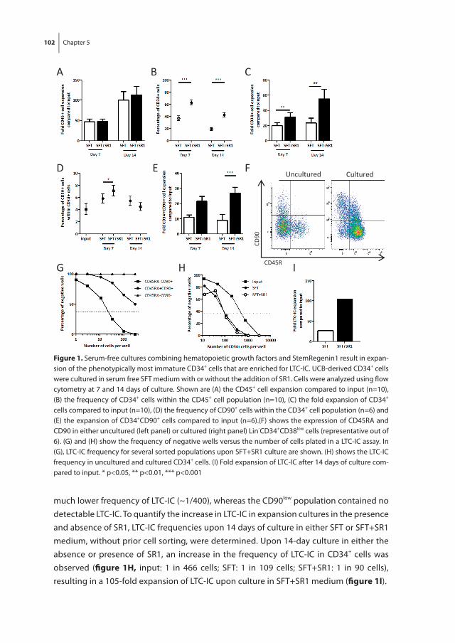
102 Chapter 5
much lower frequency of LTC-IC (~1/400), whereas the CD90low population contained no detectable LTC-IC. To quantify the increase in LTC-IC in expansion cultures in the presence and absence of SR1, LTC-IC frequencies upon 14 days of culture in either SFT or SFT+SR1 medium, without prior cell sorting, were determined. Upon 14-day culture in either the absence or presence of SR1, an increase in the frequency of LTC-IC in CD34+ cells was observed (fi gure 1H, input: 1 in 466 cells; SFT: 1 in 109 cells; SFT+SR1: 1 in 90 cells), resulting in a 105-fold expansion of LTC-IC upon culture in SFT+SR1 medium (fi gure 1I).
CD90
CD45RA
Uncultured Cultured
A B C
D E F
G IH
figure 1. Serum-free cultures combining hematopoietic growth factors and StemRegenin1 result in expan-sion of the phenotypically most immature CD34+ cells that are enriched for LTC-IC. UCB-derived CD34+ cells were cultured in serum free SFT medium with or without the addition of SR1. Cells were analyzed using fl ow cytometry at 7 and 14 days of culture. Shown are (A) the CD45+ cell expansion compared to input (n=10), (B) the frequency of CD34+ cells within the CD45+ cell population (n=10), (C) the fold expansion of CD34+ cells compared to input (n=10), (D) the frequency of CD90+ cells within the CD34+ cell population (n=6) and (E) the expansion of CD34+CD90+ cells compared to input (n=6).(F) shows the expression of CD45RA and CD90 in either uncultured (left panel) or cultured (right panel) Lin-CD34+CD38low cells (representative out of 6). (G) and (H) show the frequency of negative wells versus the number of cells plated in a LTC-IC assay. In (G), LTC-IC frequency for several sorted populations upon SFT+SR1 culture are shown. (H) shows the LTC-IC frequency in uncultured and cultured CD34+ cells. (I) Fold expansion of LTC-IC after 14 days of culture com-pared to input. * p<0.05, ** p<0.01, *** p<0.001
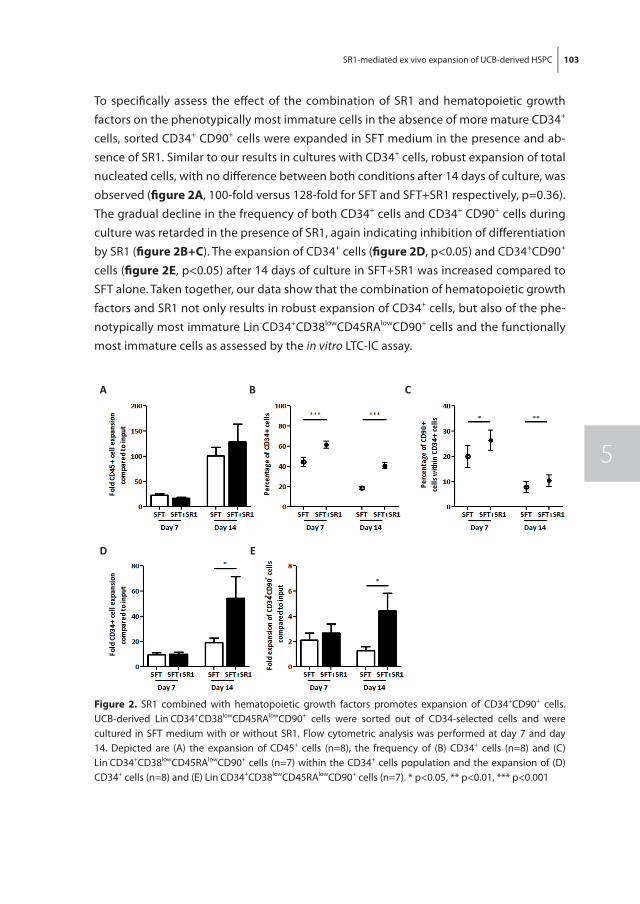
SR1-mediated ex vivo expansion of UCB-derived HSPC 103
5
To specifi cally assess the eff ect of the combination of SR1 and hematopoietic growth factors on the phenotypically most immature cells in the absence of more mature CD34+ cells, sorted CD34+ CD90+ cells were expanded in SFT medium in the presence and ab-sence of SR1. Similar to our results in cultures with CD34+ cells, robust expansion of total nucleated cells, with no diff erence between both conditions after 14 days of culture, was observed (fi gure 2a, 100-fold versus 128-fold for SFT and SFT+SR1 respectively, p=0.36). The gradual decline in the frequency of both CD34+ cells and CD34+ CD90+ cells during culture was retarded in the presence of SR1, again indicating inhibition of diff erentiation by SR1 (fi gure 2B+C). The expansion of CD34+ cells (fi gure 2D, p<0.05) and CD34+CD90+ cells (fi gure 2E, p<0.05) after 14 days of culture in SFT+SR1 was increased compared to SFT alone. Taken together, our data show that the combination of hematopoietic growth factors and SR1 not only results in robust expansion of CD34+ cells, but also of the phe-notypically most immature Lin-CD34+CD38lowCD45RAlowCD90+ cells and the functionally most immature cells as assessed by the in vitro LTC-IC assay.
A B
D E
C
figure 2. SR1 combined with hematopoietic growth factors promotes expansion of CD34+CD90+ cells. UCB-derived Lin-CD34+CD38lowCD45RAlowCD90+ cells were sorted out of CD34-selected cells and were cultured in SFT medium with or without SR1. Flow cytometric analysis was performed at day 7 and day 14. Depicted are (A) the expansion of CD45+ cells (n=8), the frequency of (B) CD34+ cells (n=8) and (C) Lin-CD34+CD38lowCD45RAlowCD90+ cells (n=7) within the CD34+ cells population and the expansion of (D) CD34+ cells (n=8) and (E) Lin-CD34+CD38lowCD45RAlowCD90+ cells (n=7). * p<0.05, ** p<0.01, *** p<0.001
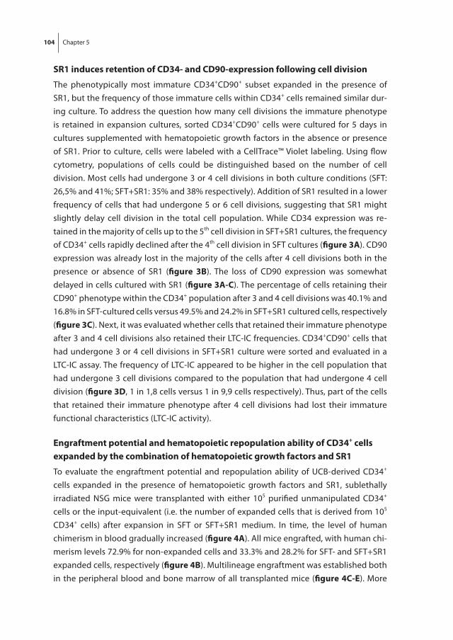
104 Chapter 5
Sr1 induces retention of CD34- and CD90-expression following cell division
The phenotypically most immature CD34+CD90+ subset expanded in the presence of SR1, but the frequency of those immature cells within CD34+ cells remained similar dur-ing culture. To address the question how many cell divisions the immature phenotype is retained in expansion cultures, sorted CD34+CD90+ cells were cultured for 5 days in cultures supplemented with hematopoietic growth factors in the absence or presence of SR1. Prior to culture, cells were labeled with a CellTrace™ Violet labeling. Using flow cytometry, populations of cells could be distinguished based on the number of cell division. Most cells had undergone 3 or 4 cell divisions in both culture conditions (SFT: 26,5% and 41%; SFT+SR1: 35% and 38% respectively). Addition of SR1 resulted in a lower frequency of cells that had undergone 5 or 6 cell divisions, suggesting that SR1 might slightly delay cell division in the total cell population. While CD34 expression was re-tained in the majority of cells up to the 5th cell division in SFT+SR1 cultures, the frequency of CD34+ cells rapidly declined after the 4th cell division in SFT cultures (figure 3a). CD90 expression was already lost in the majority of the cells after 4 cell divisions both in the presence or absence of SR1 (figure 3B). The loss of CD90 expression was somewhat delayed in cells cultured with SR1 (figure 3a-C). The percentage of cells retaining their CD90+ phenotype within the CD34+ population after 3 and 4 cell divisions was 40.1% and 16.8% in SFT-cultured cells versus 49.5% and 24.2% in SFT+SR1 cultured cells, respectively (figure 3C). Next, it was evaluated whether cells that retained their immature phenotype after 3 and 4 cell divisions also retained their LTC-IC frequencies. CD34+CD90+ cells that had undergone 3 or 4 cell divisions in SFT+SR1 culture were sorted and evaluated in a LTC-IC assay. The frequency of LTC-IC appeared to be higher in the cell population that had undergone 3 cell divisions compared to the population that had undergone 4 cell division (figure 3D, 1 in 1,8 cells versus 1 in 9,9 cells respectively). Thus, part of the cells that retained their immature phenotype after 4 cell divisions had lost their immature functional characteristics (LTC-IC activity).
Engraftment potential and hematopoietic repopulation ability of CD34+ cells expanded by the combination of hematopoietic growth factors and Sr1
To evaluate the engraftment potential and repopulation ability of UCB-derived CD34+ cells expanded in the presence of hematopoietic growth factors and SR1, sublethally irradiated NSG mice were transplanted with either 105 purified unmanipulated CD34+ cells or the input-equivalent (i.e. the number of expanded cells that is derived from 105 CD34+ cells) after expansion in SFT or SFT+SR1 medium. In time, the level of human chimerism in blood gradually increased (figure 4a). All mice engrafted, with human chi-merism levels 72.9% for non-expanded cells and 33.3% and 28.2% for SFT- and SFT+SR1 expanded cells, respectively (figure 4B). Multilineage engraftment was established both in the peripheral blood and bone marrow of all transplanted mice (figure 4C-E). More
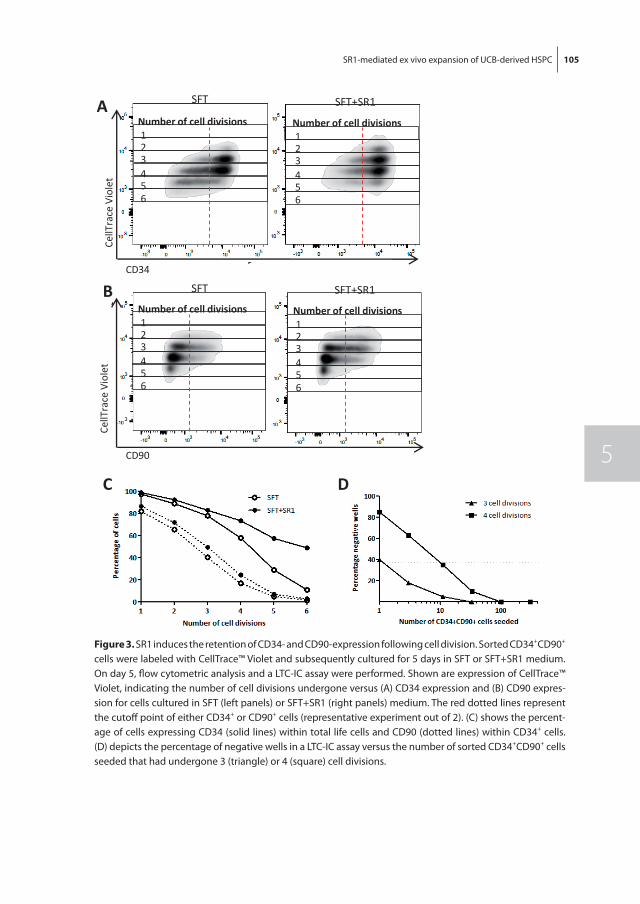
SR1-mediated ex vivo expansion of UCB-derived HSPC 105
5
A
CD34
CellT
race
Vio
let
B
CD90
CellT
race
Vio
letNumber of cell divisions123456
Number of cell divisions123456
SFT
SFT
Number of cell divisions123456
SFT+SR1
Number of cell divisions123456
SFT+SR1
C D
figure 3. SR1 induces the retention of CD34- and CD90-expression following cell division. Sorted CD34+CD90+ cells were labeled with CellTrace™ Violet and subsequently cultured for 5 days in SFT or SFT+SR1 medium. On day 5, flow cytometric analysis and a LTC-IC assay were performed. Shown are expression of CellTrace™ Violet, indicating the number of cell divisions undergone versus (A) CD34 expression and (B) CD90 expres-sion for cells cultured in SFT (left panels) or SFT+SR1 (right panels) medium. The red dotted lines represent the cutoff point of either CD34+ or CD90+ cells (representative experiment out of 2). (C) shows the percent-age of cells expressing CD34 (solid lines) within total life cells and CD90 (dotted lines) within CD34+ cells. (D) depicts the percentage of negative wells in a LTC-IC assay versus the number of sorted CD34+CD90+ cells seeded that had undergone 3 (triangle) or 4 (square) cell divisions.
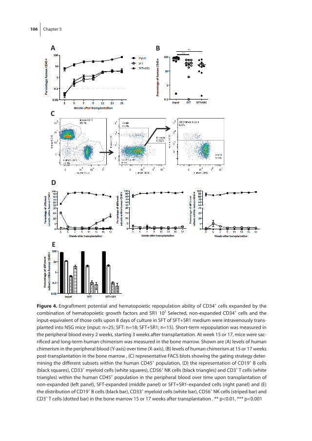
106 Chapter 5
D
E
C
A B
figure 4. Engraftment potential and hematopoietic repopulation ability of CD34+ cells expanded by the combination of hematopoietic growth factors and SR1 105 Selected, non-expanded CD34+ cells and the input-equivalent of those cells upon 8 days of culture in SFT of SFT+SR1 medium were intravenously trans-planted into NSG mice (input: n=25; SFT: n=18; SFT+SR1; n=15). Short-term repopulation was measured in the peripheral blood every 2 weeks, starting 3 weeks after transplantation. At week 15 or 17, mice were sac-rificed and long-term human chimerism was measured in the bone marrow. Shown are (A) levels of human chimerism in the peripheral blood (Y-axis) over time (X-axis), (B) levels of human chimerism at 15 or 17 weeks post-transplantation in the bone marrow , (C) representative FACS blots showing the gating strategy deter-mining the different subsets within the human CD45+ population, (D) the representation of CD19+ B cells (black squares), CD33+ myeloid cells (white squares), CD56+ NK cells (black triangles) and CD3+ T cells (white triangles) within the human CD45+ population in the peripheral blood over time upon transplantation of non-expanded (left panel), SFT-expanded (middle panel) or SFT+SR1-expanded cells (right panel) and (E) the distribution of CD19+ B cells (black bar), CD33+ myeloid cells (white bar), CD56+ NK cells (striped bar) and CD3+ T cells (dotted bar) in the bone marrow 15 or 17 weeks after transplantation . ** p<0.01, *** p<0.001
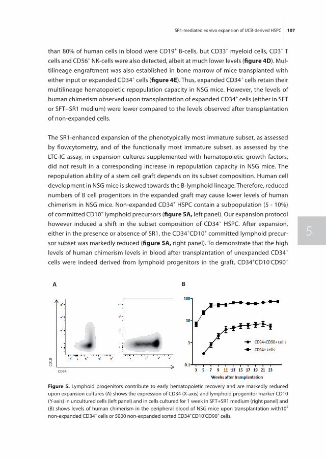
SR1-mediated ex vivo expansion of UCB-derived HSPC 107
5
than 80% of human cells in blood were CD19+ B-cells, but CD33+ myeloid cells, CD3+ T cells and CD56+ NK-cells were also detected, albeit at much lower levels (figure 4D). Mul-tilineage engraftment was also established in bone marrow of mice transplanted with either input or expanded CD34+ cells (figure 4E). Thus, expanded CD34+ cells retain their multilineage hematopoietic repopulation capacity in NSG mice. However, the levels of human chimerism observed upon transplantation of expanded CD34+ cells (either in SFT or SFT+SR1 medium) were lower compared to the levels observed after transplantation of non-expanded cells.
The SR1-enhanced expansion of the phenotypically most immature subset, as assessed by flowcytometry, and of the functionally most immature subset, as assessed by the LTC-IC assay, in expansion cultures supplemented with hematopoietic growth factors, did not result in a corresponding increase in repopulation capacity in NSG mice. The repopulation ability of a stem cell graft depends on its subset composition. Human cell development in NSG mice is skewed towards the B-lymphoid lineage. Therefore, reduced numbers of B cell progenitors in the expanded graft may cause lower levels of human chimerism in NSG mice. Non-expanded CD34+ HSPC contain a subpopulation (5 - 10%) of committed CD10+ lymphoid precursors (figure 5a, left panel). Our expansion protocol however induced a shift in the subset composition of CD34+ HSPC. After expansion, either in the presence or absence of SR1, the CD34+CD10+ committed lymphoid precur-sor subset was markedly reduced (figure 5a, right panel). To demonstrate that the high levels of human chimerism levels in blood after transplantation of unexpanded CD34+ cells were indeed derived from lymphoid progenitors in the graft, CD34+CD10-CD90+
CD34
CD10
A B
figure 5. Lymphoid progenitors contribute to early hematopoietic recovery and are markedly reduced upon expansion cultures (A) shows the expression of CD34 (X-axis) and lymphoid progenitor marker CD10 (Y-axis) in uncultured cells (left panel) and in cells cultured for 1 week in SFT+SR1 medium (right panel) and (B) shows levels of human chimerism in the peripheral blood of NSG mice upon transplantation with105 non-expanded CD34+ cells or 5000 non-expanded sorted CD34+CD10-CD90+ cells.
108 Chapter 5
cells were sorted and transplanted into NSG mice (5000 cells per mice, total of 15 mice). As expected, the removal of committed progenitors from the fresh CD34+ HSPC resulted in a delayed recovery of human cells and lower levels of human chimerism in blood of transplanted mice (figure 5B). These results demonstrate that the observed high levels of chimerism after transplantation of non-expanded CD34+ cells are mainly derived from lymphoid progenitors, which are markedly reduced after culture in SFT+SR1 medium.
DISCUSSIon
SR1 has been shown to promote the expansion of human HSPC capable of hematopoi-etic reconstitution in mice (15) and men (23). However, some studies suggested that SR1 predominantly affects committed progenitor cells with short-term repopulation potential, rather than the true long-term repopulating HSC (27, 28). Therefore, this study evaluated whether the combination of SCF, Flt3L and TPO (SFT) with SR1 would be able to expand a very immature subset of human UCB-derived phenotypic HSPC, characterized as Lin-CD34+CD38lowCD45RAlowCD90+ cells, which are highly enriched for HSC (29). In addition, functional evaluation included LTC-IC assays as well as an in vivo mice model. It is shown that the combination of SFT and SR1 expands purified immature Lin-CD34+CD38lowCD45RAlowCD90+ cells in serum-free expansion cultures. Most stud-ies claiming expansion used either the number of CD34+ cells or the more immature population of CD34+CD38low cells as an endpoint (11-13), but few studies addressed the effect on the phenotypically most immature subset (Lin-CD34+CD38low CD45RAlowCD90+ cells). Boitano et al. observed a ten-fold expansion of human CD34+CD90+ cells in cultures supplemented with SCF,Flt3L, TPO, IL-6 and SR1 compared to cultures supplemented with SFT6 only. Fold expansion compared to input was not shown. These cultures were initiated with mobilized peripheral blood-derived CD34+ cells from adult patients (15). However, Gu et al. (26) reported a reduced frequency of CD34+CD90+ cells upon 1 week culture of mobilized adult peripheral blood CD34+ cells in a serum-free culture system with a combination of SR1 and the growth factors SCF, Flt3L, TPO and IL-6. Despite that reduced frequency, absolute number of CD34+CD90+ cells were increased compared to input cell numbers due to strong expansion of the total CD34+ population. Fares et al. observed only a two-fold increase in CD34+CD38lowCD45RAlowCD90+ cells in cul-tures using a combination of SFT and SR1 compared to cultures initiated with growth factors only (27). In contrast to those studies, cell sorting of a more primitive subset (Lin-CD34+CD38lowCD45RAlowCD90+ cells) was performed prior to initiate cultures in the present study enabling the evaluation of proliferation and inhibition of differentiation of HSPC more closely. It is shown that a combination of SFT and SR1 enhanced both the fre-quency and absolute number of Lin-CD34+CD38lowCD45RAlowCD90+ cells in cultures initi-
SR1-mediated ex vivo expansion of UCB-derived HSPC 109
5
ated with purified immature cells. Cell division analysis showed a retention of both CD34 and CD90 expression following cell division, suggesting a delay of the differentiation of actively dividing cells by SR1. Moreover, a robust expansion of LTC-IC was observed fol-lowing evaluation of expanded cells in a 6-week LTC-IC system. Virtually all LTC-IC activity resided in the phenotypically most immature Lin-CD34+CD38lowCD45RAlowCD90+ cells. The subset of Lin-CD34+CD38lowCD45RA+CD90+ cells, which was generated during culture as a more differentiated subset, hardly contained any LTC-IC activity. These results suggest that expansion of phenotypically immature CD34+CD90+ cells cannot be taken as a surro-gate marker for expansion of functional HSC with LTC-IC capacity. Apart from expanding the subset of Lin-CD34+CD38lowCD45RAlowCD90+ cells our data indicate that SR1 is able to inhibit the differentiation of actively dividing Lin-CD34+CD38lowCD45RAlowCD90+ cells with retention of most LTC-IC activity.
Although SR1-expanded cells retained long-term multilineage repopulation ability in NSG mice, lower levels of human chimerism were observed upon intravenous transplan-tation of SR1-expanded cells compared to transplantation of unexpanded cells. These results may seem in contrast to those by Boitano et al., who reported a 17-fold increase in SCID-repopulating cells (SRC) (15). In addition, several other groups have reported that SR1 induces at best maintenance of SRC rather than an increase (26, 27) and Fares et al. proposed that SR1 does not affect the true long-term HSC, but rather a less primitive sub-type, the short-term HSC (28). How to explain these contradictory results? A possible clue may come from the differences of cytokines and time periods used in expansion cultures. The growth factors used in the present study included SCF, FLT3L and TPO, while IL-6 was left out considering its predominant effect on committed lymphoid progenitors and the absence of the IL-6 receptor on human HSC. Fares et al. evaluated the combination of SCF, Flt3L, TPO and SR1, which compares more closely to the present study (27). Their results indicated a two-fold expansion of CD34+CD38lowCD45RAlowCD90+ cells compared to the growth factor only condition. Gu et al. (26), however, also included IL-6 in their ex-pansion cultures and reported a 10-fold decrease in SRC upon 7 day expansion cultures using SFT6+SR1 compared to overnight SFT6+SR1-culture. This clearly differs from the 17-fold increase in SRC that was described by Boitano et al. Of note, transplantation of non-expanded cells was not included in the latter study, excluding the comparison of hu-man chimerism levels to unmanipulated cell transplantation, as presented here. Several publications have suggested that IL-6 has a positive effect on the maintenance of long-term HSC in in vitro expansion cultures (31, 32). However, these results may need to be interpreted cautiously, because the endpoints are related to either committed progeni-tor cells (CFC activity directly upon culture (32)) or related indirectly to HSC by evaluating secondary transplantations (31). SRC numbers assessed in primary transplantations did not differ between the conditions with and without IL-6 in the study by Duchez et al (31),
110 Chapter 5
while the effect on the expansion of Lin-CD34+CD38lowCD45RAlowCD90+ cells and LTC-IC was not addressed. Collectively, these results suggest that the contributory effect of IL-6 in expansion cultures is questionable. In addition, the use of IL-6 might explain the dif-ferent in vivo results in the present study as compared to the study by Boitano. The effect of IL-6 on the in vivo repopulating capacity of expanded UCB-derived progenitor cells needs to be interpreted cautiously, because of severe limitations of the model to assess SRC numbers. Increased SRC numbers are generally considered the ‘golden standard’ to address whether the true long-term HSC has actually been expanded. However, early hematopoietic recovery after transplantation in NSG mice originates from committed progenitor cells. In the NSG mouse model human repopulation rapidly starts after transplantation and 80-90% of human cells are CD19+ B cells, indicating that commit-ted lymphoid progenitors predominantly account for human engraftment in NSG mice. The present study showed that ex vivo CD34+ cell expansion without IL-6 resulted in an almost complete loss of lymphoid progenitors, which may explain the lower levels of human chimerism in NSG mice as compared to transplantation of uncultured cells and as compared to earlier studies that included IL-6 in their expansion protocol (15). In order to assess the contribution of lymphoid progenitors to early recovery, CD34+CD90+CD10- cells were sorted and thereby the CD10+ lymphoid progenitor subset was removed to specifically study repopulation capacity of immature HSPC. A delayed recovery and lower levels of human chimerism were observed demonstrating early hematopoietic recovery and chimerism is predominantly accounted for by lymphoid progenitors in the graft. Collectively, these results question whether numbers of SRC as assessed in NSG mice upon transplantation reflect the number of HSC in the graft. As a result, increasing SRC number cannot be interpreted as evidence of expansion of HSC and the LTC-IC analysis may be preferred to functionally evaluate expanded cell fractions. However, secondary transplantation and long-term T cell reconstitution might alternatively be considered as primary endpoint in NSG mice and might reflect more closely the number and functional capacity of expanded human HSC. In addition, the use of a more myeloid skewed mouse model, such as the MISTRG mice (33) may be preferred over the current NSG mouse mod-el, allowing to evaluate both myeloid and lymphoid recovery in addition to long-term recovery, which might yield a more complete picture of HSC content of the transplanted and expanded graft. Despite the limitations of the in vivo model, both the expansion of the immature subset of Lin-CD34+CD38lowCD45RAlowCD90+ cells and the strong increase of LTC-IC in the present study suggest effective expansion of HSC.
Recently, the safety and feasibility of SR1-expanded HSPC was evaluated clinically in a dUCBT setting. Wagner and colleagues observed rapid neutrophil and platelet recovery upon transplantation of SR1-expanded cells (15 days and 49 days, respectively) in 17 patients. Recovery originated from the expanded graft in 11 out of 17 patients. Of note,
SR1-mediated ex vivo expansion of UCB-derived HSPC 111
5
no graft failures were observed, indicating long-term engraftment capacity of the SR1-expanded cells. These favorable results have set the stage to study SR1-expanded UCB in a single UCBT setting, which is currently underway by us and others. Ultimately, prospec-tive randomized trials comparing transplantation of (SR1-)expanded and non-expanded cells are needed to assess whether transplantation with expanded UCB may result in more rapid and qualitative better recovery that may result in a reduction of non-relapse mortality. The encouraging results of the present study, showing robust expansion of the phenotypic most immature cells and of LTC-IC cells, combined with the work by Wagner et al. in a dUCBT setting, may lead the way to improved hematopoietic recovery following UCBT, that may match the recovery currently observed with haplo-identical allo-SCT.
112 Chapter 5
rEfErEnCES
1. Robinson TM, O’Donnell PV, Fuchs EJ, Luznik L. Haploidentical bone marrow and stem cell transplantation: experience with post-transplantation cyclophosphamide. Semin Hematol. 2016;53(2):90-7.
2. Fuchs EJ. HLA-haploidentical blood or marrow transplantation with high-dose, post-transplanta-tion cyclophosphamide. Bone Marrow Transplant. 2015;50 Suppl 2:S31-6.
3. Barker JN, Scaradavou A, Stevens CE. Combined effect of total nucleated cell dose and HLA match on transplantation outcome in 1061 cord blood recipients with hematologic malignancies. Blood. 2010;115(9):1843-9.
4. Wagner JE, Barker JN, DeFor TE, Baker KS, Blazar BR, Eide C, et al. Transplantation of unrelated donor umbilical cord blood in 102 patients with malignant and nonmalignant diseases: influ-ence of CD34 cell dose and HLA disparity on treatment-related mortality and survival. Blood. 2002;100(5):1611-8.
5. Gluckman E, Rocha V, Boyer-Chammard A, Locatelli F, Arcese W, Pasquini R, et al. Outcome of cord-blood transplantation from related and unrelated donors. Eurocord Transplant Group and the European Blood and Marrow Transplantation Group. N Engl J Med. 1997;337(6):373-81.
6. Eapen M, Rubinstein P, Zhang MJ, Stevens C, Kurtzberg J, Scaradavou A, et al. Outcomes of transplantation of unrelated donor umbilical cord blood and bone marrow in children with acute leukaemia: a comparison study. Lancet. 2007;369(9577):1947-54.
7. Sauvageau G, Iscove NN, Humphries RK. In vitro and in vivo expansion of hematopoietic stem cells. Oncogene. 2004;23(43):7223-32.
8. Goff JP, Shields DS, Greenberger JS. Influence of cytokines on the growth kinetics and immuno-phenotype of daughter cells resulting from the first division of single CD34(+)Thy-1(+)lin- cells. Blood. 1998;92(11):4098-107.
9. Glimm H, Eaves CJ. Direct evidence for multiple self-renewal divisions of human in vivo repopulat-ing hematopoietic cells in short-term culture. Blood. 1999;94(7):2161-8.
10. Ivanovic Z, Duchez P, Chevaleyre J, Vlaski M, Lafarge X, Dazey B, et al. Clinical-scale cultures of cord blood CD34(+) cells to amplify committed progenitors and maintain stem cell activity. Cell Transplant. 2011;20(9):1453-63.
11. Peled T, Landau E, Mandel J, Glukhman E, Goudsmid NR, Nagler A, et al. Linear polyamine cop-per chelator tetraethylenepentamine augments long-term ex vivo expansion of cord blood-derived CD34+ cells and increases their engraftment potential in NOD/SCID mice. Exp Hematol. 2004;32(6):547-55.
12. Peled T, Shoham H, Aschengrau D, Yackoubov D, Frei G, Rosenheimer GN, et al. Nicotinamide, a SIRT1 inhibitor, inhibits differentiation and facilitates expansion of hematopoietic progenitor cells with enhanced bone marrow homing and engraftment. Exp Hematol. 2012;40(4):342-55 e1.
13. Delaney C, Varnum-Finney B, Aoyama K, Brashem-Stein C, Bernstein ID. Dose-dependent effects of the Notch ligand Delta1 on ex vivo differentiation and in vivo marrow repopulating ability of cord blood cells. Blood. 2005;106(8):2693-9.
14. Varnum-Finney B, Brashem-Stein C, Bernstein ID. Combined effects of Notch signaling and cy-tokines induce a multiple log increase in precursors with lymphoid and myeloid reconstituting ability. Blood. 2003;101(5):1784-9.
15. Boitano AE, Wang J, Romeo R, Bouchez LC, Parker AE, Sutton SE, et al. Aryl hydrocarbon receptor antagonists promote the expansion of human hematopoietic stem cells. Science. 2010;329(5997):1345-8.
SR1-mediated ex vivo expansion of UCB-derived HSPC 113
5
16. Robinson SN, Ng J, Niu T, Yang H, McMannis JD, Karandish S, et al. Superior ex vivo cord blood expansion following co-culture with bone marrow-derived mesenchymal stem cells. Bone Marrow Transplant. 2006;37(4):359-66.
17. Duinhouwer LE, Tuysuz N, Rombouts EW, Ter Borg MN, Mastrobattista E, Spanholtz J, et al. Wnt3a protein reduces growth factor-driven expansion of human hematopoietic stem and progenitor cells in serum-free cultures. PLoS One. 2015;10(3):e0119086.
18. Horwitz ME, Chao NJ, Rizzieri DA, Long GD, Sullivan KM, Gasparetto C, et al. Umbilical cord blood expansion with nicotinamide provides long-term multilineage engraftment. J Clin Invest. 2014;124(7):3121-8.
19. de Lima M, McMannis J, Gee A, Komanduri K, Couriel D, Andersson BS, et al. Transplantation of ex vivo expanded cord blood cells using the copper chelator tetraethylenepentamine: a phase I/II clinical trial. Bone Marrow Transplant. 2008;41(9):771-8.
20. Delaney C, Heimfeld S, Brashem-Stein C, Voorhies H, Manger RL, Bernstein ID. Notch-mediated expansion of human cord blood progenitor cells capable of rapid myeloid reconstitution. Nat Med. 2010;16(2):232-6.
21. Gasiewicz TA, Singh KP, Casado FL. The aryl hydrocarbon receptor has an important role in the regulation of hematopoiesis: implications for benzene-induced hematopoietic toxicity. Chem Biol Interact. 2010;184(1-2):246-51.
22. Singh KP, Wyman A, Casado FL, Garrett RW, Gasiewicz TA. Treatment of mice with the Ah receptor agonist and human carcinogen dioxin results in altered numbers and function of hematopoietic stem cells. Carcinogenesis. 2009;30(1):11-9.
23. Wagner JE, Jr., Brunstein CG, Boitano AE, DeFor TE, McKenna D, Sumstad D, et al. Phase I/II Trial of StemRegenin-1 Expanded Umbilical Cord Blood Hematopoietic Stem Cells Supports Testing as a Stand-Alone Graft. Cell Stem Cell. 2016;18(1):144-55.
24. Carlin SM, Ma DD, Moore JJ. T-cell potential of human adult and cord blood hemopoietic stem cells expanded with the use of aryl hydrocarbon receptor antagonists. Cytotherapy. 2013;15(2):224-30.
25. Wils EJ, van der Holt B, Broers AE, Posthumus-van Sluijs SJ, Gratama JW, Braakman E, et al. Insuf-ficient recovery of thymopoiesis predicts for opportunistic infections in allogeneic hematopoietic stem cell transplant recipients. Haematologica. 2011;96(12):1846-54.
26. Gu A, Torres-Coronado M, Tran CA, Vu H, Epps EW, Chung J, et al. Engraftment and lineage po-tential of adult hematopoietic stem and progenitor cells is compromised following short-term culture in the presence of an aryl hydrocarbon receptor antagonist. Hum Gene Ther Methods. 2014;25(4):221-31.
27. Fares I, Chagraoui J, Gareau Y, Gingras S, Ruel R, Mayotte N, et al. Cord blood expansion. Py-rimidoindole derivatives are agonists of human hematopoietic stem cell self-renewal. Science. 2014;345(6203):1509-12.
28. Fares I, Rivest-Khan L, Cohen S, Sauvageau G. Small molecule regulation of normal and leukemic stem cells. Curr Opin Hematol. 2015;22(4):309-16.
29. Majeti R, Park CY, Weissman IL. Identification of a hierarchy of multipotent hematopoietic progeni-tors in human cord blood. Cell Stem Cell. 2007;1(6):635-45.
30. van Os RP, Dethmers-Ausema B, de Haan G. In vitro assays for cobblestone area-forming cells, LTC-IC, and CFU-C. Methods Mol Biol. 2008;430:143-57.
31. Duchez P, Rodriguez L, Chevaleyre J, Lapostolle V, Vlaski M, Brunet de la Grange P, et al. Inter-leukin-6 enhances the activity of in vivo long-term reconstituting hematopoietic stem cells in “hypoxic-like” expansion cultures ex vivo. Transfusion. 2015;55(11):2684-91.

114 Chapter 5
32. Kovacevic-Filipovic M, Petakov M, Hermitte F, Debeissat C, Krstic A, Jovcic G, et al. Interleukin-6 (IL-6) and low O(2) concentration (1%) synergize to improve the maintenance of hematopoietic stem cells (pre-CFC). J Cell Physiol. 2007;212(1):68-75.
33. Rongvaux A, Willinger T, Martinek J, Strowig T, Gearty SV, Teichmann LL, et al. Development and function of human innate immune cells in a humanized mouse model. Nat Biotechnol. 2014;32(4):364-72.


A
Chapter 6Magnetic resonance detection of CD34+ cells from umbilical cord blood using a 19F label
Lucia E. Duinhouwer1*, Bernard J.M. van Rossum2, Sandra T. van Tiel2, Ramon M. van der Werf2, Gabriela N. Doeswijk2, Joost C. Haeck2, Elwin W.J.C. Rombouts1, Mariëtte N.D. ter Borg1, Gyula Kotek2, Eric Braakman1, Jan J. Cornelissen1, Monique R. Bernsen2,3
1 Department of Hematology, Erasmus University Medical Centre, Rotterdam, The
Netherlands 2 Department of Radiology, Erasmus University Medical Centre, Rotterdam, The Netherlands 3 Department of Nuclear Medicine, Erasmus University Medical Centre, Rotterdam, The
Netherlands
PLoS One 2015 Sep 22;10(9):e0138572
118 Chapter 6
aBStraCt
Impaired homing and delayed recovery upon hematopoietic stem cell transplantation (HSCT) with hematopoietic stem cells (HSC) derived from umbilical cord blood (UCB) is a major problem. Tracking transplanted cells in vivo will be helpful to detect impaired homing at an early stage and allows early interventions to improve engraftment and outcome after transplantation. In this study, we show sufficient intracellular labeling of UCB-derived CD34+ cells, with 19F-containing Poly(Lactic-co-Glycolic Acid) (PLGA) nanoparticles which were detectable with both flow cytometry and magnetic resonance spectroscopy (MRS). In addition, labeled CD34+ cells maintain their capacity to proliferate and differentiate, which is pivotal for successful engraftment after transplantation in vivo. These results set the stage for in vivo tracking experiments, through which the homing efficiency of transplanted cells can be studied.
119
6
Magnetic resonance detection of CD34+ cells from umbilical cord blood using a 19F label
IntroDUCtIon
Cell transplantation is an important therapeutic strategy for various malignant and non-malignant diseases. Migration of transplanted cells to their designated organs (‘homing’) is pivotal for treatment success. Information about transplanted cell localization can be of great value in the evaluation and development of stem cell-based therapies [1]. This information follows from magnetic resonance imaging (MRI) data, when cells of interest are labeled so that they can be discriminated from surrounding tissue. The stable nature of MRI cell labels facilitates longitudinal measurements, respecting the dynamic process of stem cell homing. Multiple studies have shown effective magnetic labeling and subse-quent in vivo imaging in a variety of medical fields, including cardiovascular disease [2], neurodegenerative disease [3], neurological trauma [4], diabetes [5] and others. Using fluorine (19F) as a label has the advantage that 19F [6] has no detectable background in vivo [7]. Therefore, detection of 19F in cell labels is highly specific.
Labeling cells with 19F is mostly done using perfluorocarbons (PFCs), because PFCs are high in fluorine content [8]. Because of their insolubility in both lipophilic and hydrophilic solvents, PFCs need to be incorporated in emulsion droplets, nanoparticles or micelles before they can be used for cell labeling. Another reported 19F labeling strategy is to fluorinate sugars or peptides [9-11] but then the 19F content of the label is low compared to the 19F content of PFCs.
Transplantation of hematopoietic stem and progenitor cells from umbilical cord blood (UCB) is an example of the need for information about homing. UCB is an important alternative stem cell source for patients lacking a sibling or matched unrelated stem cell donor, because of its rapid availability and less stringent matching criteria [12]. However, adult patients who receive a UCB transplantation have a delayed neutrophil and platelet recovery time and a higher incidence of graft failure as compared to patients who receive CD34+ cells from adult donors [13, 14]. During the delayed recovery period, patients are at high risk for severe complications such as infections and bleeding, resulting in a high mortality rate. Several factors may contribute to the delayed hematopoietic recovery following UCB transplantation. Besides the relative higher immaturity of UCB-derived CD34+ cells as compared to adult bone marrow-derived CD34+ cells [15] , delayed recov-ery may also be due to the relatively low number of CD34+ cells in UCB grafts [12, 16]. In addition, it is known that CD34+ cells derived from UCB do not home as efficiently to the bone marrow as their adult-donor-derived counterparts, due to a lack of binding of UCB-HSC to the P- and E-selectin adhesion molecules expressed by the recipients bone marrow endothelial cells [17]. Tracking stem cell homing after transplantation, as a means to study engraftment kinetics, will be helpful to detect impaired homing at an early stage after transplantation, allowing interventions to improve engraftment and outcome after transplantation.
120 Chapter 6
In this preclinical study, the aim is to label umbilical cord blood (UCB)-derived CD34+ cells with fluorine (19F)-containing nanoparticles while maintaining cell viability and functionality. This will set the stage for further in vivo studies in order to track the homing of CD34+ cells upon hematopoietic stem cell transplantation.
matErIaL anD mEtHoDS
Synthesis and characterization of 19f- Poly(Lactic-co-Glycolic acid) (PLGa) nanoparticles
Nanoparticles were produced as described [18]. Subsequently, the nanoparticles were resuspended in PBS (Invitrogen, the Netherlands) and stored at 4°C until use. The final con-centration of 19F-PLGA nanoparticles was 76 mg/ml, as measured from a lyophilized sample.
Analysis of the 19F -PLGA nanoparticles using dynamic light scattering (Zetasizer Nano Series, Malvern Instruments, Worcestershire, UK) showed a mean particle diameter ± SD (n = 4) of 290 nm ± 56 nm. The mean polydispersity index ± SD was 0.17 ± 0.04, indicating particles were fairly homogenous in diameter.
Umbilical cord blood processing and cell selection
Umbilical cord blood was collected in several hospitals using Stemcare®/CB collect blood bag system (Fresenius Kabi Norge AS) containing citrate phosphate dextrose (CPD) as an anticoagulant. The Medical Ethical Committee of the Erasmus University Medical Centre approved collection of the cord blood (MEC-2009-410) and written informed consent from the mother was obtained prior to donation. Within 48 hours after collection, mono-nuclear cells were isolated using ficoll (Lymphoprep™, Fresenius Kabi Norge AS). CD34+ cells were isolated with positive immunomagnetic selection using Magnetic Activated Cell Sorting (MACS) technology according instructions of the manufacturer (Miltenyi Biotech GmBH, Bergisch Gladbach, Germany).
Labeling CD34+ cells with 19f -PLGa nanoparticles
Cells were resuspended at 200,000 cells/ml in serum-free Glycostem Basal Growth Medi-um (GBGM®, Glycostem Therapeutics, 's Hertogenbosch, The Netherlands) supplemented with thrombopoietin (TPO), stem cell factor (SCF) and Flt3 ligand (Flt3L) (Cellgenix, Freiburg, Germany) at 50 ng/ml each. Labeling of cells was performed at concentrations ranging from 5 to 40 μl/ml of 19F -PLGA nanoparticles. Standard concentration was 20 µl/ml. After addition of the label, cells were incubated in the dark at 37°C and 5.0% CO2 for 4 or 20 hours. Control cells were mock-labeled, i.e. treated identically until the end of the incubation time, but without the addition of nanoparticles. After incubation, cells were processed as required for further analysis.
121
6
Magnetic resonance detection of CD34+ cells from umbilical cord blood using a 19F label
flow cytometry
Labeling efficiency and median labeling intensity were determined using flow cytometry. Cells were stained with anti-CD45-APC-H7, anti-CD34-PE-Cy7 (both from BD Biosciences, San Jose, CA, USA) and diamidinophenylindole (DAPI) (Sigma-Aldrich, St Louis, MO, USA). Only viable (DAPI-) CD45+CD34+ cells were included in the analysis. The maximal fluores-cence intensity of mock-labeled control cells in the FITC-channel was set as threshold for considering a cell labeled. Flow cytometric analysis was performed using a BD FACS-Canto™ (BD Biosciences, San Jose, CA, USA) and data was analyzed using FlowJo software (Tree Star Inc, Ashland, OR, USA).
To assess 19F-PLGA labeling intensity in cells that divided 0-3 times, cells were labeled with 5µM CellTrace Violet (Molecular Probes, Eugene, Oregon, USA) upon 19F-PLGA label-ing and subsequently cultured for 4 days in our culture medium as described above.
Confocal microscopy
Cells were labeled at a 19F -PLGA nanoparticle concentration of 20 μl/ml with an incuba-tion time of 4 hours. Labeled-cells were separated from free 19F -PLGA particles using ficoll separation. Washed labeled or mock-labeled control cells were transferred to microscopy slides by centrifugation. Subsequently, slides were air dried and mounted in Prolong® Gold Antifade Reagent with DAPI (Molecular Probes, Eugene, Oregon, USA). Cells were imaged using a Leica SP5 CLSM equipped with Ar-He/Ne lasers (Leica Microsystems, Wetzlar, Germany) and a Zeiss 63x Plan-Apochromat oil immersion objective (Carl Zeiss, Oberkochen, Germany). A 405 nm laser was used for DAPI excitation (with a 413-476 nm acousto-optical beam splitter (AOBS)), and a 488 nm laser was used for FITC excitation (with a 503-596 nm AOBS).
Colony forming unit (CfU) assays
Washed labeled or mock-labeled cells were resuspended in methylcellulose containing medium (Methocult GF H84434, Stemcell Techonologies, Vancouver, BC, Canada) and seeded in triplicate at 500 cells per 35 mm dish. Dishes were incubated for 14 days in a humidified tray at 37°C and 5.0% CO2, after which two trained non-blinded observers enumerated the colonies. We accepted an interobserver variation of 10%. Three types of colonies were distinguished: burst forming unit-erythroid (BFU-E), colony forming unit-granulocyte/monocyte (CFU-GM) and colony forming unit-granulocyte/erythrocyte/monocyte/megakaryocyte (CFU-GEMM).
magnetic resonance spectroscopy
Cells were labeled at a concentration of 20 µl/ml for 20 hours and subsequently fixed in 4% formaldehyde solution for at least 15 minutes at room temperature, washed in PBS, and resuspended in agar solution (0.3%). An MR 901 Discovery 7T magnet (Agilent
122 Chapter 6
Technologies, Santa Clara, CA, USA) with a preclinical front-end (GE Healthcare, Little Chalfont, UK) was used for MRS acquisition. The system is equipped with a gradient set with a maximum gradient strength of 300 mT m-1, a rise-time of 600 T m-1 s-1 and an inner diameter of 310 mm. For transmission and reception, an in-house-built dual tuned 1H/19F single channel surface coil with a diameter of 2 cm was used. The 19F MRS spectrum was recorded using a EchoSCI sequence (TR/TE = 1250/15 ms, NEX = 128, FOV = 6 cm, slice thickness = 2,5 cm).
MRS processing was performed in SAGE 7.6.2 (GE Healthcare, Little Chalfont, UK) on the MR 901 Discovery system. For processing of the data, time domain signals were apodized with a 17.6 Hz line broadening function, after which the signal was zerofilled to 4096 points. Subsequently the time domain signal was Fourier transformed and the resulting spectrum was properly phased to show an absorption mode resonance line. For signal intensity the maximum intensity of the resonance line was determined, and noise was estimated from the standard deviation of the signal intensity of the baseline. 19F in the sample was quantified by reference to a standard curve, which was obtained by measuring a dilution series of PFCE with known 19F content.
Statistics
Unpaired two-tailed t-tests were performed to test the difference in median labeling intensity between different incubation times and to test the difference in number of colonies between labeled and mock-labeled CD34+ cells for both incubation times in CFU experiments. Differences were considered to be statistically significant if p < 0.05. Statistical tests were performed in Excel (Microsoft Corporation, Redmond, WA, USA).
rESULtS
CD34+ cells are efficiently labeled with 19f -PLGa nanoparticles
Cells were labeled with 5, 10, 20 and 40 µl/ml 19F -PLGA nanoparticles for 4 or 20 hours respectively, in order to address whether CD34+ cells could be labeled using 19F -PLGA nanoparticles and which labeling time and concentration would be most optimal. In all conditions tested, incubation of CD34+ cells with 19F -PLGA nanoparticles resulted in labeling of nearly all cells. The lowest percentage of labeled cells (94.5%) was observed following incubation with a 19F -PLGA nanoparticle concentration of 5 μl/ml for 4 hours; all other conditions resulted in > 99% labeled cells (table 1). However, the median fluorescence intensity of labeled cells varied across labeling conditions (table 1, figure 1a). Higher 19F -PLGA nanoparticle concentrations were associated with higher median fluorescence intensity of viable CD45+CD34+ cells. At each labeling concentration, a longer incubation time was associated with higher median fluorescence intensity of
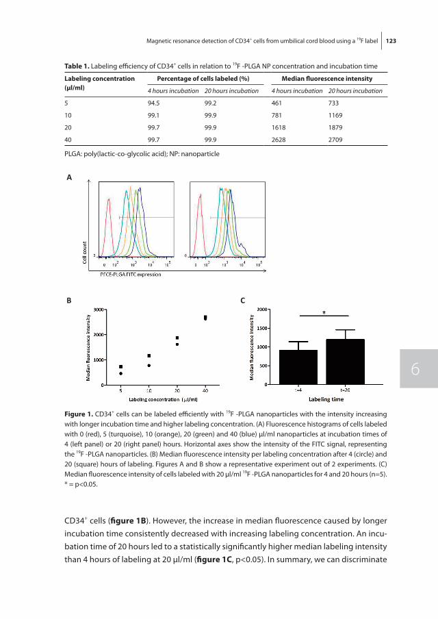
123
6
Magnetic resonance detection of CD34+ cells from umbilical cord blood using a 19F label
CD34+ cells (figure 1B). However, the increase in median fluorescence caused by longer incubation time consistently decreased with increasing labeling concentration. An incu-bation time of 20 hours led to a statistically significantly higher median labeling intensity than 4 hours of labeling at 20 μl/ml (figure 1C, p<0.05). In summary, we can discriminate
table 1. Labeling efficiency of CD34+ cells in relation to 19F -PLGA NP concentration and incubation time
Labeling concentration (µl/ml)
Percentage of cells labeled (%) median fluorescence intensity
4 hours incubation 20 hours incubation 4 hours incubation 20 hours incubation
5 94.5 99.2 461 733
10 99.1 99.9 781 1169
20 99.7 99.9 1618 1879
40 99.7 99.9 2628 2709
PLGA: poly(lactic-co-glycolic acid); NP: nanoparticle
A
B C
figure 1. CD34+ cells can be labeled efficiently with 19F -PLGA nanoparticles with the intensity increasing with longer incubation time and higher labeling concentration. (A) Fluorescence histograms of cells labeled with 0 (red), 5 (turquoise), 10 (orange), 20 (green) and 40 (blue) µl/ml nanoparticles at incubation times of 4 (left panel) or 20 (right panel) hours. Horizontal axes show the intensity of the FITC signal, representing the 19F -PLGA nanoparticles. (B) Median fluorescence intensity per labeling concentration after 4 (circle) and 20 (square) hours of labeling. Figures A and B show a representative experiment out of 2 experiments. (C) Median fluorescence intensity of cells labeled with 20 µl/ml 19F -PLGA nanoparticles for 4 and 20 hours (n=5). * = p<0.05.
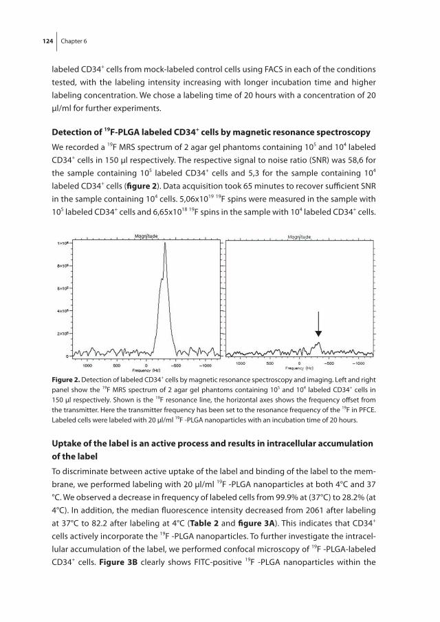
124 Chapter 6
labeled CD34+ cells from mock-labeled control cells using FACS in each of the conditions tested, with the labeling intensity increasing with longer incubation time and higher labeling concentration. We chose a labeling time of 20 hours with a concentration of 20 μl/ml for further experiments.
Detection of 19f-PLGa labeled CD34+ cells by magnetic resonance spectroscopy
We recorded a 19F MRS spectrum of 2 agar gel phantoms containing 105 and 104 labeled CD34+ cells in 150 µl respectively. The respective signal to noise ratio (SNR) was 58,6 for the sample containing 105 labeled CD34+ cells and 5,3 for the sample containing 104 labeled CD34+ cells (figure 2). Data acquisition took 65 minutes to recover sufficient SNR in the sample containing 104 cells. 5,06x10¹⁹ 19F spins were measured in the sample with 105 labeled CD34+ cells and 6,65x10¹⁸ 19F spins in the sample with 104 labeled CD34+ cells.
Uptake of the label is an active process and results in intracellular accumulation of the label
To discriminate between active uptake of the label and binding of the label to the mem-brane, we performed labeling with 20 µl/ml 19F -PLGA nanoparticles at both 4°C and 37 °C. We observed a decrease in frequency of labeled cells from 99.9% at (37°C) to 28.2% (at 4°C). In addition, the median fluorescence intensity decreased from 2061 after labeling at 37°C to 82.2 after labeling at 4°C (table 2 and figure 3a). This indicates that CD34+ cells actively incorporate the 19F -PLGA nanoparticles. To further investigate the intracel-lular accumulation of the label, we performed confocal microscopy of 19F -PLGA-labeled CD34+ cells. figure 3B clearly shows FITC-positive 19F -PLGA nanoparticles within the
figure 2. Detection of labeled CD34+ cells by magnetic resonance spectroscopy and imaging. Left and right panel show the 19F MRS spectrum of 2 agar gel phantoms containing 105 and 104 labeled CD34+ cells in 150 µl respectively. Shown is the 19F resonance line, the horizontal axes shows the frequency offset from the transmitter. Here the transmitter frequency has been set to the resonance frequency of the 19F in PFCE. Labeled cells were labeled with 20 µl/ml 19F -PLGA nanoparticles with an incubation time of 20 hours.
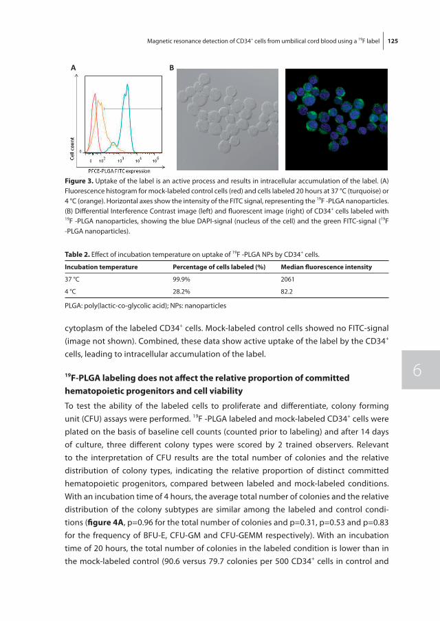
125
6
Magnetic resonance detection of CD34+ cells from umbilical cord blood using a 19F label
cytoplasm of the labeled CD34+ cells. Mock-labeled control cells showed no FITC-signal (image not shown). Combined, these data show active uptake of the label by the CD34+ cells, leading to intracellular accumulation of the label.
19f-PLGa labeling does not affect the relative proportion of committed hematopoietic progenitors and cell viability
To test the ability of the labeled cells to proliferate and differentiate, colony forming unit (CFU) assays were performed. 19F -PLGA labeled and mock-labeled CD34+ cells were plated on the basis of baseline cell counts (counted prior to labeling) and after 14 days of culture, three different colony types were scored by 2 trained observers. Relevant to the interpretation of CFU results are the total number of colonies and the relative distribution of colony types, indicating the relative proportion of distinct committed hematopoietic progenitors, compared between labeled and mock-labeled conditions. With an incubation time of 4 hours, the average total number of colonies and the relative distribution of the colony subtypes are similar among the labeled and control condi-tions (figure 4a, p=0.96 for the total number of colonies and p=0.31, p=0.53 and p=0.83 for the frequency of BFU-E, CFU-GM and CFU-GEMM respectively). With an incubation time of 20 hours, the total number of colonies in the labeled condition is lower than in the mock-labeled control (90.6 versus 79.7 colonies per 500 CD34+ cells in control and
A B
figure 3. Uptake of the label is an active process and results in intracellular accumulation of the label. (A) Fluorescence histogram for mock-labeled control cells (red) and cells labeled 20 hours at 37 °C (turquoise) or 4 °C (orange). Horizontal axes show the intensity of the FITC signal, representing the 19F -PLGA nanoparticles. (B) Differential Interference Contrast image (left) and fluorescent image (right) of CD34+ cells labeled with 19F -PLGA nanoparticles, showing the blue DAPI-signal (nucleus of the cell) and the green FITC-signal (19F -PLGA nanoparticles).
table 2. Effect of incubation temperature on uptake of 19F -PLGA NPs by CD34+ cells.
Incubation temperature Percentage of cells labeled (%) median fluorescence intensity
37 °C 99.9% 2061
4 °C 28.2% 82.2
PLGA: poly(lactic-co-glycolic acid); NPs: nanoparticles
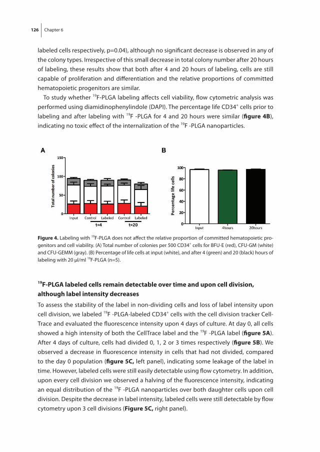
126 Chapter 6
labeled cells respectively, p=0.04), although no significant decrease is observed in any of the colony types. Irrespective of this small decrease in total colony number after 20 hours of labeling, these results show that both after 4 and 20 hours of labeling, cells are still capable of proliferation and differentiation and the relative proportions of committed hematopoietic progenitors are similar.
To study whether 19F-PLGA labeling affects cell viability, flow cytometric analysis was performed using diamidinophenylindole (DAPI). The percentage life CD34+ cells prior to labeling and after labeling with 19F -PLGA for 4 and 20 hours were similar (figure 4B), indicating no toxic effect of the internalization of the 19F -PLGA nanoparticles.
19f-PLGa labeled cells remain detectable over time and upon cell division, although label intensity decreases
To assess the stability of the label in non-dividing cells and loss of label intensity upon cell division, we labeled 19F -PLGA-labeled CD34+ cells with the cell division tracker Cell-Trace and evaluated the fluorescence intensity upon 4 days of culture. At day 0, all cells showed a high intensity of both the CellTrace label and the 19F -PLGA label (figure 5a). After 4 days of culture, cells had divided 0, 1, 2 or 3 times respectively (figure 5B). We observed a decrease in fluorescence intensity in cells that had not divided, compared to the day 0 population (figure 5C, left panel), indicating some leakage of the label in time. However, labeled cells were still easily detectable using flow cytometry. In addition, upon every cell division we observed a halving of the fluorescence intensity, indicating an equal distribution of the 19F -PLGA nanoparticles over both daughter cells upon cell division. Despite the decrease in label intensity, labeled cells were still detectable by flow cytometry upon 3 cell divisions (figure 5C, right panel).
figure 4. Labeling with 19F-PLGA does not affect the relative proportion of committed hematopoietic pro-genitors and cell viability. (A) Total number of colonies per 500 CD34+ cells for BFU-E (red), CFU-GM (white) and CFU-GEMM (gray). (B) Percentage of life cells at input (white), and after 4 (green) and 20 (black) hours of labeling with 20 µl/ml 19F-PLGA (n=5).
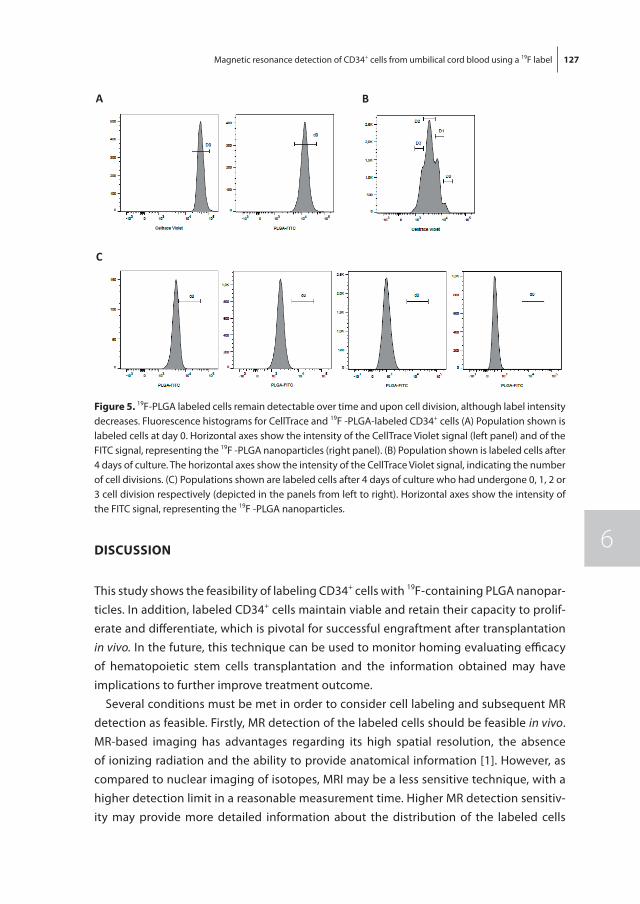
127
6
Magnetic resonance detection of CD34+ cells from umbilical cord blood using a 19F label
DISCUSSIon
This study shows the feasibility of labeling CD34+ cells with 19F-containing PLGA nanopar-ticles. In addition, labeled CD34+ cells maintain viable and retain their capacity to prolif-erate and differentiate, which is pivotal for successful engraftment after transplantation in vivo. In the future, this technique can be used to monitor homing evaluating efficacy of hematopoietic stem cells transplantation and the information obtained may have implications to further improve treatment outcome.
Several conditions must be met in order to consider cell labeling and subsequent MR detection as feasible. Firstly, MR detection of the labeled cells should be feasible in vivo. MR-based imaging has advantages regarding its high spatial resolution, the absence of ionizing radiation and the ability to provide anatomical information [1]. However, as compared to nuclear imaging of isotopes, MRI may be a less sensitive technique, with a higher detection limit in a reasonable measurement time. Higher MR detection sensitiv-ity may provide more detailed information about the distribution of the labeled cells
A B
C
figure 5. 19F-PLGA labeled cells remain detectable over time and upon cell division, although label intensity decreases. Fluorescence histograms for CellTrace and 19F -PLGA-labeled CD34+ cells (A) Population shown is labeled cells at day 0. Horizontal axes show the intensity of the CellTrace Violet signal (left panel) and of the FITC signal, representing the 19F -PLGA nanoparticles (right panel). (B) Population shown is labeled cells after 4 days of culture. The horizontal axes show the intensity of the CellTrace Violet signal, indicating the number of cell divisions. (C) Populations shown are labeled cells after 4 days of culture who had undergone 0, 1, 2 or 3 cell division respectively (depicted in the panels from left to right). Horizontal axes show the intensity of the FITC signal, representing the 19F -PLGA nanoparticles.
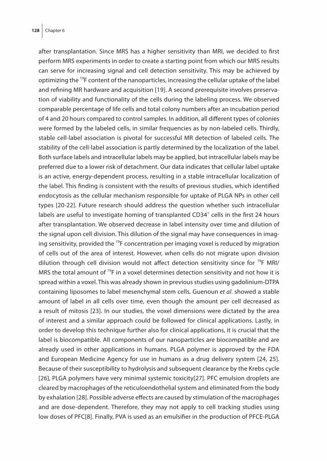
128 Chapter 6
after transplantation. Since MRS has a higher sensitivity than MRI, we decided to first perform MRS experiments in order to create a starting point from which our MRS results can serve for increasing signal and cell detection sensitivity. This may be achieved by optimizing the 19F content of the nanoparticles, increasing the cellular uptake of the label and refining MR hardware and acquisition [19]. A second prerequisite involves preserva-tion of viability and functionality of the cells during the labeling process. We observed comparable percentage of life cells and total colony numbers after an incubation period of 4 and 20 hours compared to control samples. In addition, all different types of colonies were formed by the labeled cells, in similar frequencies as by non-labeled cells. Thirdly, stable cell-label association is pivotal for successful MR detection of labeled cells. The stability of the cell-label association is partly determined by the localization of the label. Both surface labels and intracellular labels may be applied, but intracellular labels may be preferred due to a lower risk of detachment. Our data indicates that cellular label uptake is an active, energy-dependent process, resulting in a stable intracellular localization of the label. This finding is consistent with the results of previous studies, which identified endocytosis as the cellular mechanism responsible for uptake of PLGA NPs in other cell types [20-22]. Future research should address the question whether such intracellular labels are useful to investigate homing of transplanted CD34+ cells in the first 24 hours after transplantation. We observed decrease in label intensity over time and dilution of the signal upon cell division. This dilution of the signal may have consequences in imag-ing sensitivity, provided the 19F concentration per imaging voxel is reduced by migration of cells out of the area of interest. However, when cells do not migrate upon division dilution through cell division would not affect detection sensitivity since for 19F MRI/MRS the total amount of 19F in a voxel determines detection sensitivity and not how it is spread within a voxel. This was already shown in previous studies using gadolinium-DTPA containing liposomes to label mesenchymal stem cells. Guenoun et al. showed a stable amount of label in all cells over time, even though the amount per cell decreased as a result of mitosis [23]. In our studies, the voxel dimensions were dictated by the area of interest and a similar approach could be followed for clinical applications. Lastly, in order to develop this technique further also for clinical applications, it is crucial that the label is biocompatible. All components of our nanoparticles are biocompatible and are already used in other applications in humans. PLGA polymer is approved by the FDA and European Medicine Agency for use in humans as a drug delivery system [24, 25]. Because of their susceptibility to hydrolysis and subsequent clearance by the Krebs cycle [26], PLGA polymers have very minimal systemic toxicity[27]. PFC emulsion droplets are cleared by macrophages of the reticuloendothelial system and eliminated from the body by exhalation [28]. Possible adverse effects are caused by stimulation of the macrophages and are dose-dependent. Therefore, they may not apply to cell tracking studies using low doses of PFC[8]. Finally, PVA is used as an emulsifier in the production of PFCE-PLGA
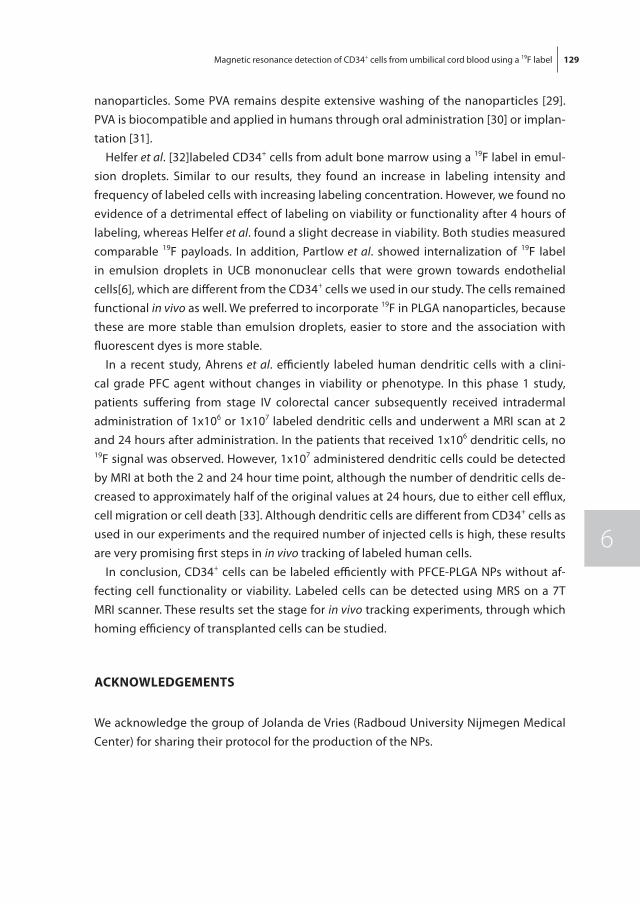
129
6
Magnetic resonance detection of CD34+ cells from umbilical cord blood using a 19F label
nanoparticles. Some PVA remains despite extensive washing of the nanoparticles [29]. PVA is biocompatible and applied in humans through oral administration [30] or implan-tation [31].
Helfer et al. [32]labeled CD34+ cells from adult bone marrow using a 19F label in emul-sion droplets. Similar to our results, they found an increase in labeling intensity and frequency of labeled cells with increasing labeling concentration. However, we found no evidence of a detrimental effect of labeling on viability or functionality after 4 hours of labeling, whereas Helfer et al. found a slight decrease in viability. Both studies measured comparable 19F payloads. In addition, Partlow et al. showed internalization of 19F label in emulsion droplets in UCB mononuclear cells that were grown towards endothelial cells[6], which are different from the CD34+ cells we used in our study. The cells remained functional in vivo as well. We preferred to incorporate 19F in PLGA nanoparticles, because these are more stable than emulsion droplets, easier to store and the association with fluorescent dyes is more stable.
In a recent study, Ahrens et al. efficiently labeled human dendritic cells with a clini-cal grade PFC agent without changes in viability or phenotype. In this phase 1 study, patients suffering from stage IV colorectal cancer subsequently received intradermal administration of 1x10⁶ or 1x10⁷ labeled dendritic cells and underwent a MRI scan at 2 and 24 hours after administration. In the patients that received 1x10⁶ dendritic cells, no 19F signal was observed. However, 1x10⁷ administered dendritic cells could be detected by MRI at both the 2 and 24 hour time point, although the number of dendritic cells de-creased to approximately half of the original values at 24 hours, due to either cell efflux, cell migration or cell death [33]. Although dendritic cells are different from CD34+ cells as used in our experiments and the required number of injected cells is high, these results are very promising first steps in in vivo tracking of labeled human cells.
In conclusion, CD34+ cells can be labeled efficiently with PFCE-PLGA NPs without af-fecting cell functionality or viability. Labeled cells can be detected using MRS on a 7T MRI scanner. These results set the stage for in vivo tracking experiments, through which homing efficiency of transplanted cells can be studied.
aCKnoWLEDGEmEntS
We acknowledge the group of Jolanda de Vries (Radboud University Nijmegen Medical Center) for sharing their protocol for the production of the NPs.
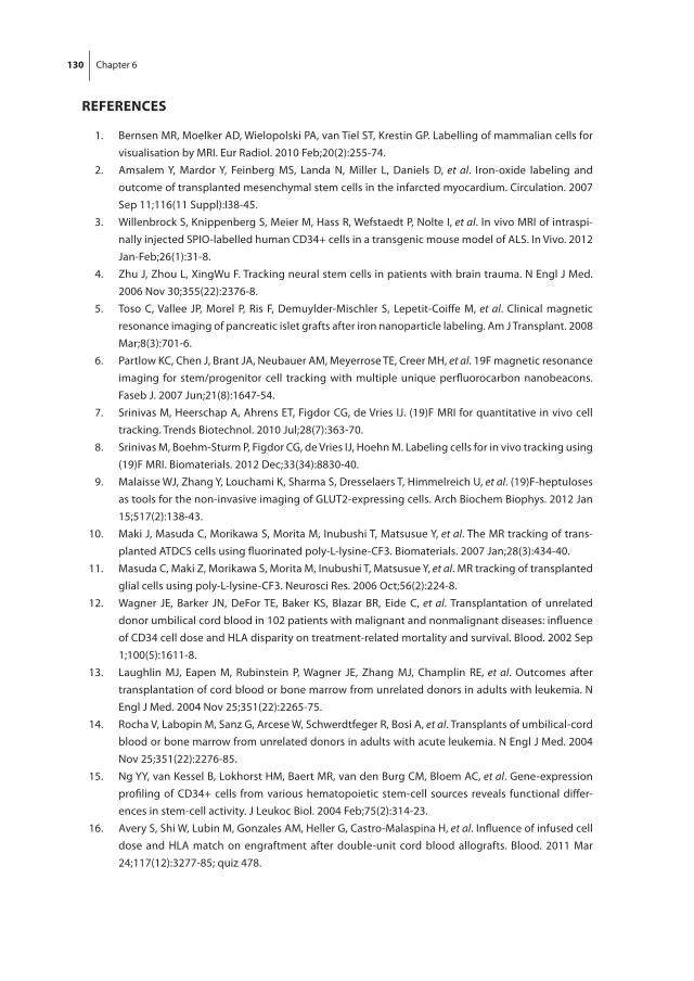
130 Chapter 6
rEfErEnCES
1. Bernsen MR, Moelker AD, Wielopolski PA, van Tiel ST, Krestin GP. Labelling of mammalian cells for visualisation by MRI. Eur Radiol. 2010 Feb;20(2):255-74.
2. Amsalem Y, Mardor Y, Feinberg MS, Landa N, Miller L, Daniels D, et al. Iron-oxide labeling and outcome of transplanted mesenchymal stem cells in the infarcted myocardium. Circulation. 2007 Sep 11;116(11 Suppl):I38-45.
3. Willenbrock S, Knippenberg S, Meier M, Hass R, Wefstaedt P, Nolte I, et al. In vivo MRI of intraspi-nally injected SPIO-labelled human CD34+ cells in a transgenic mouse model of ALS. In Vivo. 2012 Jan-Feb;26(1):31-8.
4. Zhu J, Zhou L, XingWu F. Tracking neural stem cells in patients with brain trauma. N Engl J Med. 2006 Nov 30;355(22):2376-8.
5. Toso C, Vallee JP, Morel P, Ris F, Demuylder-Mischler S, Lepetit-Coiffe M, et al. Clinical magnetic resonance imaging of pancreatic islet grafts after iron nanoparticle labeling. Am J Transplant. 2008 Mar;8(3):701-6.
6. Partlow KC, Chen J, Brant JA, Neubauer AM, Meyerrose TE, Creer MH, et al. 19F magnetic resonance imaging for stem/progenitor cell tracking with multiple unique perfluorocarbon nanobeacons. Faseb J. 2007 Jun;21(8):1647-54.
7. Srinivas M, Heerschap A, Ahrens ET, Figdor CG, de Vries IJ. (19)F MRI for quantitative in vivo cell tracking. Trends Biotechnol. 2010 Jul;28(7):363-70.
8. Srinivas M, Boehm-Sturm P, Figdor CG, de Vries IJ, Hoehn M. Labeling cells for in vivo tracking using (19)F MRI. Biomaterials. 2012 Dec;33(34):8830-40.
9. Malaisse WJ, Zhang Y, Louchami K, Sharma S, Dresselaers T, Himmelreich U, et al. (19)F-heptuloses as tools for the non-invasive imaging of GLUT2-expressing cells. Arch Biochem Biophys. 2012 Jan 15;517(2):138-43.
10. Maki J, Masuda C, Morikawa S, Morita M, Inubushi T, Matsusue Y, et al. The MR tracking of trans-planted ATDC5 cells using fluorinated poly-L-lysine-CF3. Biomaterials. 2007 Jan;28(3):434-40.
11. Masuda C, Maki Z, Morikawa S, Morita M, Inubushi T, Matsusue Y, et al. MR tracking of transplanted glial cells using poly-L-lysine-CF3. Neurosci Res. 2006 Oct;56(2):224-8.
12. Wagner JE, Barker JN, DeFor TE, Baker KS, Blazar BR, Eide C, et al. Transplantation of unrelated donor umbilical cord blood in 102 patients with malignant and nonmalignant diseases: influence of CD34 cell dose and HLA disparity on treatment-related mortality and survival. Blood. 2002 Sep 1;100(5):1611-8.
13. Laughlin MJ, Eapen M, Rubinstein P, Wagner JE, Zhang MJ, Champlin RE, et al. Outcomes after transplantation of cord blood or bone marrow from unrelated donors in adults with leukemia. N Engl J Med. 2004 Nov 25;351(22):2265-75.
14. Rocha V, Labopin M, Sanz G, Arcese W, Schwerdtfeger R, Bosi A, et al. Transplants of umbilical-cord blood or bone marrow from unrelated donors in adults with acute leukemia. N Engl J Med. 2004 Nov 25;351(22):2276-85.
15. Ng YY, van Kessel B, Lokhorst HM, Baert MR, van den Burg CM, Bloem AC, et al. Gene-expression profiling of CD34+ cells from various hematopoietic stem-cell sources reveals functional differ-ences in stem-cell activity. J Leukoc Biol. 2004 Feb;75(2):314-23.
16. Avery S, Shi W, Lubin M, Gonzales AM, Heller G, Castro-Malaspina H, et al. Influence of infused cell dose and HLA match on engraftment after double-unit cord blood allografts. Blood. 2011 Mar 24;117(12):3277-85; quiz 478.
131
6
Magnetic resonance detection of CD34+ cells from umbilical cord blood using a 19F label
17. Hidalgo A, Weiss LA, Frenette PS. Functional selectin ligands mediating human CD34(+) cell interactions with bone marrow endothelium are enhanced postnatally. J Clin Invest. 2002 Aug;110(4):559-69.
18. Srinivas M, Cruz LJ, Bonetto F, Heerschap A, Figdor CG, de Vries IJ. Customizable, multi-functional fluorocarbon nanoparticles for quantitative in vivo imaging using 19F MRI and optical imaging. Biomaterials. 2010 Sep;31(27):7070-7.
19. Amiri H, Srinivas M, Veltien A, van Uden MJ, de Vries IJ, Heerschap A. Cell tracking using (19)F magnetic resonance imaging: technical aspects and challenges towards clinical applications. Eur Radiol. 2015 Mar;25(3):726-35.
20. Qaddoumi MG, Ueda H, Yang J, Davda J, Labhasetwar V, Lee VH. The characteristics and mecha-nisms of uptake of PLGA nanoparticles in rabbit conjunctival epithelial cell layers. Pharm Res. 2004 Apr;21(4):641-8.
21. Cartiera MS, Johnson KM, Rajendran V, Caplan MJ, Saltzman WM. The uptake and intracellular fate of PLGA nanoparticles in epithelial cells. Biomaterials. 2009 May;30(14):2790-8.
22. Panyam J, Labhasetwar V. Dynamics of endocytosis and exocytosis of poly(D,L-lactide-co-glycolide) nanoparticles in vascular smooth muscle cells. Pharm Res. 2003 Feb;20(2):212-20.
23. Guenoun J, Koning GA, Doeswijk G, Bosman L, Wielopolski PA, Krestin GP, et al. Cationic Gd-DTPA liposomes for highly efficient labeling of mesenchymal stem cells and cell tracking with MRI. Cell Transplant. 2012;21(1):191-205.
24. Danhier F, Ansorena E, Silva JM, Coco R, Le Breton A, Preat V. PLGA-based nanoparticles: an over-view of biomedical applications. J Control Release. 2012 Jul 20;161(2):505-22.
25. Mundargi RC, Babu VR, Rangaswamy V, Patel P, Aminabhavi TM. Nano/micro technologies for delivering macromolecular therapeutics using poly(D,L-lactide-co-glycolide) and its derivatives. J Control Release. 2008 Feb 11;125(3):193-209.
26. Houchin ML, Topp EM. Chemical degradation of peptides and proteins in PLGA: a review of reac-tions and mechanisms. J Pharm Sci. 2008 Jul;97(7):2395-404.
27. Kumari A, Yadav SK, Yadav SC. Biodegradable polymeric nanoparticles based drug delivery sys-tems. Colloids Surf B Biointerfaces. 2010 Jan 1;75(1):1-18.
28. Flaim SF. Pharmacokinetics and side effects of perfluorocarbon-based blood substitutes. Artif Cells Blood Substit Immobil Biotechnol. 1994;22(4):1043-54.
29. Sahoo SK, Panyam J, Prabha S, Labhasetwar V. Residual polyvinyl alcohol associated with poly (D,L-lactide-co-glycolide) nanoparticles affects their physical properties and cellular uptake. J Control Release. 2002 Jul 18;82(1):105-14.
30. DeMerlis CC, Schoneker DR. Review of the oral toxicity of polyvinyl alcohol (PVA). Food Chem Toxicol. 2003 Mar;41(3):319-26.
31. Baker MI, Walsh SP, Schwartz Z, Boyan BD. A review of polyvinyl alcohol and its uses in cartilage and orthopedic applications. J Biomed Mater Res B Appl Biomater. 2012 Jul;100(5):1451-7.
32. Helfer BM, Balducci A, Sadeghi Z, O’Hanlon C, Hijaz A, Flask CA, et al. (1)(9)F MRI tracer preserves in vitro and in vivo properties of hematopoietic stem cells. Cell Transplant. 2013;22(1):87-97.
33. Ahrens ET, Helfer BM, O’Hanlon CF, Schirda C. Clinical cell therapy imaging using a perfluorocarbon tracer and fluorine-19 MRI. Magn Reson Med. 2014 Dec;72(6):1696-701.

A
Chapter 7Towards clinical application of StemRegenin1-expanded umbilical cord blood-derived hematopoietic stem and progenitor cells: manufacture and clinical study design
Lucia E. Duinhouwer1, Mariëtte ter Borg1, Eric Braakman1 and Jan J. Cornelissen1
1Department of Hematology, Erasmus Medical Centre, Rotterdam, the Netherlands

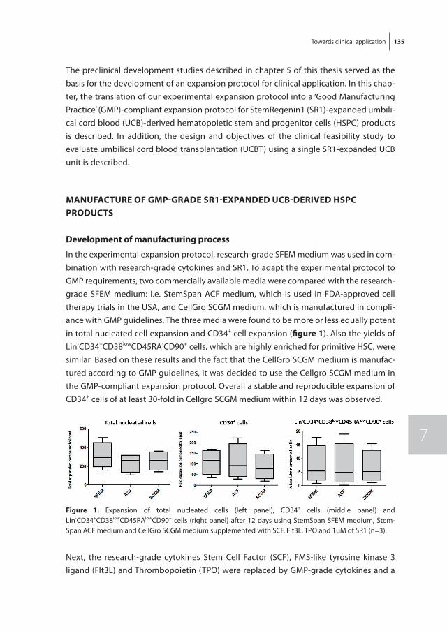
Towards clinical application 135
7
The preclinical development studies described in chapter 5 of this thesis served as the basis for the development of an expansion protocol for clinical application. In this chap-ter, the translation of our experimental expansion protocol into a ‘Good Manufacturing Practice’ (GMP)-compliant expansion protocol for StemRegenin1 (SR1)-expanded umbili-cal cord blood (UCB)-derived hematopoietic stem and progenitor cells (HSPC) products is described. In addition, the design and objectives of the clinical feasibility study to evaluate umbilical cord blood transplantation (UCBT) using a single SR1-expanded UCB unit is described.
manUfaCtUrE of GmP-GraDE Sr1-ExPanDED UCB-DErIVED HSPC ProDUCtS
Development of manufacturing process
In the experimental expansion protocol, research-grade SFEM medium was used in com-bination with research-grade cytokines and SR1. To adapt the experimental protocol to GMP requirements, two commercially available media were compared with the research-grade SFEM medium: i.e. StemSpan ACF medium, which is used in FDA-approved cell therapy trials in the USA, and CellGro SCGM medium, which is manufactured in compli-ance with GMP guidelines. The three media were found to be more or less equally potent in total nucleated cell expansion and CD34+ cell expansion (figure 1). Also the yields of Lin-CD34+CD38lowCD45RA-CD90+ cells, which are highly enriched for primitive HSC, were similar. Based on these results and the fact that the CellGro SCGM medium is manufac-tured according to GMP guidelines, it was decided to use the Cellgro SCGM medium in the GMP-compliant expansion protocol. Overall a stable and reproducible expansion of CD34+ cells of at least 30-fold in Cellgro SCGM medium within 12 days was observed.
figure 1. Expansion of total nucleated cells (left panel), CD34+ cells (middle panel) and Lin-CD34+CD38lowCD45RAlowCD90+ cells (right panel) after 12 days using StemSpan SFEM medium, Stem-Span ACF medium and CellGro SCGM medium supplemented with SCF, Flt3L, TPO and 1µM of SR1 (n=3).
Next, the research-grade cytokines Stem Cell Factor (SCF), FMS-like tyrosine kinase 3 ligand (Flt3L) and Thrombopoietin (TPO) were replaced by GMP-grade cytokines and a
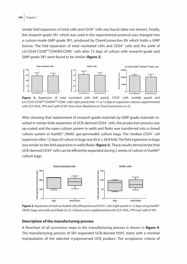
136 Chapter 7
similar fold expansion of total cells and CD34+ cells was found (data not shown). Finally, the research-grade SR1 which was used in the experimental protocol was changed into a custom-made GMP-grade SR1, produced by ChemConnection BV which holds a GMP license. The fold expansion of total nucleated cells and CD34+ cells and the yield of Lin-CD34+CD38lowCD45RA-CD90+ cells after 12 days of culture with research-grade and GMP-grade SR1 were found to be similar (figure 2).
figure 2. Expansion of total nucleated cells (left panel), CD34+ cells (middle panel) and Lin-CD34+CD38lowCD45RAlowCD90+ cells (right panel) after 11 or 12 days in expansion cultures supplemented with SCF, Flt3L, TPO and 1µM of SR1 from Axon Medchem or ChemConnection (n=3)
After showing that replacement of research-grade materials by GMP-grade materials re-sulted in similar folds expansion of UCB-derived CD34+ cells, the production process was up-scaled and the open culture system in wells and flasks was transferred into a closed culture system in Vuelife® 290AC gas-permeable culture bags. The median CD34+ cell expansion after 12 days of culture in bags was 85.6 ± 28.8 fold. The fold expansion in bags was similar to the fold expansion in wells/flasks (figure 3). These results demonstrate that UCB-derived CD34+ cells can be efficiently expanded during 2 weeks of culture in Vuelife® culture bags.
figure 3. Expansion of total nucleated cells (left panel) and CD34+ cells (right panel) in 12 days using Vuelife® 290AC bags and wells and flasks (n=5). Cultures were supplemented with SCF, Flt3L, TPO and 1µM of SR1.
Description of the manufacturing process
A flowchart of all successive steps in the manufacturing process is shown in figure 4. The manufacturing process of SR1-expanded UCB-derived HSPC starts with a minimal manipulation of the selected cryopreserved UCB product. The acceptance criteria of
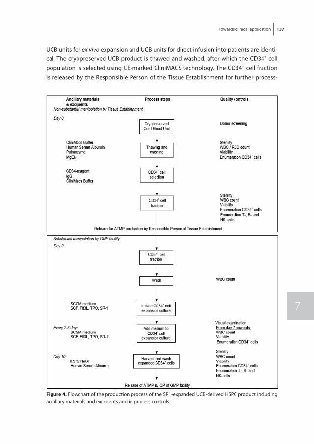
Towards clinical application 137
7
UCB units for ex vivo expansion and UCB units for direct infusion into patients are identi-cal. The cryopreserved UCB product is thawed and washed, after which the CD34+ cell population is selected using CE-marked CliniMACS technology. The CD34+ cell fraction is released by the Responsible Person of the Tissue Establishment for further process-
figure 4. Flowchart of the production process of the SR1-expanded UCB-derived HSPC product including ancillary materials and excipients and in process controls.
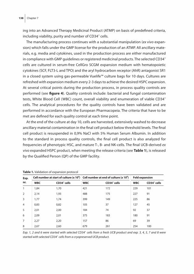
138 Chapter 7
ing into an Advanced Therapy Medicinal Product (ATMP) on basis of predefined criteria, including viability, purity and number of CD34+ cells.
The manufacturing process continues with a substantial manipulation (ex vivo expan-sion) which falls under the GMP license for the production of an ATMP. All ancillary mate-rials, e.g. media and cytokines, used in the production process are either manufactured in compliance with GMP guidelines or registered medicinal products. The selected CD34+ cells are cultured in serum-free CellGro SCGM expansion medium with hematopoietic cytokines (SCF, FLT3-L and TPO) and the aryl hydrocarbon receptor (AhR) antagonist SR1 in a closed system using gas-permeable Vuelife™ culture bags for 10 days. Cultures are refreshed with expansion medium every 2-3 days to achieve the desired HSPC expansion. At several critical points during the production process, in process quality controls are performed (see figure 4). Quality controls include: bacterial and fungal contamination tests, White Blood Cell (WBC) count, overall viability and enumeration of viable CD34+ cells. The analytical procedures for the quality controls have been validated and are performed in accordance with the European Pharmacopeia. The criteria that have to be met are defined for each quality control at each time point.
At the end of the culture at day 10, cells are harvested, extensively washed to decrease ancillary material contamination in the final cell product below threshold levels. The final cell product is resuspended in 0,9% NaCl with 5% Human Serum Albumin. In addition to the standard in process quality controls, the final cell product is also analyzed for frequencies of phenotypic HSC, and mature T-, B- and NK-cells. The final UCB-derived ex vivo expanded HSPC product, when meeting the release criteria (see table 1), is released by the Qualified Person (QP) of the GMP facility.
table 1. Validation of expansion protocol
Exp. nr.
Cell number at start of culture (x 106) Cell number at end of culture (x 106) fold expansion
WBC CD34+ cells WBC CD34+ cells WBC CD34+ cells
1 1,84 1,70 421 172 229 101
2 2,14 1,93 488 175 227 91
3 1,77 1,74 399 149 225 86
4 0,83 0,82 105 37 127 45
5 2,01 2,00 184 74 92 37
6 2,09 2,01 375 183 180 91
7 2,27 2,20 157 86 69 39
8 2,67 2,60 679 261 254 100
Exp. 1, 2 and 6 were started with selected CD34+ cells from a fresh UCB product and exp. 3, 4, 5, 7 and 8 were started with selected CD34+ cells from a cryopreserved UCB product.
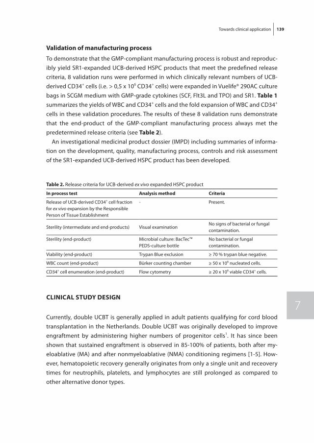
Towards clinical application 139
7
Validation of manufacturing process
To demonstrate that the GMP-compliant manufacturing process is robust and reproduc-ibly yield SR1-expanded UCB-derived HSPC products that meet the predefined release criteria, 8 validation runs were performed in which clinically relevant numbers of UCB-derived CD34+ cells (i.e. > 0,5 x 10⁶ CD34+ cells) were expanded in Vuelife® 290AC culture bags in SCGM medium with GMP-grade cytokines (SCF, Flt3L and TPO) and SR1. table 1 summarizes the yields of WBC and CD34+ cells and the fold expansion of WBC and CD34+ cells in these validation procedures. The results of these 8 validation runs demonstrate that the end-product of the GMP-compliant manufacturing process always met the predetermined release criteria (see table 2).
An investigational medicinal product dossier (IMPD) including summaries of informa-tion on the development, quality, manufacturing process, controls and risk assessment of the SR1-expanded UCB-derived HSPC product has been developed.
CLInICaL StUDy DESIGn
Currently, double UCBT is generally applied in adult patients qualifying for cord blood transplantation in the Netherlands. Double UCBT was originally developed to improve engraftment by administering higher numbers of progenitor cells1. It has since been shown that sustained engraftment is observed in 85-100% of patients, both after my-eloablative (MA) and after nonmyeloablative (NMA) conditioning regimens [1-5]. How-ever, hematopoietic recovery generally originates from only a single unit and receovery times for neutrophils, platelets, and lymphocytes are still prolonged as compared to other alternative donor types.
table 2. Release criteria for UCB-derived ex vivo expanded HSPC product
In process test analysis method Criteria
Release of UCB-derived CD34+ cell fraction for ex vivo expansion by the Responsible Person of Tissue Establishment
- Present.
Sterility (intermediate and end-products) Visual examination No signs of bacterial or fungal contamination.
Sterility (end-product) Microbial culture: BacTec™ PEDS-culture bottle
No bacterial or fungal contamination.
Viability (end-product) Trypan Blue exclusion ≥ 70 % trypan blue negative.
WBC count (end-product) Bürker counting chamber ≥ 50 x 106 nucleated cells.
CD34+ cell enumeration (end-product) Flow cytometry ≥ 20 x 106 viable CD34+ cells.
140 Chapter 7
Preceding studies: HoVon studies 106 and 115
Recently, results from the HOVON 106 study were published, showing the feasibility of double (dUCBT) preceded by a 4 Gy TBI containing reduced intensity conditioning (RIC) regimen in patients with poor-risk underlying hematological malignancies [6,7]. Data indicated that dUCBT following the above mentioned RIC regimen is feasible and may provide an important alternative source of hematopoietic stem cells in recipients lack-ing a sibling or well matched unrelated donor (MUD). With a median follow-up of 35 months, 2-year OS was 57%, 2 year TRM was 19%, and 2 year PFS was 42%. Thereafter, the HOVON-106 protocol has become the standard protocol for application of UCBT in adult patients in The
Netherlands. Sustained hematopoiesis in dUCBT recipients was derived from a single donor in 90% of patients and early predominance was already discernible at days 7-14 [6,7]. The mechanism of predominance was unclear, but subject of investigation in the succeeding HOVON 115 study. It was hypothesized that HLA class II specific CD4+ T cells of the ultimate engrafting UCB unit elicit an immediate and targeted alloreactive im-mune response towards mismatched HLA class II molecules of the non-engrafting UCB cells, resulting in rapid rejection of the non-engrafting unit. The HOVON-115 trial was designed to test that hypothesis. It was shown that the majority of patients indeed de-veloped an early expansion of CD4+ T-cells, including effector T-cells with specificity for mismatched class II alleles of the rejected cord. The majority of mismatched class II alleles between the 2 cords in each individual patient evoked an alloreactive response, which appeared most prominent early after transplantation [8]. These findings suggested that matching of the 2 cords at the allele level for class II is no longer obligatory and can be omitted, which expands the choices for dUCBT. Despite the low incidence of graft failure following dUCBT, hematopoietic recovery is usually slow with neutrophil recovery (>0.5 x 10⁻⁹/L) around day 26 after transplantation and platelet recovery (>50) around day 50. Retarded hematopoietic recovery is mainly associated with the low number of HSPC in the graft. In order to increase these numbers, several investigators have initiated studies to expand HSPC ex vivo and improve hematopoietic recovery [9-21].
Expansion of hematopoietic stem cells
In this thesis, the history and current practice on the expansion of HSC are comprehen-sively described. Currently, the hypothesis that ex vivo expansion of HSC requires a com-bination of proliferative signals provided by hematopoietic growth factors with a factor that inhibits differentiation during culture has been studied extensively. Compounds including StemRegenin1 [20-21], copper chelation [14-15], mesenchymal stem cells [18-19], Nicotinamide [12-13], and Notch ligand Delta-1 [16-17], were studied in order to test that hypothesis. Of these, especially SR1 was shown to be associated with inhibition of
Towards clinical application 141
7
differentiation and when used with early acting cytokines to result in strong expansion of HSPC (this thesis (Chapter 5); [20-21]).
rationale of the study
Transplantation of SR1-expanded CD34+ cells in a clinical double cord blood setting ap-peared safe and feasible and resulted in engraftment in all 17 transplanted patients [21]. The median time to neutrophil and platelet recovery was 15 and 49 days, respectively, which is faster compared to an historical control group receiving unmanipulated dUCBT. In 11/17 patients, hematopoiesis was primarily derived from the expanded unit and in those patients, neutrophil recovery was even more rapid with a median of 11 days versus 23 days in the 6/17 patients engrafting with the unmanipulated unit [21]. These results have set the stage for clinical studies in a single UCBT setting, which is proposed here. Collectively, in view of 1. the rejection of one UCB graft in the majority of recipients of double UCBT, 2. the absent contribution of the rejected unit to hematopoietic recovery, and 3. the potency of SR1 to expand hematopoietic progenitor cells and thereby to improve engraftment and recovery, it is proposed to perform a feasibility study in 10-15 patients evaluating single UCBT using one SR1 expanded unit.
Study objectives
• To study the feasibility of single UCBT using one SR1-expanded unit• To assess engraftment and engraftment kinetics; to evaluate immune reconstitution,
acute and chronic Graft-Versus-Host Disease (GVHD), chimerism, toxicity, progres-sion-free survival and overall survival after single unit UCBT, using with one ex vivo expanded unit.
Study design
This is a prospective phase I-II feasibility study. Patients lacking a matched unrelated donor and patients for whom a matched unrelated donor cannot be identified within 2 months and for whom an allogeneic transplant is urgently needed, are eligible for UCBT if a suitable UCB unit is available. Transplantation will be preceded by a reduced-intensity conditioning regimen, irrespective of patient age. Post grafting immunosuppression is performed by mycophenolate mofetil (30 days) and cyclosporine A (90 days, taper there-after).
Study population
Eligibility for registrationAll patients must be registered before start of treatment and must meet all of the follow-ing eligibility criteria.

142 Chapter 7
Inclusion criteria• Age 18-70 years inclusive• Diagnosis of poor-risk hematological malignancy or (Very) Severe Anaplastic Anemia
relapsing after or failing immunosuppressive therapy and meeting the criteria for a MUD allo-SCT
• Lacking a sufficiently matched volunteer unrelated donor or lacking such a donor within the required time period of ≤ 2 months in case of urgently needed allo-SCT
• Availability of 1 (≥5/6) matched UCB graft with a nuclear cell count > 2,7 x 10⁷/kg (see paragraph 8.2).
• Availability of a back-up autograft, harvested and frozen earlier in the course of treat-ment, (harvest according to local apheresis policies)
• WHO performance status 0-2• Written informed consent
Exclusion criteria• Bilirubin and/or transaminases > 2.5 x normal value • Creatinine clearance < 40 ml/min• Cardiac dysfunction as defined by:- Reduced left ventricular function with an ejection fraction < 45% as measured by
MUGA scan or echocardiogram (another method for measuring cardiac function is acceptable)
- Unstable angina- Unstable cardiac arrhythmias• Pulmonary function test with VC, FEV1 and/ or DCO < 50%• Active, uncontrolled infection• History of high dose (≥ 8 Gy) total body irradiation• Pregnant or lactating females• HIV positivity
UCB unit selection
UCB units will be selected according to the following criteria: 1. The total amount of total nucleated cells present in the UCB unit must be > 2.7 x 10⁷/
kg recipient body weight.2. HLA-matching is performed for HLA-A and HLA-B at the serological split resolution
level and for HLA-DRB1 at the 4-digit resolution level. The minimal match grade required is a 5/6 match both between recipient and the UCB unit.
3. Absence of HLA-antibodies in the recipient directed against HLA class I and class II mismatches on the cord blood cells is required.
4. Preferably, RBC- and plasma reduced UCB units are selected.
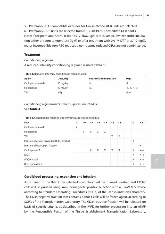
Towards clinical application 143
7
5. Preferably, ABO-compatible or minor ABO-mismatched UCB units are selected.6. Preferably, UCB units are selected from NETCORD/FACT accredited UCB banksNote: If recipient anti-A/anti-B titer >512, (NaCl gel card (Diamed, Switzerland)); incuba-tion either at room temperature (IgM) or after treatment with 0.01M DTT at 37° C (IgG), major incompatible non-RBC reduced / non-plasma reduced CBUs are not administered.
treatment
Conditioning regimenA reduced-intensity conditioning regimen is used (table 3).
table 3. Reduced-intensity conditioning regimen used
agent Dose/day route of administration Days
Cyclophosphamide 60 mg/kg i.v. -7
Fludarabine 40 mg/m2 i.v. -6, -5, -4, -3
TBI 2 Gy -2, -1
Conditioning regimen and immunosuppression scheduleSee table 4.
table 4. Conditioning regimen and immunosuppression schedule
Day - 7 - 6 - 5 - 4 - 3 - 2 - 1 0 + 1
Cyclophosphamide X
Fludarabine X X X X
TBI X X
Infusion of ex vivo expanded HSPC product X
Infusion of UCB-CD34- fraction X
Cyclosporine A X X X X X X X→
MMF X X →
(Val)acyclovir X X →
Benzylpenicilline X X →
Cord blood processing, expansion and infusion
As outlined in the IMPD, the selected cord blood will be thawed, washed and CD34+ cells will be purified using immunomagnetic positive selection with a CliniMACS device according to Standard Operating Procedures (SOP’s) of the Transplantation Laboratory. The CD34 negative fraction that contains donor T-cells will be frozen again, according to SOP’s of the Transplantation Laboratory. The CD34 positive fraction will be released on basis of specific criteria, as described in the IMPD for further processing into an ATMP by the Responsible Person of the Tissue Establishment Transplantation Laboratory.
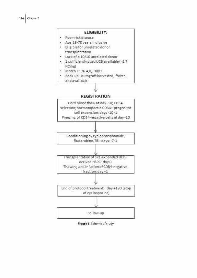
144 Chapter 7
figure 5. Scheme of study
Towards clinical application 145
7
Subsequently, CD34+ cells will be expanded in ex vivo cultures using a combination of cytokines and SR1, as detailed in the IMPD. The UCB-derived ex vivo expanded HSPC end product will be released by the QP of the GMP facility on basis of prespecified criteria (as outlined in detail in the IMPD). The manipulated UCB unit will contain after culture no red blood cells and will also be devoid of donor plasma, which obviates the need for additional precautions in case of major and minor ABO-incompatibility. The expanded graft will be infused on day 0, followed by thawing and infusion of the cryopreserved CD34 negative fraction on day +1.
The CD34 negative fraction contains RBC and residual donor plasma, which necessi-tates the need for monitoring of hemolysis (haptoglobin, bilirubin, LDH, creatinine, CBC, DAT and plasma-Hb) in case of major- or minor AB0 blood group incompatibility.
approval
As outlined above, a clinical study protocol and the IMPD have been submitted for ap-proval to the competent authorities in the Netherlands, the Centrale Commissie Mensge-bonden Onderzoek (CCMO).
146 Chapter 7
rEfErEnCES
1. Barker JN, Weisdorf DJ, DeFor TE, Blazar BR, Mc Glave PB, Miller JS, et al. Transplantation of 2 par-tially HLA-matched umbilical cord blood units to enhance engraftment in adults with hematologic malignancy. Blood. 2005;105(3):1343-1347.
2. Barker, JN, Weisdorf DJ, DeFor TE, Blazar BR, Miller JS, Wagner JE. Rapid and complete donor chi-merism in adult recipients of unrelated donor umbilical cord blood transplantation after reduced-intensity conditioning. Blood. 2003;102(5):1915-1919
3. Ballen KK, Spitzer TR, Yeap BY, McAfee S, Dey BR, Attar E, et al. Double unrelated reduced-intensity umbilical cord blood transplantation in adults. Biol Blood Marrow Transplant. 2007;13(1):82-89.
4. Brunstein CG, Barker JN, Weisdorf DJ, DeFor TE, Miller JS, Blazar BR, et al. Umbilical cord blood transplantation after nonmyeloablative conditioning: impact on transplantation outcomes in 110 adults with hematologic disease. Blood. 2007;110(8):3064-3070.
5. Verneris MR, Brunstein CG, Barker JN, MacMillan ML, DeFor TE, McKenna DH, et al. Relapse risk after umbilical cord blood transplantation: enhanced graft-versus-leukemia effect in recipients of 2 units. Blood. 2009;114(19):4293-4299.
6. Somers JA, Braakman E, van der Holt B, Petersen EJ, Marijt EW, Huisman C, Sintnicolaas K, Oudshoorn M, Groenendijk-Sijnke ME, Brand A, Cornelissen JJ. Rapid induction of single donor chimerism after double umbilical cord blood transplantation preceded by reduced intensity conditioning: results of the HOVON 106 phase II study. Haematologica. 2014;99(11):1753-61.
7. Somers JA, Brand A, van Hensbergen Y, Mulder A, Oudshoorn M, Sintnicolaas K, Gratama JW, Falkenburg JH, Braakman E, Cornelissen JJ. Double umbilical cord blood transplantation: a study of early engraftment kinetics in leukocyte subsets using HLA-specific monoclonal antibodies. Biol Blood Marrow Transplant. 2013;19(2):266-73.
8. Cornelissen JJ, Wijers R, van Bergen CAM, Somers JA, Braakman E, Gratama JW, et al. Early CD4+ T-Cell Effector Alloreactivity Towards Multiple Mismatched HLA Class II Alleles Is Associated with Graft Predominance after Double Umbilical Cord Blood Transplantation (dUCBT). 57th ASH Annual Meeting & Exposition; December 03, 2015; Orlando, United States of America: Blood; 2015. p. 387.
9. Sauvageau G, Iscove NN, Humphries RK. In vitro and in vivo expansion of hematopoietic stem cells. Oncogene. 2004;23(43):7223-32.
10. Ivanovic Z, Duchez P, Chevaleyre J, Vlaski M, Lafarge X, Dazey B, et al. Clinical-scale cultures of cord blood CD34(+) cells to amplify committed progenitors and maintain stem cell activity. Cell Transplant. 2011;20(9):1453-63.
11. Walasek MA, van Os R, de Haan G. Hematopoietic stem cell expansion: challenges and opportuni-ties. Ann N Y Acad Sci. 2012 Aug;1266:138-50.
12. Peled T, Shoham H, Aschengrau D, Yackoubov D, Frei G, Rosenheimer GN, et al. Nicotinamide, a SIRT1 inhibitor, inhibits differentiation and facilitates expansion of hematopoietic progenitor cells with enhanced bone marrow homing and engraftment. Exp Hematol. 2012;40(4):342-55 e1.
13. Horwitz ME, Chao NJ, Rizzieri DA, Long GD, Sullivan KM, Gasparetto C, et al. Umbilical cord blood expansion with nicotinamide provides long-term multilineage engraftment. J Clin Invest. 2014;124(7):3121-8.
14. Peled T, Landau E, Mandel J, Glukhman E, Goudsmid NR, Nagler A, et al. Linear polyamine copper chelator tetraethylenepentamine augments long-term ex vivo expansion of cord blood-derived CD34+ cells and increases their engraftment potential in NOD/SCID mice. Exp Hematol. 2004 Jun;32(6):547-55.
Towards clinical application 147
7
15. de Lima M, McMannis J, Gee A, Komanduri K, Couriel D, Andersson BS, et al. Transplantation of ex vivo expanded cord blood cells using the copper chelator tetraethylenepentamine: a phase I/II clinical trial. Bone Marrow Transplant. 2008 May;41(9):771-8.
16. Delaney C, Varnum-Finney B, Aoyama K, Brashem-Stein C, Bernstein ID. Dose-dependent effects of the Notch ligand Delta1 on ex vivo differentiation and in vivo marrow repopulating ability of cord blood cells. Blood. 2005 Oct 15;106(8):2693-9.
17. Delaney C, Heimfeld S, Brashem-Stein C, Voorhies H, Manger RL, Bernstein ID. Notch-mediated expansion of human cord blood progenitor cells capable of rapid myeloid reconstitution. Nat Med. 2010 Feb;16(2):232-6.
18. Robinson SN, Ng J, Niu T, Yang H, McMannis JD, Karandish S, et al. Superior ex vivo cord blood expansion following co-culture with bone marrow-derived mesenchymal stem cells. Bone Marrow Transplant. 2006 Feb;37(4):359-66.
19. de Lima M, McNiece I, Robinson SN, Munsell M, Eapen M, Horowitz M, et al. Cord-blood engraft-ment with ex vivo mesenchymal-cell coculture. N Engl J Med. 2012 Dec 13;367(24):2305-15
20. Boitano AE, Wang J, Romeo R, Bouchez LC, Parker AE, Sutton SE, et al. Aryl hydrocarbon recep-tor antagonists promote the expansion of human hematopoietic stem cells. Science. 2010 Sep 10;329(5997):1345-8.
21. Wagner JE, Jr., Brunstein CG, Boitano AE, DeFor TE, McKenna D, Sumstad D, et al. Phase I/II Trial of StemRegenin-1 Expanded Umbilical Cord Blood Hematopoietic Stem Cells Supports Testing as a Stand-Alone Graft. Cell Stem Cell. 2016 Jan 7;18(1):144-55.


A
Chapter 8General discussion

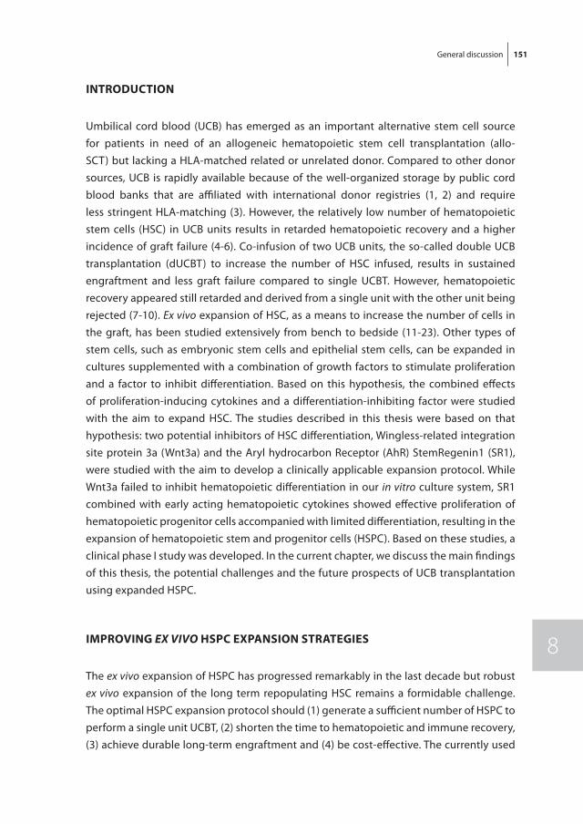
General discussion 151
8
IntroDUCtIon
Umbilical cord blood (UCB) has emerged as an important alternative stem cell source for patients in need of an allogeneic hematopoietic stem cell transplantation (allo-SCT) but lacking a HLA-matched related or unrelated donor. Compared to other donor sources, UCB is rapidly available because of the well-organized storage by public cord blood banks that are affiliated with international donor registries (1, 2) and require less stringent HLA-matching (3). However, the relatively low number of hematopoietic stem cells (HSC) in UCB units results in retarded hematopoietic recovery and a higher incidence of graft failure (4-6). Co-infusion of two UCB units, the so-called double UCB transplantation (dUCBT) to increase the number of HSC infused, results in sustained engraftment and less graft failure compared to single UCBT. However, hematopoietic recovery appeared still retarded and derived from a single unit with the other unit being rejected (7-10). Ex vivo expansion of HSC, as a means to increase the number of cells in the graft, has been studied extensively from bench to bedside (11-23). Other types of stem cells, such as embryonic stem cells and epithelial stem cells, can be expanded in cultures supplemented with a combination of growth factors to stimulate proliferation and a factor to inhibit differentiation. Based on this hypothesis, the combined effects of proliferation-inducing cytokines and a differentiation-inhibiting factor were studied with the aim to expand HSC. The studies described in this thesis were based on that hypothesis: two potential inhibitors of HSC differentiation, Wingless-related integration site protein 3a (Wnt3a) and the Aryl hydrocarbon Receptor (AhR) StemRegenin1 (SR1), were studied with the aim to develop a clinically applicable expansion protocol. While Wnt3a failed to inhibit hematopoietic differentiation in our in vitro culture system, SR1 combined with early acting hematopoietic cytokines showed effective proliferation of hematopoietic progenitor cells accompanied with limited differentiation, resulting in the expansion of hematopoietic stem and progenitor cells (HSPC). Based on these studies, a clinical phase I study was developed. In the current chapter, we discuss the main findings of this thesis, the potential challenges and the future prospects of UCB transplantation using expanded HSPC.
ImProVInG ex vIvo HSPC ExPanSIon StratEGIES
The ex vivo expansion of HSPC has progressed remarkably in the last decade but robust ex vivo expansion of the long term repopulating HSC remains a formidable challenge. The optimal HSPC expansion protocol should (1) generate a sufficient number of HSPC to perform a single unit UCBT, (2) shorten the time to hematopoietic and immune recovery, (3) achieve durable long-term engraftment and (4) be cost-effective. The currently used
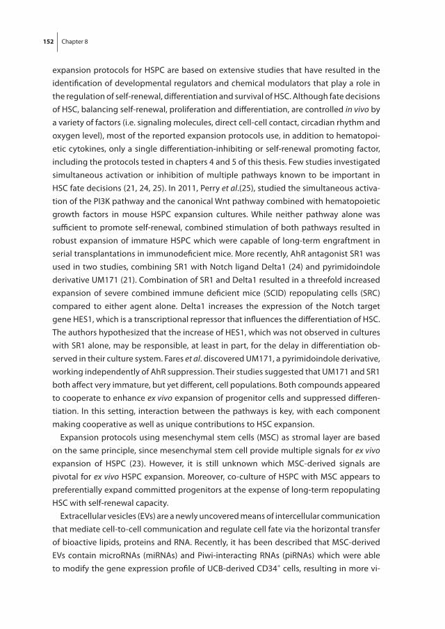
152 Chapter 8
expansion protocols for HSPC are based on extensive studies that have resulted in the identification of developmental regulators and chemical modulators that play a role in the regulation of self-renewal, differentiation and survival of HSC. Although fate decisions of HSC, balancing self-renewal, proliferation and differentiation, are controlled in vivo by a variety of factors (i.e. signaling molecules, direct cell-cell contact, circadian rhythm and oxygen level), most of the reported expansion protocols use, in addition to hematopoi-etic cytokines, only a single differentiation-inhibiting or self-renewal promoting factor, including the protocols tested in chapters 4 and 5 of this thesis. Few studies investigated simultaneous activation or inhibition of multiple pathways known to be important in HSC fate decisions (21, 24, 25). In 2011, Perry et al.(25), studied the simultaneous activa-tion of the PI3K pathway and the canonical Wnt pathway combined with hematopoietic growth factors in mouse HSPC expansion cultures. While neither pathway alone was sufficient to promote self-renewal, combined stimulation of both pathways resulted in robust expansion of immature HSPC which were capable of long-term engraftment in serial transplantations in immunodeficient mice. More recently, AhR antagonist SR1 was used in two studies, combining SR1 with Notch ligand Delta1 (24) and pyrimidoindole derivative UM171 (21). Combination of SR1 and Delta1 resulted in a threefold increased expansion of severe combined immune deficient mice (SCID) repopulating cells (SRC) compared to either agent alone. Delta1 increases the expression of the Notch target gene HES1, which is a transcriptional repressor that influences the differentiation of HSC. The authors hypothesized that the increase of HES1, which was not observed in cultures with SR1 alone, may be responsible, at least in part, for the delay in differentiation ob-served in their culture system. Fares et al. discovered UM171, a pyrimidoindole derivative, working independently of AhR suppression. Their studies suggested that UM171 and SR1 both affect very immature, but yet different, cell populations. Both compounds appeared to cooperate to enhance ex vivo expansion of progenitor cells and suppressed differen-tiation. In this setting, interaction between the pathways is key, with each component making cooperative as well as unique contributions to HSC expansion.
Expansion protocols using mesenchymal stem cells (MSC) as stromal layer are based on the same principle, since mesenchymal stem cell provide multiple signals for ex vivo expansion of HSPC (23). However, it is still unknown which MSC-derived signals are pivotal for ex vivo HSPC expansion. Moreover, co-culture of HSPC with MSC appears to preferentially expand committed progenitors at the expense of long-term repopulating HSC with self-renewal capacity.
Extracellular vesicles (EVs) are a newly uncovered means of intercellular communication that mediate cell-to-cell communication and regulate cell fate via the horizontal transfer of bioactive lipids, proteins and RNA. Recently, it has been described that MSC-derived EVs contain microRNAs (miRNAs) and Piwi-interacting RNAs (piRNAs) which were able to modify the gene expression profile of UCB-derived CD34+ cells, resulting in more vi-
General discussion 153
8
able, less differentiated cells (26). Furthermore, human osteoblast-derived EVs promoted proliferation and cell cycle progression of human UCB-derived HSPC and enhanced the ex vivo expansion of human UCB-derived HSPC, including the most immature subset in growth factor supported expansion cultures. EV-expanded HSPC retained their full differ-entiation capacity in vitro and successfully engrafted in immune-deficient NSG mice (27). Interestingly, the expansion promoting effects of osteoblast-derived EVs on UCB-derived HSPC remained evident even in the presence of the differentiation inhibiting factor SR1. However, the molecular mechanisms underlying the EVs mediated expansion of HSPC still remain to be explored.
Another strategy to improve engraftment and time to hematopoietic and immune recovery is to expose HSPC to factors that improve homing of HSPC towards the bone marrow niche. It is known that UCB-derived HSPC do not home as efficiently to the bone marrow as HSPC derived from adult bone marrow (28) and in addition, cytokine exposure of HSPC leads to a rapid loss of bone marrow homing capacity (29). Several strategies have been reported to enhance the homing capacity of HSPC and thereby enhance hematopoietic recovery post-transplantation, such as fucosylation of the HSPC ligands binding to P- and E-selectin adhesion molecules expressed on bone marrow endothelial cells (30, 31) or activation of ‘homing receptor’ CXCR4 on HSPC by treatment with 16,16-dimethyl prostaglandin E2 (dmPGE2) (32, 33). Practical advantage of these techniques is that they require minimal processing as the maximum incubation time is 120 minutes. Strategies aiming to combine an optimized expansion protocol with an approach to improve the homing capacity of the expanded cells may result in a graft with optimal engraftment capacity and hematopoietic recovery potential.
ImmUnE rEConStItUtIon aftEr ExPanDED (D)UCBt
Of all the different expansion protocols, only few were tested clinically (14, 16, 18, 20, 23). The studies by Horwitz et al. (14), Wagner et al. (20) and de Lima et al. (23), with lim-ited patient numbers, showed significant enhanced hematopoietic recovery in terms of neutrophil and platelet recovery. Neutrophil recovery was accelerated by approximately 10 days, while the time to platelet recovery (> 50 x 109/L) was reduced with 4 to 5 days. However, apart from protracted neutrophil and platelet recovery, especially retarded immune recovery is of concern, as non-relapse mortality (NRM) is especially associated with insufficient T cell recovery rather than protracted neutrophil recovery(34). T cell recovery after dUCBT was studied and described in chapter 2. A higher risk of severe infections in patients failing to recover thymopoiesis at 3 months post-transplantation was described, which compares well to our earlier observations in unrelated donor allo-SCT recipients (34). Sofar, only one clinical study addressed the recovery of B and T
154 Chapter 8
cells after expanded UCB transplantation (20), which failed to demonstrate improved T cell recovery upon engraftment of the expanded UCB unit compared to T cell recovery originating from the unmanipulated unit in a dUCBT setting. Of note, T cell recovery was even suggested to occur at a slower rate after transplantation using an expanded UCB unit (20). How to explain the latter findings? Different factors may be involved. First, it is still possible that expansion of true HSC was limited, while more committed progenitors were expanded at a greater degree, especially myeloid committed progenitors (35). The rapid neutrophil recovery rather than lymphoid recovery might support that explana-tion. Secondly, T cell recovery not only depends on hematopoietic HSC engraftment but also depends on generation of lymphoid precursors, their entry into the thymus, and ef-fective thymopoiesis. It is well known that thymopoiesis in adult patients can be severely compromised (36-38) despite efficient engraftment and early hematopoietic recovery. However, Wils et al. (36) showed that T cell recovery may be improved upon expansion of HSC and lymphoid progenitors in vivo by treatment with Flt3ligand of immunodeficient mice having received a bone marrow transplantation. Lastly, many transplant centers use G-SCF post-transplant to accelerate neutrophil recovery. That myeloid growth factor may skew hematopoietic recovery towards the myeloid lineage. Therefore, a slow rate of T cell recovery after UCBT with expanded HSC/HPC may be due to a combination of insufficient thymic function and insufficient generation and supply of lymphoid progenitors. Since T cell recovery is pivotal to reduce the toxic and potential lethal complications of allo-SCT, it is of the utmost importance to develop expansion strategies that also expand T cell precursors. Apart from the cytokines we applied in our expansion protocol (SCF, Flt3L and TPO), lymphopoietic cytokines including IL-7 and IL-15 might be considered. Both cytokines are acting as survival factors for (developing) T cells by the prevention of apop-tosis through up- and downregulation of anti- and pro-apoptotic factors, respectively (39, 40). An alternative approach might the use of stromal cells, such as DL-1 expressing cells, in the expansion protocol. Interaction of HPC with DL-1 results in the generation of T cell progenitors, that may directly enter the thymus (41). Given the pivotal role of the thymic epithelium in T-cell recovery, another strategy to improve T cell recovery might be the application of strategies that favorably affect the thymic epithelium. Possible strategies may include protection of the nursing stromal compartment by keratinocyte growth factor or by regeneration of the thymic epithelium (34). Altogether, it is clear that expansion of true hematopoietic stem cells is an important first step, but it might be nec-essary to add strategies specially aiming to improve T cell recovery and thymic function. Therefore, in future clinical trials with ex vivo expanded stem cell grafts, endpoints should include improvement of T cell reconstitution and thymopoiesis, apart from neutrophil and platelet recovery. In addition, immune reconstitution data should be correlated to HSC and HPC numbers infused as well as to subsets of lymphoid and myeloid progenitors obtained after expansion.
General discussion 155
8
aLtErnatIVE DonorS
HLA-matched related and HLA 10/10 matched unrelated donors are currently considered as preferred donors for patients eligible for allo-SCT. However, many patients lack such a preferred donor and depend on alternative stem cell sources. Currently, alternative donors or stem cell sources include mismatched unrelated donors, haplo-identical family donors, and UCB. While the large retrospective registry study by Brunstein et al. suggested that disease free survival (DFS) in recipients of UCBT may come close to DFS following matched unrelated allo-SCT (51% versus 48%, respectively), NRM remains of concern (34% versus 14%, respectively)(42). The use of UCB as an alternative stem cell source for adult patients is hampered by the limited number of HSPC in most of the UCB grafts available, leading to susceptibility for infections and also NRM. In order to increase the number of HSPC, double UCBT was developed , which however insufficiently accelerated recovery and also appeared to be associated with high costs. Other alterna-tive donor sources for patients lacking a matched related or unrelated donor include HLA-mismatched unrelated donors (MMUD, defined as 1 or 2 mismatches at class I and/or class II) or haplo-identical family donors (haplo-SCT). Currently, no consensus has been reached as regards the hierarchy of alternative donors. Recently, several studies have been published discussing and comparing alternative transplant possibilities (43-47). An important concern for the use of MMUD grafts is the higher risk of GVHD due to HLA-incompatibility. However, HLA-incompatibility may also be associated with a somewhat higher graft versus leukemia (GVL) effect, preventing disease relapse. A recent meta-analysis comparing overall survival after MUD and MMUD showed an increased mortality risk of 27% for patients receiving a MMUD (43). It indicates that the excess rate of NRM was insufficiently compensated by a lower risk of relapse, leading to significantly reduced OS. In the past, results obtained with haplo-SCT suggested a similar pattern of increased NRM associated with increased degree of HLA-incompatibility, which is insuf-ficiently counteracted by a reduced relapse rate (48, 49). As a result of observed high NRM, also autologous transplantation has been brought forward as an alternative stem cell source (50, 51). While the latter stem cell source lacks allogenic T cells and thereby is devoid of the allogeneic GVL-effect, the very low rate of NRM may favor that mode of transplantation. Especially in leukemia patients, autografting may provide for an alter-native stem cells source if no HLA-identical sibling or MUD is available (52). However, the last decade has witnessed an enormous surge in haplo-SCT, because of significant progress in the prevention of GVHD and thereby reduction of NRM. Especially the use of post-transplant cyclophosphamide, as explored by the Baltimore group (53, 54) appeared associated with effective prevention of GVHD and limited NRM. Of note, an accompany-ing increase of relapse, as may be expected by inhibition and elimination of allogeneic haplo-identical T-cells, has sofar not been demonstrated. These results suggest that the
156 Chapter 8
net result of low NRM and an acceptable risk of relapse may favor haplo-SCT. In addition, benefits of haplo-SCT include the rapid availability of the donor, the option for subse-quent donor lymphocyte infusions and the relatively low costs. When compared to UCBT, clinical outcome after haplo-SCT was reported to be largely similar in terms of relapse incidence, NRM and relapse-free survival (RFS) in a retrospective study including patients with acute leukemia (46). However, higher rates of relapse and lower rates of NRM, result-ing in similar OS, have been reported as well (47). Preliminary findings in a large group of high-risk AML patients showed a lower relapse rate and similar NRM upon haplo-SCT (using post-transplant cyclophosphamide) compared to UCBT, resulting in better OS and RFS at 2 years post-transplantation (55). Collectively, recent studies have shown a clear improvement in haplo-SCT, while NRM following UCB remains of concern. Future studies using expanded UCB will answer the question whether manipulation of UCB grafts may be associated with less NRM and to what extent that will compare to haplo-SCT. Until results of those studies become available, allo-SCT using sibling and MUD donors are still to be preferred, while haploidentical donors and UCB can be considered as preferred alternative donors and mismatched unrelated donors as third alternative, which then, however, should also be weighed versus autologous transplantation.
ConCLUSIon
The field of alternative donor transplantation is currently rapidly evolving. Although hampered by the small size of the grafts, UCB is an important stem cell source for patients in high need of an allogenic HSC donor. Ex vivo expansion protocols using a combination of signals to inhibit differentiation in combination with proliferation inducing growth factors are promising. Preclinical studies as described in this thesis and clinical studies in dUCBT settings have set the stage for a clinical feasibility study of expanded HSPC in a single unit setting. Results of that study as well as those addressing other approaches to expand HSC are eagerly awaited.
General discussion 157
8
rEfErEnCES
1. Barker JN, Krepski TP, DeFor TE, Davies SM, Wagner JE, Weisdorf DJ. Searching for unrelated donor hematopoietic stem cells: availability and speed of umbilical cord blood versus bone marrow. Biol Blood Marrow Transplant. 2002;8(5):257-60.
2. Hakenberg P, Kogler G, Wernet P. NETCORD: a cord blood allocation network. Bone Marrow Trans-plant. 1998 Jul;22 Suppl 1:S17-8.
3. Petersdorf EW, Gooley TA, Anasetti C, Martin PJ, Smith AG, Mickelson EM, et al. Optimizing outcome after unrelated marrow transplantation by comprehensive matching of HLA class I and II alleles in the donor and recipient. Blood. 1998 Nov 15;92(10):3515-20.
4. Barker JN, Scaradavou A, Stevens CE. Combined effect of total nucleated cell dose and HLA match on transplantation outcome in 1061 cord blood recipients with hematologic malignancies. Blood. 2010 Mar 4;115(9):1843-9.
5. Wagner JE, Barker JN, DeFor TE, Baker KS, Blazar BR, Eide C, et al. Transplantation of unrelated donor umbilical cord blood in 102 patients with malignant and nonmalignant diseases: influence of CD34 cell dose and HLA disparity on treatment-related mortality and survival. Blood. 2002 Sep 1;100(5):1611-8.
6. Gluckman E, Rocha V, Boyer-Chammard A, Locatelli F, Arcese W, Pasquini R, et al. Outcome of cord-blood transplantation from related and unrelated donors. Eurocord Transplant Group and the European Blood and Marrow Transplantation Group. N Engl J Med. 1997 Aug 7;337(6):373-81.
7. Barker JN, Weisdorf DJ, DeFor TE, Blazar BR, McGlave PB, Miller JS, et al. Transplantation of 2 par-tially HLA-matched umbilical cord blood units to enhance engraftment in adults with hematologic malignancy. Blood. 2005 Feb 1;105(3):1343-7.
8. Brunstein CG, Barker JN, Weisdorf DJ, DeFor TE, Miller JS, Blazar BR, et al. Umbilical cord blood transplantation after nonmyeloablative conditioning: impact on transplantation outcomes in 110 adults with hematologic disease. Blood. 2007 Oct 15;110(8):3064-70.
9. Ballen KK, Spitzer TR, Yeap BY, McAfee S, Dey BR, Attar E, et al. Double unrelated reduced-intensity umbilical cord blood transplantation in adults. Biol Blood Marrow Transplant. 2007 Jan;13(1):82-9.
10. Somers JA, Braakman E, van der Holt B, Petersen EJ, Marijt EW, Huisman C, et al. Rapid induc-tion of single donor chimerism after double umbilical cord blood transplantation preceded by reduced intensity conditioning: results of the HOVON 106 phase II study. Haematologica. 2014 Nov;99(11):1753-61.
11. Sauvageau G, Iscove NN, Humphries RK. In vitro and in vivo expansion of hematopoietic stem cells. Oncogene. 2004 Sep 20;23(43):7223-32.
12. Ivanovic Z, Duchez P, Chevaleyre J, Vlaski M, Lafarge X, Dazey B, et al. Clinical-scale cultures of cord blood CD34(+) cells to amplify committed progenitors and maintain stem cell activity. Cell Transplant. 2011;20(9):1453-63.
13. Peled T, Shoham H, Aschengrau D, Yackoubov D, Frei G, Rosenheimer GN, et al. Nicotinamide, a SIRT1 inhibitor, inhibits differentiation and facilitates expansion of hematopoietic progenitor cells with enhanced bone marrow homing and engraftment. Exp Hematol. 2012 Apr;40(4):342-55 e1.
14. Horwitz ME, Chao NJ, Rizzieri DA, Long GD, Sullivan KM, Gasparetto C, et al. Umbilical cord blood expansion with nicotinamide provides long-term multilineage engraftment. J Clin Invest. 2014 Jul;124(7):3121-8.
15. Peled T, Landau E, Mandel J, Glukhman E, Goudsmid NR, Nagler A, et al. Linear polyamine copper chelator tetraethylenepentamine augments long-term ex vivo expansion of cord blood-derived
158 Chapter 8
CD34+ cells and increases their engraftment potential in NOD/SCID mice. Exp Hematol. 2004 Jun;32(6):547-55.
16. de Lima M, McMannis J, Gee A, Komanduri K, Couriel D, Andersson BS, et al. Transplantation of ex vivo expanded cord blood cells using the copper chelator tetraethylenepentamine: a phase I/II clinical trial. Bone Marrow Transplant. 2008 May;41(9):771-8.
17. Delaney C, Varnum-Finney B, Aoyama K, Brashem-Stein C, Bernstein ID. Dose-dependent effects of the Notch ligand Delta1 on ex vivo differentiation and in vivo marrow repopulating ability of cord blood cells. Blood. 2005 Oct 15;106(8):2693-9.
18. Delaney C, Heimfeld S, Brashem-Stein C, Voorhies H, Manger RL, Bernstein ID. Notch-mediated expansion of human cord blood progenitor cells capable of rapid myeloid reconstitution. Nat Med. 2010 Feb;16(2):232-6.
19. Boitano AE, Wang J, Romeo R, Bouchez LC, Parker AE, Sutton SE, et al. Aryl hydrocarbon recep-tor antagonists promote the expansion of human hematopoietic stem cells. Science. 2010 Sep 10;329(5997):1345-8.
20. Wagner JE, Jr., Brunstein CG, Boitano AE, DeFor TE, McKenna D, Sumstad D, et al. Phase I/II Trial of StemRegenin-1 Expanded Umbilical Cord Blood Hematopoietic Stem Cells Supports Testing as a Stand-Alone Graft. Cell Stem Cell. 2016 Jan 7;18(1):144-55.
21. Fares I, Chagraoui J, Gareau Y, Gingras S, Ruel R, Mayotte N, et al. Cord blood expansion. Pyrimi-doindole derivatives are agonists of human hematopoietic stem cell self-renewal. Science. 2014 Sep 19;345(6203):1509-12.
22. Robinson SN, Ng J, Niu T, Yang H, McMannis JD, Karandish S, et al. Superior ex vivo cord blood expansion following co-culture with bone marrow-derived mesenchymal stem cells. Bone Marrow Transplant. 2006 Feb;37(4):359-66.
23. de Lima M, McNiece I, Robinson SN, Munsell M, Eapen M, Horowitz M, et al. Cord-blood engraft-ment with ex vivo mesenchymal-cell coculture. N Engl J Med. 2012 Dec 13;367(24):2305-15.
24. Dahlberg A, Brashem-Stein C, Delaney C, Bernstein ID. Enhanced generation of cord blood hema-topoietic stem and progenitor cells by culture with StemRegenin1 and Delta1(Ext-IgG.). Leukemia. 2014 Oct;28(10):2097-101.
25. Perry JM, He XC, Sugimura R, Grindley JC, Haug JS, Ding S, et al. Cooperation between both Wnt/{beta}-catenin and PTEN/PI3K/Akt signaling promotes primitive hematopoietic stem cell self-renewal and expansion. Genes Dev. 2011 Sep 15;25(18):1928-42.
26. De Luca L, Trino S, Laurenzana I, Simeon V, Calice G, Raimondo S, et al. MiRNAs and piRNAs from bone marrow mesenchymal stem cell extracellular vesicles induce cell survival and inhibit cell dif-ferentiation of cord blood hematopoietic stem cells: a new insight in transplantation. Oncotarget. 2016 Feb 9;7(6):6676-92.
27. Morhayim J, van de Peppel J, Braakman E, Rombouts W, ter Borg M, Dudakovic A, et al. Osteoblasts secrete miRNA-containing extracellular vesicles that enhance expansion of human umbilical cord blood cells Nat Sci Rep, accepted for publication. 2016.
28. Hidalgo A, Weiss LA, Frenette PS. Functional selectin ligands mediating human CD34(+) cell interactions with bone marrow endothelium are enhanced postnatally. J Clin Invest. 2002 Aug;110(4):559-69.
29. Ahmed F, Ings SJ, Pizzey AR, Blundell MP, Thrasher AJ, Ye HT, et al. Impaired bone marrow homing of cytokine-activated CD34+ cells in the NOD/SCID model. Blood. 2004 Mar 15;103(6):2079-87.
30. Xia L, McDaniel JM, Yago T, Doeden A, McEver RP. Surface fucosylation of human cord blood cells augments binding to P-selectin and E-selectin and enhances engraftment in bone marrow. Blood. 2004 Nov 15;104(10):3091-6.
General discussion 159
8
31. Robinson SN, Simmons PJ, Thomas MW, Brouard N, Javni JA, Trilok S, et al. Ex vivo fucosylation improves human cord blood engraftment in NOD-SCID IL-2Rgamma(null) mice. Exp Hematol. 2012 Jun;40(6):445-56.
32. Hoggatt J, Singh P, Sampath J, Pelus LM. Prostaglandin E2 enhances hematopoietic stem cell hom-ing, survival, and proliferation. Blood. 2009 May 28;113(22):5444-55.
33. Cutler C, Multani P, Robbins D, Kim HT, Le T, Hoggatt J, et al. Prostaglandin-modulated umbilical cord blood hematopoietic stem cell transplantation. Blood. 2013 Oct 24;122(17):3074-81.
34. Wils EJ, Cornelissen JJ. Thymopoiesis following allogeneic stem cell transplantation: new possibili-ties for improvement. Blood Rev. 2005 Mar;19(2):89-98.
35. Fares I, Rivest-Khan L, Cohen S, Sauvageau G. Small molecule regulation of normal and leukemic stem cells. Curr Opin Hematol. 2015 Jul;22(4):309-16.
36. Wils EJ, Braakman E, Verjans GM, Rombouts EJ, Broers AE, Niesters HG, et al. Flt3 ligand expands lymphoid progenitors prior to recovery of thymopoiesis and accelerates T cell reconstitution after bone marrow transplantation. J Immunol. 2007 Mar 15;178(6):3551-7.
37. Weinberg K, Blazar BR, Wagner JE, Agura E, Hill BJ, Smogorzewska M, et al. Factors affecting thymic function after allogeneic hematopoietic stem cell transplantation. Blood. 2001 Mar 1;97(5):1458-66.
38. Chung B, Barbara-Burnham L, Barsky L, Weinberg K. Radiosensitivity of thymic interleukin-7 pro-duction and thymopoiesis after bone marrow transplantation. Blood. 2001 Sep 1;98(5):1601-6.
39. Broers AE, Gratama JW, Lowenberg B, Cornelissen JJ. Lymphocyte recovery following allogeneic stem cell transplantation: new possibilities for improvement. Clin Appl Immunol Rev. 2002 July-September 2002;2(4-5):217-27.
40. Patidar M, Yadav N, Dalai SK. Interleukin 15: A key cytokine for immunotherapy. Cytokine Growth Factor Rev. 2016 Jun 7.
41. Holmes R, Zuniga-Pflucker JC. The OP9-DL1 system: generation of T-lymphocytes from embryonic or hematopoietic stem cells in vitro. Cold Spring Harb Protoc. 2009 Feb;2009(2):pdb prot5156.
42. Brunstein CG, Gutman JA, Weisdorf DJ, Woolfrey AE, Defor TE, Gooley TA, et al. Allogeneic hema-topoietic cell transplantation for hematologic malignancy: relative risks and benefits of double umbilical cord blood. Blood. 2010 Nov 25;116(22):4693-9.
43. Kekre N, Mak KS, Stopsack KH, Binder M, Ishii K, Branvall E, et al. Impact of HLA-Mismatch in Unrelated Donor Hematopoietic Stem Cell Transplantation: A Meta-Analysis. Am J Hematol. 2016 Jun;91(6):551-5.
44. Ringden O, Labopin M, Ciceri F, Velardi A, Bacigalupo A, Arcese W, et al. Is there a stronger graft-versus-leukemia effect using HLA-haploidentical donors compared with HLA-identical siblings? Leukemia. 2016 Feb;30(2):447-55.
45. Ciurea SO, Zhang MJ, Bacigalupo AA, Bashey A, Appelbaum FR, Aljitawi OS, et al. Haploidentical transplant with posttransplant cyclophosphamide vs matched unrelated donor transplant for acute myeloid leukemia. Blood. 2015 Aug 20;126(8):1033-40.
46. Ruggeri A, Labopin M, Sanz G, Piemontese S, Arcese W, Bacigalupo A, et al. Comparison of out-comes after unrelated cord blood and unmanipulated haploidentical stem cell transplantation in adults with acute leukemia. Leukemia. 2015 Sep;29(9):1891-900.
47. Brunstein CG, Fuchs EJ, Carter SL, Karanes C, Costa LJ, Wu J, et al. Alternative donor transplanta-tion after reduced intensity conditioning: results of parallel phase 2 trials using partially HLA-mismatched related bone marrow or unrelated double umbilical cord blood grafts. Blood. 2011 Jul 14;118(2):282-8.
160 Chapter 8
48. Aversa F. Haploidentical haematopoietic stem cell transplantation for acute leukaemia in adults: experience in Europe and the United States. Bone Marrow Transplant. 2008 Mar;41(5):473-81.
49. Aversa F, Terenzi A, Tabilio A, Falzetti F, Carotti A, Ballanti S, et al. Full haplotype-mismatched hematopoietic stem-cell transplantation: a phase II study in patients with acute leukemia at high risk of relapse. J Clin Oncol. 2005 May 20;23(15):3447-54.
50. Gorin NC, Labopin M, Piemontese S, Arcese W, Santarone S, Huang H, et al. T-cell-replete haploi-dentical transplantation versus autologous stem cell transplantation in adult acute leukemia: a matched pair analysis. Haematologica. 2015 Apr;100(4):558-64.
51. Gorin NC, Giebel S, Labopin M, Savani BN, Mohty M, Nagler A. Autologous stem cell transplanta-tion for adult acute leukemia in 2015: time to rethink? Present status and future prospects. Bone Marrow Transplant. 2015 Dec;50(12):1495-502.
52. Cornelissen JJ, Blaise D. Hematopoietic stem cell transplantation for patients with AML in first complete remission. Blood. 2016 Jan 7;127(1):62-70.
53. Robinson TM, O’Donnell PV, Fuchs EJ, Luznik L. Haploidentical bone marrow and stem cell transplantation: experience with post-transplantation cyclophosphamide. Semin Hematol. 2016 Apr;53(2):90-7.
54. Fuchs EJ. HLA-haploidentical blood or marrow transplantation with high-dose, post-transplanta-tion cyclophosphamide. Bone Marrow Transplant. 2015 Jun;50 Suppl 2:S31-6.
55. Versluis JL, M; Socie, G; Depei, W; Volin, L; Blaise, D; Milpied, N; Craddock, C; Yakoub-Agha, I; Maertens, J; Ljungman, P.T.; Huynh, A; Michalet, M; Deconinck, E; Chevallier, P; Russell, N; Passweg, J.R.; Ossenkoppele, G.J.; Lowenberg, B; Mohty, M; Cornelissen, J.J.; Nagler, A. The Hierarchy of Al-ternative Donors for Allogeneic Hematopoietic Stem Cell Transplantation in Poor Risk AML in CR1: 10/10 Matched Unrelated Donors Still to be Preferred over Haplo-Identical Donors or Umbilical Cord Blood. 56th ASH Annual Meeting and Exposition. San Francisco, CA, USA, 2014.


A
appendicesSummary
Samenvatting
Publications in this thesis
PhD Portfolio
Curriculum vitae
Dankwoord

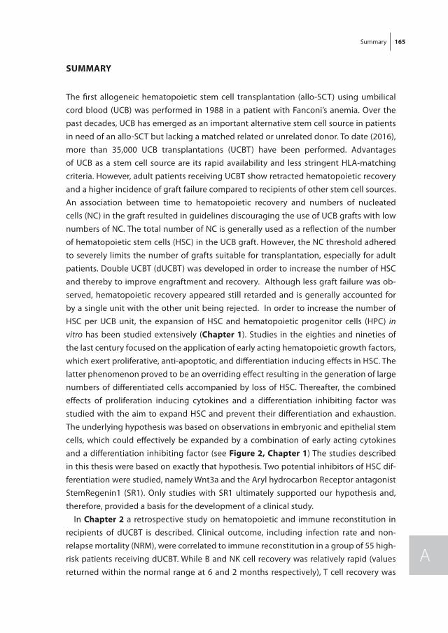
Summary 165
A
SUmmary
The first allogeneic hematopoietic stem cell transplantation (allo-SCT) using umbilical cord blood (UCB) was performed in 1988 in a patient with Fanconi’s anemia. Over the past decades, UCB has emerged as an important alternative stem cell source in patients in need of an allo-SCT but lacking a matched related or unrelated donor. To date (2016), more than 35,000 UCB transplantations (UCBT) have been performed. Advantages of UCB as a stem cell source are its rapid availability and less stringent HLA-matching criteria. However, adult patients receiving UCBT show retracted hematopoietic recovery and a higher incidence of graft failure compared to recipients of other stem cell sources. An association between time to hematopoietic recovery and numbers of nucleated cells (NC) in the graft resulted in guidelines discouraging the use of UCB grafts with low numbers of NC. The total number of NC is generally used as a reflection of the number of hematopoietic stem cells (HSC) in the UCB graft. However, the NC threshold adhered to severely limits the number of grafts suitable for transplantation, especially for adult patients. Double UCBT (dUCBT) was developed in order to increase the number of HSC and thereby to improve engraftment and recovery. Although less graft failure was ob-served, hematopoietic recovery appeared still retarded and is generally accounted for by a single unit with the other unit being rejected. In order to increase the number of HSC per UCB unit, the expansion of HSC and hematopoietic progenitor cells (HPC) in vitro has been studied extensively (Chapter 1). Studies in the eighties and nineties of the last century focused on the application of early acting hematopoietic growth factors, which exert proliferative, anti-apoptotic, and differentiation inducing effects in HSC. The latter phenomenon proved to be an overriding effect resulting in the generation of large numbers of differentiated cells accompanied by loss of HSC. Thereafter, the combined effects of proliferation inducing cytokines and a differentiation inhibiting factor was studied with the aim to expand HSC and prevent their differentiation and exhaustion. The underlying hypothesis was based on observations in embryonic and epithelial stem cells, which could effectively be expanded by a combination of early acting cytokines and a differentiation inhibiting factor (see figure 2, Chapter 1) The studies described in this thesis were based on exactly that hypothesis. Two potential inhibitors of HSC dif-ferentiation were studied, namely Wnt3a and the Aryl hydrocarbon Receptor antagonist StemRegenin1 (SR1). Only studies with SR1 ultimately supported our hypothesis and, therefore, provided a basis for the development of a clinical study.
In Chapter 2 a retrospective study on hematopoietic and immune reconstitution in recipients of dUCBT is described. Clinical outcome, including infection rate and non-relapse mortality (NRM), were correlated to immune reconstitution in a group of 55 high-risk patients receiving dUCBT. While B and NK cell recovery was relatively rapid (values returned within the normal range at 6 and 2 months respectively), T cell recovery was

166 Appendices
severely retarded. At 24 months post-transplantation, the median level of CD3+ T cells had not reached the lower normal range limit. Although T cell numbers were very low, newly formed sjTREC+ T cells were detectable in the majority of patients at 3, 6 and 12 months post-transplantation. Median frequency of sjTREC+ T cells per 105 CD3+ T cells and median sjTREC+ T cell content per milliliter blood increased over time, but at 12 months post-transplantation, both frequency and content had not recovered to normal values. The absence of sjTREC+ T cells at 3 months post-transplantation appeared associ-ated with a higher risk for severe infections after that time point. These results show that a retarded T-cell recovery and subsequent susceptibility for infections are a pronounced feature of dUCBT.
In Chapter 3 the protein Wingless-int3a (Wnt3a) in combination with the cytokines Stem Cell Factor (SCF) and Thrombopoietin (TPO) is studied using mouse HSPC. Wnt proteins have been implicated to play an important role in the regulation of stem cells, including embryonal stem cells and HSC. Based on the potential differentiating inhibi-tory effects of Wnt3a, we investigated whether the combination of the tyrosine kinase receptor activating growth factors SCF, Flt3L, and TPO and Wnt3a would result in expan-sion of HSC. Addition of Wnt3a to our culture system resulted in a reduced number of HSPC, as measured by flow cytometry. To increase the stability of Wnt3a and prolong its half-life, Wnt3a was tethered onto liposomes resulting in the formation of ‘liposomal Wnt3a’. Similar to purified Wnt3a, liposomal Wnt3a resulted in a reduced number of HSPC. Overexpression of the anti-apoptotic BCL2 protein failed to rescue the negative impact of Wnt signals on HSPC numbers, suggesting that the loss of HSPC was not due to apoptosis. An alternative explanation for the reduced number of HSPC is increased differentiation by the combination of the growth factors used and Wnt3a.
Chapter 4 describes the evaluation of the combined effects of Wnt3a and early act-ing cytokines, SCF, TPO and FMS-like tyrosine kinase 3 ligand (Flt3L) with respect to the expansion of human UCB-derived HSPC. Similar to observations with murine HSPC, com-bining Wnt3a with the hematopoietic growth factors SCF, Flt3L and TPO resulted in less expansion of CD34+ cells compared to these growth factors alone. The reduced expan-sion was also observed in cultures initiated with Lin-CD34+CD38lowCD45RAlowCD90+ cells, which are highly enriched for HSC. The effect was due to the activation of the canonical Wnt pathway by binding of Wnt3a to its receptor, as was shown by blockade of Wnt3a binding to its receptor using a Wnt antagonist. Stabilization of Wnt3a in liposomes effec-tively increased its half-life, but failed to increase expansion of HSPC. In addition, Wnt3a reduced the frequency of multilineage CFU-GEMM and the long-term repopulation abil-ity of the expanded cells in immuno-deficient mice. In conclusion, the results described in Chapters 3 and 4 show that, in contrast to our hypothesis, Wnt3a failed to inhibit the enormous differentiation induced by the growth factors. Rather, the combination of Wnt3a and the growth factors SCF, TPO and Flt3L resulted in increased differentiation.
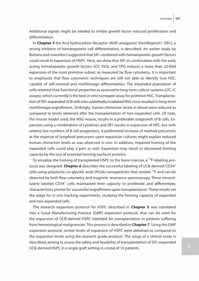
Summary 167
A
Additional signals might be needed to inhibit growth factor induced proliferation and differentiation.
In Chapter 5 the Aryl hydrocarbon Receptor (AhR) antagonist StemRegenin1 (SR1), a strong inhibitor of hematopoietic cell differentiation, is described. An earlier study by Boitano and coworkers suggested that SR1 combined with hematopoietic growth factors could result in expansion of HSPC. Here, we show that SR1 in combination with the early acting hematopoietic growth factors SCF, Flt3L and TPO induces a more than 25-fold expansion of the most primitive subset, as measured by flow cytometry. It is important to emphasize that flow cytometric techniques are still not able to identify true HSC, capable of self-renewal and multilineage differentiation. The expanded population of cells retained their functional properties as assessed in long-term culture systems (LTC-IC assays), which currently is the best in vitro surrogate assay for primitive HSC. Transplanta-tion of SR1-expanded UCB cells into sublethally irradiated NSG mice resulted in long-term multilineage engraftment. Strikingly, human chimerism levels in blood were reduced as compared to levels obtained after the transplantation of non-expanded cells. Of note, the mouse model used, the NSG mouse, results in a preferable outgrowth of B cells. Ex-pansion using a combination of cytokines and SR1 results in expansion of HPC, but with relative low numbers of B cell progenitors. A preferential increase of myeloid precursors at the expense of lymphoid precursors upon expansion cultures might explain reduced human chimerism levels as was observed in vivo. In addition, impaired homing of the expanded cells could play a part as well. Expansion may result in decreased homing capacity by the loss of essential homing (surface) proteins.
To visualize the homing of transplanted HSPC to the bone marrow, a 19F-labeling pro-tocol was designed. Chapter 6 describes the successful labeling of UCB-derived CD34+ cells using poly(lactic-co-glycolic acid) (PLGA) nanoparticles that contain 19F and can be detected by both flow cytometry and magnetic resonance spectroscopy. These intracel-lularly labeled CD34+ cells maintained their capacity to proliferate and differentiate, characteristics pivotal for successful engraftment upon transplantation. These results set the stage for in vivo tracking experiments, studying the homing capacity of expanded and non-expanded cells.
The research expansion protocol for HSPC described in Chapter 5 was translated into a Good Manufacturing Practice (GMP) expansion protocol, that can be used for the expansion of UCB-derived HSPC intended for transplantation in patients suffering from hematological malignancies. This process is described in Chapter 7. Using the GMP expansion protocol, similar levels of expansion of HSPC were obtained as compared to the expansion levels using the research grade protocol. The setup of a clinical study is described, aiming to assess the safety and feasibility of transplantation of SR1-expanded UCB-derived HSPC in a single graft setting in a total of 10 patients.

168 Appendices
Finally, the implications of the findings described in this thesis for the future are discussed in Chapter 8. The quest for the optimal protocol for ex vivo expansion and the study of its effects on subsequent in vivo engraftment will continue. In addition, early clinical studies are pivotal to address whether the current practice using double transplants can be abandoned and if transplantations using expanded single units result in similar, or better, clinical outcome. Finally, UCB should be compared with other alternative donor sources, such as adult mismatched unrelated donors and non-matched (haplo-identical) family donors.
Samenvatting 169
A
SamEnVattInG
De eerste allogene hematopoëtische stamceltransplantatie (allo-SCT) waarbij gebruik gemaakt werd van navelstrengbloed werd in 1988 uitgevoerd bij een patiënt met Fanconi Anemie. De afgelopen decennia heeft navelstrengbloed zich ontwikkeld tot een belang-rijke alternatieve stamcelbron voor patiënten die een allo-SCT nodig hebben, maar waar-bij geen passende verwante of niet-verwante donor beschikbaar is. Momenteel (2016) zijn er wereldwijd al meer dan 35.000 navelstrengbloedtransplantaties uitgevoerd. Bij volwassen patiënten, die een navelstrengbloedtransplantatie krijgen, duurt het herstel van witte bloedcellen en bloedplaatjes vaak lang en komt uitblijvend herstel ("graft failure") vaker voor in vergelijking tot andere stamcelbronnen voor allo-SCT. Dit bleek terug te voeren op het geringe aantal bloedstamcellen (verder 'stamcellen') in navel-strengbloed en heeft geleid tot richtlijnen, waarin het gebruik van navelstrengbloedeen-heden met een klein aantal stamcellen afgeraden wordt. Hierdoor vermindert het aantal eenheden dat geschikt is voor transplantatie, met name in volwassen patiënten, echter aanzienlijk. Teneinde het aantal stamcellen te verhogen werd de dubbele navelstreng-bloedtransplantatie ontwikkeld. "Graft failure" werd minder vaak waargenomen, maar het hematopoëtisch herstel bleek niet verbeterd, mogelijk als gevolg van afstoting van een van de twee gegeven eenheden. Teneinde het aantal stamcellen per navelstreng-bloed eenheid te doen toenemen, is internationaal veel onderzoek gedaan naar expansie van navelstrengbloedstamcellen in het laboratorium (hoofdstuk 1). In de jaren '80 en '90 werden expansieprotocollen ontwikkeld met daarin hematopoëtische groeifactoren, welke zorgden voor proliferatie en overleving, maar ook uitrijping van de stamcellen. Dit resulteerde in de productie van grote aantallen uitgerijpte voorlopercellen, terwijl de stamcellen niet meer aanwezig waren. Op basis hiervan, werd de combinatie proliferatie stimulerende groeifactoren met een factor die de uitrijping remt bestudeerd met als doel stamcellen te expanderen zonder dat zij uitrijpen of uitputten. De onderliggende hypothese was gebaseerd op observaties gedaan in studies met embryonale en epitheli-ale stamcellen, welke geëxpandeerd konden worden door de combinatie van specifieke groeifactoren en een factor die de uitrijping remt (zie figuur 2, hoofdstuk 1) De in dit proefschrift beschreven studies zijn gebaseerd op exact die hypothese. Twee potentiele remmers van stamcel uitrijping zijn onderzocht, namelijk Wnt3a en Aryl hydrocarbon Receptor antagonist StemRegenin1 (SR1). Alleen het onderzoek naar SR1 ondersteunde onze hypothese en heeft de basis gelegd voor de ontwikkeling van een protocol voor een klinische studie.
In hoofdstuk 2 wordt een retrospectief onderzoek naar het hematopoëtisch en immu-nologisch herstel na transplantatie met twee navelstrengbloedeenheden beschreven. Klinische uitkomstmaten, inclusief het aantal infecties, werden gerelateerd aan het im-munologisch herstel in 55 ontvangers van een dubbele navelstrengbloedtransplantatie.
170 Appendices
Terwijl herstel van B- en NK-cellen relatief vlot plaatsvond na transplantatie (respectievelijk na 6 en na 2 maanden), bleek het T-cel herstel erg traag. Na 24 maanden lag het mediane aantal T-cellen nog steeds onder de normaalwaarde. Hoewel de T-cel aantallen erg laag waren, werden bij de meeste patiënten op 3, 6 en 12 maanden na transplantatie nieuwe, in de thymus gevormde sjTREC+ T-cellen aangetroffen. De mediane sjTREC frequentie per 105 CD3+ T-cellen en het mediane aantal sjTREC+ T-cellen per millimeter bloed nam in de loop van de tijd toe, maar 12 maanden na transplantatie waren zowel de frequentie als het aantal nog niet terug op de normale waardes. Onderzocht werd of het herstel van de thymusfunctie, gemeten aan de aanwezigheid van sjTREC+ T-cellen, verband hield met ernstige infecties en sterfte als gevolg van complicaties. Met name uitblijven van herstel van nieuwgevormde T-cellen in de thymus op 3 maanden na transplantatie bleek voorspellend voor ernstige opportunistische infecties daarna. Hoe hoger het aantal sjTREC+ T-cellen in het bloed, hoe lager de kans op infectie. Deze resultaten tonen aan dat het trage hematopoëtische herstel na dubbele navelstrengbloedtransplantatie het meest uitgesproken is wat betreft het T-cel herstel. Naast het geven van meer stamcellen zouden ook interventies gericht op verbetering van thymusfunctie verder onderzocht moeten worden.
In hoofdstuk 3 wordt effect van het eiwit Wingless-int3a (Wnt3a) op de proliferatie en differentiatie van muizen stam- en voorlopercellen onderzocht. Wnt eiwitten spelen een belangrijke rol bij de deling en rijping van stamcellen, waaronder embryonale stamcellen en bloedstamcellen. De hypothese was dat een combinatie van tyrosine kinase receptor activerende groeifactoren in combinatie met Wnt3a expansie van stamcellen zou kunnen geven, zoals eerder gezien met embryonale stamcellen, waarbij het Wnt-effect berustte op remming van de uitrijping. Toevoeging van Wnt3a aan ons kweeksysteem, (met daarin ook hematopoëtische groeifactoren die aangrijpen op tyrosine kinase receptoren) leidde echter tot een vermindering van het aantal stam- en voorlopercellen, zoals gemeten door middel van flowcytometrie. Teneinde de stabiliteit te verhogen en de halfwaardetijd te verlengen werd Wnt3a geladen op liposomen (liposomaal-Wnt3a), hetgeen echter op-nieuw leidde tot een vermindering van het aantal stam- en voorlopercellen. Overexpres-sie van het anti-apoptotische BCL2 proteïne in stam- en voorlopercellen had geen effect op de vermindering van het aantal stam- en voorlopercellen door Wnt3a. Dit suggereert dat de Wnt3a-geïnduceerde vermindering van het aantal stam- en voorlopercellen niet het gevolg is van apoptose. Een mogelijke verklaring voor het lagere aantal stam- en voorlopercellen is onvoldoende remming en zelfs toename van uitrijping onder invloed van de combinatie van groeifactoren en Wnt3a.
In hoofdstuk 4 wordt het effect van Wnt3a beschreven op expansie van humane CD34+ cellen, een populatie van cellen bestaande uit stam- en voorlopercellen. Gelijk aan de observatie bij muizen stam-en voorlopercellen, bleek te combinatie van groeifactoren en Wnt3a te leiden tot minder expansie van het aantal CD34-positieve cellen in vergelijking
Samenvatting 171
A
met stimulatie d.m.v. groeifactoren alleen. Dit effect werd ook waargenomen in kweken met Lin-CD34+CD38lowCD45RA-CD90+ cellen, die in hoge mate verrijkt zijn voor stamcel-len. Het waargenomen effect was te wijten aan de activering van de Wnt signaalroute, waarbij Wnt3a aan diens receptor bond en stabilisering van Wnt3a tot liposomaal Wnt3a leidde andermaal niet tot een verhoogde expansie van stam- en voorlopercellen. Wnt3a verminderde ook het aantal multilineage kolonies (CFU-GEMM) en het vermogen tot succesvolle transplantatie in immuun gecompromitteerde NSG muizen. Concluderend wordt gesteld dat de in de hoofdstukken 3 en 4 beschreven data, in tegenstelling tot onze hypothese, aantonen dat Wnt3a de uitrijping geïnduceerd door de groeifactoren niet kan remmen. Daarentegen zagen we juist iets meer uitrijping door de combinatie van groeifactoren en Wnt3a. De door hematopoëtische groeifactoren geïnduceerde celdeling en celrijping benodigen derhalve aanvullende signalen om de rijping te blok-keren.
In hoofdstuk 5 wordt een studie beschreven, waarin gebruik gemaakt werd van een sterke remmer van de celrijping, namelijk de Aryl hydrocarbon Receptor (AhR) antagonist StemRegenin1 (SR1). Een eerdere studie in een ander centrum suggereerde dat SR1 in combinatie met hematopoëtische groeifactoren wel zou kunnen leiden tot stamcelexpansie. Wij laten zien dat SR1 de expansie van humane stam- en voorlopercel-len inderdaad bevordert. De celpopulatie die het meest verrijkt is voor echte stamcellen (Lin-CD34+CD38lowCD45RA-CD90+ cellen) expandeerde meer dan 25 maal in 2 weken. Overigens dient benadrukt te worden, dat de werkelijke bloedstamcel, die zichzelf kan vermeerderen en ook alle cellijnen als nakomelingen kan genereren, nog steeds niet met flowcytometrische technieken precies aangetoond kan worden. De geëxpandeerde stam- en voorlopercellen behielden hun functionele eigenschappen, zoals o.a. gemeten in lange termijn kweeksystemen (de zogenaamde LTC-IC testen), welke momenteel de beste in vitro surrogaattest voor primitieve stamcellen vertegenwoordigt. Transplantatie van SR1-geëxpandeerde cellen in bestraalde NSG-muizen resulteerde in herstel van rijpe humane cellen in het perifere bloed. Opvallend genoeg bleek het percentage humaan chimerisme na transplantatie van SR1-geëxpandeerde humane stam- en voorlopercellen lager dan na transplantatie van niet-geëxpandeerde humane stam- en voorlopercellen. De meest waarschijnlijke verklaring hiervoor is het gekozen muizenmodel, waarin met name rijpe humane B-cellen ontstaan. Expansie met groeifactoren en SR1 leidt weliswaar tot stam- en voorlopercel expansie, maar met een relatief laag aantal voorloper B-cellen en juist een hoog aantal myeloïde voorlopercellen. Deze verdeling van voorlopercellen na expansie is een mogelijke verklaring van het lagere percentage humaan chimerisme. Daarnaast zou ook de nesteling ("homing") van de stam- en voorlopercellen een rol kunnen spelen. Expansie zou kunnen leiden tot een verminderde homing-capaciteit, als gevolg van verlies van daartoe benodigde (oppervlakte) eiwitten op de geëxpandeerde stam- en voorlopercellen.
172 Appendices
Om homing van getransplanteerde stam- en voorlopercellen naar het beenmerg zicht-baar te kunnen maken, werd een Fluor-19-labeling protocol voor stam- en voorlopercel-len ontworpen. In hoofdstuk 6 wordt beschreven hoe stamcellen uit navelstrengbloed met succes gelabeld kunnen worden met poly(lactic-co-glycolic acid) (PLGA) nanodeel-tjes die Fluor-19 bevatten. Fluor-19-gelabelde cellen kunnen worden opgespoord met behulp van zowel flowcytometrie als magnetische resonantie spectroscopie (MRS). Deze intracellulair gelabelde stamcellen behielden hun capaciteit om in vitro te prolifereren en differentiëren, eigenschappen die essentieel zijn voor een geslaagde uitkomst na trans-plantatie. Deze studie heeft de weg vrijgemaakt voor toekomstige in vivo experimenten, waarbij de homing-capaciteit van geëxpandeerde en niet-geëxpandeerde cellen naar het beenmerg wordt onderzocht.
In hoofdstuk 7 wordt beschreven hoe het in hoofdstuk 5 ontwikkelde laboratorium expansieprotocol voor het expanderen van stam- en voorlopercellen werd vertaald in een Good Manufacturing Practice (GMP)-expansieprotocol dat gebruikt kan worden voor transplantatie in patiënten met bloedkanker. Met het GMP-expansieprotocol werd een vergelijkbare expansie van stam- en voorlopercellen verkregen als het met labora-torium expansieprotocol als beschreven in hoofdstuk 5. Daarnaast beschrijven we de opzet van een klinische studie waarvan het doel is om de veiligheid van transplantatie van SR1-geëxpandeerde van stam- en voorlopercellen afkomstig van één navelstreng-bloedeenhied te evalueren. In deze studie zullen 10 patiënten geïncludeerd worden.
Ten slotte worden in hoofdstuk 8 van dit proefschrift de implicaties van onze bevin-dingen voor de toekomst beschreven. De zoektocht naar het optimale ex vivo expansie protocol gaat verder. Daarnaast zijn vroeg-klinische studies nodig om na te gaan of de huidige praktijk met dubbele navelstrengbloedtransplantaten verlaten kan worden in volwassen patiënten en transplantaties met enkele geëxpandeerde navelstrengbloed eenheden dezelfde, zo niet betere, klinische resultaten zullen geven. Tenslotte zal navelstrengbloed afgewogen dienen te worden tegen andere alternatieve stamcelbron-nen zoals die van volwassen onverwante donoren of niet-passende (haplo-identieke) familiedonoren.
Publications in this thesis 173
A
PUBLICatIonS In tHIS tHESIS
1. Duinhouwer LE, ter Borg MND, Rombouts WJC, Cornelissen JJ, Braakman E. In vitro and in vivo evaluation of StemRegenin1-expanded UCB-derived hematopoietic stem and progenitor cells. Submitted
2. Duinhouwer LE*, Beije N*, van der Holt B, Rijken-Schelen A, Lamers CH, Somers J, Braakman E, Cornelissen JJ. Impaired thymopoiesis predicts for a high risk of severe infections after double umbilical cord blood transplantation . Submitted. *Shared first authorship
3. Tüysüz N, Duinhouwer LE, Rombouts WJC, Kurek D, van Bloois L, van der Linden R, Mastrobattista E, Cornelissen JJ, Braakman E, ten Berge D. Wnt3a protein reduces the number of mouse phenotypic hematopoietic stem and progenitor cells in serum-free cultures. Submitted
4. Duinhouwer LE, van Rossum BJM, van Tiel ST, van der Werf RM, Kotek G, ter Borg M, Rombouts WJC, Braakman E, Cornelissen JJ, Bernsen MR. Magnetic Resonance Detec-tion of CD34+ Cells from Umbilical Cord Clood Using a 19F Label. PLoS ONE. 2015 Sep 22;10(9)
5. Duinhouwer LE, Tüysüz N, Rombouts WJC, ter Borg MND, Spanholtz J, Mastrobattista E, Cornelissen JJ, ten Berge D, Braakman E. Wnt3a protein reduces growth factor-driven expansion of human hematopoietic stem and progenitor cells in serum-free cultures. PLoS ONE. 2015 Mar 25;10(3)
otHEr PUBLICatIonS
1. Duinhouwer LE*, van Mil SR*, Mannaerts GH, Dunkelgrun M, Biter LU, Apers JA. The standardized postoperative checklist for bariatric surgery: what are the predictors of complications? Submitted. *Shared first authorship
2. Duinhouwer LE, Deerenberg E, Rociu E, Kortekaas RTJ. Herniation of the colon through the foramen of Winslow: a case report. Int J Surg Case Rep. 2016 May 4;24:14-17
3. Duinhouwer LE, Biter LU, Wijnhoven BP, Mannaerts GH. Treatment of giant hiatal her-nia by laparoscopic Roux-en-Y gastric bypass. Int J Surg Case Rep. 2015 Feb 19;9C:44-46

174 Appendices
4. Lodewyck T, Oudshoorn M, van der Holt B, Petersen E, Spierings E, von dem Borne PA, Schattenberg A, Allebes W, Groenendijk-Sijnke M, Duinhouwer L, Willemze R, Lowen-berg B, Verdonck LF, Meijer E, Cornelissen JJ. Predictive impact of allele-matching and EBMT risk score for outcome after T-cell depleted unrelated donor transplantation in poor-risk acute leukemia and myelodysplasia. Leukemia. 2011 Oct;25(10):1548-54
PhD Portfolio 175
A
PHD PortfoLIo
Summary of PhD training and teaching activitiesName PhD Candidate: Lucia E. Duinhouwer
PhD period: September 2010 – November 2016
Erasmus MC department: Hematology
Promotor: prof. dr. J.J. Cornelissen
Co-promotor: dr. E. Braakman
Research School: Molecular Medicine
Activities Year ECTS
1. PHD traInInG
General academic/research skills
Biomedical Research Techniques (MolMed) 2010 1.6
Basic and Translation Oncology (MolMed) 2010 1.6
Course on Molecular Medicine (MolMed) 2011 0.7
Laboratory Animal Science (Article 9 course; Erasmus MC) 2011 3
Workshop Presenting Skills for PhD students (MolMed) 2011 1
Research Management for PhD Students (MolMed) 2011 1
Workshop on Photoshop and Illustrator CS (MolMed) 2013 0.3
Research Integrity (Erasmus MC) 2013 2
In-depth courses
Workshop Optical and Molecular Imaging (MolMed) 2013 0.6
2nd Scientific Workshop on Leukemic and Cancer Stem Cells, Mandelieu, 2013 1
France (EHA)
national and international conferences
Annual Dutch Hematology Congress (4x; Arnhem, the Netherlands) 2010-2013 4
Biannual World Cord Blood Congress (2x; Rome, Italy; Monaco, France) 2011, 2015 2
17th Annual European Hematology Association Congress (Amsterdam, 2012 1
the Netherlands)
54th American Society of Hematology Meeting and Exposition (Atlanta, USA) 2012 1
Molecular Medicine Day (3x; Rotterdam, the Netherlands) 2011-2013 1
176 Appendices
Scientific presentations
Dutch Hematology Congress (2x; oral) 2012-2013 2
Top Institute Pharma Spring Meeting (poster) 2012 1
54th American Society of Hematology Meeting and Exposition 2012 1
(Atlanta, USA; poster)
Scientific meetings
Work discussions at the Department of Hematology, Erasmus MC 2010-2013 3
AIO/Post doc meetings at the Department of Hematology, Erasmus MC 2010-2013 2
Erasmus Hematology Lectures 2010-2013 2
2. tEaCHInG aCtIVItIES
Co-supervising Master of Science student ‘Clinical Research’ (52 weeks) 2012-2013 7.5
Medical student VO Bone marrow and Leukemia (2x) 2012-2013 0.5
total 40.8
Curriculum vitae 177
A
CUrrICULUm VItaE
Lucia Duinhouwer werd op 18 april 1988 geboren in Rotterdam en bracht haar geluk-kige jeugd door onder de rook van deze prachtige stad. In 2006 behaalde zij cum laude haar gymnasiumdiploma aan het Einstein Lyceum te Hoogvliet Rotterdam. In datzelfde jaar begon zij aan de studie Geneeskunde aan de Erasmus Universiteit Rotterdam. Zij behaalde haar doctoraal diploma in augustus 2010 en startte aansluitend met een pro-motietraject op de afdeling Hematologie onder leiding van dr. E. Braakman en prof. dr. J.J. Cornelissen, waarvan de resultaten staan beschreven in dit proefschrift. In oktober 2013 pakte zij haar studie Geneeskunde weer op en startte met haar coschappen. In november 2015 behaalde zij cum laude haar artsexamen, waarop zij als ANIOS Chirurgie aan de slag ging in het IJsselland Ziekenhuis te Capelle aan den IJssel (dr. I. Dawson). Op 1 januari 2017 start zij met haar opleiding tot chirurg in de regio Rotterdam (dr. B. Wijnhoven).

Dankwoord 179
A
DanKWoorD
Eindelijk, na jaren werk, is het zover: het is af. Ik had dit proefschrift nooit kunnen schrijven zonder de hulp van velen en in dit laatste, meest gelezen, deel van mijn proefschrift pro-beer ik iedereen te bedanken die mij de afgelopen jaren heeft geholpen en bijgestaan.
Prof.dr. Cornelissen, beste Jan, het is ondertussen alweer meer dan 8 jaar geleden dat ik als tweedejaars geneeskundestudent, in het kader van keuzeonderwijs, onder jouw hoede een review schreef over de rol van Imatinib in de behandeling van CML. Via wat databasewerk en mijn keuzeonderzoek over chimerisme na stamceltransplantaties kwa-men we uiteindelijk bij dit promotietraject aan. Een fantastische kans die jij me bood en wat ben ik blij dat ik hem met beide handen heb aangegrepen. Er waren ups en uiteraard ook downs en we konden zeker van mening verschillen, maar uiteindelijk wist je mij altijd weer te motiveren verder te gaan. Ik waardeer zeer dat je tijdens de laatste loodjes zelfs op je vrije zondag naar het lab kwam om nog even wat laatste revisies met mij door te nemen. Ik kijk uit naar de eerste resultaten van de klinische fase van het project! Ook al ga ik in de kliniek een andere kant op, ik hoop je nog zeker tegen te komen in de toekomst en van je te mogen leren.
Dr. Braakman, beste Eric, waar moet ik beginnen jou te bedanken? Dankzij jouw dage-lijkse begeleiding is dit proefschrift uiteindelijk geworden wat het is. Precies tot op de komma, en daar werd ik heus weleens gek van, maar uiteindelijk heeft het zijn vruchten afgeworpen. Jouw deur stond altijd open, zeker in de laatste fase, toen ik vaak onaange-kondigd kwam binnenvallen. Ik heb ontzettend veel van je geleerd de afgelopen jaren en daar ben ik je heel dankbaar voor. Ik wens je ontzettend veel succes met het nieuwe project en hoop je nog zeker tegen te komen in de toekomst!
Ik wil graag prof.dr. Löwenberg, prof.dr. Van Leeuwen en prof.dr. Zwaginga bedan-ken voor het plaatsnemen in de leescommissie en het kritisch lezen van mijn proefschrift. Beste Bob, ik wil u bedanken voor de fijne gesprekken toen we ‘kamergenoten’ waren in EE1300. Ondanks uw drukke schema nam u altijd even de tijd hiervoor en ik heb zeer veel gehad aan uw inzichten en adviezen.
De leden van de grote commissie, dr. ten Berge, dr. Bernsen en dr. Boelens, wil ik graag bedanken voor het plaatsnemen hierin.
Elwin, aan jou 6 jaar geleden de ondankbare taak om deze geneeskundestudent, die nog nooit een pipet had vastgehouden, alle kneepjes van het vak te leren. En dat heb je gedaan! Celisolaties, kweken, FACSen, bijna alle labtechnieken gebruikt in dit proefschrift
180 Appendices
heb ik van jou geleerd. Daarnaast heb je zelf ook nog ontzettend veel werk verricht voor het project, zonder jou was dit proefschrift niet mogelijk geweest. Ik wil je hiervoor, en voor onze gezellige praatjes in de gang, ontzettend bedanken en ik wens jou en je gezin alle goeds voor de toekomst! mariëtte, wat was het een schot in de roos om jou aan te nemen als analist! Je hebt ontzettend veel werk verricht voor het project, en als ik je naar een bepaald experiment vroeg, wist je door je nauwkeurige manier van werken precies waar je de data kon vinden. Ik kan ook niemand bedenken die beter geschikt om de transplantaten in de clean room te bewerken voor de klinische fase van het project dan jij. Ik wens je veel geluk met je mooie gezin en hopelijk tot ziens! Oud-collega’s van de Braakman/Cornelissen groep, Eric, Irene en amiet wil ik bedanken voor hun hulp op het lab, input tijdens werkbesprekingen en gezelligheid tussen het werken door!
De collega’s van het transplantatielab wil ik bedanken voor het feit dat ik altijd gebruik mocht maken van hun apparatuur en de leuke gesprekken die we ondertussen hadden. Het was heel fijn samenwerken met jullie! Speciale dank gaat naar anita, bedankt voor al het werk dat je verzet hebt voor het IMRETREC project, zonder jouw kritische blik was het stuk niet zo geworden als het nu is!
The (old) members of the Cupedo Lab, natalie, rogier, ferry, Julien, Patricia, monica, Jurre and tom. I would like to thank you for the nice atmosphere in the lab, the moti-vating conversations we had and your critical views and good ideas for my work in our work discussions. I wish you all many Nature publications and I hope we will meet again.
Twee kamers heb ik versleten, en daarmee ook vele kamergenoten. Davine, ik ben zeer onder de indruk van jouw motivatie voor het onderzoek en het prachtige project waar je je al zoveel jaar voor inzet. Ik wil je bedanken voor de vele gesprekken die we had-den in EE1300, zowel over werk als over andere dingen en ik wens je alle goeds voor de toekomst. avinash, ik wens je veel succes bij de IKNL en wie weet zit er ooit nog een samenwerking in! Jurjen, het waren maar een aantal maanden, maar ik vond ze wel heel gezellig! Ik wens je ontzettend veel succes met de laatste loodjes van jouw proefschrift en wellicht kruisen onze paden nog in de kliniek! Kasia, how I loved your enthusiastic vibes in our room! Never a dull moment with you! Thank you for the nice time we had and I hope we will meet again! Good luck finishing your thesis! roberto, although you had so much lab work to do that you were not much around in EE1300, I always enjoyed your presence. I wish you all the best and good luck finishing your thesis! mark, af en aan kamergenoten de laatste jaren en dan vooral de laatste maanden, soms zelfs ’s avonds of in het weekend. Ik heb ontzettend met je gelachen en als er nieuws is over Sylvie, ben jij de eerste die het hoort! roel, van kamergenoten naar hele goede vrienden. We begon-nen tegelijk aan dit avontuur en wat een avontuur was het. Eindeloze gesprekken over
Dankwoord 181
A
geneeskunde, wetenschap en alle dingen die belangrijk zijn daarbuiten. Ik heb ontzet-tend veel bewondering voor jouw werkethos, hoewel je daar soms wel wat in afgeremd mag worden. Je hebt het mooiste meisje van het lab geschaakt en wat een prachtig stel zijn jullie! Ook nog jullie heerlijke zoon erbij, wat een rijkdom. Ik ben ontzettend blij dat we elkaar nog zien (hoewel eigenlijk veel te weinig!) ook al zijn we allebei al een aantal jaar weg van de 13e. Succes met de laatste loodjes (oh, wat wegen ze zwaar!) en probeer niet alles ’s nachts te doen. En als je Roel zegt, zeg je merel. Ondanks dat we op het lab niet zoveel met elkaar te maken hadden, hadden we direct een klik. Liters koffie hebben we versleten, en de meeste eigenlijk nog nadat we allebei niet meer op de 13e rondlie-pen. Het was altijd heerlijk om de onderzoeksfrustaties te kunnen delen met iemand die precies begrijpt wat je bedoelt! Je bent een hele goede vriendin geworden en daar ben ik ontzettend dankbaar voor, dat ik dat heb mogen overhouden aan mijn promotietraject. Ik wens jou met Roel en Max een prachtig leven! Gaan we trouwens nog een keer naar dat Cheesecake festival?
Alle overige promovendi van de Hematologie wil ik bedanken voor de gezelligheid en jullie input tijdens werkbesprekingen. Succes met het afronden van jullie proefschriften!
Verder wil ik prof.dr. Delwel, prof.dr. touw, prof.dr. Sonneveld, dr. raaijmakers en dr. Jongen-Lavrencic bedanken voor hun input en kritische blik tijdens de werkbesprekin-gen op vrijdagochtend, die het onderzoek op onze afdeling altijd naar een hoger niveau brengt.
De dames van het secretariaat, ans, Leenke en annelies, wil ik bedanken voor hun ondersteuning! Zonder jullie zou de afdeling een stuk minder soepel functioneren! mariska, ontzettend bedankt voor jouw steun tijdens de laatste fase van mijn traject. Je hebt me heel veel werk uit handen genomen en er mede voor gezorgd dat alles op tijd af was. Dank je wel!
De basis van dit proefschrift ligt in de TI Pharma samenwerking met het Stem Cell Insti-tute en de afdeling Radiologie. Derk and nesrin, thank you for all the work you put into the Wnt part of the project! I enjoyed our meetings and even though outcomes were not always as we hoped, I think we can be proud on what has been achieved! I wish you both all the best! monique, ramon, Gyula, Gaby, Sandra en Joost, bedankt voor de vruchtbare samenwerking met betrekking tot de 19F-labeling, het heeft een mooie publicatie en inspiratie voor de toekomst opgeleverd! Bernard, ik vond het ontzettend leuk om jou als student te begeleiden, het was een mooi project! Ik wens je veel succes als dokter!
182 Appendices
Het IMRETREC project was meer werk dan we gedacht hadden, nick, en ik wil je bedan-ken voor alle tijd die je erin gestopt hebt! Wellicht tot ziens in de kliniek!
Het onderzoek uitgevoerd in dit proefschrift was absoluut onmogelijk geweest zonder alle moeders in de verschillende ziekenhuizen, die zo onbaatzuchtig hun navelstreng-bloed hebben afgestaan. Mijn dank is ontzettend groot. Verder gaat mijn dank uit naar régine Steegers-theunissen, Hanneke de Graaf en Gretel van Hoecke voor hun hulp met het organiseren van de afnames in hun centra en aan alle medewerkers van de afdelingen Verloskunde van het albert Schweitzer Ziekenhuis en het Erasmus mC en van het Geboortecentum Sophia voor hun inzet ten aanzien van het verzamelen van navelstrengbloed. Tenslotte wil ik de Sanquin navelstrengbloedbank bedanken voor het ter beschikking stellen van navelstrengbloeddonaties, welke niet geschikt waren voor klinisch gebruik. Zonder de inzet van al deze mensen, had dit proefschrift niet kun-nen bestaan.
Mijn fijne collega’s van de afdeling Chirurgie van het IJsselland Ziekenhuis wil ik bedanken voor de fantastische sfeer, veel te gezellige borrels en alle ruimte die jullie mij gegeven hebben om dit proefschrift onder werktijd te voltooien! Zonder jullie, had ik hier niet nu al kunnen staan.
Onderzoek doen is hard werken, en zeker de laatste jaren in combinatie met mijn klinische taken was het niet altijd een feestje. Gelukkig heb ik een heel palet aan fijne vrienden en vriendinnen die altijd klaar stonden om mijn verhalen aan te horen, maar ook mijn aandacht af te leiden met kopjes koffies, gezellige etentjes, weekendjes weg en al die andere dingen de het leven leuk maken. Dank jullie wel daarvoor!
Stephanie, lieve kip. Alweer 10 jaar zijn we vriendinnen, en samen zijn we geneeskun-destudent, promovendus en coassistent geweest en nu zijn we ook nog samen dokter en doctor. Bij alle belangrijke momenten was je er en ik ben ontzettend dankbaar dat jij op deze dag naast me wilt staan. Het was heerlijk om alles te kunnen delen met iemand die in precies hetzelfde schuitje zat, maar om daarnaast ook met elkaar de dingen die er echt toe doen in het leven te delen. Ik wens je ontzettend veel succes met je opleiding tot KNO-arts, al weet ik nu al dat je dat ook weer op je sloffen gaat doen, en heel veel geluk samen met Lorenzo!
Sanne, we zaten al samen in de zandbak en sinds 6VWO zijn we echt onafscheidelijk. Jouw gedachten zijn mijn gedachten, dus natuurlijk moet jij als paranimf naast me staan. Lieve Sann, ik ben ontzettend dankbaar voor je vriendschap al die jaren, voor het feit dat je het altijd met me eens bent en dat we aan een half woord genoeg hebben. Ook
Dankwoord 183
A
al woon je ondertussen helemaal in Gouda, dat heeft niks veranderd. Op naar nog heel veel sushi!
Jan, nettie en marlies, mijn favoriete schoonouders en schoonzus. Bedankt voor alle gezellige avonden en jullie steun tijdens al die jaren dat ik hier mee bezig was.
Lieve opa en oma, wat een rijkdom dat jullie dit nog meemaken! Dank jullie wel voor jullie onvoorwaardelijke trots en liefde! Op naar de 100!
Papa en mama, waar het allemaal begon. Jullie hebben me altijd gestimuleerd het beste uit mezelf te halen, met als basis jullie onvoorwaardelijke trots. Zonder jullie steun was ik nooit zover gekomen. Papa, hoe ouder, hoe gekker, maar je staat altijd klaar met wijze raad. mama, de stabiele factor, je begrijpt me altijd precies. Ik kan hier nog 1000 zinnen schrijven maar jullie weten het al. Dank jullie wel.
Edwin, het is af. Net toen je dacht dat het nooit meer ging gebeuren. Al die avonden en weekenden. Dank je wel voor je begrip en geduld, voor je grote liefde en voor je humor. Je bent het beste wat er is en ik kan niet wachten om samen oud, grijs en lelijk te worden.


![hernia of the umbilical cord [وضع التوافق] of the umbilical cord.pdf · Umbilical cord hernia…cont Conclusion: ¾Hernia of the umbilical cord is a rare entityy, of the](https://static.documents.pub/doc/80x56/5ea7ce695a148409cd011fd0/hernia-of-the-umbilical-cord-of-the-umbilical-cordpdf.jpg)


