69
IMRT for Gynecologic Malignancies: The University of Chicago Experience Bulent Aydogan, PhD John C. Roeske, PhD The University of Chicago

IMRT for Gynecologic Malignancies:
The University of ChicagoExperience
Bulent Aydogan, PhDJohn C. Roeske, PhD
The University of Chicago
JCR – 7/2005
RT in Gynecologic Tumors
Typically a combination of external beam whole pelvic RT (WPRT) and intracavitary brachytherapy (ICB)WPRT is used to treat the primary tumor/tumor bed plus the regional lymphaticsICB is used to boost the primary tumor/tumor bed safely to high doses

JCR – 7/2005
Gynecologic RTHighly efficacious and well tolerated in most patientsExcellent pelvic control particularly in early stage cervical and endometrial cancerAdjuvant RT improves outcome of women with high risk features following surgery
JCR – 7/2005
GYN-IMRT RationaleRT →potential toxicities due to the treatment of considerable volumes of normal tissues
Small bowel→ diarrhea, SBO, enteritis, malabsorptionRectum → diarrhea, proctitis, rectal bleedingBone Marrow → ↓WBC, ↓platelets, anemiaPelvic Bones → Insufficiency fractures, necrosis
Reduction in the volume of normal tissues irradiated with IMRT may thus ↓risk of acute and chronic RT sequelae
JCR – 7/2005
Gynecologic IMRTPractical Issues
SimulationTarget and Tissue DelineationTreatment PlanningDelivery and Quality Assurance
JCR – 7/2005
Patient SelectionMost gynecology patients can be treated with IMRTPoor candidates
Uncooperative patientsUnable to tolerate ↑time on the table
Markedly obese patients not idealInability to capture entire external contourDifficulties with daily setup
Dosimetric benefits may be less in the obese*
*Ahamad et al. Int J Radiat Oncol Biol Phys 2002;54:42

JCR – 7/2005
Simulation and CT Scanning
Patients in supine positionImmobilized using a customized devicePatient scanned from L2 to below ischial tuberositiesOral, IV and rectal contrast
JCR – 7/2005
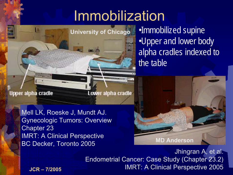
Immobilization•Immobilized supine•Upper and lower body alpha cradles indexed to the table
Mell LK, Roeske J, Mundt AJ.Gynecologic Tumors: Overview Chapter 23IMRT: A Clinical PerspectiveBC Decker, Toronto 2005
University of Chicago
MD Anderson
Jhingran A, et al.Endometrial Cancer: Case Study (Chapter 23.2)
IMRT: A Clinical Perspective 2005

JCR – 7/2005
Planning CT Scan
Scan extent:L2 vertebral body to 3 cm below the ischial tuberosities
Thin slice thickness,e.g. 3 mm
Larger volumes only used if treating extended field, whole abdomen or pelvic-inguinal IMRT

JCR – 7/2005
Helps delineate normal and target tissues
Oral, rectal and IV contrast
Contrast Administration
Bladder contrast not needed
IV contrast is important (vessels serve as surrogates for nodes)*With experience, IV contrast less needed
A vaginal marker is also placed(be careful not to distort)
JCR – 7/2005
Target DefinitionClinical target volume (CTV) drawn on axial CT slicesCTV components depend on the pathologyIn all patients:
Upper ½ of the vaginaParametrial tissuesPelvic lymph nodes regions (common, internal and external iliacs)
In cervical cancer and endometrial cancer patients with positive cervical involvement, include the presacral region
JCR – 7/2005
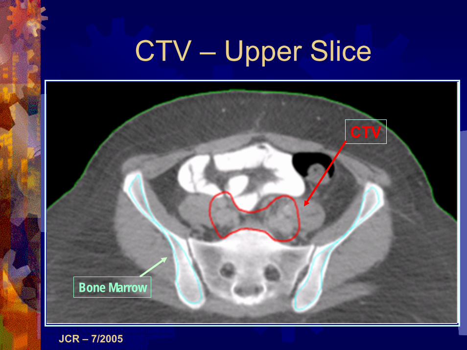
CTV – Upper Slice
CTV
Bone Marrow
JCR – 7/2005
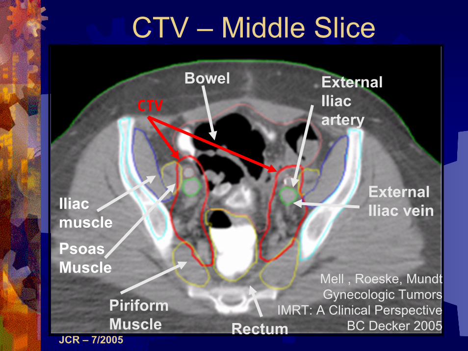
Iliac musclePsoasMuscle
PiriformMuscle
ExternalIliac artery
ExternalIliac vein
Bowel
Rectum
Mell , Roeske, Mundt Gynecologic Tumors
IMRT: A Clinical PerspectiveBC Decker 2005
CTV – Middle Slice
CTV

JCR – 7/2005
CTV – Lower Slice
External Iliac ArteryExternal Iliac Vein
Parametria
ObturatorInternus
Bladder
Rectum
Iliopsoas
Coccyx
GreaterTrochanter
CTV
JCR – 7/2005
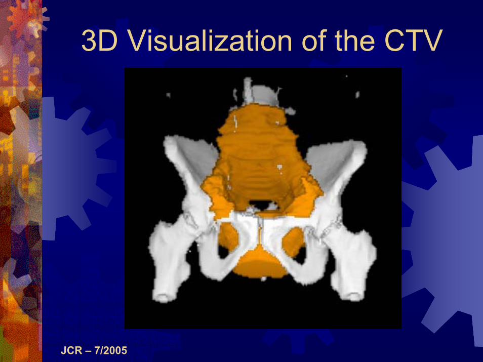
3D Visualization of the CTV
JCR – 7/2005
Normal TissuesNormal tissues delineated depends on the clinical caseIn most cases, include:
Small bowel, rectum, bladderIn patients receiving concomitant or sequential chemotherapy, include the bone marrowOthers include the femoral headsKidneys and liver included only if treating more comprehensive fields
JCR – 7/2005
Normal TissuesBe consistent with contouring
Helps with DVH interpretation
Rectum: Outer wall ?mm (anus to the sigmoid flexure)Small bowel: Outermost loops from the L4-5 interspace
Include the colon above the sigmoid flexure as well in the “small bowel” volume
Bone marrow: Intramedullary space of the iliac crests
Stop at the top of the acetabulumNote that this approach ignores marrow in other pelvic bones
JCR – 7/2005
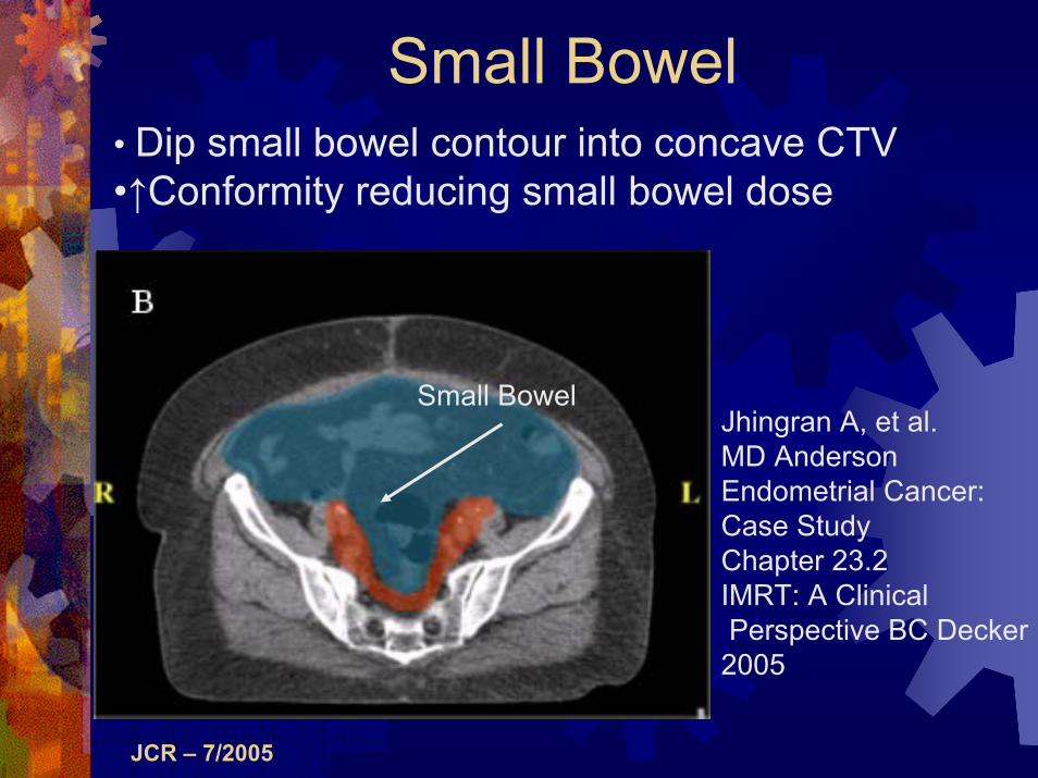
Small Bowel• Dip small bowel contour into concave CTV•↑Conformity reducing small bowel dose
Small BowelJhingran A, et al. MD AndersonEndometrial Cancer:Case StudyChapter 23.2IMRT: A ClinicalPerspective BC Decker
2005

JCR – 7/2005
Contour the intramedullary canal of the CrestsAlternatively, contour the outer surface of theiliac crests (certainly faster!)
Iliac Crests
Sacrum
Bone Marrow
JCR – 7/2005
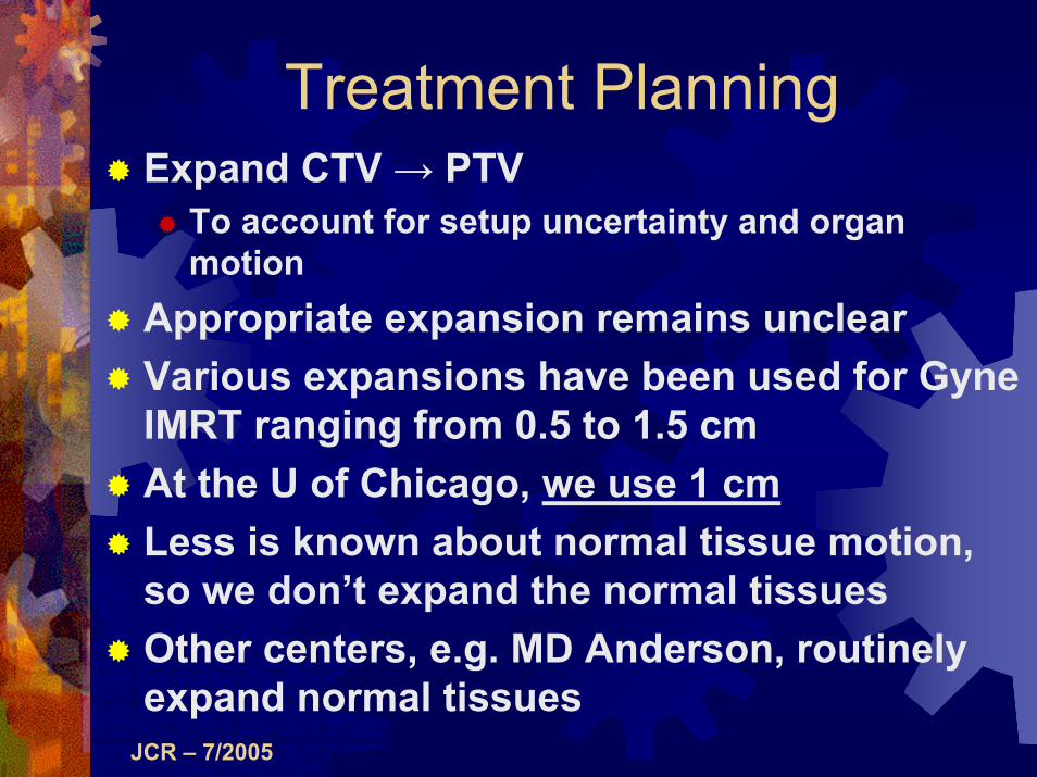
Treatment PlanningExpand CTV → PTV
To account for setup uncertainty and organ motion
Appropriate expansion remains unclearVarious expansions have been used for Gyne IMRT ranging from 0.5 to 1.5 cmAt the U of Chicago, we use 1 cmLess is known about normal tissue motion, so we don’t expand the normal tissues Other centers, e.g. MD Anderson, routinely expand normal tissues

JCR – 7/2005
Setup UncertaintiesDigitized weekly setup films of 50 patients. These patients were immobilized using upper and lower alpha cradles.Measured setup position using image-registration interface (Balter, et al.)Compared digitized images to DRR’s representing patient planning positionRecorded setup uncertainties in AP, LR, and SI directions
Mundt, Roeske and Lujan. Intensity modulated radiation therapy in gynecologic malignancies. In Medical Dosimetry, June 2002.

JCR – 7/2005
FITAPPLYMARKERS CLEAR
dx: 0.0 mm dy: 3.0 mm RMS: 29.2

JCR – 7/2005
Measured Setup Uncertainties
Immobilization: Alpha cradle under legs and upper body with arms above head*
σLR = 3.2 mmσSI = 3.7 mmσAP = 4.1 mm
* Analysis of 50 patients treated with IM-WPRT

JCR – 7/2005
Organ MotionA concern in the region of the vaginal cuffTwo approaches are being studied at our institution to address this:
IGRT (Varian OBI unit)Vaginal immobilization
Now we simply avoid tight CTV volumes and use a 1 cm CTV→PTV expansion
Produces very generous volumes around the vaginal cuff
JCR – 7/2005
Organ MotionUsing this approach, no failures in the vaginal cuff have been seen in patients treated with adjuvant IM-PRT at our institution (>80 pts treated)Nonetheless, tighter volumes could result in ↓toxicityTighter margins are also needed if higher than conventional doses are used
JCR – 7/2005
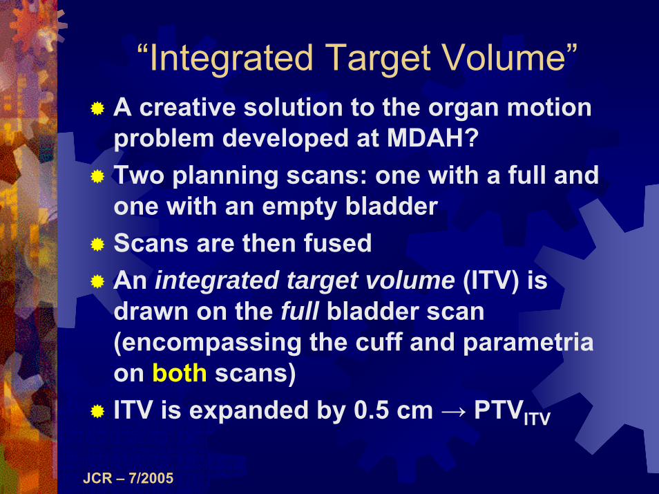
“Integrated Target Volume”A creative solution to the organ motion problem developed at MDAH?Two planning scans: one with a full and one with an empty bladderScans are then fusedAn integrated target volume (ITV) is drawn on the full bladder scan (encompassing the cuff and parametria on both scans)ITV is expanded by 0.5 cm → PTVITV

JCR – 7/2005
Normal Tissue Organ Motion
Bladder
Rectum
Small bowel
Rectum
Bladder
Week 3 scan Treatment planning scan

JCR – 7/2005
Bladder and Rectal Volumes
0
20
40
60
80
100
120
140
160
0 1 2 3 4 5
Week
RectumBladder
JCR – 7/2005
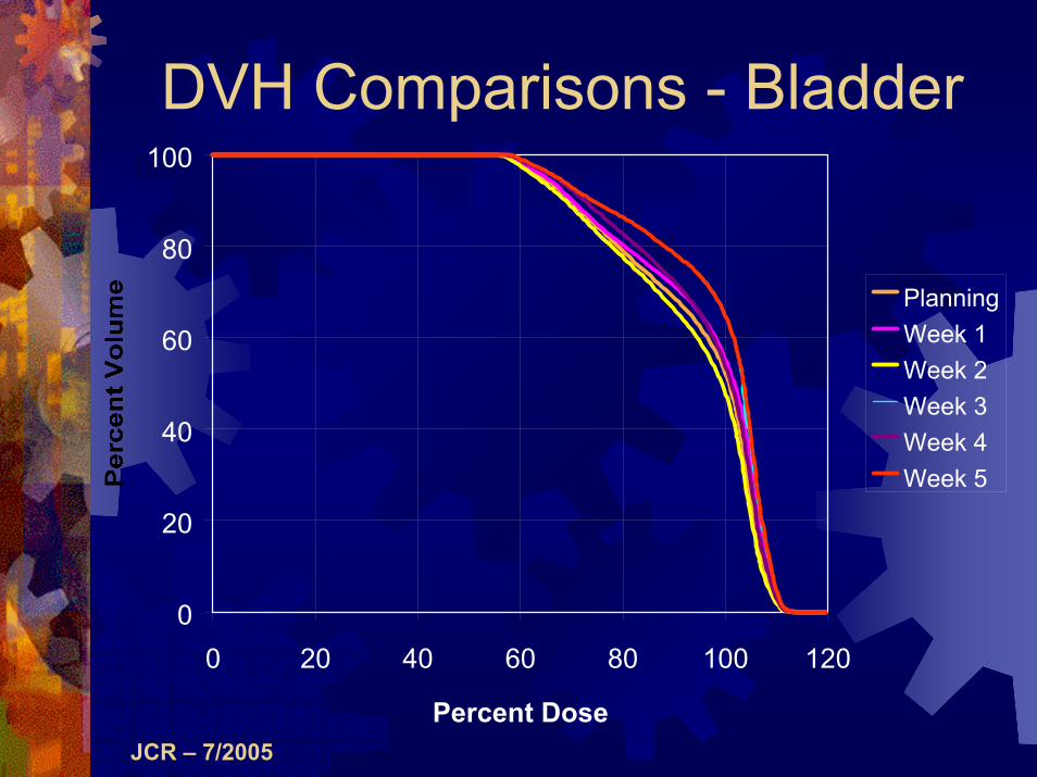
DVH Comparisons - Bladder
0
20
40
60
80
100
0 20 40 60 80 100 120
Percent Dose
PlanningWeek 1Week 2Week 3Week 4Week 5

JCR – 7/2005
DVH Comparisons - Rectum
0
20
40
60
80
100
0 20 40 60 80 100 120
Percent Dose
PlanningWeek 1Week 2Week 3Week 4Week 5
JCR – 7/2005
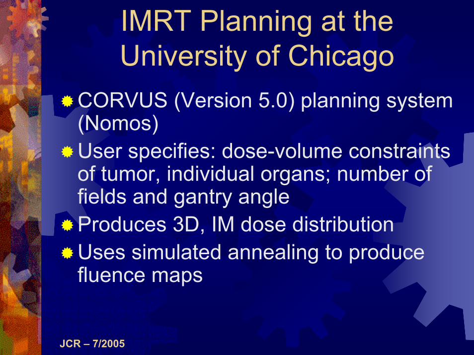
IMRT Planning at the University of Chicago
CORVUS (Version 5.0) planning system (Nomos)User specifies: dose-volume constraints of tumor, individual organs; number of fields and gantry angleProduces 3D, IM dose distributionUses simulated annealing to produce fluence maps
JCR – 7/2005
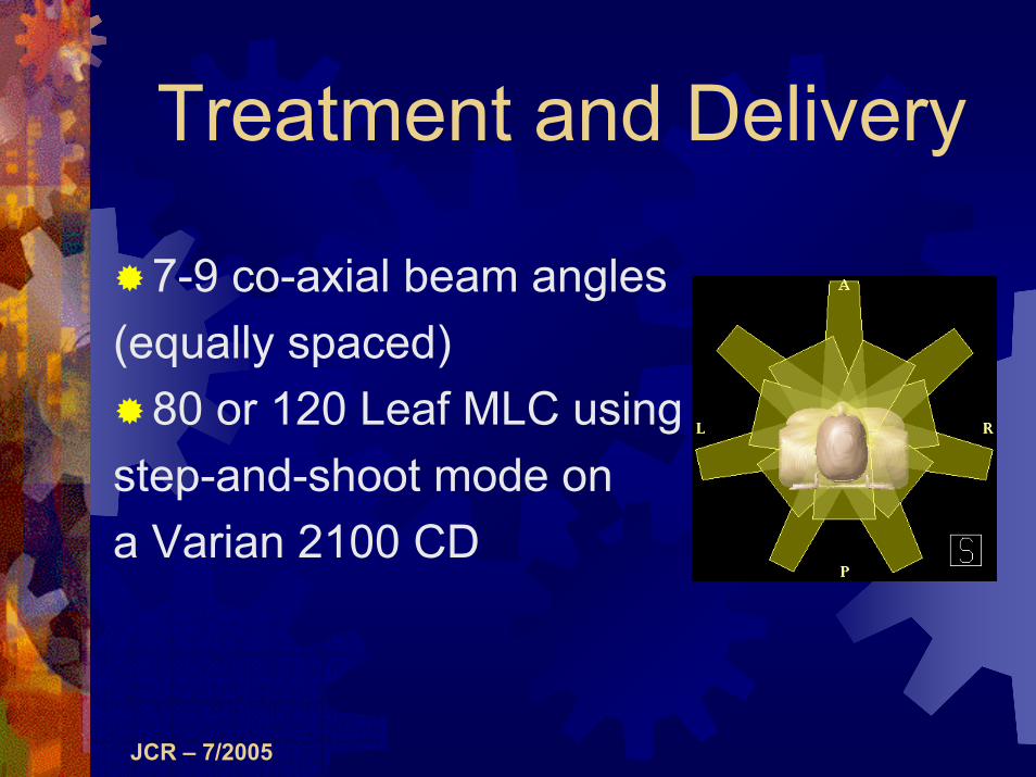
Treatment and Delivery
7-9 co-axial beam angles (equally spaced)
80 or 120 Leaf MLC using step-and-shoot mode ona Varian 2100 CD
JCR – 7/2005
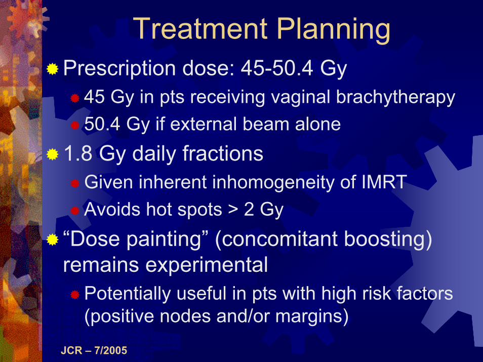
Treatment PlanningPrescription dose: 45-50.4 Gy
45 Gy in pts receiving vaginal brachytherapy50.4 Gy if external beam alone
1.8 Gy daily fractions Given inherent inhomogeneity of IMRT Avoids hot spots > 2 Gy
“Dose painting” (concomitant boosting) remains experimental
Potentially useful in pts with high risk factors (positive nodes and/or margins)

JCR – 7/2005
Treatment PlanningIncreasing number of planning systems now commercially availableDespite inherent differences, no one system appears superiorAcceptable gynecologic IMRT plans have been produced on all major planning systems

JCR – 7/2005
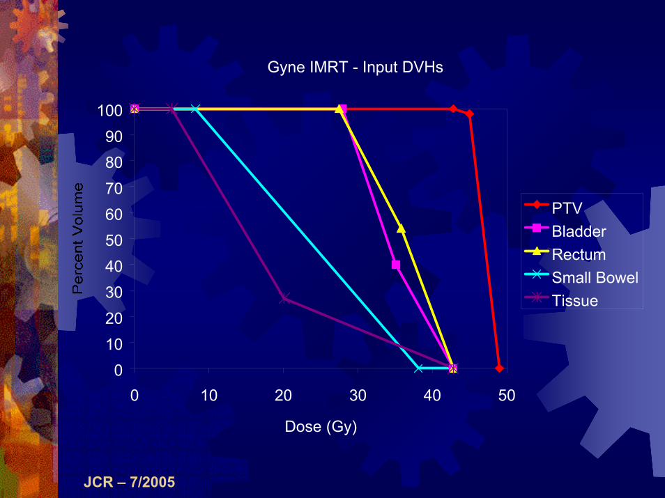
Treatment PlanningInput parameters are next entered for the PTV and normal tissuesOptimal input parameters not knownDerived iterativelyDiffer from one system to anotherPriority should be given to coverage of the PTV (over sparing of normal tissues)Strive for ≥ 97% PTV coverage
JCR – 7/2005
Gyne IMRT - Input DVHs
0102030405060708090
100
0 10 20 30 40 50
Dose (Gy)
PTVBladderRectumSmall BowelTissue
JCR – 7/2005
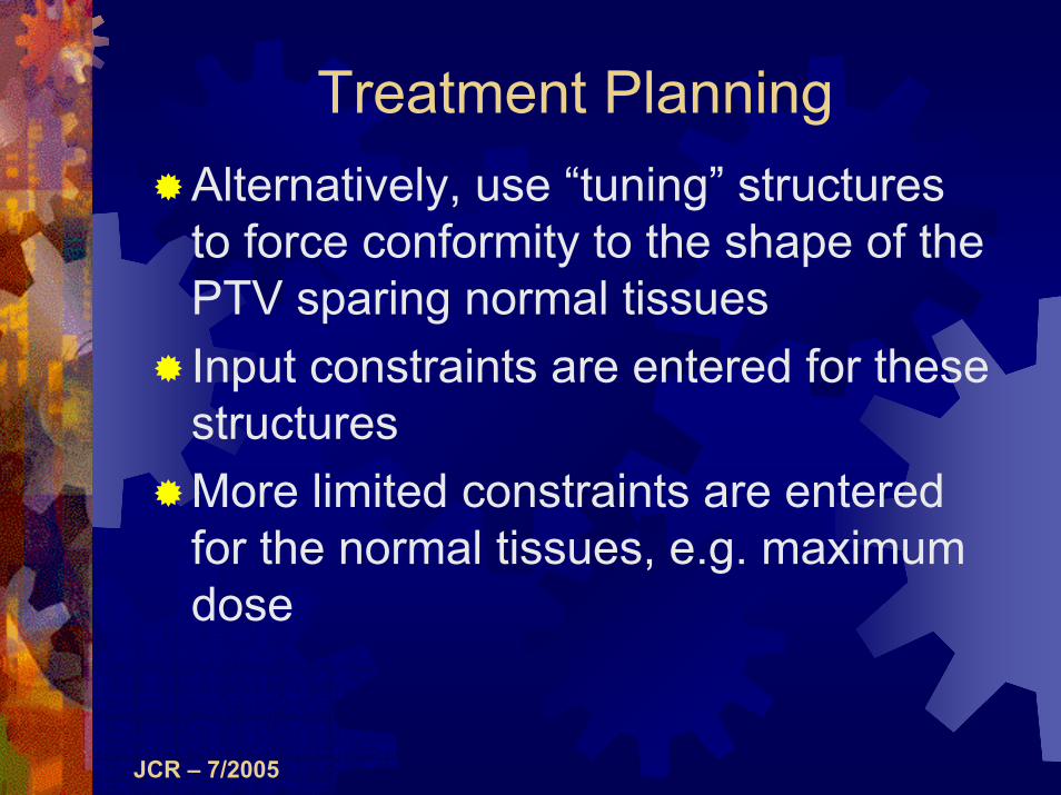
Treatment PlanningAlternatively, use “tuning” structures to force conformity to the shape of the PTV sparing normal tissuesInput constraints are entered for these structuresMore limited constraints are entered for the normal tissues, e.g. maximum dose
JCR – 7/2005
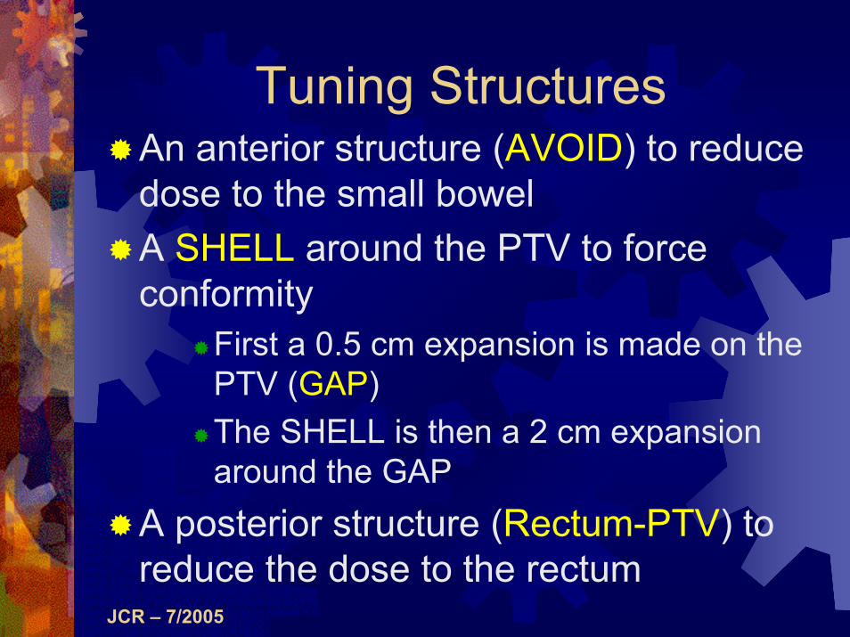
Tuning StructuresAn anterior structure (AVOID) to reduce dose to the small bowelA SHELL around the PTV to force conformity
First a 0.5 cm expansion is made on the PTV (GAP)The SHELL is then a 2 cm expansion around the GAP
A posterior structure (Rectum-PTV) to reduce the dose to the rectum
JCR – 7/2005
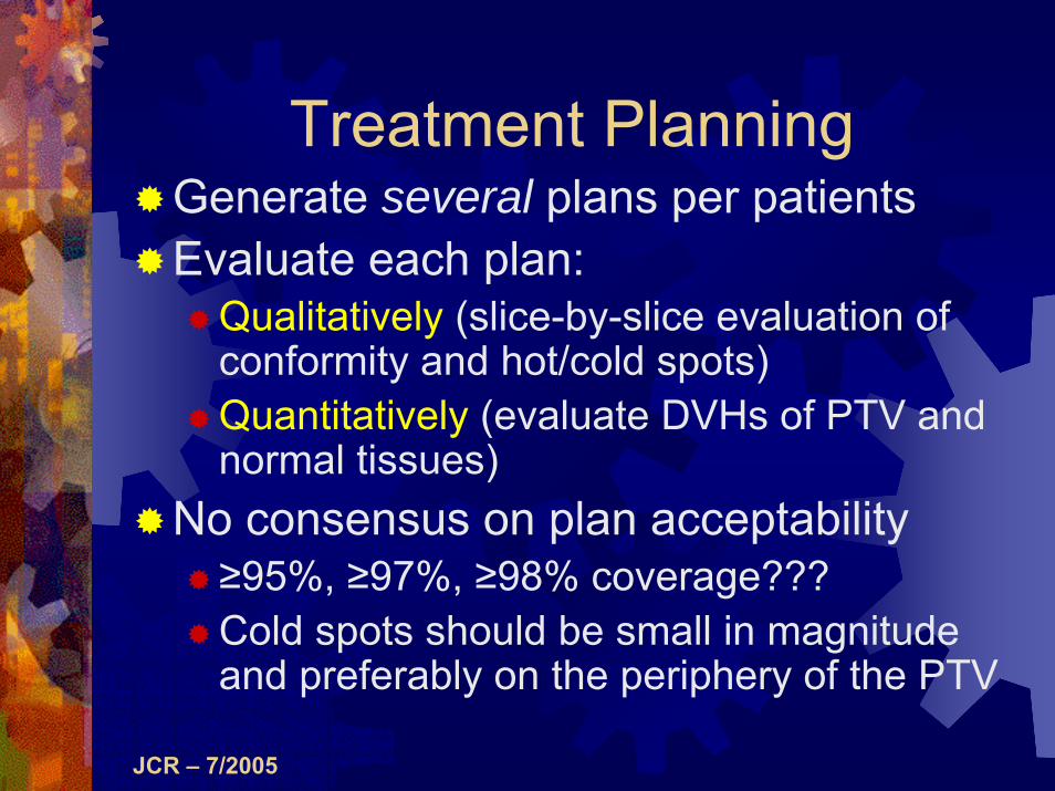
Treatment PlanningGenerate several plans per patientsEvaluate each plan:
Qualitatively (slice-by-slice evaluation of conformity and hot/cold spots)Quantitatively (evaluate DVHs of PTV and normal tissues)
No consensus on plan acceptability≥95%, ≥97%, ≥98% coverage???Cold spots should be small in magnitude and preferably on the periphery of the PTV
JCR – 7/2005
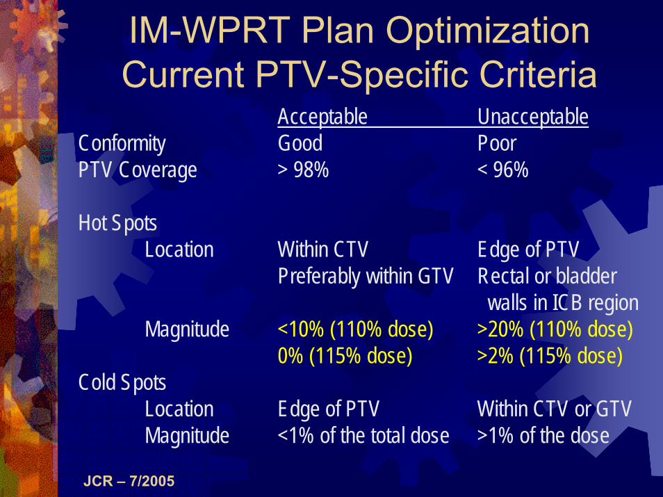
IM-WPRT Plan OptimizationCurrent PTV-Specific Criteria
Acceptable UnacceptableConformity Good PoorPTV Coverage > 98% < 96%
Hot SpotsLocation Within CTV Edge of PTV
Preferably within GTV Rectal or bladderwalls in ICB region
Magnitude <10% (110% dose) >20% (110% dose)0% (115% dose) >2% (115% dose)
Cold SpotsLocation Edge of PTV Within CTV or GTVMagnitude <1% of the total dose >1% of the dose

JCR – 7/2005
IM-WPRT Plan OptimizationNormal Tissue Specific Criteria
A more difficult question is whatmakes a normal tissue DVHacceptable.
IM-WPRT plans achieve betternormal tissue DVHs than WPRT plans. But how good does a normal tissue DVH need to be?
The answer is not clear

JCR – 7/2005
DVH Acceptance Criteria for Small Bowel
Dosimetric analysis of acute GI toxicity in our Gyne IMRT pts was performedOn multivariate analysis, the strongest predictor of acute GI toxicity was the small bowel volume receiving the prescription dose or higher (SBvol100%)
Roeske et al.Radiother Oncol 2003;69:201-7.

JCR – 7/2005
2.3
100
4101
1
⎟⎟⎠
⎞⎜⎜⎝
⎛+
=
V
NTCP
0
0.1
0.2
0.3
0.4
0.5
0.6
0.7
0.8
0.9
1
0 100 200 300 400 500 600
Volume (cc)
NTCP Analysis?Gynecologic IMRT Patients
Roeske et al. Radiother Oncol 2003;69:201-7.
ConventionalPelvic RT
IMRT

JCR – 7/2005
Conventional Isodose Distribution
PTV
100% 70%

JCR – 7/2005
IMRT Isodose Distribution
PTV
100% 70%

JCR – 7/2005
PTV
0
20
40
60
80
100
0 20 40 60 80 100 120
Percent Dose
IMRT
Conv

JCR – 7/2005
Rectum
0
20
40
60
80
100
0 20 40 60 80 100 120
Percent Dose
Conv
IMRT

JCR – 7/2005
Bladder
0
20
40
60
80
100
0 20 40 60 80 100 120
Percent Dose
Conv
IMRT

JCR – 7/2005
Small Bowel
0
20
40
60
80
100
0 20 40 60 80 100 120
Percent Dose
Conv
IMRT

JCR – 7/2005
Absolute Volume (cc) of SBRReceiving 45 Gy
0
200
400
600
800
1000
1200
1 2 3 4 5 6 7 8 9 10
Patient Number
ConvIMRT

JCR – 7/2005
IM-WPRT Planning Studies↓Volume Receiving Prescription Dose
Author Bowel Bladder RectumRoeske ↓50% ↓23% ↓23%Ahamad ↓40-63%* NS NSChen ↓70% ↓** ↓**Selvaraj ↓51%*** ↓31%*** ↓66%***
*dependent on PTV expansion used**data not shown***reduction in percent volume receiving 30 Gy or higher

JCR – 7/2005
PositioningAll of our studies (set-up uncertainty, organ motion) are based on patients in the supine positionThe prone position may offer some additional dosimetric sparing
Small bowel DVHs
Adli N, Mayr N et al. Int J Radiat Oncol Biol Phys 57: 230-238, 2003.

JCR – 7/2005
Quality Assurance

JCR – 7/2005
Treatment Delivery/QAAt U Chicago:
Varian CL2100 CD accelerators120-leaf MLCAutomatic beam sequencing software. Step and shoot mode
All major delivery systems have been used successfully
Elekta, Siemens, Varian, TomotherapyAlternatively, fabricated customized physical modulators could be used
Southeastern Radiation Productswww.seradiation.com

JCR – 7/2005
Treatment Delivery/QANo clear best delivery approachIncreasingly important factor, however, is treatment duration
↑time → ↓efficacyEffort should be directed to minimize treatment duration
Joseph Deasy, Jack F. FowlerRadiobiology of IMRT Chapter 3IMRT: A Clinical Perspective 2005

JCR – 7/2005
Treatment Delivery/QAPrior to (and throughout) treatment, rigorous QA is essentialVerify setup accuracy on day 1 and then weekly with orthogonal x-ray filmsSpecial QA problem is that field sizes may exceed MLC travel limits
Fields must be split into ≥ 2 carriage movements
Kamath S et al. Med Phys 2004;31:3314Hong L et al. Int J Radiat Oncol Biol Phys 2002;54:278

JCR – 7/2005
Comparison of Ion Chamber with Calculation
y = 0.990xR2 = 0.995
80
100
120
140
160
180
200
220
240
80 100 120 140 160 180 200 220 240
Corvus (cGy)
Mea
sure
men
t (cG
y)

JCR – 7/2005
Independent MU Verification
Use RadCalc Software (Lifeline Medical)*Uses a modified Clarkson integration algorithm to calculate dose to isocenterProgram exploits rotational symmetry of scatter to make computation efficient
*MUVC code is licensed by the University of Chicago to Lifeline Medical

JCR – 7/2005
Comparison of Radcalc to CorvusAll Treatments
0
20
40
60
80
100
120
-3 -1 1 3 5
Percent Disparity
Num
ber o
f Occ
uren
ces
Mean = 1.4%, Standard deviation = 1.2%, N = 504
J. Haslam et al. Comparison of dose calculated by an intensity modulated radiotherapy treatment planning system and an independent monitor unit verification program.In Press J Appl Clin Med Phys.

JCR – 7/2005
Treatment Delivery/QA
In our Gyne IMRT patients, we compared doses calculated by CORVUS and the RadCalc MUVC program
Lifeline Software, Inc., lifelinesoftware.com/Mean disparity was 0.2% (standard deviation 1.1%)Disparities ≥ 3% result in additional QA (ion chamber and film measurements)
Haslam J. J Appl Clin Med Phys 2003; 4:224-30.

JCR – 7/2005
Film Dosimetry

JCR – 7/2005
Clinical ExperienceBetween 2/00 and 7/05, >150 women were treated with IM-WPRT in our clinicMost had cervical cancer, primarily stage IBMost underwent definitive RT and, in stages IB2-IIIB, concomitant cisplatin-based chemotherapyEndometrial cancer patients were treated following primary surgeryICB was administered in ~50% of women following IM-WPRT
Mundt, Roeske, et al. Gyne Oncol 82(3): 456-463, 2001.Mundt et al. Int J Radiat Oncol Biol Phys 52(5):1330-1337, 2002.

JCR – 7/2005
Clinical Experience
How do results compare to conventional treatments?Acute GI toxicities (Grade 2)
WPRT: 91%IM-WPRT: 60% p = 0.002
Acute GU toxicities (Grade 2)WPRT: 20%IM-WPRT: 10% p = 0.22
Mundt et al. Int J Radiat Oncol Biol Phys 52(5):1330-1337, 2002.

JCR – 7/2005
Acute GI toxicity in IM-WPRT Patients vs. WPRT
0102030405060708090
100
Grade 0 Grade 1 Grade 2 Grade 3
IM-WPRTWPRT

JCR – 7/2005
Chronic GI Toxicity
0%10%20%30%40%50%60%70%80%90%
0 1 2 3
IM-WPRTWPRT
On multivariate analysis controlling for age, chemo, stage and site,IMRT remained statistically significant ( p = 0.01; odds ratio 0.16, 95% confidence interval 0.04, 0.67)

JCR – 7/2005
Cervical CancerKochanski J, Mundt AJ. ASCO (2004)34 stage I-II cervical cancer pts
21 intact uterus, 13 postoperativeMedian follow-up = 26.2 months3-year actuarial pelvic control = 92%
Endometrial CancerKnab B, Mundt AJ. ASTRO (2004)31 stage I-III endometrial cancer pts
All treated postoperativelyMedian follow-up = 24.1 months3-year actuarial pelvic control = 100%
Excellent Pelvic Control Rates

JCR – 7/2005
Pelvic ControlWhile encouraging, follow-up remains relatively short and the number of patients treated remains smallOnly with longer follow-up and larger patient cohorts can more definitive statements be madeCooperative groups (RTOG, GOG) are currently developing protocols to evaluate IMRT in gynecology patients

JCR – 7/2005
ConclusionsIMRT is a useful means of reducing the volume of normal tissues irradiated in gynecologic patients receiving WPRTOur initial evaluation indicate a significant reduction in GI toxicity relative to patients receiving conventional therapyContinued follow-up and critical evaluation are required to validate the long term merits of this approach

JCR – 7/2005
What about the negatives?
IMRT results in higher volumes of normal tissue receiving lower dosesIncreased MUs result in higher total body dosesTarget and tissue delineation are time-consumingNo guidelines exist regarding how targets should be contoured and plans optimizedLong-term follow-up is not available assessing tumor control and unexpected sequelaeClinical data are available from only one institution and while prospective no randomized comparisons have been performed


