6/24/2014 1 In Vivo Dosimetry in Brachytherapy: Feasible and Needed? Feasible and Needed? Annette Haworth, PhD, FACPSEM Peter MacCallum Cancer Centre, East Melbourne, Australia; University of Melbourne, East Melbourne, Australia Disclosure Annette Haworth, PhD, FACPSEM, does not have any financial relationships or products or devices with any commercial interest related to the content of this activity of any amount during the past 12 months.
Transcript
6/24/2014
1
In Vivo Dosimetry in Brachytherapy: Feasible and Needed?Feasible and Needed?
Annette Haworth, PhD, FACPSEMPeter MacCallum Cancer Centre, East Melbourne, Australia;
University of Melbourne, East Melbourne, Australia
Disclosure
Annette Haworth, PhD, FACPSEM, does not have any financial relationships or products or devices with any commercial interest related to the content of this activity of any amount during the past 12 months.
6/24/2014
2
In Vivo Dosimetry in Brachytherapy: Feasible and Needed?
• Is it needed?– Is there any evidence that in vivo dosimetry couldIs there any evidence that in vivo dosimetry could have prevented accidents/mis‐administrations?
– Will it be of benefit for future developments in brachytherapy?
• Is it feasible?Wh l il bl ?– What systems are currently available?
– What’s in development?
Needed?
We deliver high doses in small numbers of fractions, often under high-pressure situations
• What can possibly go wrong?– ICRP Publication 97 & 86, IAEA RS17, WHO 2009
• Incidents are mostly related to procedural errors
• Malfunctions are noted
– We have minimal R&V• Might tell us what the system thought it delivered, but doesn’t tell us if
d h h l l d h i dwe connected the catheters correctly, selected the correct indexer length, etc
– Conclude• Lack of good systematic evidence makes it difficult to justify to administrators, but there is sufficient evidence to show errors can and do happen in brachytherapy
6/24/2014
3
Is in Vivo Dosimetry Needed to Further the Developments in Brachytherapy?
– Are our current dose constraints based on accurate knowledge of the dose we delivered to our patients?
– Can we safely dose escalate?
– Can we be confident it is safe to deliver increasingly conformal dose distributions?
– Do we want to assure our patient’s brachytherapy is a safe and accurate treatment option?
Feasible?
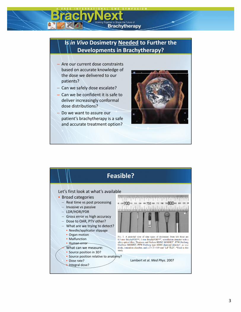
Let’s first look at what’s available• Broad categories
– Real time vs post processingReal time vs post processing– Invasive vs passive– LDR/HDR/PDR– Gross error vs high accuracy– Dose to OAR, PTV other?– What are we trying to detect?
• Needle/applicator slippage• Organ motion• Malfunction• Malfunction• Human error
– What can we measure:• Source position in 3D?• Source position relative to anatomy?• Dose rate?• Integral dose?
Lambert et al. Med Phys. 2007
6/24/2014
4
Challenges in in Vivo Dosimetry in Brachytherapy
• High dose gradients– Slight detector displacement = large dose discrepancy
– Close to source = high signal + large dose gradient
– Far from source = low signal
• Detectors placed within tumor or cavity– Clinically relevant dose (rate) readings
– Risk needs to be offset by benefit• Additional needle insertion• Infection• Prolonged workflow
• Threshold for error detection– Compromise on sensitivity and specificity
What Systems Have Been Reported in the Literature?
• Few commercial systems
• Few clinical trials (mostly phantom studies)• Few clinical trials (mostly phantom studies)
• Cost largely unknown
• General vs site specific
• Single vs multiple detectors
• Refer to Vision 20/20 paper (next slide) for• Refer to Vision 20/20 paper (next slide) for comprehensive overview
6/24/2014
5
*
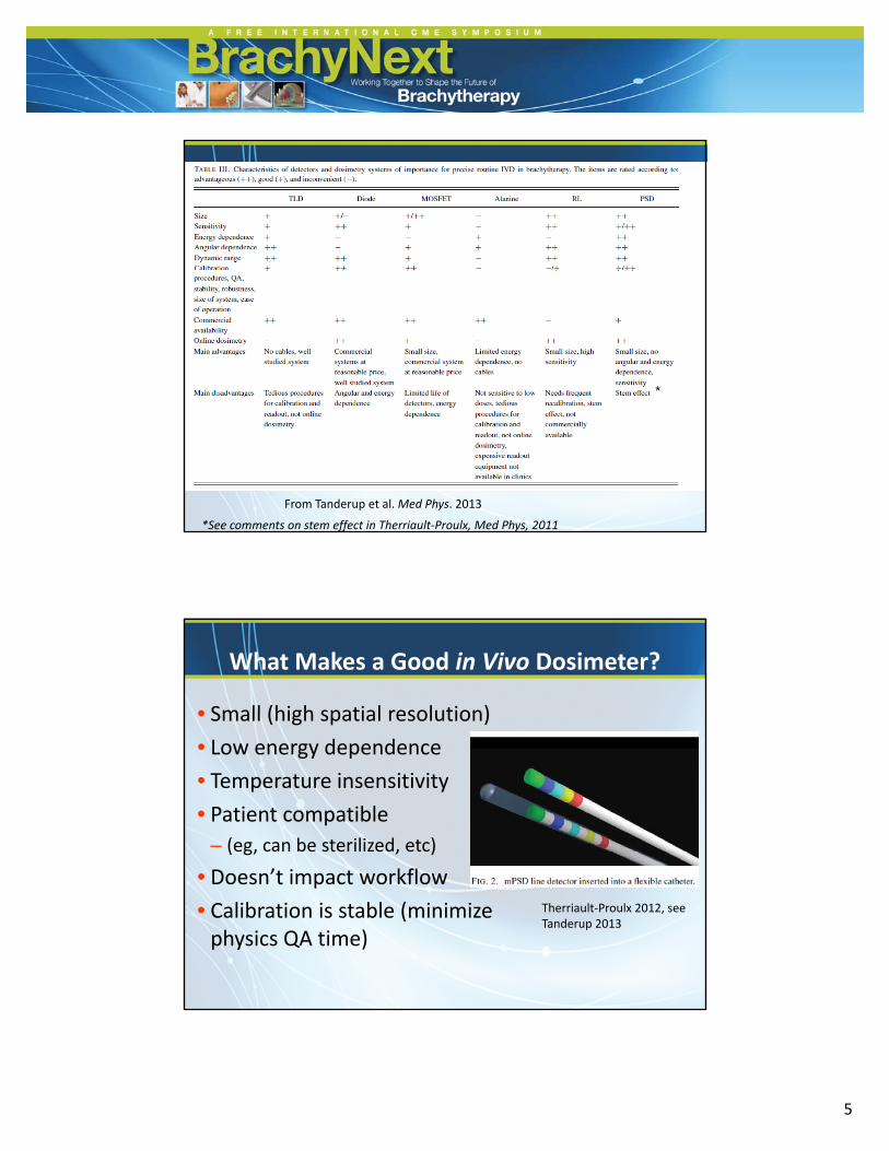
From Tanderup et al. Med Phys. 2013
*See comments on stem effect in Therriault‐Proulx, Med Phys, 2011
What Makes a Good in Vivo Dosimeter?
• Small (high spatial resolution)
• Low energy dependence• Low energy dependence
• Temperature insensitivity
• Patient compatible
– (eg, can be sterilized, etc)
• Doesn’t impact workflowDoesn t impact workflow
• Calibration is stable (minimize physics QA time)
Therriault‐Proulx 2012, see Tanderup 2013
6/24/2014
6
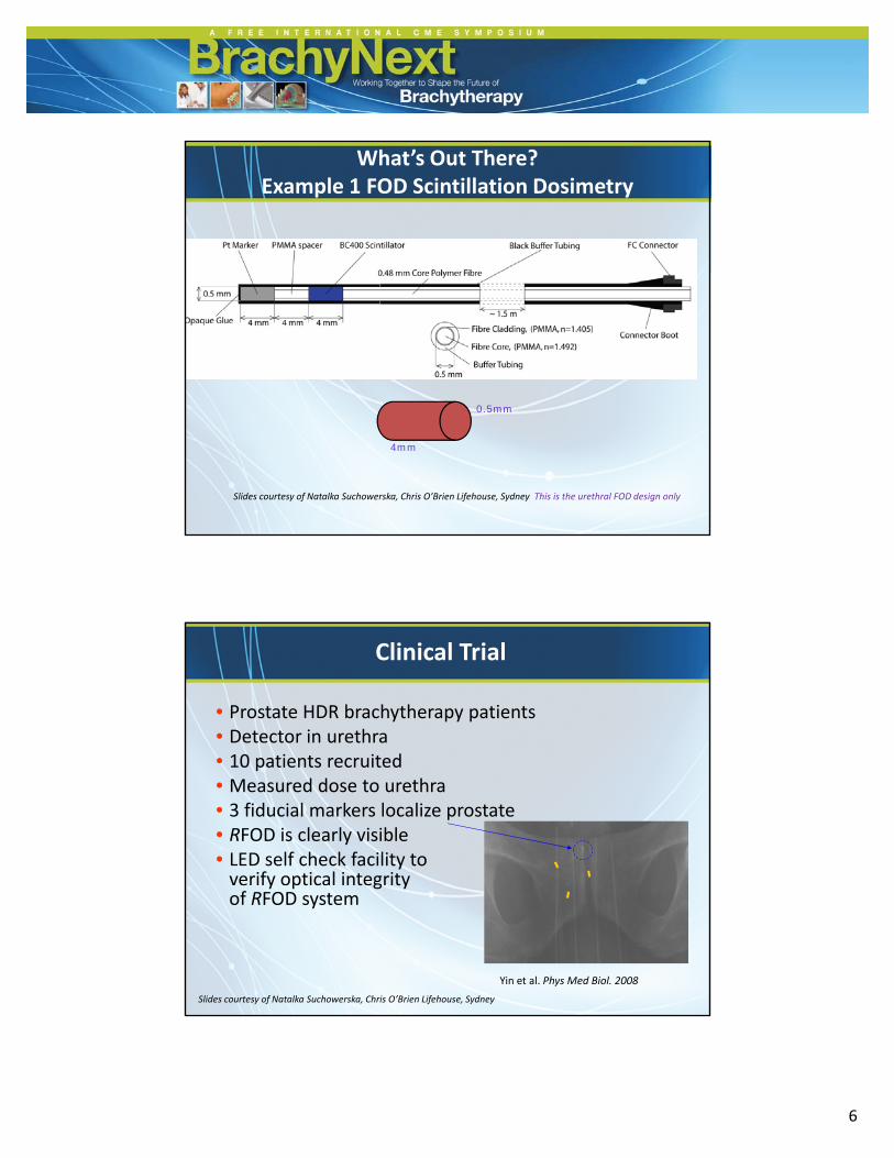
What’s Out There? Example 1 FOD Scintillation Dosimetry
4mm
0.5mm
Slides courtesy of Natalka Suchowerska, Chris O’Brien Lifehouse, Sydney This is the urethral FOD design only
Clinical Trial
• Prostate HDR brachytherapy patients• Detector in urethra
d• 10 patients recruited • Measured dose to urethra• 3 fiducial markers localize prostate• RFOD is clearly visible• LED self check facility to verify optical integrity
Yin et al. Phys Med Biol. 2008
verify optical integrity of RFOD system
Slides courtesy of Natalka Suchowerska, Chris O’Brien Lifehouse, Sydney
6/24/2014
7
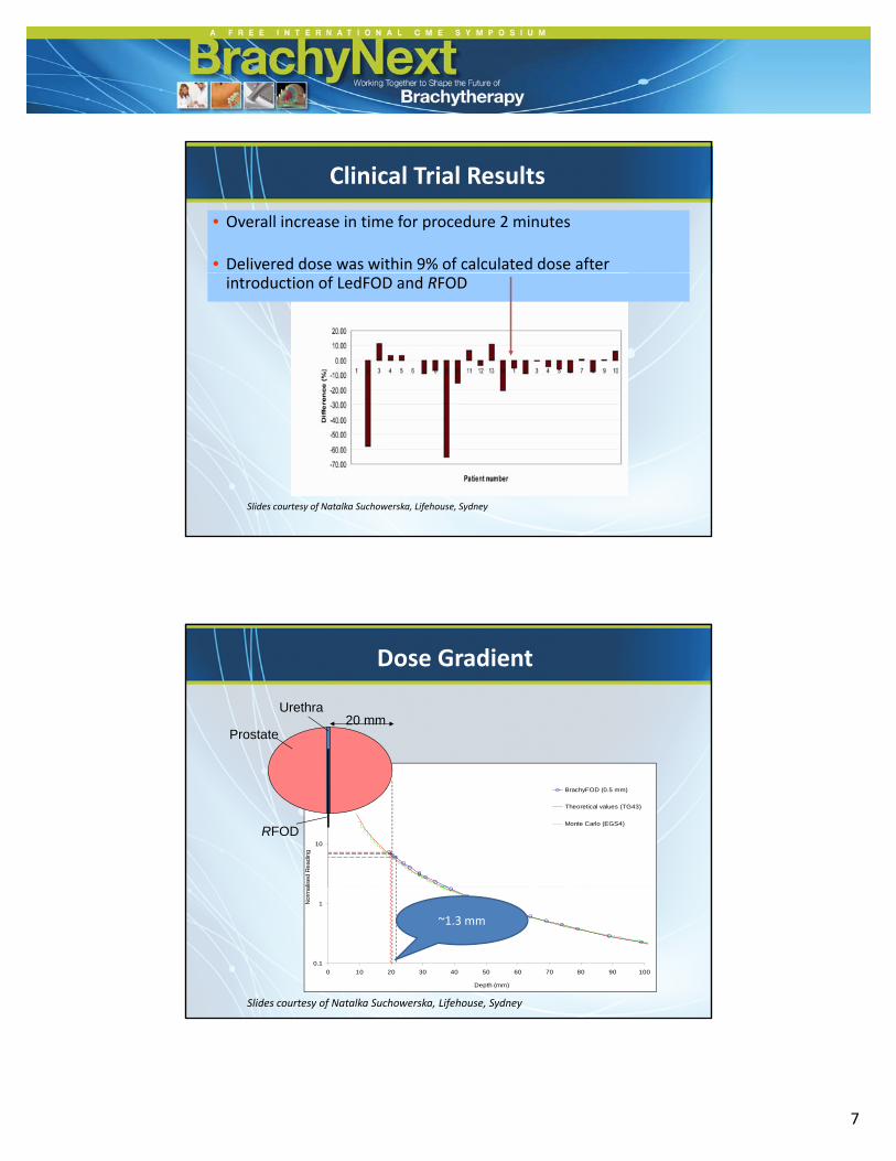
Clinical Trial Results
• Overall increase in time for procedure 2 minutes
• Delivered dose was within 9% of calculated dose after introduction of LedFOD and RFOD
Slides courtesy of Natalka Suchowerska, Lifehouse, Sydney
Dose Gradient
Urethra20 mm
Prostate
10
100
alis
ed R
eadin
g
BrachyFOD (0.5 mm)
Theoretical values (TG43)
Monte Carlo (EGS4)
RFOD
0.1
1
0 10 20 30 40 50 60 70 80 90 100
Depth (mm)
Norm
a
Slides courtesy of Natalka Suchowerska, Lifehouse, Sydney
~1.3 mm
6/24/2014
8
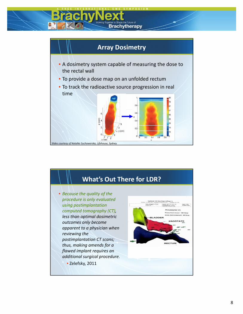
Array Dosimetry
• A dosimetry system capable of measuring the dose to the rectal wall
• To provide a dose map on an unfolded rectum
• To track the radioactive source progression in real time
Slides courtesy of Natalka Suchowerska, Lifehouse, Sydney
What’s Out There for LDR?
• Because the quality of the procedure is only evaluated i ti l t tiusing postimplantation
computed tomography (CT), less than optimal dosimetric outcomes only become apparent to a physician when reviewing the postimplantation CT scans;postimplantation CT scans; thus, making amends for a flawed implant requires an additional surgical procedure.
• Zelefsky, 2011
6/24/2014
9
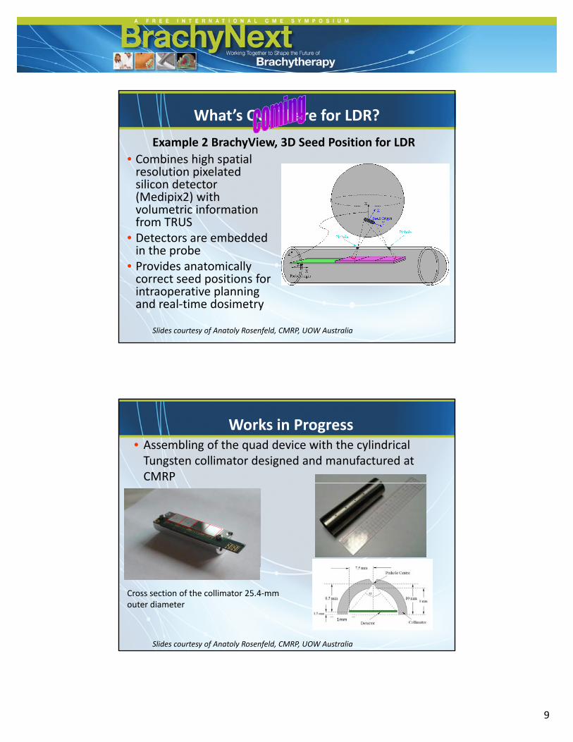
What’s Out There for LDR?
• Combines high spatial resolution pixelated
Example 2 BrachyView, 3D Seed Position for LDR
resolution pixelated silicon detector (Medipix2) with volumetric information from TRUS
• Detectors are embedded in the probe
• Provides anatomically correct seed positions for intraoperative planning and real‐time dosimetry
Slides courtesy of Anatoly Rosenfeld, CMRP, UOW Australia
• Assembling of the quad device with the cylindrical Tungsten collimator designed and manufactured at CMRP
Works in Progress
Cross section of the collimator 25.4‐mm outer diameter
1mm
Slides courtesy of Anatoly Rosenfeld, CMRP, UOW Australia
6/24/2014
10
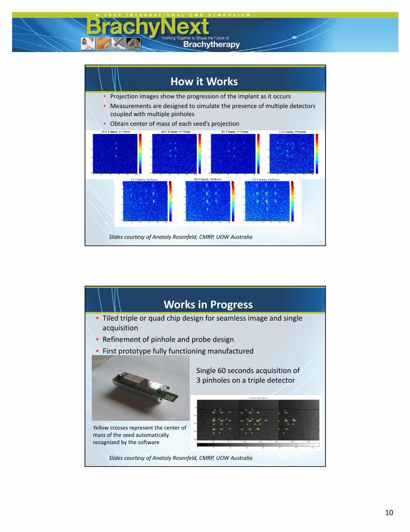
• Projection images show the progression of the implant as it occurs
• Measurements are designed to simulate the presence of multiple detectors coupled with multiple pinholes
• Obtain center of mass of each seed’s projection
How it Works
Obtain center of mass of each seed s projection
Slides courtesy of Anatoly Rosenfeld, CMRP, UOW Australia
• Tiled triple or quad chip design for seamless image and single acquisition
• Refinement of pinhole and probe design
Works in Progress
• First prototype fully functioning manufactured
Single 60 seconds acquisition of 3 pinholes on a triple detector
Yellow crosses represent the center of mass of the seed automatically recognized by the software
Slides courtesy of Anatoly Rosenfeld, CMRP, UOW Australia
6/24/2014
11
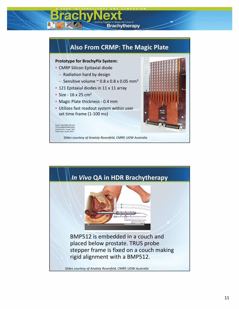
Prototype for BrachyPix System:
• CMRP Silicon Epitaxial diode
Also From CRMP: The Magic Plate
– Radiation hard by design
– Sensitive volume ~ 0.8 x 0.8 x 0.05 mm3
• 121 Epitaxial diodes in 11 x 11 array
• Size ‐ 16 x 25 cm2
• Magic Plate thickness ‐ 0.4 mm
• Utilizes fast readout system within user• Utilizes fast readout system within user set time frame (1‐100 ms)
Patent: WO2008/148150 AlPCT/au2008 000788 (2008)Patent China , issued , 2012Patent USA , issued, 2014
Slides courtesy of Anatoly Rosenfeld, CMRP, UOW Australia
In Vivo QA in HDR Brachytherapy
BMP512 is embedded in a couch andBMP512 is embedded in a couch and placed below prostate. TRUS probe stepper frame is fixed on a couch making rigid alignment with a BMP512.
Slides courtesy of Anatoly Rosenfeld, CMRP, UOW Australia
6/24/2014
12
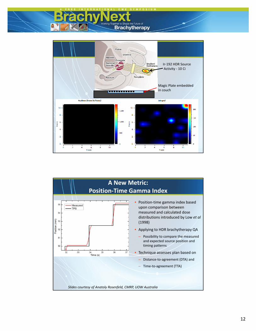
Ir‐192 HDR SourceActivity ‐ 10 Ci
Magic Plate embedded in couch
• Position‐time gamma index based upon comparison between measured and calculated dose
A New Metric: Position‐Time Gamma Index
distributions introduced by Low et al (1998)
• Applying to HDR brachytherapy QA
– Possibility to compare the measured and expected source position and timing patterns
• Technique assesses plan based onTechnique assesses plan based on
– Distance‐to‐agreement (DTA) and
– Time‐to‐agreement (TTA)
Slides courtesy of Anatoly Rosenfeld, CMRP, UOW Australia
6/24/2014
13
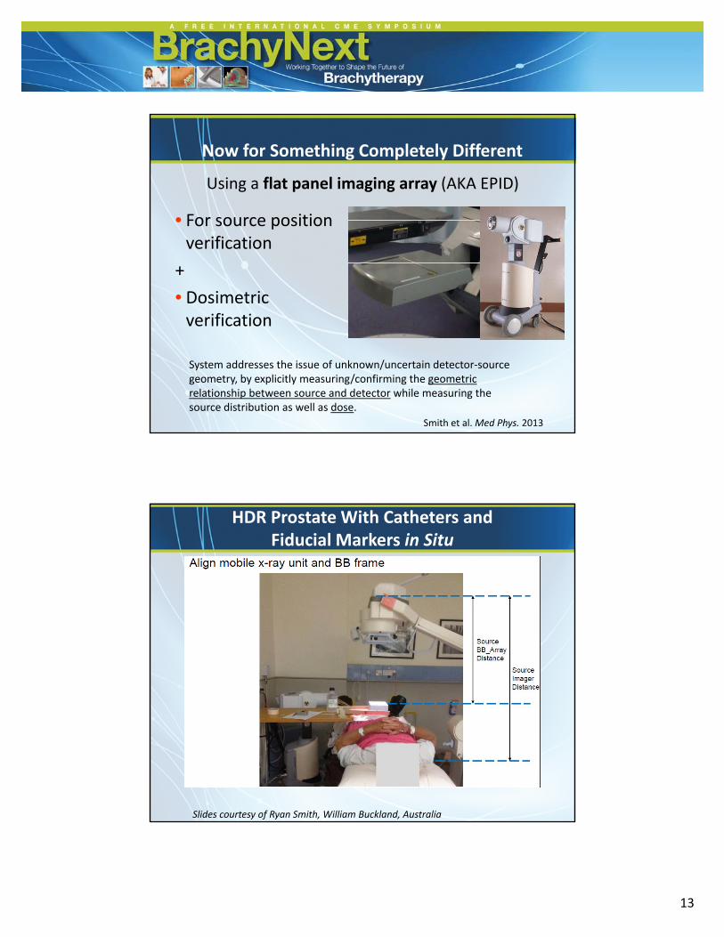
Now for Something Completely Different
• For source position
Using a flat panel imaging array (AKA EPID)
• For source position verification
+
• Dosimetric verification
Smith et al. Med Phys. 2013
System addresses the issue of unknown/uncertain detector‐source geometry, by explicitly measuring/confirming the geometric relationship between source and detector while measuring the source distribution as well as dose.
HDR Prostate With Catheters and Fiducial Markers in Situ
Slides courtesy of Ryan Smith, William Buckland, Australia
6/24/2014
14
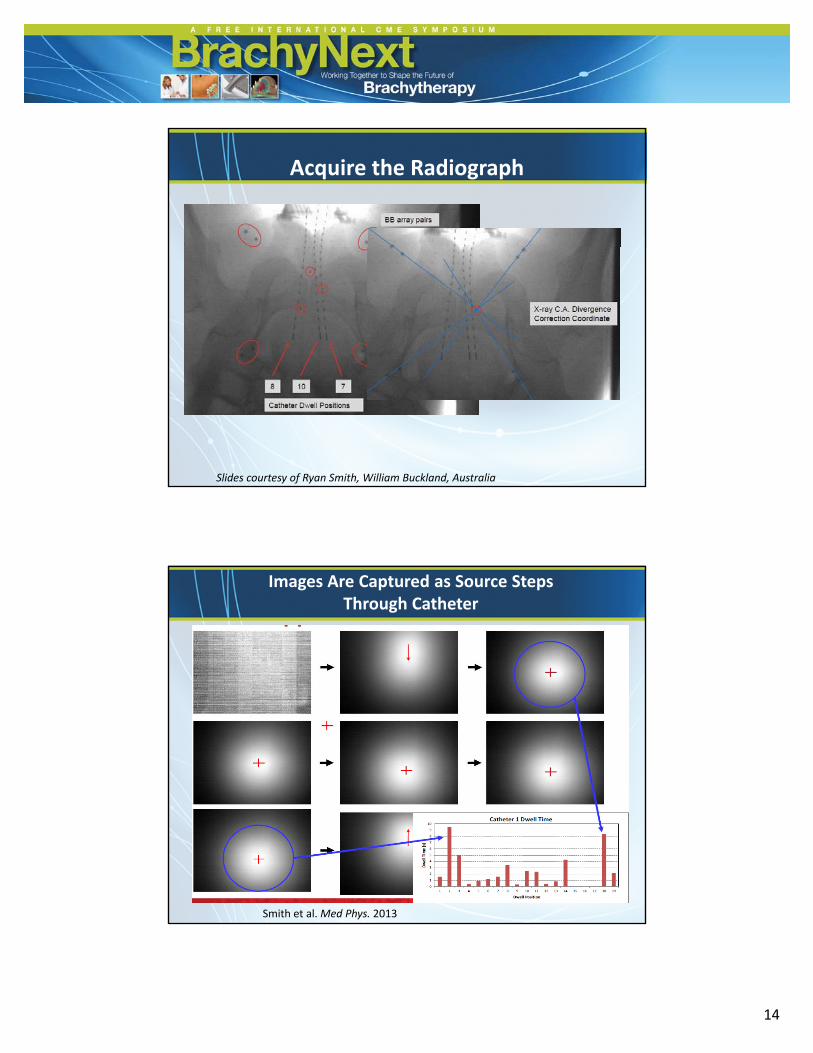
Acquire the Radiograph
Slides courtesy of Ryan Smith, William Buckland, Australia
Images Are Captured as Source Steps Through Catheter
Smith et al. Med Phys. 2013
6/24/2014
15
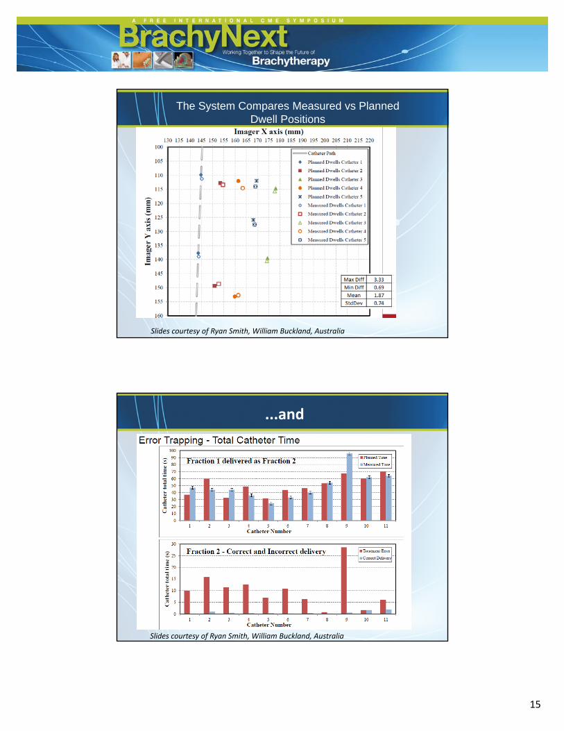
The System Compares Measured vs Planned Dwell Positions
Slides courtesy of Ryan Smith, William Buckland, Australia
...and
Slides courtesy of Ryan Smith, William Buckland, Australia
6/24/2014
16
Feasible? Summary
• No perfect system exists
• Define what you are trying to do and what• Define what you are trying to do and what accuracy you need
– How does it compliment your existing QA program?
Remember: "treatment verification" doesn't only mean in vivo dosimetry,
and in vivo dosimetry doesn't necessarily achieve treatment verification
You really need to do both.
Rick Franich
Conclusions
• Needed?– Incidents have been reported that could have beenIncidents have been reported that could have been prevented by use of in vivo dosimetry
• But under‐reporting and lack of systematic data collection makes developing business cases challenging
– Our patients need to be assured that brachytherapy is safe and accurate
– Future developments in brachytherapy require precise knowledge of the treatment that was actually delivered
6/24/2014
17
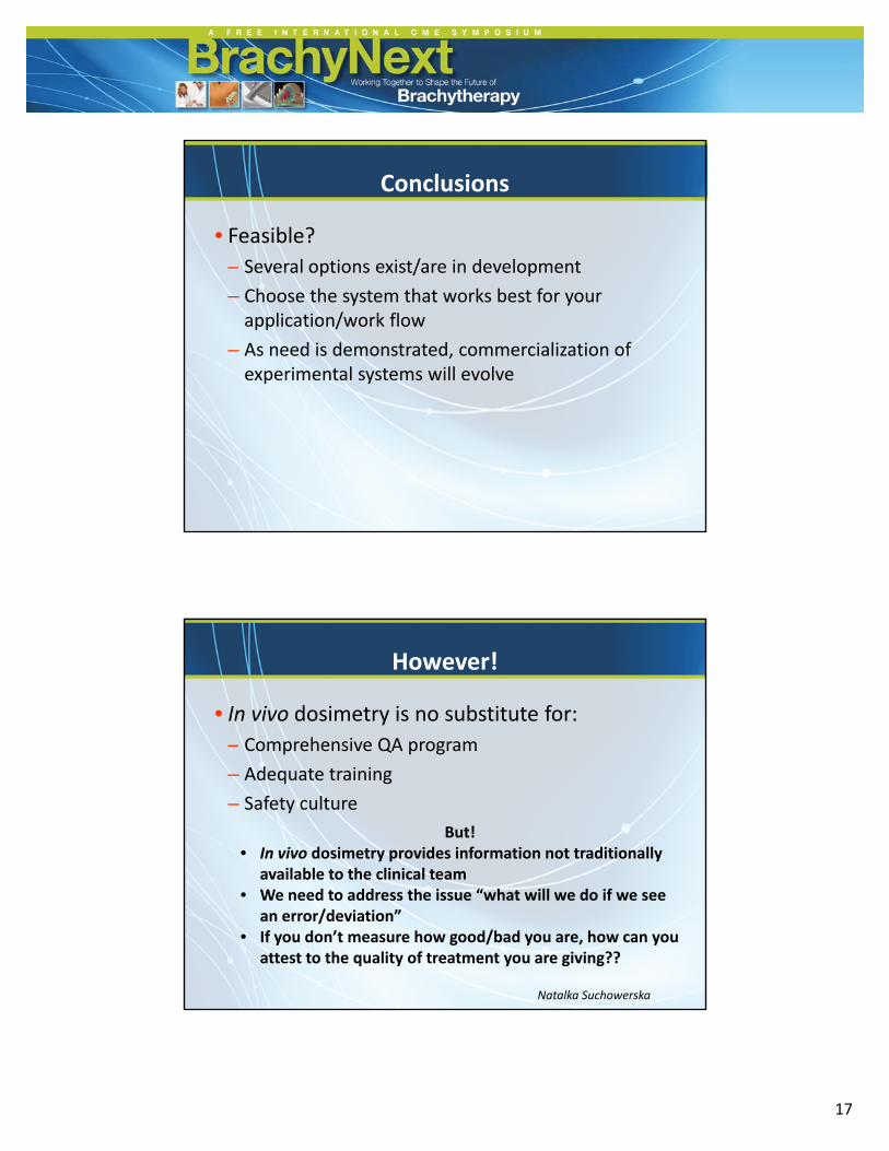
Conclusions
• Feasible?– Several options exist/are in developmentSeveral options exist/are in development
– Choose the system that works best for your application/work flow
– As need is demonstrated, commercialization of experimental systems will evolve
However!
• In vivo dosimetry is no substitute for:
– Comprehensive QA programComprehensive QA program
– Adequate training
– Safety culture
But! • In vivo dosimetry provides information not traditionally
available to the clinical team• We need to address the issue “what will we do if we see
an error/deviation” • If you don’t measure how good/bad you are, how can you
attest to the quality of treatment you are giving??
Natalka Suchowerska
6/24/2014
18
My Thanks to:– A/Prof Natalka Suchowerska
– Prof Anatoly Rosenfeld
Ryan Smith– Ryan Smith
– A/Prof Rick Franich
For their slides, resources, and many comments that I’ve incorporated into this presentation
http://mmnd-ipct.com
References• Kertzscher G, et al Adaptive error detection for HDR/PDR brachytherapy: Guidance for decision making during real‐time in vivo point dosimetry. Med
Phys. 2014 May;41(5):052102.
• Dempsey C, et al. ACPSEM brachytherapy working group recommendations for quality assurance in brachytherapy. Australas Phys Eng Sci Med. 2013 Dec;36(4):387‐96
• Smith RL, et al. Source position verification and dosimetry in HDR brachytherapy using an EPID. Med Phys. 2013 Nov;40(11):111706.
• Haworth A, et al. Australasian brachytherapy audit: results of the 'end‐to‐end' dosimetry pilot study. J Med Imaging Radiat Oncol. 2013 Aug;57(4):490‐8
• Tanderup K et al In vivo dosimetry in brachytherapy Med Phys 2013 Jul;40(7):070902• Tanderup K, et al. In vivo dosimetry in brachytherapy. Med Phys. 2013 Jul;40(7):070902.
• Gambarini G, et al. Online in vivo dosimetry in high dose rate prostate brachytherapy with MOSkin detectors: in phantom feasibility study. Appl RadiatIsot. 2014 Jan;83 Pt C:222‐6
• Therriault‐Proulx F, et al On the use of a single‐fiber multipoint plastic scintillation detector for 192Ir high‐dose‐rate brachytherapy. Med Phys. 2013 Jun;40(6):062101
• Haworth A, et al. Comparison of TLD calibration methods for 192Ir dosimetry. J Appl Clin Med Phys. 2013 Jan 7;14(1):4037
• Liu PZ, et al. Real‐time scintillation array dosimetry for radiotherapy: the advantages of photomultiplier detectors. Med Phys. 2012 Apr;39(4):1688‐95.
• Hayton A, et al. Lack of backscatter factor measurements in HDR applications with MOSkins. Australas Phys Eng Sci Med. 2011 Dec;34(4):545‐52.
• Kertzscher G, et al. Identifying afterloading PDR and HDR brachytherapy errors using real‐time fiber‐coupled Al(2)O(3):C dosimetry and a novel statistical error decision criterion. Radiother Oncol. 2011 Sep;100(3):456‐62..
• Therriault‐Proulx F, et al. A phantom study of an in vivo dosimetry system using plastic scintillation detectors for real‐time verification of 192Ir HDR brachytherapy Med Phys. 2011 May;38(5):2542‐51.
• Therriault‐Proulx F, et al Technical note: removing the stem effect when performing Ir‐192 HDR brachytherapy in vivo dosimetry using plastic ll d l d d h ( )scintillation detectors: a relevant and necessary step. Med Phys. 2011 Apr;38(4):2176‐9.
• SuchowerskaN, et al. Clinical trials of a urethral dose measurement system in brachytherapy using scintillation detectors. Int J Radiat Oncol Biol Phys. 2011 Feb 1;79(2):609‐15.
• Cartwright LE, et al. Dose mapping of the rectal wall during brachytherapy with an array of scintillation dosimeters. Med Phys. 2010 May;37(5):2247‐55.
• WHO Radiotherapy Risk Profile. Geneva, World Health Organisation. 2008
• Lambert J et al. In vivo dosimeters for HDR brachytherapy: a comparison of a diamond detector, MOSFET, TLD, and scintillation detector. Med Phys. 2007 May;34(5):1759‐65.
• Lambert J, et al. A plastic scintillation dosimeter for high dose rate brachytherapy. Phys Med Biol. 2006 Nov 7;51(21):5505‐16.
• Ashton L, et al. Prevention of high‐dose‐rate brachytherapy accidents,” ICRP Publication 97, Annals of the ICRP (Pergamon, New York, 2004).
• IAEA, Lessons learned from accidental exposures in radiotherapy. IAEA Safety Report Series 17 (IAEA, Vienna, 2000).
• Lopez P, et al. Prevention of accidental exposures to patients undergoing radiation therapy,”ICRP Publication 86, Annals of the ICRP (Pergamon, New York, 2000).


![Volume-8 | Issue-5 | May-2019 | PRINT ISSN No. 2250 - 1991 ... · brachytherapy are as per the American Brachytherapy Society (ABS) consensus guidelines for sarcoma brachytherapy[7]](https://static.documents.pub/doc/80x56/5ee1cc87ad6a402d666c8c3a/volume-8-issue-5-may-2019-print-issn-no-2250-1991-brachytherapy-are.jpg)
