Proc. Nat. Acad. Sci. USA Vol. 70, No. 1, pp. 11-14, January 1973 In Situ Hybridization of RD114-Virus RNA with Human Metaphase Chromosomes (density gradient centrifugation/karyotype/RNA-dependent DNA polymerase) PETER M. PRICE*, KURT HIRSCHHORN*, NORMAN GABELMANt, AND SAMUEL WAXMANt * Division of Human Genetics, Department of Pediatrics; and t Cancer Chemotherapy Laboratory, Department of Medicine, Mount Sinai School of Medicine of the City University of New York, N.Y. 10029 Communicated by Robert J. Huebner, October 17, 1972 ABSTRACT In situ hybridization was used to detect regions of human metaphase chromosomes exhibiting complementarity with radioactively-labeled RNA isolated from RD114-virus. The region of homology, as identified by autoradiography, was confined to one D-group chromo- some. In a previous paper (1), we reported the probable localization of hemoglobin genes on human chromosomes. Hemoglobin structural genes were tentatively identified by autoradio- graphic detection of molecular hybrids formed in situ between purified, radiolabeled hemoglobin message and the DNA of metaphase chromosomes. The present communication reports the detection of regions of homology between the RNA extracted from RD114-virus (2), isolated from a serially cultured line of human rhabdo- myosarcoma cells (3), and a group-D chromosome from a human lymphoid cell line cultured serially. Methods were as described (2, 3). MATERIALS AND METHODS Virus and Cells. RNA was isolated from virus released into the culture medium from RD114 cells (RD114-virus). The RD 114 cell line (gift from Dr. Aaron Freeman) was grown in 75-cm2 tissue culture flasks (Falcon Plastics) or in roller bot- tles (670-cm2 surface area, Bellco) in complete medium [me- dium RPMI-1640 (GIBCO) supplemented with 10% fetal- calf serum, containing 4 mM glutamine and 50 jg of genta- mycin per ml]. Cells were fed with fresh complete medium every fourth day and subcultured every eighth day. Morphology and growth characteristics of RD114 cells appeared to be similar to those reported by McAllister et al. (3). The cells extruded virus into the culture medium that incorporated [3H1 U, demonstrated RNA-directed DNA poly- merase (reverse transcriptase) activity (4. 5), and had a buoy- ant density of about 1.16 g/ml in a 15-65% sucrose gradient (Fig. 1). These findings are similar to those reported by McAl- lister et al. (2). Radioactive virus was prepared (6) in complete medium containing 20 ,Ci/ml of [3H]U (28 Ci/mmol). The purified, labeled virus was isolated from supernatant tissue-culture fluid clarified and pooled as follows: An equal volume of satu- rated ammonium sulfate was added and the precipitate was collected by centrifugation (30,000 X g, 5 min at 40) and dissolved in 5 ml of NTE (100 mM NaCl-10 mM Tris HCl- 1 mM Na2EDTA, pH 7.4). The dissolved ammonium sulfate Abbreviation: SSC, 0.15 M NaCl-0.015 M Na-acetate. precipitate was then layered over a discontinuous gradient containing 0.7 ml of 65% sucrose and 3 ml of 20% sucrose in NTE and centrifuged in a Spinco SW39 rotor for 2 hr at 32,000 rpm. The interphase was collected, diluted 10-20 times, and centrifuged (37,000 X g, 120 min at 4°). The pellet was then dissolved in 0.4 ml of NTE, layered onto a 5-ml 20-65% su- crose gradient, and spun in an SW39 rotor (37,500 rpm, 120 min at 40). 0.3-ml Fractions were collected from the bottom of the tube. Each fraction was assayed for density, radioactiv- ity, and RNA-directed DNA polymerase activity (3, 5, 6). Isoltion of RD114 [3H] RNA. Fractions of the sucrose gra- dient with a density between 1.13 and 1.19 and demonstrating incorporation of [3H ]U and/or RNA-directed DNA polymerase activity (fractions 3-7, Fig. 1) were pooled and diluted with an equal volume of NTE. To this were added 2-mercaptoethanol (final concentration 0.01 M) and sodium dodecyl sulfate (final concentration 1%) with stirring (15 min at 4°). The labeled RNA was then extracted by stirring the virus solution with 0.5 volume of phenol-cresol for 30 min at 250. The resulting emulsion was then centrifuged (18,000 X g, 20 min at 40) and the aqueous phase was retained. The phenol layer was 2000 g/cm3 -1100 1.20 1. 5 15 1500 a 75 tr t layre ovraim ursegaint(06No nNE ufr L~J I 500 2 I 5 1 0 1 5 FRACTION NUMBER FIG. 1. Equilibrium sucrose density gradient centrifugation of ['H]U-labeled virus. Centrifugation for hr at 37,500 rpm and 40 in a Spinco SW39 rotor after the virus wa's layered over a 5-nil sucrose gradient (20-65%) in NTE buffer. A 0.01-ml aliquot of each fraction was assayed for radioac- tivity and a 0.1-ml aliquot of each fraction was assayed for RNA-directed DNA polymerase activity. Radioactivity, cpm of [3H]U, * -; RNA-dependent DNA polymerase, [3H]TTP incorporation/Mg of protein, 0-O; solution density, X X . 11
Transcript
Proc. Nat. Acad. Sci. USAVol. 70, No. 1, pp. 11-14, January 1973
In Situ Hybridization of RD114-Virus RNA with HumanMetaphase Chromosomes
(density gradient centrifugation/karyotype/RNA-dependent DNA polymerase)
PETER M. PRICE*, KURT HIRSCHHORN*, NORMAN GABELMANt, AND SAMUEL WAXMANt* Division of Human Genetics, Department of Pediatrics; and t Cancer Chemotherapy Laboratory, Department of Medicine,Mount Sinai School of Medicine of the City University of New York, N.Y. 10029
Communicated by Robert J. Huebner, October 17, 1972
ABSTRACT In situ hybridization was used to detectregions of human metaphase chromosomes exhibitingcomplementarity with radioactively-labeled RNA isolatedfrom RD114-virus. The region of homology, as identifiedby autoradiography, was confined to one D-group chromo-some.
In a previous paper (1), we reported the probable localizationof hemoglobin genes on human chromosomes. Hemoglobinstructural genes were tentatively identified by autoradio-graphic detection of molecular hybrids formed in situ betweenpurified, radiolabeled hemoglobin message and the DNA ofmetaphase chromosomes.The present communication reports the detection of regions
of homology between the RNA extracted from RD114-virus(2), isolated from a serially cultured line of human rhabdo-myosarcoma cells (3), and a group-D chromosome from ahuman lymphoid cell line cultured serially. Methods were asdescribed (2, 3).
MATERIALS AND METHODS
Virus and Cells. RNA was isolated from virus released intothe culture medium from RD114 cells (RD114-virus). TheRD 114 cell line (gift from Dr. Aaron Freeman) was grown in75-cm2 tissue culture flasks (Falcon Plastics) or in roller bot-tles (670-cm2 surface area, Bellco) in complete medium [me-dium RPMI-1640 (GIBCO) supplemented with 10% fetal-calf serum, containing 4 mM glutamine and 50 jg of genta-mycin per ml]. Cells were fed with fresh complete mediumevery fourth day and subcultured every eighth day.Morphology and growth characteristics of RD114 cells
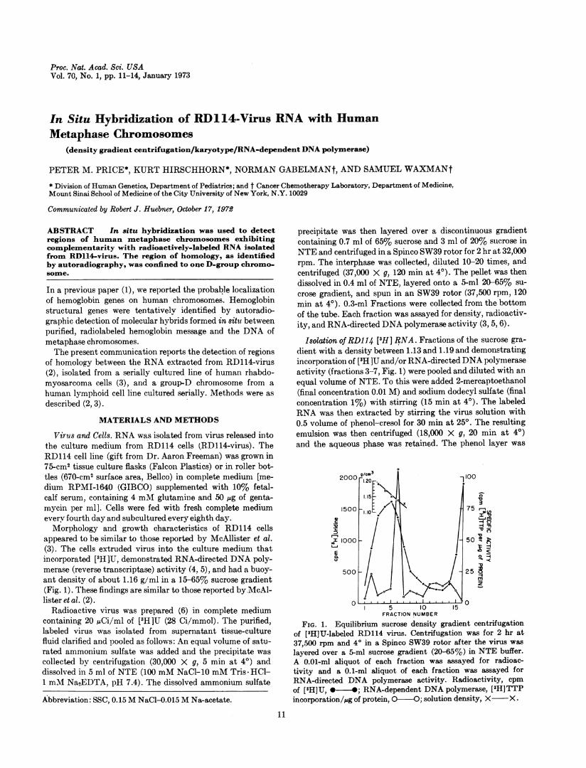
appeared to be similar to those reported by McAllister et al.(3). The cells extruded virus into the culture medium thatincorporated [3H1 U, demonstrated RNA-directed DNA poly-merase (reverse transcriptase) activity (4. 5), and had a buoy-ant density of about 1.16 g/ml in a 15-65% sucrose gradient(Fig. 1). These findings are similar to those reported byMcAl-lister et al. (2).
Radioactive virus was prepared (6) in complete mediumcontaining 20 ,Ci/ml of [3H]U (28 Ci/mmol). The purified,labeled virus was isolated from supernatant tissue-culturefluid clarified and pooled as follows: An equal volume of satu-rated ammonium sulfate was added and the precipitate wascollected by centrifugation (30,000 X g, 5 min at 40) anddissolved in 5 ml of NTE (100 mM NaCl-10 mM Tris HCl-1 mM Na2EDTA, pH 7.4). The dissolved ammonium sulfate
Abbreviation: SSC, 0.15 M NaCl-0.015 M Na-acetate.
precipitate was then layered over a discontinuous gradientcontaining 0.7 ml of 65% sucrose and 3 ml of 20% sucrose inNTE and centrifuged in a Spinco SW39 rotor for 2 hr at 32,000rpm. The interphase was collected, diluted 10-20 times, andcentrifuged (37,000 X g, 120 min at 4°). The pellet was thendissolved in 0.4 ml of NTE, layered onto a 5-ml 20-65% su-crose gradient, and spun in an SW39 rotor (37,500 rpm, 120min at 40). 0.3-ml Fractions were collected from the bottomof the tube. Each fraction was assayed for density, radioactiv-ity, and RNA-directed DNA polymerase activity (3, 5, 6).
Isoltion of RD114 [3H] RNA. Fractions of the sucrose gra-dient with a density between 1.13 and 1.19 and demonstratingincorporation of [3H ]U and/or RNA-directed DNA polymeraseactivity (fractions 3-7, Fig. 1) were pooled and diluted with an
equal volume of NTE. To this were added 2-mercaptoethanol(final concentration 0.01 M) and sodium dodecyl sulfate (finalconcentration 1%) with stirring (15 min at 4°). The labeledRNA was then extracted by stirring the virus solution with0.5 volume of phenol-cresol for 30 min at 250. The resultingemulsion was then centrifuged (18,000 X g, 20 min at 40)and the aqueous phase was retained. The phenol layer was
2000 g/cm3 -11001.20
1. 515
1500 a 75trt
layreovraim ursegaint(06No nNE ufr
L~J I
500 2
I 5 10 15FRACTION NUMBER
FIG. 1. Equilibrium sucrose density gradient centrifugationof ['H]U-labeled virus. Centrifugation for hr at
37,500 rpm and 40 in a Spinco SW39 rotor after the virus wa'slayered over a 5-nil sucrose gradient (20-65%) in NTE buffer.A 0.01-ml aliquot of each fraction was assayed for radioac-tivity and a 0.1-ml aliquot of each fraction was assayed forRNA-directed DNA polymerase activity. Radioactivity, cpmof [3H]U, * -; RNA-dependent DNA polymerase, [3H]TTPincorporation/Mg of protein, 0-O; solution density, X X .
11
Proc. Nat. Acad. Sci. USA 70 (1978)
a*.A
.
(.4
/V., _
I
0~¶1vD
pI
bC
V . 0e
V
-S.
A
£
alb31
AOF
I
VAIi
WA
i.0
*
2
USA-7
-IA '14
4.1
- 8
li 0
3 4
I4-_ 9 - 10
15'a1 6
aa
- 5 X -
- I I - 12
ta LA17 - 18
a,&
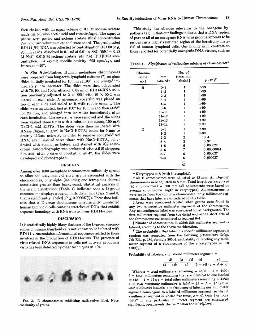
19 - 20 21 - 22 YFIG. 2. Karyotype and chromosome spread showing significant label on both long arms of D-group chromosome (arrow). Silver
grains on the labeled D chromosomes were so dense as to make accurate grain counts impossible. For this reason the statisticalanalysis is based on whether or not a millimeter segment of a chromosome has grains. Dashes between numbers under chromosomesindicate inability to distinguish individual chromosome groups on the basis of morphology alone.
1 710
04
-6
13-
a'4
12 Genetics: Price et al.
0 a
In Situ Hybridization of Virus RNA to Human Chromosomes 13
then shaken with an equal volume of 0.1 M sodium acetatemade pH 5.0 with acetic acid and recentrifuged. The aqueous
phases were pooled and sodium acetate (final concentration2%) and two volumes of ethanol were added. The precipitatedRD114[3H] RNA was collected by centrifugation (18,000 X g,
20 min at 40), dissolved in 0.1 mil of 0.01 X SSC (SSC = 0.15M NaCI-0.015 M sodium acetate, pH 7.4) ([3H]RNA con-
centration, 1.4 ,g/ml; specific activity, 695 cpm/,ug), andfrozen at -20° .
In Situ Hybridization. Human metaphase chromosomeswere prepared from long-term lymphoid cultures (7) on glassslides, initially incubated for 10 min at 100°, and plunged im-mediately into ice-water. The slides were then dehydratedwith 70, 90, and 100% ethanol. 0.05 ml of RD114-RNA solu-tion previously adjusted to 2 X SSC with 10 X SSC was
placed on each slide. A siliconized coverslip was placed on
top of each slide and sealed to it with rubber cement. Theslides were incukated, first at 1000 for 10 min and then at 650for 20 min, and plunged into ice-water immediately aftereach incubation. The coverslips were removed and the slideswere washed three times with a solution containing 100 mMNaCl-1 mM EDTA. The slides were then incubated withRNase (Sigma, 1 ;Lg/ml in NaCl-EDTA) boiled for 5 min todestroy DNase activity, in order to remove nonhybridizedRNA, again washed three times with NaCl-EDTA, dehy-drated with ethanol as before, and stained with 2% aceto-orcein. Autoradiography was performed with AR10 strippingfilm and, after 6 days of incubation at 40. the slides were
developed and photographed.
RESULTS
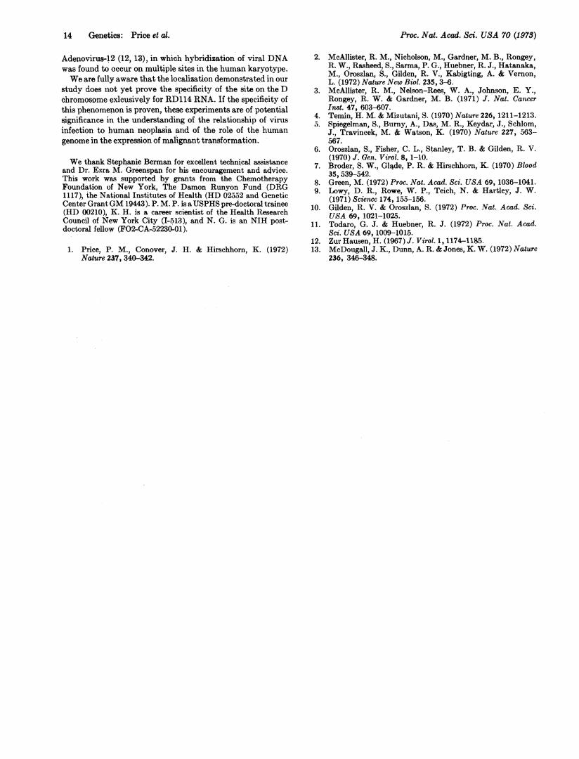
Among over 1000 metaphase chromosomes sufficiently spreadto allow the assignment of silver grains associated with thechromosomes, only eight (including one tetraploid) showedassociation greater than background. Statistical analysis ofthe grain distribution (Table 1) indicates that a D-groupchromosome displays a region in its distal half (Figs. 2 and 3)that is significantly labeled (P < 0.000027%). These data indi-cate that a D-group chromosome in apparently uninfectedhuman lymphoid cells contains stretches of DNA that exhibitsequence homology with RNA isolated fron RD114-virus.
DISCUSSION
It is statistically highly likely that one of the D-group chromo-somes of human lymphoid cells not known to be infected withRD114-virus contains informational sequences related to thoseinvolved in the production of RD114-virus. The presence ofvirus-related DNA sequences in cells not actively producingvirus has been detected by other techniques (8-10).
4A
FIG. 3. D chromosomes exhibiting radioactive label. Note
continuity of grains.
This study has obvious relevance to the oncogene hy-pothesis (11) in that our findings indicate that a DNA replicaof part or all of an oncogenic RNA virus genome appears to beresident in a highly restricted region of the hereditary mate-rial of human lymphoid cells. Our finding is in contrast tothose reported for potentially oncogenic DNA viruses, such as
TABLE 1. Significance of radioactive labeling of chromosomes*
Chromo- No. ofsome mm times mmno.t labeledt labeled§ P (%)
* Karyotypes = 8 (with 1 tetraploid).t All B chromosomes were adjusted to 15 mm. All D-group
chromosomes were adjusted to 8 mmi. Total length per karyotype(46 chromosomes) = 500 mm (all adjustments were based on
average chromosome length in karyotypes). All measurementswere made from the top of a chromosome; only millimeter seg-ments that have label are considered in this table.
t Areas were considered labeled when grains were found inany two consecutive millimeter segments of the chromosome.Any noncontiguous label was considered to be background. Thefirst millimeter segment from the distal end of the short arm ofthe chromosome was considered as segment 0-1.
§ Number of chromosomes in which this millimeter segment islabeled, according to the above consideration.
¶ The probability that label in a specific millimeter segment israndom was computed from the following (Documenta Geigy.7th Ed., p. 189, formula 805b): probability of labeling any milli-meter segment of a chromosome of the 8 karyotypes = 1.0
(100%).Probability of labeling any labeled millimeter segment =
d! (a-d)! b! c!(d-e)!e! a! (b-e)! (c-d + e)!
Where a = total millimeters remaining = 4500 - 1 = 4499;b = total millimeters remaining that are identical to one labeled= (18 - 1 = 17); c = total other millimeters remaining = 4482;d = total remaining millimeters to label = 42 - I = 41 (42 =total millimeters labeled); e = frequency of labeling any millimetersegment homologous to a labeled millimeter segment (so that ifa millimeter segment is labeled five times, e = 4). Only 4 or more"hits in any particular millimeter segment are consideredsignificant, because only then is P below the 0.01% level.
Proc. Nat. Acad. Sci. USA 70 (1973)
Proc. Nat. Acad. S4. USA 70 (1973)
Adenovirus-12 (12, 13), in which hybridization of viral DNAwas found to occur on multiple sites in the human karyotype.We are fully aware that the localization demonstrated in our
study does not yet prove the specificity of the site on the Dchromosome exlcusively for RD114 RNA. If the specificity ofthis phenomenon is proven, these experiments are of potentialsignificance in the understanding of the relationship of virusinfection to human neoplasia and of the role of the humangenome in the expression of malignant transformation.
We thank Stephanie Berman for excellent technical assistanceand Dr. Ezra M. Greenspan for his encouragement and advice.This work was supported by grants from the ChemotherapyFoundation of New York, The Damon Runyon Fund (DRG1117), the National Institutes of Health (HD 02552 and GeneticCenter GrantGM 19443). P. M. P. is aUSPHS pre-doctoral trainee(HD 00210), K. H. is a career scientist of the Health ResearchCouncil of New York City (I-513), and N. G. is an NIH post-doctoral fellow (FO2-CA-.52230-01).
1. Price, P. M., Conover, J. H. & Hirschhorn, K. (1972)Nature 237, 340-342.
2. McAllister, R. M., Nicholson, M., Gardner, M. B., Rongey,R. W., Rasheed, S., Sarma, P. G., Huebner, R. J., Hatanaka,M., Oroszlan, S., Gilden, R. V., Kabigting, A. & Vernon,L. (1972) Nature New Biol. 235, 3-6.
3. McAllister, R. M., Nelson-Rees, W. A., Johnson, E. Y.,Rongey, R.. W. & Gardner, M. B. (1971) J. Nat. CamcerInst. 47, 603-607.
4. Temin, H. M. & Mizutani, S. (1970) Nature 226, 1211-1213.). Spiegelman, S., Burny, A., Da.s, M. R., Keydar, J., Schlom,
J., Travincek, M. & Watson, K. (1970) Nature 227, 563-567.
6. Oroszlan, S., Fisher, C. L., Stanley, T. B. & Gilden, R. V.(1970) J. Gen. Virot. 8, 1-10.
7. Broder, S. W., Glade, P. R. & Hirschhorn, K. (1970) Blood35, 539-542.
8. Green, M. (1972) Proc. Nat. Acad. Sci. USA 69, 1036-1041.9. Lowy, D. R., Rowe, W. P., Teich, N. & Hartley, J. W.
(1971) Science 174, 155-156.10. Gilden, R. V. & Oroszlan, S. (1972) Proc. Nat. Acad. Sci.
USA 69, 1021-1025.11. Todaro, G. J. & Huebner, R. J. (1972) Proc. Nat. Acad.
Sci. USA 69, 1009-1015.12. Zur Hausen, H. (1967) J. Virol. 1, 1174-1185.13. McDougall, J. K., Dunn, A. R. & Jones, K. W. (1972) Nature