Page 1/18 Transplantation of neural stem cells encapsulated in hydrogels improve functional recovery in a cauda equina lesion model Zhiyi Fu Shanghai Ninth People's Hospital Huidong Wang Shanghai Ninth People‘s Hospital Yujie Wu ( [email protected]) Shanghai Ninth People's Hospital,Shanghai Jiao Tong University School of Medicine Tong Zhu Shanghai Ninth People's Hospital Research article Keywords: cauda equina syndrome, cauda equina lesion model, neural stem cells, hydrogels, functional recovery Posted Date: January 2nd, 2020 DOI: https://doi.org/10.21203/rs.2.19916/v1 License: This work is licensed under a Creative Commons Attribution 4.0 International License. Read Full License
Transcript
Page 1/18
Transplantation of neural stem cells encapsulatedin hydrogels improve functional recovery in a caudaequina lesion modelZhiyi Fu
AbstractBackground This study explored the therapeutic effects of transplantation of neural stem cells (NSCs)encapsulated in hydrogels in a cauda equina lesion model.
Methods NSCs were isolated from neonatal dorsal root ganglion (DRG) and cultured in three-dimensionalporous hydrogel scaffolds. Immunohistochemistry, transmission electron microscopy, Luxol fast bluestaining, TUNEL assay were performed to detect the differentiation capability, ultrastructural andpathological changes, and apoptosis of NSCs. Furthermore, the functional recovery of sensorimotorre�exes was determined using the tail-�ick test.
Results NSCs derived from DRG were able to proliferate to form neurospheres and mainly differentiateinto oligodendrocytes in the three-dimensional hydrogel culture system. After transplantation of NSCsencapsulated in hydrogels, NSCs differentiated into oligodendrocytes, neurons or astrocytes in vivo .Moreover, NSCs engrafted on the hydrogels decreased apoptosis and alleviated the ultrastructural andpathological changes of injured cauda equina. Behavioral analysis showed that transplanted hydrogel-encapsulated NSCs decreased the tail-�ick latency and showed a neuroprotective role on injured caudaequina.
Conclusions Our results indicate transplantation of hydrogel-encapsulated NSCs promotes stem celldifferentiation into oligodendrocytes, neurons or astrocytes and contributes to the functional recovery ofinjured cauda equina, suggesting that NSCs encapsulated in hydrogels may be applied for the treatmentof cauda equina injury.
BackgroundCauda equina syndrome (CES) is a neurological disease which is usually caused by central lumbar discherniation. The consequences of CES, such as neuropathic pain, lower extremity dysfunction, and sexualdysfunction are extremely frustrating [1]. It greatly affects the patient’s mental and physical condition andcauses enormous economic impact to society. Although this disease has a low incidence in thepopulation, ranging from 1:33,000 to 1:100,000 inhabitants, its sequelae still generate high publichealthcare costs [2]. Current treatment strategies include application of corticosteroid, surgicalstabilization and decompression, although effective but with unsatis�ed therapeutic e�cacy [3, 4].Because of the poor self-repair ability of nerve tissue, especially those in the central nervous system, thetherapeutic effects of surgery and neurotrophic drugs on cauda equina injury-induced CES is limited.
Neural stem cells (NSCs) can make copies of themselves and generate different mature cell types. Theyare promising candidate cells for neural transplantation treatment of neurological disorders, such asbrain trauma, spinal cord injury, peripheral nerve injury [5–7]. Many studies indicate that stem cells fosterhost axons to grow into the grafted spinal cord [8–10]. In addition, induced pluripotent stem cellsdifferentiate into astrocytes, neurons and oligodendrocytes and further improve functional recovery afterspinal cord injury [11]. Nevertheless, application of stem cell transplantation therapy is limited by the poor
Page 3/18
cell survival at the injury site. Thus, development of novel approaches to maintain neural stem cellviability is important to achieve ideal therapeutic outcomes.
Recently, tissue engineering has been developed that could provide solutions to the problem of stem celldeaths during transplantation [12]. Biopolymer hydrogels are designed to promote stem cell survival aftercerebral transplantation, exhibiting promising therapeutic role in central nervous system damage [13]. Aspreviously reported by Singh et al, neural stem cells derived from adult dorsal root ganglia not only retainthe multi-differentiation potential, but also have a tendency to differentiate into sensory neurons aftertransplantation, supporting that dorsal root ganglion neural stem cells (DRG NSCs) may be useful forrepair of damaged cauda equina [14]. In the present study, NSCs were isolated from neonatal dorsal rootganglion (DRG), encapsulated in three-dimensional porous hydrogel scaffolds, and used to repairdamaged cauda equina in a rat model of CES.
Materials And MethodsAnimals
Male Sprague-Dawley rats weighing 200-250 g and aged 6-8 weeks were purchased from the AnimalCenter of the Second Military Medical University. The surgical interventions for animal experiments wereapproved by by the Ethical Committee of the Shanghai Jiao Tong University School of Medicine, and theanimals were cared for in accordance with the Guide for the Care and Use of Laboratory Animals after thesurgery.
Culture of DRG-NSCs
DRG were dissected from postnatal day 2 rats, mechanically dissociated in Hank’s balanced salinesolution, pH 7.4, and seeded in Dulbecco’s modi�ed Eagle’s medium (DMEM)/F12 (Invitrogen)supplemented with 2% B27, 10 ng/mL epidermal growth factor (EGF), and 10 ng/mL bFGF. The cells wereincubated in a humidi�ed atmosphere with 5% CO2 at 37°C. The medium was changed every 3 days. Thedissociated DRG cells formed clusters or neurospheres within 1-2 weeks. Afterwards, the neurosphereswere collected, mechanically aspirated, and resuspended in the proliferation culture medium every 4 daysto reduce the cell heterogeneity by maintaining small spheres (50-100 cells/sphere). After 3 generationsof subcloning, the NSCs derived from the neurospheres were subjected to lentivirus transduction [15].
Hydrogel preparation
Hydrogels were purchased from Beaver Nano-Technologies Co., Ltd, China and were cross-linked withpolyethylene glycol diacrylate. Solutions of this hydrogel form a transparent gel when mixed with a cross-
Page 4/18
linker over a period of 30 minutes. Then, NSCs were mixed with hydrogels to form a stem-cell–hydrogelcomplex.
Model establishment and NSC transplantation
Forty-eight SD rats were randomly divided into sham, CES model and NSCs transplantation groups. Afteranesthesia with chloral hydrate, laminectomy was performed at the lumbar 4 and a silicone band (10mm long, 1 mm wide, and 1 mm thick) was placed under the laminae of the L5-6 vertebra to produce theCES animal model [15,16]. Sixteen rats were subjected to NSC transplantation, and another 16 rats wereused as control. A sham operation was performed with a simple laminectomy but without contusioninjury (n = 16).
For transplantation of NSCs, we removed the silicone 7 days after the compression injury and thenintrathecally injected 12 μL 0.5% hydrogels containing approximately 1000,000 NSCs (transduced withlentivirus) using a micropulled pipette connected to a Hamilton syringe. As control, 12 μL 0.5% hydrogelscontaining no NSCs was injected into the subarachnoid space.
Immunohistochemistry assay
The cauda equina was extracted, placed in EDTA solution, and heated in an oven for antigen retrieval.Then, 15-μm thick sections of the cauda equina around the lesion site were prepared longitudinally. Thetissue sections were permeabilized with 0.2% Triton X-100 and blocked in blocking solution for 1 h atroom temperature. After rinsing with PBS, the sections were incubated with primary antibodies againstO4, βIII-tubulin, glial �brillary acidic protein (GFAP) overnight at 4°C followed by incubation withsecondary antibodies for 1 h at 37°C. The slices were stained with Hoechst for 10 min and images werephotographed by inverted �uorescence microscopy.
Ultrastructural imaging
For transmission electron microscopic (TEM) studies, the sections were fractured with liquid nitrogen andquenched in hydrogen peroxide solution. After rinsing in PBS, the sections were prepared for ultra-thinsectioning. Tissue sections were �xed in osmium tetroxide, dehydrated in ethanol, and embedded in resin.All samples were observed under transmission electron microscope (TEM).
Luxol fast blue staining
Page 5/18
Tissue sections were stained with Luxol fast blue to detect myelin damage as previously described [17].Brie�y, the cauda equina sections were immersed in Luxol fast blue solution in an oven overnight. Afterrinsing in 95% ethanol and distilled water, the pathological changes of nerve �bers were observed under alight microscope.
TdT-mediated dUTP-biotin nick end labeling (TUNEL) staining
Apoptosis of cauda equina were measured by using a TUNEL detection kit according to themanufacturer’s instructions (Sigma, USA). In brief, para�n-embedded tissue sections (4-mm-thick) weredewaxed, rehydrated, and incubated with reaction mixture of terminal deoxynucleotidyl transferase for 1h. After rinsing in PBS, the sections were incubated with biotinylated antibody and ABC complex, andphotographed in a light microscope (Zeiss) equipped with a digital camera.
Behavioral analysis
For the tail-�ick test, the rats were immobilized 20 min before the test, and the tail was placed over a slit.A beam of light from a projection lamp (voltage of 18.5 V) was focused on the tail skin at the junctionbetween the middle and distal 1/3 of the tail. The latency to respond was recorded with a maximal 15sradiant heat stimulus [15].
Statistical analysis
Experimental data are presented as mean ± SD. One-Way ANOVA was used for comparison of differentgroups. Results were considered statistically signi�cant when the P value was less than 0.05.
ResultsCharacterization of NSCs in hydrogel scaffolds
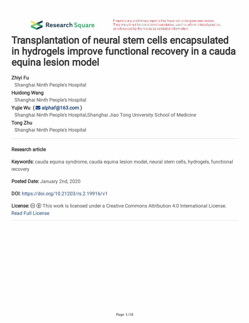
NSCs were isolated from the neonatal rat DRG and cultured in proliferation culture medium for differentdays. We observed that many cells were �oated in the medium and formed neurospheres (Figure 1A).Then, NSCs were successfully grown in 0.25% and 0.5% hydrogels (Figure 1A). Immunohistochemicalstaining showed that cells were positive for nestin and P75R, two markers of NSCs (Figure 1B).
Differentiation of NSCs in hydrogel scaffolds
Page 6/18
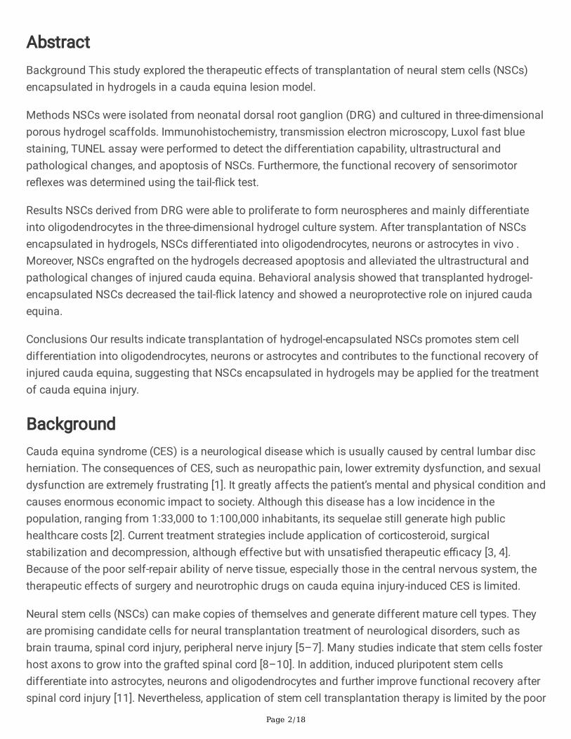
To more easily track cells, NSCs were transfected with lentivirus vectors carrying green �uorescent protein(GFP) and cultured in differentiation medium (Figure 2A). After 7 days of differentiation in vitro, theneural progenitors in the spheres attached to the hydrogel scaffolds and differentiated into differenttypes of cells. Immunohistochemical staining showed that most of the GFP labelled NSCs differentiatedinto O4+ oligodendrocytes, and only a very few cells differentiated into S100+ Schwann cells, βIII-tubulin+neurons and GFAP+ astrocytes (Figure 2B).
Differentiation of NSCs following transplantation in the injured cauda equina
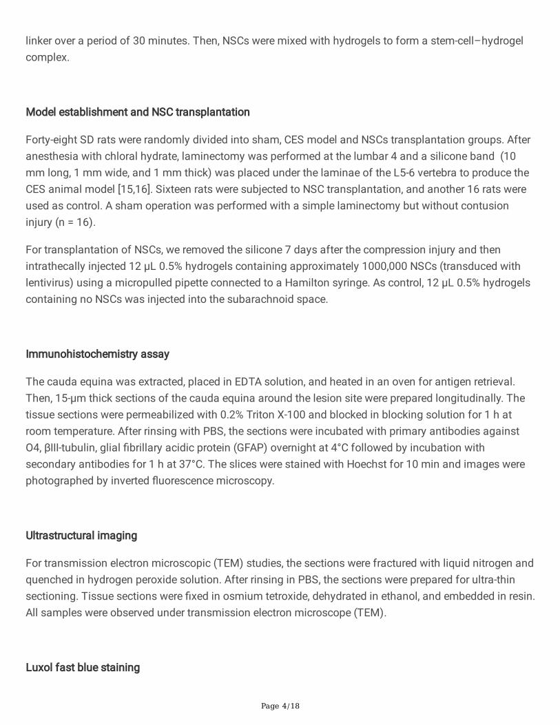
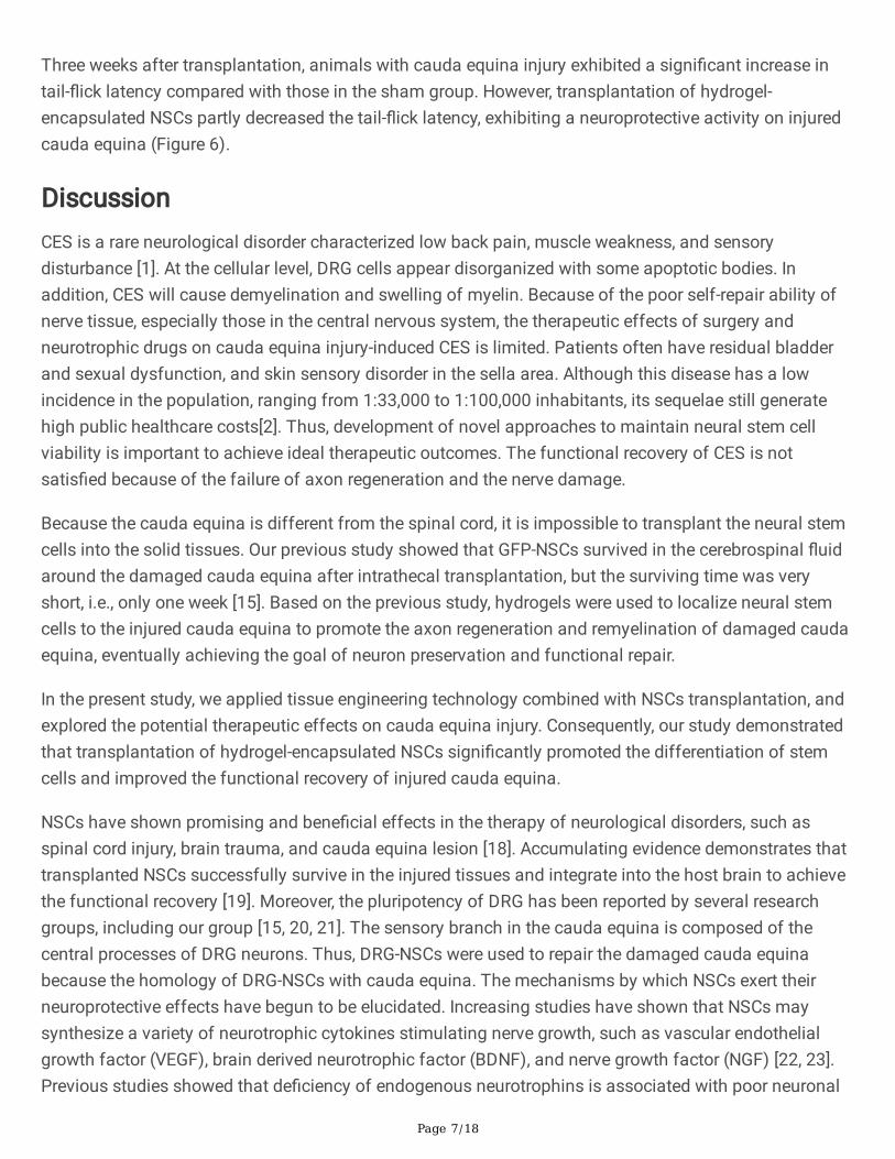
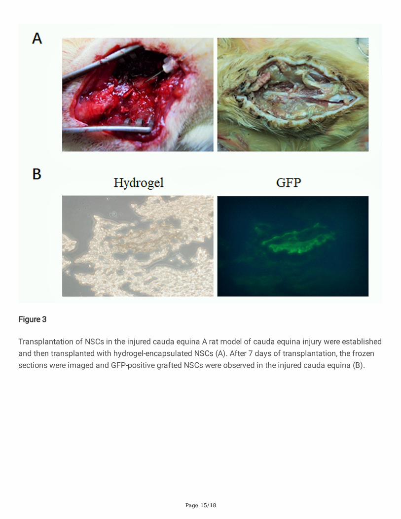
Next, we successful established the rat model of cauda equina injury and transplanted hydrogel-encapsulated NSCs (Figure 3A). To detect the viability of the transplanted NSCs in the cauda equina, thefrozen sections were imaged by confocal microscopy after 7 days of transplantation. As expected, GFP-positive grafted NSCs were present in the injured cauda equina (Figure 3B). Furthermore, immunostainingof the sagittal sections of the cauda equina showed that GFP-positive cells were co-stained with O4, βIII-tubulin or GFAP, but not with S100, suggesting that NSCs differentiated into oligodendrocytes, neurons orastrocytes in vivo (Figure 4).
Functional recovery following NSC transplantation encapsulated in three-dimensional hydrogels
To explore the regenerative effect of the transplanted NSCs, the functional recovery of sensorimotorre�exes was determined using the tail-�ick test. Fourteen days after transplantation, rat bilateral L5-6DRG tissues were isolated and subject to TUNEL staining. Results showed that cauda equina injury led toa signi�cant increase in apoptotic cells compared to sham group (P<0.01). By contrast, NSCstransplantation with three-dimensional hydrogels obviously decreased the apoptosis rate (P<0.05) (Figure5A). Two weeks after surgery, LFB staining and TEM analysis showed cauda equina nerve �bers in anorganized state, normal axons, and intact myelin sheath. However, compression of cauda equina resultedin disorganized nerve �bers, swollen axons and myelin sheaths, and demyelination. These observationswere alleviated after transplantation of NSCs encapsulated in hydrogels (Figure 5A). Additionally, G-ratio(inner diameter/outer diameter of myelinated axons) was signi�cantly higher in model group than shamgroup, and then decreased in the grafted NSCs animals (Figure 5B).
The rat model procedure was described and modi�ed by our research team, which has been successfullyused in many studies. When we established the model, we proved that: at day 1, 3, and 7 postcompression of cauda equina, we observed higher the tail �ick latency (TFL) of the compression groupthan that of the sham group. The compression group was signi�cantly prolonged. After 7 days,immuno�uorescence staining showed dense and organized cauda equina nerve �bers, no swollen axons,and intact myelin in the sham group. However, in the compression group, we observed disorganizedcauda equina nerve �bers, swollen axons and demyelinated myelin.
Page 7/18
Three weeks after transplantation, animals with cauda equina injury exhibited a signi�cant increase intail-�ick latency compared with those in the sham group. However, transplantation of hydrogel-encapsulated NSCs partly decreased the tail-�ick latency, exhibiting a neuroprotective activity on injuredcauda equina (Figure 6).
DiscussionCES is a rare neurological disorder characterized low back pain, muscle weakness, and sensorydisturbance [1]. At the cellular level, DRG cells appear disorganized with some apoptotic bodies. Inaddition, CES will cause demyelination and swelling of myelin. Because of the poor self-repair ability ofnerve tissue, especially those in the central nervous system, the therapeutic effects of surgery andneurotrophic drugs on cauda equina injury-induced CES is limited. Patients often have residual bladderand sexual dysfunction, and skin sensory disorder in the sella area. Although this disease has a lowincidence in the population, ranging from 1:33,000 to 1:100,000 inhabitants, its sequelae still generatehigh public healthcare costs[2]. Thus, development of novel approaches to maintain neural stem cellviability is important to achieve ideal therapeutic outcomes. The functional recovery of CES is notsatis�ed because of the failure of axon regeneration and the nerve damage.
Because the cauda equina is different from the spinal cord, it is impossible to transplant the neural stemcells into the solid tissues. Our previous study showed that GFP-NSCs survived in the cerebrospinal �uidaround the damaged cauda equina after intrathecal transplantation, but the surviving time was veryshort, i.e., only one week [15]. Based on the previous study, hydrogels were used to localize neural stemcells to the injured cauda equina to promote the axon regeneration and remyelination of damaged caudaequina, eventually achieving the goal of neuron preservation and functional repair.
In the present study, we applied tissue engineering technology combined with NSCs transplantation, andexplored the potential therapeutic effects on cauda equina injury. Consequently, our study demonstratedthat transplantation of hydrogel-encapsulated NSCs signi�cantly promoted the differentiation of stemcells and improved the functional recovery of injured cauda equina.
NSCs have shown promising and bene�cial effects in the therapy of neurological disorders, such asspinal cord injury, brain trauma, and cauda equina lesion [18]. Accumulating evidence demonstrates thattransplanted NSCs successfully survive in the injured tissues and integrate into the host brain to achievethe functional recovery [19]. Moreover, the pluripotency of DRG has been reported by several researchgroups, including our group [15, 20, 21]. The sensory branch in the cauda equina is composed of thecentral processes of DRG neurons. Thus, DRG-NSCs were used to repair the damaged cauda equinabecause the homology of DRG-NSCs with cauda equina. The mechanisms by which NSCs exert theirneuroprotective effects have begun to be elucidated. Increasing studies have shown that NSCs maysynthesize a variety of neurotrophic cytokines stimulating nerve growth, such as vascular endothelialgrowth factor (VEGF), brain derived neurotrophic factor (BDNF), and nerve growth factor (NGF) [22, 23].Previous studies showed that de�ciency of endogenous neurotrophins is associated with poor neuronal
Page 8/18
survival and cell death [24]. BDNF has very extensive neurotrophy and can maintain the survival ofvarious kinds of neurons and directly promote their axon growth [25]. Following a cervical spinal cordinjury, administration of BDNF into the site of spinal cord injury promoted axonal regeneration andprevented axotomy-induced atrophy and/or death of rubrospinal neurons [26, 27]. Furthermore, celltransplantation may also enhance endogenous repair processes including neurogenesis, axonalsprouting, and angiogenesis [28, 29]. However, NSCs application is limited due to the poor cell survival inhost tissues. In our study, NSCs were successfully isolated and cultured in hydrogels. Moreover, we foundthat NSCs could differentiate into oligodendrocytes, Schwann cells, neurons and astrocytes.
Tissue engineering may provide solutions to the challenges of stem cell death and damage associatedwith transplantation [30]. Biopolymer hydrogels can promote stem cell survival, enhance stem cellengraftment, and minimize wound scar formation [13]. Published studies have shown that hydrogels alterthe survival and differentiation of stem cells both in vitro and in vivo [31, 32]. In the present study, weisolated NSCs from neonatal DRG to repair damaged cauda equina in a rat model of lumbar spinal canalstenosis. As a result, hydrogel-encapsulated NSCs presented high viability in the injured cauda equinaand mainly differentiated to oligodendrocytes. Oligodendrocytes are known to be susceptible to spinalcord contusion and loss of oligodendrocytes may induce demyelination, disturb the functional recoveryof damaged nerve tissues, and damage the conductive capacity of sensory nerves [33]. Therefore, stemcell transplantation is helpful to improve myelination and enhance functional recovery after CNS injury[34]. To evaluate the neuroprotective role of the hydrogel encapsulated NSCs, the tail-�ick test wasperformed to measure the functional recovery of sensorimotor re�exes. As expected, NSCs engrafted onthe hydrogels obviously decreased apoptosis of injured cauda equina tissue. Moreover, cauda equinanerve �bers presented an organized state, normal axons, and intact myelin sheath. Additionally,transplanted hydrogel-encapsulated NSCs decreased the tail-�ick latency and showed a neuroprotectiverole on injured cauda equina.
In summary, our study demonstrates that transplantation of hydrogel-encapsulated NSCs enhances theviability of transplanted cells, promotes stem cell differentiation into oligodendrocytes, therebycontributing to the functional recovery of injured cauda equina. These results implied that NSCsencapsulated in three-dimensional hydrogels may be used for the treatment of cauda equina disorder.
ConclusionsOur results indicate transplantation of hydrogel-encapsulated NSCs promotes stem cell differentiationinto oligodendrocytes, neurons or astrocytes and contributes to the functional recovery of injured caudaequina, suggesting that NSCs encapsulated in hydrogels may be applied for the treatment of caudaequina injury.
DeclarationsAvailability of data and materials
Page 9/18
All data generated or analyzed during this study are included in this published article.
Abbreviations
CES
Cauda equina syndrome
NSCs:
neural stem cells
DRG:
dorsal root ganglion
GFAP:
glial �brillary acidic protein
TEM:
transmission electron microscopic
GFP:
green �uorescent protein
Acknowledgements
Not applicable.
Funding
This research is supported by the National Natural Science Foundation of China (grant #81400997),Shanghai Municipal Commission of Health and Family Planning (grant #201440326).
Author information
Page 10/18
A�liations
Shanghai Key Laboratory of Orthopaedic Implants, Department of Orthopaedic Surgery, Shanghai NinthPeople’s Hospital, Shanghai Jiao Tong University School of Medicine, 639 Zhizaoju Rd, Shanghai 200011,China.
Author Contributions Statement
Zhiyi Fu Manuscript Preparation, Literature Search, Funds Collection
Huidong Wang Data Collection
Yujie Wu Study Design, Data Interpretation
Tong Zhu Statistical Analysis
Corresponding author
Correspondence to Yujie Wu
Ethics declarationsEthics approval and consent to participate
The study protocol was approved by by the Ethical Committee of the Shanghai Jiao Tong UniversitySchool of Medicine. Informed consent was provided by all participating individuals.
1. Ma B, Wu H, Jia LS, et al. Cauda equina syndrome: a review of clinical progress. Chin Med J (Engl).2009;122:1214-1222.
2. Dias ALN, Araújo FF, Cristante AF, et al. Epidemiology of cauda equina syndrome. What changeduntil 2015. Rev Bras Ortop. 2017;53(1):107-112.
3. Li X, Dou Q, Hu S, et al. Treatment of cauda equina syndrome caused by lumbar disc herniation withpercutaneous endoscopic lumbar discectomy. Acta Neurol Belg. 2016;116(2):185-190.
4. Korse NS, Pijpers JA, Zwet EV, et al. Cauda Equina Syndrome: presentation, outcome, and predictorswith focus on micturition, defecation, and sexual dysfunction. Eur Spine J. 2017;26(3):894-904.
5. Imitola J, Raddassi K, Park KI, et al. Directed migration of neural stem cells to sites of CNS injury bythe stromal cell-derived factor 1alpha/CXC chemokine receptor 4 pathway. Proc Natl Acad Sci U S A.2004;101(52):18117-18122.
�. Abe K. Therapeutic potential of neurotrophic factors and neural stem cells against ischemic braininjury. J Cereb Blood Flow Metab. 2000;20(10):1393-1408.
7. Haus DL, López-Velázquez L, Gold EM, et al. Transplantation of human neural stem cells restorescognition in an immunode�cient rodent model of traumatic brain injury. Exp Neurol. 2016;281:1-16.
�. Marei HE, Shouman Z, Althani A, et al. Differentiation of human olfactory bulb-derived neural stemcells toward oligodendrocyte. J Cell Physiol. 2018;233(2):1321-1329.
9. Zhang LQ, Zhang WM, Deng L, et al. Transplantation of a Peripheral Nerve with Neural Stem CellsPlus Lithium Chloride Injection Promote the Recovery of Rat Spinal Cord Injury. Cell Transplant.2018;27(3):471-484.
10. Méndezolivos EE, Muñoz R, Larraín J. Spinal Cord Cells from Pre-metamorphic Stages Differentiateinto Neurons and Promote Axon Growth and Regeneration after Transplantation into the InjuredSpinal Cord of Non-regenerativeXenopus laevisFroglets. Front Cell Neurosci. 2017;11:398.
11. Levison SW, Druckman SK, Young GM, et al. Neural stem cells in the subventricular zone are a sourceof astrocytes and oligodendrocytes, but not microglia. Dev Neurosci. 2003;25(2-4):184-196.
12. Wang A, Tang Z, Park IH, et al. Induced pluripotent stem cells for neural tissue engineering.Biomaterials. 2011;32(22):5023-5032.
13. Chen XY, Zhuang SJ, Hou XM, et al. Research Progress on Tissue Engineering of Neural Stem Cells.Adv Mat Res. 2012;535-537:1104-1107.
14. Singh RP, Cheng YH, Nelson P, et al. Retentive multipotency of adult dorsal root ganglia stem cells.Cell Transplant. 2009;18(1):55-68.
15. Fu ZY, Shi JG, Liu N, et al. Differentiation of neonatal dorsal root ganglion-derived neural stem cellsinto oligodendrocytes after intrathecal transplantation into a cauda equina lesion model. Genet MolRes. 2013;12(4):6092-6102.
1�. Liu X, Fu Z, Wu Y, et al. Neuroprotective effect of hydrogen sul�de on acute cauda equina injury inrats. Spine J. 2016;16(3):402-7.
17. Kim JH, Budde MD, Liang HF, et al. Detecting axon damage in spinal cord from a mouse model ofmultiple sclerosis. Neurobiol Dis. 2006;21(3):626-632.
1�. Pluchino S, Zanotti L, Deleidi M, et al. Neural stem cells and their use as therapeutic tool inneurological disorders. Brain Res Rev. 2005;48(2):211-219.
19. Uchida K, Momiyama T, Okano H, et al. Potential functional neural repair with grafted neural stemcells of early embryonic neuroepithelial origin. Neurosci Res. 2005;52(3):276.
20. Ogawa R, Fujita K, Ito K. Mouse embryonic dorsal root ganglia contain pluripotent stem cells thatshow features similar to ES cells and induced pluripotentstem Biol Open. 2017;6(5):602-618.
21. Hu H, Ding Y, Mu W, et al. DRG-Derived Neural Progenitors Differentiate into Functional EntericNeurons Following Transplantation in the Postnatal Colon. Cell Transplant. 2019;28(2):157-169.
22. Salewski RP, Mitchell RA, Li L, et al. Transplantation of Induced Pluripotent Stem Cell-Derived NeuralStem Cells Mediate Functional Recovery Following Thoracic Spinal Cord Injury ThroughRemyelination of Axons. Stem Cells Transl Med. 2015;4(7):743-754.
24. Nakamura M, Bregman BS. Differences in neurotrophic factor gene expression pro�les betweenneonate and adult rat spinal cord after injury. Exp Neurol. 2001;169(2):407-15.
25. McCall J, Weidner N, Blesch A. Neurotrophic factors in combinatorial approaches for spinal cordregeneration. Cell Tissue Res. 2012;349(1):27-37.
2�. Liu Y, Himes BT, Murray M, et al. Grafts of BDNF-producing �broblasts rescue axotomizedrubrospinal neurons and prevent their atrophy. Exp Neurol. 2002;178(2):150-64.
27. Tan J, Shi J, Shi G, et al. Changes in compressed neurons from dogs with acute and severe caudaequina constrictions following intrathecal injection of brain-derived neurotrophic factor-conjugatedpolymer nanoparticles. Neural Regen Res. 2013;8(3):233-43.
2�. Andres RH, Horie N, Slikker W, et al. Human neural stem cells enhance structural plasticity and axonaltransport in the ischaemic brain. Brain. 2011;134(Pt6):1777-1789.
29. Daadi MM, Davis AS, Arac A, et al. Human neural stem cell grafts modify microglial response andenhance axonal sprouting in neonatal hypoxic-ischemic brain injury. Stroke. 2010;41(3):516-523.
30. Kim H, Cooke MJ, Shoichet MS. Creating permissive microenvironments for stem cell transplantationinto the central nervous system. Trends Biotechnol. 2012;30(1):55-63.
31. Führmann T, Tam RY, Ballarin B, et al. Injectable hydrogel promotes early survival of inducedpluripotent stem cell-derived oligodendrocytes and attenuates longterm teratoma formation in aspinal cord injury model. Biomaterials. 2016;83:23-36.
32. Naghdi P, Tiraihi T, Ganji F, et al. Survival, proliferation and differentiation enhancement of neuralstem cells cultured in three-dimensional polyethylene glycol-RGD hydrogel with tenascin. J TissueEng Regen Med. 2016;10(3):199-208.
33. Nistor GI, Totoiu MO, Haque N, et al. Human embryonic stem cells differentiate into oligodendrocytesin high purity and myelinate after spinal cord transplantation. Glia. 2005;49(3):385-396.
34. Armstrong RC, Mierzwa AJ, Sullivan GM, et al. Myelin and Oligodendrocyte Lineage Cells in WhiteMatter Pathology and Plasticity after Traumatic Brain Injury. Neuropharmacology.2016;110(Pt8):654-659.
Figures
Figure 1
Characterization of NSCs in hydrogel scaffolds NSCs were isolated from the neonatal rat DRG andcultured in proliferation culture medium for different time points at 0, 3, 6, and 10 days (A). Foridenti�cation of NSCs, stem cells were immunohistochemically stained with antibodies against nestinand P75R, and nucleus was stained with Hoechst (B).
Page 14/18
Figure 2
Differentiation of NSCs in vitro NSCs were transfected with lentivirus vectors carrying green �uorescentprotein (GFP) and cultured in differentiation medium (A). After 7 days of differentiation, stem cells wereimmunohistochemically stained with antibodies against O4, S100, βIII-tubulin, and GFAP, and nucleuswas stained with Hoechst (B). Scale bar=50μm.
Page 15/18
Figure 3
Transplantation of NSCs in the injured cauda equina A rat model of cauda equina injury were establishedand then transplanted with hydrogel-encapsulated NSCs (A). After 7 days of transplantation, the frozensections were imaged and GFP-positive grafted NSCs were observed in the injured cauda equina (B).
Page 16/18
Figure 4
Differentiation of NSCs in vivo After 7 days of transplantation, the frozen sections wereimmunohistochemically stained with antibodies against O4, S100, βIII-tubulin, and GFAP, and imaged byconfocal microscopy. Scale bar=50μm.
Page 17/18
Figure 5
Ultrastructural and pathological changes of DRG after NSC transplantation The rat DRG tissues weresubject to TUNEL and LFB staining, and TEM (A). Then, G-ratio (inner diameter/outer diameter ofmyelinated axons) was calculated (B). * P<0.05, compared to sham group; # P<0.05, compared to CESmodel group. Data are reported as means ± SD.
Figure 6
Page 18/18
Functional recovery following transplantation of NSCs encapsulated in hydrogels The functional recoveryof sensorimotor re�exes in sham, CES model and NSCs groups was determined using the tail-�ick test. **P<0.01, compared to sham group; # P<0.05, compared to CES model group. Data are reported as means ±SD.