0270.6474/84/0404-0908$02.00/O The Journal of Neuroscience Copyright 0 Society for Neuroscience Vol. 4, No. 4, pp. 908-917 Printed in U.S.A. April 1984 EFFERENT NEUROTRANSMISSION OF CIRCADIAN RHYTHMS IN LIMULUS LATERAL EYE I. Octopamine-induced Increases in Retinal Sensitivity1 LEONARD KASS’ AND ROBERT B. BARLOW, JR. Institute for Sensory Research, Syracuse University, Syracuse, New York 13210 Received March 29, 1983; Revised October 25, 1983; Accepted October 29,1983 Abstract Octopamine increases the sensitivity of the Limulus lateral eye in situ when injected beneath the cornea during the day. The effect of octopamine is dose-dependent with a threshold concentration of about 0.1 PM injected at 1 pl/min for 15 min. Injection of 40 PM octopamine increases lateral eye sensitivity to approximately 70% of the nighttime level normally caused by the efferent output of a circadian clock. Injections of octopamine analogues and other candidate neurotransmitters indicate that the postsynaptic receptor mediating the increase of retinal sensitivity is relatively specific for the structure of octopamine. The postsynaptic receptor is tentatively classified as a type 2B octopamine receptor (Evans, P. D. (1981) J. Physiol. (Lond.) 318: 99-122). Clozapine suppresses the effects of both exogenous octopamine and the endogenous efferent neurotransmitter. Together with the results from Barlow et al. (Barlow, R. B., Jr., S. J. Bolanowski, Jr., and M. L. Brachman (1977) Science 197: 86-89) and Battelle et al. (Batelle, B. -A., J. A. Evans, and S. C. Chamberlain (1982) Science 216: 1250-1252) our study leads to the following conclusion: retinal efferents, driven by a circadian clock in Limulus brain, release octopamine that increases visual sensitivity. A circadian clock in Limulus brain generates efferent activity in lateral optic nerve fibers at night (Barlow et al., 1977). The efferent fibers terminate in the retina (Fahrenbach, 1971; 1981) and presumably release one or more neurotransmitters (Battelle, 1980). Efferent neu- rotransmission in the lateral eye produces marked changes in fundamental processes, such as photoreceptor noise (Kaplan and Barlow, 1980), sensitivity to light (Barlow et al., 1977; Kaplan and Barlow, 1980), and the photon-to-spike transduction mechanism (Barlow et al., 1977; Barlow, 1983). The functional changes may result in part from circadian alterations in retinal morphology and ultrastructure (Chamberlain and Barlow, 1977,1979; Barlow et al., 1980; Barlow and Chamberlain, 1980). The circadian clock plays an important role in Limulus ’ We thank Dr. Barbara-Anne Battelle for helpful suggestions and Janice Pelletier for editorial comments. We also thank Joseph Fladd, Jerome Brophy, Steven Swales, Michael Serafini, and the late Bernard Klock for technical assistance and Betty Zahora and Michael Rock for typing the manuscript. This work was supported by National Institutes of Health Grants EY-00667 and EY-05443, National Science Founda- tion Grant BNS 81-04669, and the Grass Foundation. * To whom correspondence should be sent, at his present address: Microsurgical Research Center, Eastern Virginia Medical School, P.O. Box 1980, Norfolk, VA 23501. vision. The ventral eyes and the median ocelli, in addi- tion to the lateral eyes, receive input at night from the circadian clock (Eisele et al., 1982; Kass et al., 1983). Illumination of the median ocellus at night increases efferent activity along the lateral optic nerve, leading to changes in lateral eye sensitivity (R. B. Barlow, Jr., L. A. Westerman, and S. C. Chamberlain, submitted for publication). Behavioral experiments reveal diurnal changes in visual sensitivity (Powers and Barlow, 1981) and a possible role of vision during mating (Barlow et al., 1982). Octopamine may be the neurotransmitter that links the circadian activity from the clock to the changes in structure and function of the lateral eye. It has recently been shown to be synthesized, stored, and released from efferent nerve terminals in the ventral and lateral eyes of Limulus (Battelle et al., 1982). The purpose of the present study in the lateral eye was to determine (I) a method for subcorneal application of candidate neuro- transmitters that mimics the physiological action of en- dogenous efferent neurotransmitter(s); (2) the threshold and effective concentration range for octopamine; (3) the transmitter specificity for the postsynaptic receptor and the receptor classification; and (4) a pharmacological agent that antagonizes the physiological actions of both exogenous octopamine and endogenous neurotransmit- 908
Transcript
0270.6474/84/0404-0908$02.00/O The Journal of Neuroscience Copyright 0 Society for Neuroscience Vol. 4, No. 4, pp. 908-917 Printed in U.S.A. April 1984
EFFERENT NEUROTRANSMISSION OF CIRCADIAN RHYTHMS IN LIMULUS LATERAL EYE
I. Octopamine-induced Increases in Retinal Sensitivity1
LEONARD KASS’ AND ROBERT B. BARLOW, JR.
Institute for Sensory Research, Syracuse University, Syracuse, New York 13210
Received March 29, 1983; Revised October 25, 1983; Accepted October 29,1983
Abstract
Octopamine increases the sensitivity of the Limulus lateral eye in situ when injected beneath the cornea during the day. The effect of octopamine is dose-dependent with a threshold concentration of about 0.1 PM injected at 1 pl/min for 15 min. Injection of 40 PM octopamine increases lateral eye sensitivity to approximately 70% of the nighttime level normally caused by the efferent output of a circadian clock. Injections of octopamine analogues and other candidate neurotransmitters indicate that the postsynaptic receptor mediating the increase of retinal sensitivity is relatively specific for the structure of octopamine. The postsynaptic receptor is tentatively classified as a type 2B octopamine receptor (Evans, P. D. (1981) J. Physiol. (Lond.) 318: 99-122). Clozapine suppresses the effects of both exogenous octopamine and the endogenous efferent neurotransmitter. Together with the results from Barlow et al. (Barlow, R. B., Jr., S. J. Bolanowski, Jr., and M. L. Brachman (1977) Science 197: 86-89) and Battelle et al. (Batelle, B. -A., J. A. Evans, and S. C. Chamberlain (1982) Science 216: 1250-1252) our study leads to the following conclusion: retinal efferents, driven by a circadian clock in Limulus brain, release octopamine that increases visual sensitivity.
A circadian clock in Limulus brain generates efferent activity in lateral optic nerve fibers at night (Barlow et al., 1977). The efferent fibers terminate in the retina (Fahrenbach, 1971; 1981) and presumably release one or more neurotransmitters (Battelle, 1980). Efferent neu- rotransmission in the lateral eye produces marked changes in fundamental processes, such as photoreceptor noise (Kaplan and Barlow, 1980), sensitivity to light (Barlow et al., 1977; Kaplan and Barlow, 1980), and the photon-to-spike transduction mechanism (Barlow et al., 1977; Barlow, 1983). The functional changes may result in part from circadian alterations in retinal morphology and ultrastructure (Chamberlain and Barlow, 1977,1979; Barlow et al., 1980; Barlow and Chamberlain, 1980).
The circadian clock plays an important role in Limulus
’ We thank Dr. Barbara-Anne Battelle for helpful suggestions and Janice Pelletier for editorial comments. We also thank Joseph Fladd, Jerome Brophy, Steven Swales, Michael Serafini, and the late Bernard Klock for technical assistance and Betty Zahora and Michael Rock for
typing the manuscript. This work was supported by National Institutes of Health Grants EY-00667 and EY-05443, National Science Founda- tion Grant BNS 81-04669, and the Grass Foundation.
* To whom correspondence should be sent, at his present address: Microsurgical Research Center, Eastern Virginia Medical School, P.O.
Box 1980, Norfolk, VA 23501.
vision. The ventral eyes and the median ocelli, in addi- tion to the lateral eyes, receive input at night from the circadian clock (Eisele et al., 1982; Kass et al., 1983). Illumination of the median ocellus at night increases efferent activity along the lateral optic nerve, leading to changes in lateral eye sensitivity (R. B. Barlow, Jr., L. A. Westerman, and S. C. Chamberlain, submitted for publication). Behavioral experiments reveal diurnal changes in visual sensitivity (Powers and Barlow, 1981) and a possible role of vision during mating (Barlow et al., 1982).
Octopamine may be the neurotransmitter that links the circadian activity from the clock to the changes in structure and function of the lateral eye. It has recently been shown to be synthesized, stored, and released from efferent nerve terminals in the ventral and lateral eyes of Limulus (Battelle et al., 1982). The purpose of the present study in the lateral eye was to determine (I) a method for subcorneal application of candidate neuro- transmitters that mimics the physiological action of en- dogenous efferent neurotransmitter(s); (2) the threshold and effective concentration range for octopamine; (3) the transmitter specificity for the postsynaptic receptor and the receptor classification; and (4) a pharmacological agent that antagonizes the physiological actions of both exogenous octopamine and endogenous neurotransmit-
908
The Journal of Neuroscience Neurotransmission of Circadian Rhythms 909
ter(s). Brief accounts of some of this work have already been published (Kass and Barlow, 1980, 1982a).
Materials and Methods
The methods are essentially the same as described by Barlow (1983), except for the addition of the section pertaining to subcorneal injections of pharmacological agents.
Animals and laboratory locations. Adult horseshoe crabs (Limulus polyphemus), measuring about 20 cm across the carapace, were supplied by the Marine Biolog- ical Laboratory (MBL) (Woods Hole, MA) and by the Gulf Speciman Company (Panacea, FL). The experi- ments were conducted at MBL during the summer and at Syracuse, New York during the rest of the year. Differences in experimental conditions between the two locations have been described by Barlow (1983). Animals freshly caught at Woods Hole generally showed more pronounced circadian rhythms; nevertheless, we found no substantial differences in experimental results ob- tained between the two locations.
Electrical recording and optical stimulation. During an experiment, animals were fastened to a rigid platform and placed into an aquarium located within a lightproof shielded cage (Barlow et al., 1977). Electroretinograms (ERGS) were recorded from each lateral eye with a wick electrode placed in contact with the cornea (Barlow, 1983). The electrode was connected to a band-pass dif- ferential amplifier (3 to 300 Hz, Electronics Laboratory, Rockefeller University). The reference input to the am- plifier was connected to a silver wire inserted beneath the carapace near the eye. Brief flashes of light (10 to 50 msec in duration) were delivered to the cornea1 surface by either a green light-emitting diode (LED) or a glass fiberoptic bundle transmitting light from a tungsten fil- ament source. When the glass fiberoptic bundle was used, a white Teflon screen was placed in front of the eye to provide uniform illumination of the retina (Barlow, 1983).
Stimulus control, data collection, and analysis. In ex- periments utilizing the tungsten filament light source, brief pulses of retinal illumination were controlled by electromagnetic shutters (Unbilitz, A. W. Vincent Asso- ciates, Rochester, NY) and digital timers (ISR Electron- ics Shop). The timers also controlled a two-channel strip chart recorder (Gould Brush, Model 220) for a permanent record of the ERGS from both eyes. The peak-to-peak amplitudes of the ERG could be directly measured and plotted as a function of light intensity or of time of day.
Experiments using LEDs for retinal stimulation were conducted with the aid of a microcomputer (AIM-65, Rockwell International), together with analogue-to-dig- ital convertors and current drivers (ISR Electronics Shop). In each experiment the microcomputer program was initialized with the cycle time for flash delivery, duration of the LED flashes, and sample rate (typically 2 msec) for the conversion of the ERG responses. The microcomputer determined and printed out the latency of the ERG, its peak-to-peak amplitude, together with the date and the time. In addition, the microcomputer stored peak-to-peak amplitudes of the ERGS for several days (256 ERGS/eye) and, on command, plotted them
versus time of day on the strip chart recorder. The program and associated electronic equipment permitted the analysis and storage of ERG data from up to three animals (six lateral eyes) during the course of a single experiment.
Subcorneal injections. Pharmacological agents were dissolved in sea water and delivered to the lateral eye by a hypodermic needle (26 gauge) connected to a syringe pump (Sage Instruments, Model 341). The needle was inserted beneath the cornea by hand or by a hydraulic drive unit (David Kopf Instruments, Tunjunga, CA). In all experiments, solutions were injected at the rate of 1 pl/min. Injection rates greater than 3 pl/min proved deleterious to the retinal responses. The pharmacological agents listed in Table I and tetrodotoxin (TTX) were obtained from Sigma Chemical Company (St. Louis, MI). The “DL” forms of octopamine, synephrine, norepineph- rine, and tyrosine were used. Clonidine was obtained from Boehringer Ingelheim (stock); clozapine was from Sandoz (stock). All solutions were freshly prepared with pH adjusted to 7.2 to 7.4.
Results
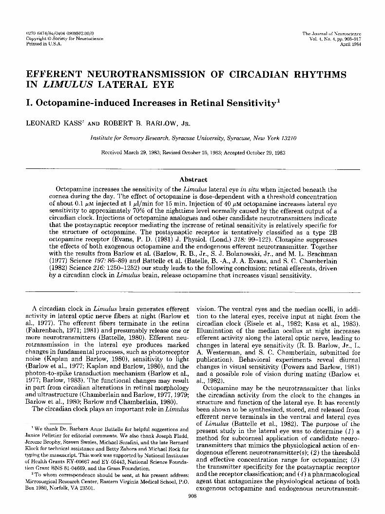
The ERG of the lateral eye. The ERG was used through- out this study as a measure of the retinal sensitivity of the lateral eye. Hartline (1928) first recorded the ERG from the lateral eye and showed that it had consisted of a simple biomodal wave form. He later showed that its waveform resembled that of the photoreceptor potential (Hartline, 1935). To investigate further the nature of the lateral eye ERG, we compared the normal ERG response to that recorded in the presence of TTX (Fig. l), a substance shown to block the sodium channels that give rise to the propagated action potential (Moore et al., 1967). In this experiment the animal was maintained in complete darkness during the simultaneous recording of
Before TTX 4f
n After TTX i’
\K
Figure 1. ERGS of the lateral eye before and after TTX injection. The animal was maintained in complete darkness during simultaneous ERG recording (shown) and optic nerve recording (not shown). TTX (1 FM) was injected (15 ~1 in 15 min) into the lateral eye until spontaneous and light-driven spike activities were abolished. The peak-to-peak amplitudes of the ERGS are 400 pV, and the records are 0.6 set long. The latency, shape, and duration of the ERG were unchanged by the TTX application. This finding supports our claim that the ERG provides a convenient measure of the primary photores- ponse, without complications from spike-initiated processes.
910 Kass and Barlow Vol. 4, No. 4, Apr. 1984
the ERG and extracellular optic nerve activity (Barlow, 1983). The optic nerve recording (not shown) consisted of spike activity from about 50 optic nerve fibers located in a horizontal strip across the eye. The ERG electrode was placed in the middle of the strip. TTX (1 pM) was injected into the eye until spontaneous and light-evoked spike activities were abolished (15 ~1 in 15 min). The latency, shape, and duration of the ERG were unaffected by TTX, indicating that the waveform of the response is not influenced by spike-initiated, secondary retinal proc- esses. In sum, the lateral eye ERG seems to provide a convenient measure of the primary photoresponse.
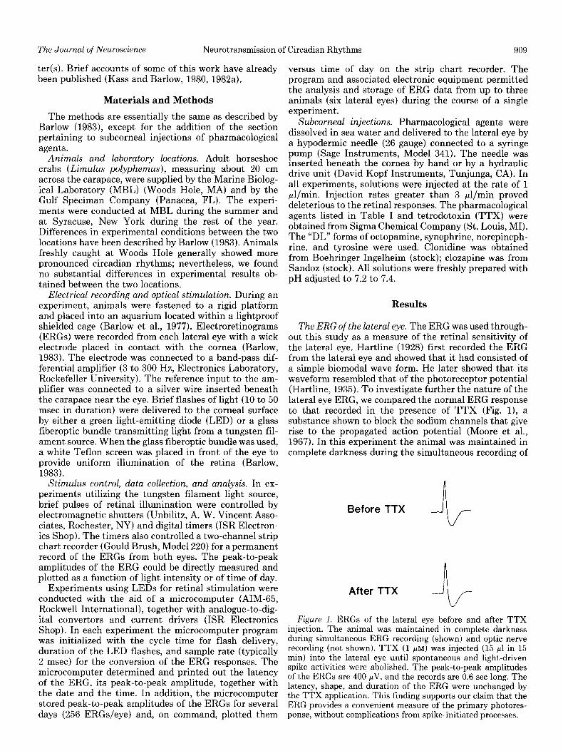
Circadian and octopamine-induced increases in ERG amplitude. Figure 2 shows the circadian rhythm in the amplitude of the ERG of the lateral eye. The animal was maintained in constant darkness for 3 days, during which time the ERG was evoked by a constant intensity flash every 20 min. As previously shown by Barlow et al. (1977) and Barlow (1983), the ERG amplitude increases at around 6 P.M., stays elevated during the nighttime hours, declines in amplitude at about 4 A.M., and remains low during the daytime until the following evening. While cutting the optic nerve abolishes the circadian changes in ERG amplitude, stimulating the distal end of the cut nerve during the day or night mimics the nighttime increase in ERG amplitude; thus, efferent optic nerve activity generated at night from a circadian clock me- diates the elevation in ERG amplitude (Barlow et al., 1977; Barlow, 1983).
Figure 2 also shows that octopamine can mimic, in part, the effects of the circadian clock. During days 2 and 3, octopamine was injected into the eye for 15 min (1 pl/min) at concentrations of 0.05 and 5 pM, respec- tively. The 0.05 pM injection had no effect, but the 5 pM
injection increased the ERG amplitude to about 50% of the natural nighttime level.
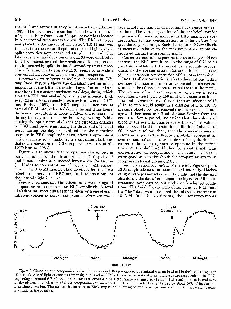
Figure 3 summarizes the effects of a wide range of octopamine concentrations on ERG amplitude. A total of 49 daytime injections was made, each with one of eight different concentrations of octopamine. Encircled num-
0.05 IJM Octopamine
+
bers denote the number of injections at various concen- trations. The vertical position of the encircled number represents the average increase in ERG amplitude cor- responding to that concentration, and the vertical bars give the response range. Each change in ERG amplitude is measured relative to the maximum ERG amplitude recorded during the preceding night.
Concentrations of octopamine less than 0.1 pM did not increase the ERG amplitude. In the range of 0.25 to 40 pM, the increase in ERG amplitude is roughly propor- tional to the concentration. Extrapolation of the data yields a threshold concentration of 0.1 PM octopamine.
Because all concentrations refer to the solutions within syringes, the question arises as to the actual concentra- tion near the efferent nerve terminals within the retina. The volume of a lateral eye into which we injected octopamine was typically 150 ~1. If one assumes no blood flow and no barriers to diffusion, then an injection of 15 ~1 in 15 min would result in a dilution of 1 to 10. To measure blood flow, we removed the cornea of the lateral eye and then measured 3 ml of blood flowing from the eye in a 15-min period, indicating that the volume of blood in the eye may change every 45 sec. This volume change would lead to an additional dilution of about 1 to 20. It would follow, then, that the concentrations of octopamine graphed in Figure 3 probably represent an overestimate of at least two orders of magnitude. The concentration of exogenous octopamine in the retinal tissue at threshold would then be about 1 nM. This concentration of octopamine in the lateral eye would correspond well to thresholds for octopamine effects at receptors in locust (Evans, 1981).
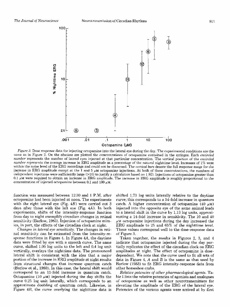
Intensity-response function of the ERG. Figure 4 plots ERG amplitude as a function of light intensity. Flashes of light were presented during the night and the day and also during the day after octopamine injection. All meas- urements were carried out under dark-adapted condi- tions. The “night” data were obtained at 11 P.M., and the “day” data were measured the following morning at 10 A.M. In both experiments, the intensity-response
5 JJM Octopamine
.a0 @---%a . + 0. . -3
1
Midnight I
Noon 1
Midnight
Time of day
I
Noon I
Midnight
Figure 2. Circadian and octopamine-induced increases in ERG amplitude. The animal was maintained in darkness except for lo-msec flashes of light at constant intensity that evoked ERGS. Circadian activity at night increases the amplitude of the ERG beginning at around 6 P.M. and continuing until about 4 A.M. Octopamine was injected (15 min; 1 pl/min) into the lateral eyes in the afternoons. Injection of 5 FM octopamine can increase the ERG amplitude during the day to about 50% of its natural nighttime elevation. The rate of the increase in ERG amplitude following octopamine injection is similar to that which occurs naturally in the evening.
Neurotransmission of Circadian Rhythms 911 The Journal of Neuroscience
51
t 0
.l 1
Octopamine @Ml
J
10 100
Figure 3. Dose response data for injecting octopamine into the lateral eye during the day. The experimental conditions are the same as in Figure 2. On the abscissa are plotted the concentrations of octopamine contained in the syringes. Each encircled number represents the number of lateral eyes injected at that particular concentration. The vertical position of the encircled number represents the average increase in ERG amplitude as. a percentage of the natural nighttime level. Increases of 1% were within the noise level of the ERG recordings and could not be discerned. The uertical bars denote the full response range for the increase in ERG amplitude except at the 1 and 5 pM octopamine injections. At both of these concentrations, the numbers of independent injections were sufficiently large (>lO) to justify a calculation based on 1 SD. Injections of octopamine greater than 0.1 PM were required to obtain an increase in ERG amplitude. The increase in ERG amplitude is roughly proportional to the concentration of injected octopamine between 0.1 and 100 pM.
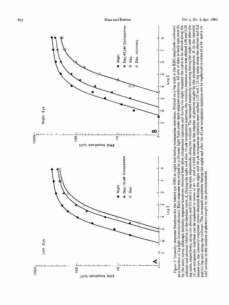
function was measured between 12:30 and 1 P.M. after octopamine had been injected at noon. The experiments with the right lateral eye (Fig. 4B) were carried out 3 days after those with the left eye (Fig. 4A). In both experiments, shifts of the intensity-response function from day to night exemplify circadian changes in retinal sensitivity (Barlow, 1983). Injection of octopamine mim- ics, in part, the effects of the circadian clock at night.
Changes in lateral eye sensitivity. The changes in reti- nal sensitivity can be estimated from the intensity-re- sponse functions in Figure 4. In Figure 4A, the daytime data were fitted by eye with a smooth curve. The same curve, shifted 1.05 log units to the left and 0.4 log unit vertically, overlays the nighttime data. The pronounced lateral shift is consistent with the idea that a major portion of the increase in ERG amplitude at night results frqm structural changes that increase quantum catch (Barlow et al., 1980). In this case, the lateral shift would correspond to an 11-fold increase in quantum catch. Octopamine (10 pM) injected during the day shifts the curve 0.25 log unit laterally, which corresponds to an approximate doubling of quantum catch. Likewise, in Figure 4B, the curve overlying the nighttime data is
shifted 1.73 log units laterally relative to the daytime curve; this corresponds to a 54-fold increase in quantum catch. A higher concentration of octopamine (40 pM) injected into the opposite eye of the same animal leads to a lateral shift in the curve by 1.13 log units, approxi- mating a 14-fold increase in sensitivity. The 10 and 40 pM octopamine injections during the day increased the ERG amplitude to 25 and 65% of the nighttime level. These values correspond well to the dose-response data of Figure 3.
Taken together, the results in Figures 2, 3, and 4 indicate that octopamine injected during the day par- tially replicates the effect of the circadian clock on ERG amplitudes at night. The effect of octopamine is dose- dependent. We note that the curve used to fit all sets of data in Figure 4, A and B is the same as that used by Barlow (1983) to fit ERG intensity-response data from other horseshoe crabs.
Relative potencies of other pharmacological agents. Ta- ble I lists the relative potencies of agonists and analogues of octopamine as well as other neurotransmitters for elevating the amplitude of the ERG of the lateral eye. Potencies of the various agents were arrived at by first
lOOO
-
Left
Eye
0 Ni
ght
A Da
y, 10
UM
O
ctop
amin
e
0 Da
y
0 2 10
Righ
t Ey
e
0 Ni
ght
A Da
y,40u
M
Oct
opam
ine
0 Da
y
A Da
y, re
cove
ry
7 6
5 4
3 2
1 0
Log
I
Figu
re
4. In
tens
ity-r
espo
nse
func
tions
for
the
lat
eral
eye
ER
G a
t ni
ght
and
durin
g oc
topa
min
e in
ject
ions
. Plo
tted
on a
log
scale
is th
e E
RG
am
plitu
de (o
rdin
ate)
a
s a fu
nctio
n of
log
light
int
ensi
ty(a
bsci
ssa)
. Eac
h re
sponse
was e
voke
d by
a 5
0-m
sec li
ght
flash
und
er d
ark-
adap
ted
cond
ition
s. A
ll se
ts of
dat
a in
bot
h ey
es w
ere
fit
by t
he s
am
e cu
rve,
alth
ough
inte
nsity
-res
pons
e fun
ctio
ns o
btai
ned
both
afte
r oc
topa
min
e in
ject
ion
and
durin
g th
e ni
ght
requ
ired
the
curv
e to
be
shift
ed a
long
the
ordi
nate
an
d ab
scis
sa
rela
tive
to t
he d
aytim
e cu
rve.
A,
Dur
ing
the
nigh
t an
d af
ter
10 pM
oc
topa
min
e inj
ectio
n, t
he in
tens
ity-r
espo
nse c
urve
was s
hifte
d 1.
05 an
d 0.
25
q g lo
g un
its, r
espe
ctiv
ely,
alo
ng th
e absc
issa
; and
0.2
and
0.1
log
unit,
res
pect
ivel
y, a
long
the
ordi
nate
. The
inc
reas
ed se
nsiti
vity
occ
urrin
g du
ring
the
nigh
t an
d af
ter
the
.JJ
10 pM
oc
topa
min
e in
ject
ion
can
be e
xpla
ined
in t
erm
s of
an
ll-
and
a 2-
fold
incr
ease
in t
he n
umbe
r of ph
oton
s ca
ught
by
the
phot
orec
epto
rs. B
, In
the
opp
osite
2
late
ral e
ye, t
he i
nten
sity
-res
pons
e cur
ves
obta
ined
dur
ing
the
nigh
t an
d 40
pM
octo
pam
ine
inje
ctio
n w
ere
shift
ed 1
.73
and
1.13
log
units
alo
ng th
e ab
scis
sa
and
0.2
9 an
d 0.
1 lo
g un
it al
ong
the
ordi
nate
. T
he in
crea
sed s
ensi
tivity
dur
ing
the
nigh
t an
d af
ter
the
40 pM
oc
topa
min
e inj
ectio
n ca
n be
exp
lain
ed in
ter
ms
of a
54-
and
a 1
4-
wti
fold
incr
ease
in t
he n
umbe
r of p
hoto
ns c
augh
t by
the
phot
orec
epto
rs.
2 z s 2
The Journal of Neuroscience Neurotransmission of Circadian Rhythms 913
TABLE I Relative potencies of various agonists and analogues of octopamine and
of other neurotransmitters that increase ERG amplitude Naphazoline, clonidine, synephrine, and tolazoline are synthetic
drugs that are generally considered agonists of octopamine. Synephrine, norepinephrine, dopamine, and tyrosine are considered analogues of octopamine because of their related structures. Norepinephrine, dopa-
mine, GABA, 5-HT, and ACh are other candidate neurotransmitters. All values are relative to an effect produced by 10 pM octopamine injected at 1 pl/min for 15 min. Naphazoline was the most potent,
whereas 5HT was the least effective. Tyrosine and ACh did not increase the ERG amplitude, even at concentrations of 10 mM. Note that the agonists of octopamine most effectively increased the ERG amplitude. These results further support the possibility that octopa-
mine may be an efferent neurotransmitter in the lateral eye.
determining the concentration required to increase the ERG amplitude by 10 to 60% of the nighttime level. Assuming that the middle range for the dose-response data for octopamine in Figure 3 is representative of the action of all agents, we then determine the concentration that would have yielded an amplitude increase of 35%, which is equal to that produced by 10 pM octopamine. We define the relative potency to be the ratio of this concentration of the agent to 10 pM octopamine. Note that in Table I only naphazoline, tolazoline, synephrine, and clonidine, which are all known agonists of octopa- mine (Evans, 1981), were relatively effective in increas- ing ERG amplitude. These agonists do not occur in nature. Norepinephrine, dopamine, y-aminobutyric acid (GABA), 5-hydroxytryptamine (5HT; serotonin), and acetylcholine (ACh) were relatively ineffective. Although these biogenic amines are neurotransmitters in many neural tissues, none has yet been found in Limulus lateral eye (Battelle, 1980). GABA can be synthesized by Lim- ulus retina from exogenously applied precursor, but the amounts of GABA synthesized are three orders of mag- nitude smaller than amounts synthesized for octopamine (Battelle, 1980). Also, GABA was two orders of magni- tude less potent than octopamine. Tyrosine, which is a precursor and analogue of octopamine, had no measura- ble effect on the amplitude of the ERG.
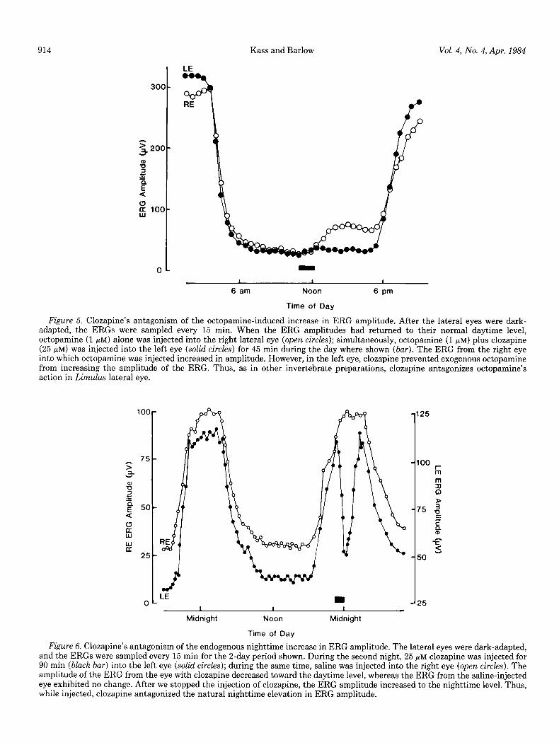
Antagonism of octopamine-induced and nighttime in- creases in ERG amplitude. If octopamine is the efferent neurotransmitter that mediates the action of the circa- dian clock on the retina, then an octopamine antagonist should suppress the clock’s action. Clozopine is a potent a-adrenergic antagonist (Berthelsen and Pettinger, 1977) and a general octopamine antagonist in other inverte- brate systems (Harmer and Horn, 1977; Dougan and
Wade, 1978; Evans, 1981). In the experiment shown in Figure 5, we injected octopamine (1 PM) plus clozapine (25 pM) during the day (bar) in the left eye (LE). Into the right eye (RE) we injected only octopamine (1 PM).
Subsequently, the ERG amplitude of the right eye in- creased, while that of the left eye did not. Neither of the injections that were made at midday impaired the natural action of the clock on ERG amplitude which began at about 6 P.M. We would expect clozapine’s action to require continuous injection because of the rapid blood flow into and out of the retina.
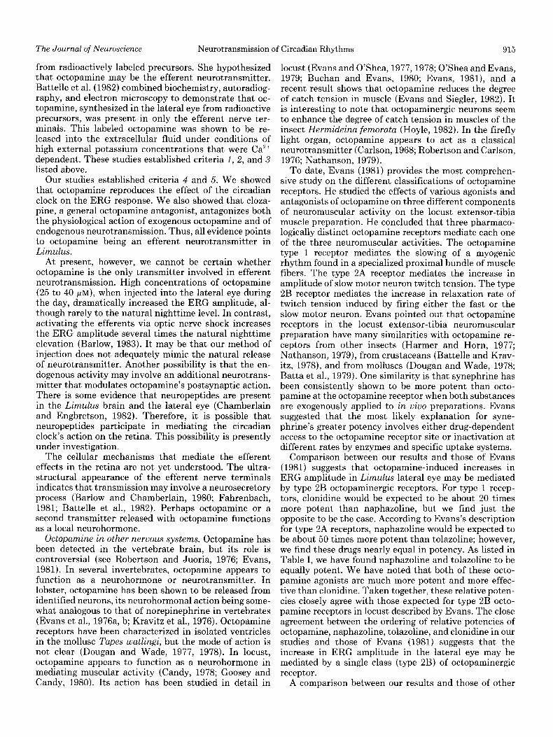
Figure 6 shows that clozapine can antagonize the en- dogenous nighttime elevation in the amplitude of the ERG. In this experiment, clozapine (25 FM) was injected at night (bar) in the left eye and normal saline into the right. Clozapine decreased the amplitude of the ERG toward the daytime level. After injection stopped, the amplitude of the ERG returned to the normal nighttime level. Thus, clozapine antagonizes the endogenous night- time and the exogenous octopamine-induced elevation in the ERG amplitude. Therefore, we conclude that octo- pamine may be an efferent neurotransmitter of the cir- cadian clock’s action on the retina.
Pharmacological controls. In most experiments, a phar- macological agent was injected into one eye while normal saline (with or without 1% dimethyl sulfoxide or ethanol) was simultaneously injected into the other (e.g., Fig. 6). The ERG response from the eye into which saline was injected served as a control against general drug- induced metabolic effects. This was important given that Evans et al. (1976a, b) and Kravitz et al. (1976) have shown that octopamine acts as a general neurohormone in lobster, and that Augustine et al. (1982) and Lumis et al. (1982) indicate in preliminary results that octopamine may also have widespread effects in Limulus. We have found that an agent injected into one eye never produced an effect in the opposite eye (n = 30). Thus, in some experiments it was possible to compare the relative po- tencies of two different agents by using both lateral eyes of the same animal. Whenever practical, the syringe needles carrying the drugs were reversed on subsequent days in order to determine the drug’s action in the opposite eye. In addition, we could inject a drug and its antagonist into one eye and directly compare their action to the drug alone in the opposite eye (e.g., Fig. 5). In sum, we used the opposite lateral eye either to control against nonlocalized drug effects or to compare the ac- tions of different drugs.
Discussion
Octopamine as an efferent neurotransmitter. Octopa- mine has fulfilled five criteria toward its identification as a neurotransmitter in Limulus lateral eye; namely, octopamine has been shown (1) to be synthesized from its precursors, (2) to be localized intracellularly, (3) to be released from efferent nerve terminals, (4) to mimic the physiological actions of the endogenous neurotrans- mitter, and (5) to be blocked pharmacologically.
Battelle (1980) originally showed that octopamine was endogenously present in the Limulus lateral eye; in ad- dition, she showed that octopamine could be synthesized
914 Kass and Barlow Vol. 4, No. 4, Apr. 1984
1
6 am
D
1
Noon
Time of Day
1
6 pm
Figure 5. Clozapine’s antagonism of the octopamine-induced increase in ERG amplitude. After the lateral eyes were dark- adapted, the ERGS were sampled every 15 min. When the ERG amplitudes had returned to their normal daytime level, octopamine (1 pM) alone was injected into the right lateral eye (open cir&s); simultaneously, octopamine (1 PM) plus clozapine (25 FM) was injected into the left eye (solid circles) for 45 min during the day where shown (bar). The ERG from the right eye into which octopamine was injected increased in amplitude. However, in the left eye, clozapine prevented exogenous octopamine from increasing the amplitude of the ERG. Thus, as in other invertebrate preparations, clozapine antagonizes octopamine’s action in Limulus lateral eye.
100
0 LE I
Midnight Noon
Time of Day
Midnight
Figure 6. Clozapine’s antagonism of the endogenous nighttime increase in ERG amplitude. The lateral eyes were dark-adapted, and the ERGS were sampled every 15 min for the 2-day period shown. During the second night, 25 pM clozapine was injected for 90 min (b&h bar) into the left eye (solid circles); during the same time, saline was injected into the right eye (open circles). The amplitude of the ERG from the eye with clozapine decreased toward the daytime level, whereas the ERG from the saline-injected eye exhibited no change. After we stopped the injection of clozapine, the ERG amplitude increased to the nighttime level. Thus, while injected, clozapine antagonized the natural nighttime elevation in ERG amplitude.
The Journal of Neuroscience Neurotransmission of Circadian Rhythms 915
from radioactively labeled precursors. She hypothesized that octopamine may be the efferent neurotransmitter. Battelle et al. (1982) combined biochemistry, autoradiog- raphy, and electron microscopy to demonstrate that oc- topamine, synthesized in the lateral eye from radioactive precursors, was present in only the efferent nerve ter- minals. This labeled octopamine was shown to be re- leased into the extracellular fluid under conditions of high external potassium concentrations that were Ca2+ dependent. These studies established criteria 1, 2, and 3 listed above.
Our studies established criteria 4 and 5. We showed that octopamine reproduces the effect of the circadian clock on the ERG response. We also showed that cloza- pine, a general octopamine antagonist, antagonizes both the physiological action of exogenous octopamine and of endogenous neurotransmission. Thus, all evidence points to octopamine being an efferent neurotransmitter in Limulus.
At present, however, we cannot be certain whether octopamine is the only transmitter involved in efferent neurotransmission. High concentrations of octopamine (25 to 40 yM), when injected into the lateral eye during the day, dramatically increased the ERG amplitude, al- though rarely to the natural nighttime level. In contrast, activating the efferents via optic nerve shock increases the ERG amplitude several times the natural nighttime elevation (Barlow, 1983). It may be that our method of injection does not adequately mimic the natural release of neurotransmitter. Another possibility is that the en- dogenous activity may involve an additional neurotrans- mitter that modulates octopamine’s postsynaptic action. There is some evidence that neuropeptides are present in the Limulus brain and the lateral eye (Chamberlain and Engbretson, 1982). Therefore, it is possible that neuropeptides participate in mediating the circadian clock’s action on the retina. This possibility is presently under investigation.
The cellular mechanisms that mediate the efferent effects in the retina are not yet understood. The ultra- structural appearance of the efferent nerve terminals indicates that transmission may involve a neurosecretory process (Barlow and Chamberlain, 1980; Fahrenbach, 1981; Battelle et al., 1982). Perhaps octopamine or a second transmitter released with octopamine functions as a local neurohormone.
Octopamine in other nervous systems. Octopamine has been detected in the vertebrate brain, but its role is controversial (see Robertson and Juoria, 1976; Evans, 1981). In several invertebrates, octopamine appears to function as a neurohormone or neurotransmitter. In lobster, octopamine has been shown to be released from identified neurons, its neurohormonal action being some- what analogous to that of norepinephrine in vertebrates (Evans et al., 1976a, b; Kravitz et al., 1976). Octopamine receptors have been characterized in isolated ventricles in the mollusc Tapes watlingi, but the mode of action is not clear (Dougan and Wade, 1977, 1978). In locust, octopamine appears to function as a neurohormone in mediating muscular activity (Candy, 1978; Goosey and Candy, 1980). Its action has been studied in detail in
locust (Evans and O’Shea, 1977,1978; O’Shea and Evans, 1979; Buchan and Evans, 1980; Evans, 1981), and a recent result shows that octopamine reduces the degree of catch tension in muscle (Evans and Siegler, 1982). It is interesting to note that octopaminergic neurons seem to enhance the degree of catch tension in muscles of the insect Hermideina femorata (Hoyle, 1982). In the firefly light organ, octopamine appears to act as a classical neurotransmitter (Carlson, 1968; Robertson and Carlson, 1976; Nathanson, 1979).
To date, Evans (1981) provides the most comprehen- sive study on the different classifications of octopamine receptors. He studied the effects of various agonists and antagonists of octopamine on three different components of neuromuscular activity on the locust extensor-tibia muscle preparation. He concluded that three pharmaco- logically distinct octopamine receptors mediate each one of the three neuromuscular activities. The octopamine type 1 receptor mediates the slowing of a myogenic rhythm found in a specialized proximal bundle of muscle fibers. The type 2A receptor mediates the increase in amplitude of slow motor neuron twitch tension. The type 2B receptor mediates the increase in relaxation rate of twitch tension induced by firing either the fast or the slow motor neuron. Evans pointed out that octopamine receptors in the locust extensor-tibia neuromuscular preparation have many similarities with octopamine re- ceptors from other insects (Harmer and Horn, 1977; Nathanson, 1979), from crustaceans (Battelle and Krav- itz, 1978), and from molluscs (Dougan and Wade, 1978; Batta et al., 1979). One similarity is that synephrine has been consistently shown to be more potent than octo- pamine at the octopamine receptor when both substances are exogenously applied to in vivo preparations. Evans suggested that the most likely explanation for syne- phrine’s greater potency involves either drug-dependent access to the octopamine receptor site or inactivation at different rates by enzymes and specific uptake systems.
Comparison between our results and those of Evans (1981) suggests that octopamine-induced increases in ERG amplitude in Limulus lateral eye may be mediated by type 2B octopaminergic receptors. For type 1 recep- tors, clonidine would be expected to be about 20 times more potent than naphazoline, but we find just the opposite to be the case. According to Evans’s description for type 2A receptors, naphazoline would be expected to be about 50 times more potent than tolazoline; however, we find these drugs nearly equal in potency. As listed in Table I, we have found naphazoline and tolazoline to be equally potent. We have noted that both of these octo- pamine agonists are much more potent and more effec- tive than clonidine. Taken together, these relative poten- cies closely agree with those expected for type 2B octo- pamine receptors in locust described by Evans. The close agreement between the ordering of relative potencies of octopamine, naphazoline, tolazoline, and clonidine in our studies and those of Evans (1981) suggests that the increase in ERG amplitude in the lateral eye may be mediated by a single class (type 2B) of octopaminergic receptor.
A comparison between our results and those of other
916 Kass and Barlow Vol. 4, No. 4, Apr. 1984
investigators who have worked on octopamine receptors Candy, D. J. (1978) The regulation of locust flight muscle involves the relative potencies between octopamine and metabolism by octopamine and other compounds. Insect
synephrine. We have found that synephrine is more Biochem. 8: 177-181.
potent than octopamine (Table I), a finding consistent Carlson, A. D. (1968) Effect of drugs on luminescence in larval
with in viva studies on octopamine receptors in all other fireflies. J. Exp. Biol. 49: 195-199.
laboratories. Chamberlain, S. C., and Barlow, R. B., Jr. (1977) Morphological
Circadian changes in structure and function of Limulus correlates of efferent circadian activity and light adaptation
lateral eye. Efferent input to the retina from the circadian in the Limulus lateral eye. Biol. Bull. 153: 418-419.
clock does more than increase the sensitivity of the Chamberlain, S. C., and R. B. Barlow, Jr. (1979) Light and
lateral eye to light. It also changes retinal function and efferent activity control rhabdom turnover in Limulus pho- toreceptors. Science 206: 361-363.
structure. Recordings from single retinal cells show that Chamberlain, S. C., and G. A. Engbretson (1982) Neuropeptide efferent neurotransmission decreases the spontaneous immunoreactivity in Limulus. I. Substance P-like immuno-
(dark) noise of photoreceptor cells (Kaplan and Barlow, reactivity in the lateral eye and protocerebrum. J. Comp.
1980) and increases the spontaneous spike rate and the Neurol. 208: 304-315.
slope of the intensity-response function of second-order Dougan, D. F. H., and D. N. Wade (1977) Octopamine receptors
retinal cells (Barlow et al., 1977). Efferent activity also and their structural specificity. Clin Exp. Pharmacol. Phys-
changes the structure of the retina in ways that may iol. 4: 219.
account for the increases in retinal sensitivity and the Dougan, D. F. H., and D. N. Wade (1978) Differential blockade
other functional changes cited above (Chamberlain and of octopamine and dopamine receptors by analogues of clo-
Barlow, 1977; Barlow et al., 1980). In preliminary reports zapine and metoclopranide. Clin. Exp. Pharmacol. Physiol. 5: 341-349.
(Kass and Barlow, 1981; 1982b), we showed that exoge- Eisele, L. E., L. Kass, and R. B. Barlow, Jr. (1982) Circadian nous octopamine during the day can also mimic these clock generates efferent optic nerve activity in the excised
nighttime changes in retinal structure and function. We Limulus brain. Biol. Bull. 163: 382.
will fully report these additional findings in a subsequent Evans, P. D. (1981) Multiple receptor types for octopamine in
paper. the locust. J. Physiol. (Land.) 318: 99-122. Evans, P. D., and M. O’Shea (1977) The identification of an
octopaminergic neurone which modulates neuromuscular References transmission in the locust. Nature 270: 257-259.
Augustine, G. J., R. Fetterer, and W. H. Watson, III (1982) Evans, P. D., and M. O’Shea (1978) The identification of an
Amine modulation of the neurogenic Limulus heart. J. Neu- octopaminergic neurone and the modulation of a myogenic
robiol. 13: 61-74. rhythm in the locust. J. Exp. Biol. 73: 235-260.
Barlow, R. B., Jr. (1983) Circadian rhythms in the Limidus Evans, P. D., and M. V. Siegler (1982) Octopamine mediated
relaxation of maintained and catch tension in locust skeletal visual system. J. Neurosci. 3: 856-870.
Barlow, R. B., Jr., and S. C. Chamberlain (1980) Light and a muscle. J. Physiol. (Lond.) 324: 93-112.
circadian clock modulate structure and function in Limulus Evans, P. D., E. A. Kravitz, and B. R. Talamo (1976a) Octo-
photoreceptors. In The Effects of Constant Light on Visual pamine release at two points along lobster nerve trunks. J.
Processes, T. P. Williams and B. N. Baker, eds., pp. 247-269, Physiol. (Lond.) 262: 71-89.
Plenum Press, New York. Evans, P. D., E. A. Kravitz, B. R. Talamo, and B. G. Wallace
Barlow, R. B., Jr., S. J. Bolanowski, Jr., and M. L. Brachman (197613) The association of octopamine with specific neurons
(1977) Efferent optic nerve fibers mediate circadian rhythms along lobster nerve trunks. J. Physiol. (Lond.) 262: 51-70.
in the Limulus eye. Science 197: 86-89. Fahrenbach, W. H. (1971) The morphology of the Limu1u.s
Barlow, R. B., Jr., S. C. Chamberlain, and J. Z. Levinson (1980) visual system. IV. The lateral optic nerve. Z. Zellforsch. 114:
Limulus brain modulates structure and function of the lateral 532-545.
eye. Science 210: 1037-1039. Fahrenbach, W. H. (1981) The morphology of the Limulus
Barlow, R. B., Jr., L. C. Ireland, and L. Kass (1982) Vision has visual system. VII. Innervation of photoreceptor neurons by
a role in Limulus mating behaviour. Nature 296: 65-66. neurosecretory efferents. Cell Tissue Res. 216: 655-659.
Goosey, M. W., and D. J. Candy (1980) The D-octopamine Batta, S., R. J. Walker, and G. N. Woodruff (1979) Pharma- content of the haemolymph of the locust, Schistocerea amer-
cological studies on helix neuron octopamine receptors. icana gregaria, and its elevation during flight. Insect Comp. Biochem. Physiol. 64C: 43-51. Biochem. 10: 393-397.
Battelle, B. -A. (1980) Neurotransmitter candidates in the Harmer, A. J., and A. S. Horn (1977) Octopamine-sensitive visual system of Limulus polyphemus: Synthesis and distri- bution of octopamine. Vision Res. 20: 911-922.
adenylate cyclase in cockroach brain: Effects of agonists, antagonists, and guanylyl nucleotides. Mol. Pharmacol. 13:
Battelle, B. -A., and E. A. Kravitz (1978) Targets of octopamine 512-520. action in the lobster: Cyclic nucleotide changes and physio- Hartline, H. D. (1928) A quantitative and descriptive study of logical effects in haemolymph, heart, and exoskeletal muscle. the electric response to illumination of the anthropod eye. J. Pharmacol. Exp. Ther. 205: 438-448. Am. J. Physiol. 83: 446-483.
Battelle, B. -A., J. A. Evans, and S. C. Chamberlain (1982) Hartline, H. K. (1935) The discharge of nerve impulses from Efferent fibers to Limulus eyes synthesize and release octo- pamine. Science 216: 1250-1252.
the single visual sense cell. Cold Spring Harbor Symp. Quant. Biol. 3: 245-250.
Berthelsen, S., and W. A. Pettinger (1977) A functional basis Hoyle, G. (1982) Postural learning in “Wetas” is mediated by for classification of oc-adrenergic receptors. Life Sci. Oxford octopamine-modulated neuromuscular events and “catch” 21: 595-606. contraction. Sot. Neurosci. Abstr. 8: 824.
Buchan, P. B., and P. D. Evans (1980) Use of an operational Kaplan, E., and R. B. Barlow, Jr. (1980) Circadian clock in amplifier signal differentiator reveals that octopamine in- Limulus brain increases response and decreases noise of creases the rate of development of neurally evoked tension retinal photoreceptors. Nature 286: 393-395. in insect muscles. J. Exp. Biol. 85: 349-352. Kass, L., and R. B. Barlow, Jr. (1980) Octopamine increases
The Journal of Neuroscience Neurotransmission of Circadian Rhythms 917
the ERG of the Limulus lateral eye. Biol. Bull. 159: 487. Kass, L., and R. B. Barlow, Jr. (1981) Pharmacological agents
partially reproduce the effects of a circadian clock on the Limulus lateral eye. Biol. Bull. 161: 348.
Kass, L., and R. B. Barlow, Jr. (1982a) Efferent neurotrans- mission of circadian rhythms in the Limulus lateral eye: Specificity for octopamine. Invest. Ophthalmol. Vis. Sci. Suppl. 22: 179.
Kass, L., and R. B. Barlow, Jr. (198213) Efferent neurotrans- mission of circadian rhythms in Limulus lateral eye: Single cell studies. Biol. Bull. 163: 386.
Kass, L., L. E. Eisele, and R. B. Barlow, Jr. (1983) Circadian clock in the excised Limulus brain transmits efferent activity to all eyes. Invest. Ophthalmol. Vis. Sci. Suppl. 24: 218.
Kravitz, E. A., B. -A. Battelle, P. D. Evans, B. R. Talamo, and B. G. Wallace (1976) Octopamine neurons in lobsters. Neu- rosci. Symp. 1: 67-81.
Lummis, S. C., P. M. O’Connor, and B. -A. Battelle (1982) Synthesis and release of ‘H-octopamine from the cardiac
ganglion of Limulus polyphemus. Biol. Bull. 163: 387. Moore, J. W., M. P. Blaustein, N. C. Anderson, and T. Nara-
hashi (1967) Basis of tetrodotoxin’s selectivity in blockage of squid axons. J. Gen. Physiol. 50: 1401-1411.
Nathanson, J. A. (1979) Octopamine receptors, adenosine, 3’,5’-monophosphate, and neural control of firefly flashing. Science 203: 65-68.
O’Shea, M., and P. D. Evans (1979) Potentiation of neuromus- cular transmission by an octopaminergic neurone in the locust. J. Exp. Biol. 79: 169-190.
Powers, M. K., and R. B. Barlow, Jr. (1981) Circadian changes in visual sensitivity of Limulus: Behavioral evidence. Biol. Bull. 161: 350.
Robertson, H. A., and A. D. Carlson (1976) Octopamine: Pres- ence in firefly lantern suggests a transmitter role. J. Exp. Zool. 195: 159-164.
Robertson, H. A., and A. V. Juoria (1976) Octopamine and some related noncatecholic amines in invertebrate nervous systems. Int. Rev. Neurobiol. 19: 173-224.