Page 1
REVIEW ARTICLE Am. J. PharmTech Res. 2012; 2(4) ISSN: 2249-3387
Please cite this article in press as: Rajoria G et al., In-Situ Gelling System: A Novel Approach for Ocular
Drug Delivery. American Journal of PharmTech Research 2012.
In-Situ Gelling System: A Novel Approach for Ocular Drug Delivery
Gourav Rajoria*, Arushi Gupta 1 Department of Pharmaceutics, ISF College of Pharmacy, Moga, Punjab, 142001 India
ABSTRACT
Eye, which is the most vital organ of the body suffer from various eye problems like glaucoma,
endopthalmitis, dry eye syndrome, trachoma, keratitis, conjunctivitis etc. Most ocular diseases
are treated by topical drug application in the form of solutions, suspensions and ointment. These
conventional dosage forms suffer from the problems of poor ocular bioavailability because of
dilution and rapid drainage. Prolonged drug delivery can be achieved by various new dosage
forms like in-situ gel, collagen shield, minidisc, ocular film, ocusert, nanosuspension,
nanoparticulate system, liposomes, niosomes, dendrimers, ocular iontophoresis etc. The most
successful of these is the in-situ forming ophthalmic drug delivery systems prepared from
polymers that exhibit reversible liquid-gel phase transition .The aim of this article is to present a
concise review of in-situ gelling system to overcome all above problems. This review also
summarizes various temperature, pH, and ion induced in-situ forming polymeric systems used to
achieve prolonged contact time of drugs with the cornea and increase their bioavailability.
Keywords: Ophthalmic Solution, In-Situ, Hydrogel, Liquid-gel transition.
*Corresponding Author Email: [email protected]
Received 22 May 2012, Accepted 14 June 2012
Journal home page: http://www.ajptr.com/
Page 2
Rajoria et. al., Am. J. PharmTech Res. 2012; 2(4) ISSN: 2249-3387
25 www.ajptr.com
INTRODUCTION
In the development of ocular drug delivery system lot of complications and difficulties are
found. The conventional drug delivery such as suspension, ointment, solution show some
drawbacks like increase pre-corneal drainage, blurred vision, low bioavailability low residence
time. Various problems encountered in poor bioavailability of the eye installed drugs are:
Binding by the lachrymal proteins,
Drainage of the instilled solutions,
Lachrimation and tear turnover,
Limited corneal area and poor corneal metabolism,
Non‐productive absorption/adsorption,
For the therapeutic treatment of most ocular problems, topical administration clearly seems to be
the preferred route, because in case of systemic administration of drugs, only a very small
fraction of their total dose reach the eye from the general circulatory system.1 Even for this
fraction, distribution to the inside of the eye is further hindered by the blood‐retinal barrier
(BRB).
Consequently, there is a window of only ~5 to 7 minutes for any topically introduced drug to be
absorbed and in many cases, not more than 2% of the medication introduced to the eye is
actually be absorbed 2. The main biological barrier to penetration of the medication is
represented by the cornea. The human cornea is composed of five tissue types with three of
them, the epithelium, the endothelium and the inner stroma, being the main barriers to
absorption. The rest is washed away and absorbed through the nasolacrimal duct and the mucosal
membranes of the nasal, oropharyngeal, and gastrointestinal tract. Figure (1) shows the pre-
corneal and intraocular drug movement from topical dosing.
The poor bioavailability and therapeutic response exhibited by conventional ophthalmic
solutions due to rapid pre-corneal elimination of the drug may be overcome by the use of a gel
system that are instilled as drops into the eye and undergo a sol‐gel transition in the cul-de-sac.
This new system developed is called in-situ gelling system 3. This system shows various
advantages like:
Like Improved Patient Compliance
Reduce Dose Frequency
Increase Bioavailability
Sustain And Controlled Delivery.
Page 3
Rajoria et. al., Am. J. PharmTech Res. 2012; 2(4) ISSN: 2249-3387
www.ajptr.com 26
Kloss KP
abs Kloss
KP
abs
Kd
Figure 1:- Model depicting pre-corneal and intraocular drug movement from topical
dosing.
The in-situ gelling occurs due to different stimuli ion activation (sodium alginate), temperature
change (poloxamer, chitosan), pH change (carbopol), environmental change, solvent exchange.
From the early 1970's natural and synthetic polymers began to be used for controlled release
formulations. Various natural and synthetic polymers are used for formulation development of
in-situ forming drug delivery systems which release the drug as they themselves degrade and are
sometimes finally absorbed within the body. Use of biodegradable and water soluble polymers
for the in-situ gel formulations can make them more acceptable and excellent drug delivery
systems4.
In-Situ Gelling System
In-situ forming hydrogels are refer to polymer solution which can be administered as liquid upon
instillation and undergo phase transition in the ocular cul-de-sac to form viscoelastic gel and this
provides a response to environmental changes. Gelation can be triggered by temperature, pH,
ions; solvent induced and may be UV induced. Three methods have been employed to cause
phase transition on the surface: change in temperature, pH, and electrolyte composition5. In-situ
hydrogels are providing such ‘sensor’ properties and can undergo reversible sol-gel phase
Anterior segment disposition
Installed
Pre-corneal area
-Tear turnover
-Drug metabolism
-Protein drug binding
-Nasolacrimal drainage
-Tear evaporation
-Corneal absorption
-Conjunctival absorption
-Poor corneal
permeability
Cornea
Aqueous humor
Page 4
Rajoria et. al., Am. J. PharmTech Res. 2012; 2(4) ISSN: 2249-3387
27 www.ajptr.com
transitions upon changes in the environmental condition6. It is widely accepted that increasing
the viscosity of a drug formulation in the pre-corneal region will lead to increased
bioavailability, due to slower drainage rate from the cornea7. Moreover, the efficacy of
ophthalmic hydrogels is mostly based on an increase of ocular residence time via enhanced
viscosity and mucoadhesive properties. Since resulted swollen hydrogel is aqueous based, it is
very comfortable in the human eye. In-situ gels are preferred since they are conveniently
dropped in the eye as a solution, where undergo transition into a gel.
Ideally, an in-situ gelling system should be a low viscous, free flowing liquid to allow for
reproducible administration to the eye as drops, and the gel formed following phase transition
should be strong enough to with stand the shear forces in the cul-de-sac and demonstrated long
residence times in the eye. In order to increase the effectiveness of the drug a dosage form should
be chosen which increases the contact time of the drug in the eye. This may then prolonged
residence time of the gel formed in-situ along with its ability to release drugs in sustained
manner will assist in enhancing the bioavailability, reduce systemic absorption and reduce the
need for frequent administration leading to improved patient compliance8. Different polymers
used for this in-situ gelling system according to their sensitivity for example- sodium alginate,
gelrite, carbopol, poloxamer.
Table 1:- Classification of In-Situ Gelling Systems:-
Sr.no In-situ gelling systems Polymers used
1. Temperature dependent systems chitosan, pluronics, tetronics, xyloglucans,
hydroxypropylmethyl cellulose or
hypromellose (HPMC).
2. pH‐triggered systems Cellulose acetate phthalate (CAP) latex,
carbopol, polymethacrilic acid(PMMA),
polyethylene glycol (PEG), pseudolatexes.
3. Ion‐activated systems (osmotically
induced gelation):
gelrite, gellan, hyaluronic acid, alginates.
THERMOREVERSIBLE HYDROGELS
In thermo sensitive systems gelling of solution is triggered by change in temperature. Sustained
drug delivery can be providing by the use of temperature sensitive polymers that change from
solution to gel at the temperature of the eye9 (37ºC). These preparations are liquid at room
temperature (20ºC-25ºC) and become gel at body temperature (35ºC-37ºC) due to change in
temperature. These temperature sensitive gels are classified into three types i.e., negative
temperature sensitive, positively thermo sensitive and thermally reversible gels10
. Negative
temperature-sensitive hydrogels have a lower critical solution temperature (LCST) and contract
Page 5
Rajoria et. al., Am. J. PharmTech Res. 2012; 2(4) ISSN: 2249-3387
www.ajptr.com 28
upon heating above the LCST i.e. Copolymers of (N-isopropylacrylamide) (NIAAm) show an
on-off drug release with on at a low temperature and off at high temperature allowing pulsatile
drug release. LCST systems are mainly relevant for controlled release of drugs, and of proteins
in particular. Thermosensitive polymers may be fixed on liposome membranes; in that case
liposomes exhibit control of their content release11
. A positive temperature-sensitive hydrogel
has an upper critical solution temperature (UCST), such hydrogel contracts upon cooling below
the UCST. Polymer networks of poly (acrylic acid) (PAA) and polyacrylamide (PAAm) or poly
(acryl amide-co-butyl methacrylate) have positive temperature dependence of swelling12
. Figure
(2) showing the mechanism of temperature sensitive gel formation.
Figure 2:- Mechanism of temperature sensitive system.
Polymers used in temperature sensitive system:-
Poloxamers
Poloxamers are a broad group of compounds that were introduced in the early 1950s as food
additives and for pharmaceutical preparations. These water-soluble surfactants are triblock co-
polymers prepared from poly (ethylene oxide)-b-poly (propylene oxide)-b-poly (ethylene oxide)
commercially available as Pluronic®. These are the most commonly used thermosetting
polymers and could be applicable for the development of effective ophthalmic drug delivery13
.
Figure (3) shows the general structure of the pluronics.
HO CH2 CH2 O CH2 CH
CH3
O CH2
H2C O H
a b a
Figure 3: - The general structure for the pluronics.
Page 6
Rajoria et. al., Am. J. PharmTech Res. 2012; 2(4) ISSN: 2249-3387
29 www.ajptr.com
Depending upon the ratio and the distribution along with the chain of the hydrophilic and
hydrophobic subunits, several molecular weights are available having different gelling
properties.
Table 2:- Different grades of Poloxamer
Poloxamer Pluronic ® A B Content of
Oxyethylene(%)
Molecular
Weight
124 L 44 NF 12 20 44.8-48.6 2200
188 F 68 NF 80 27 79.9-83.7 8400
237 F 87 NF 64 37 70.5-74.3 7959
338 F 108 NF 141 44 81.4-84.9 14600
407 F 127 NF 101 56 71.5-74.9 12600
Pluronics F127, which gives colourless and transparent gel so commonly used in the
pharmaceutical industries. Pluronic F 127 is no more damaging to the mouse or rabbit cornea
than a physiological saline. The Poloxamers are reported to be well tolerated and non-toxic even
though large amounts (25-30%) of polymers are required to obtained a suitable gel. At
concentrations of 20% w/v and higher aqueous solutions of Poloxamer-407 remain as a liquid at
low temperatures [<15ºC] and yield a highly viscous semisolid gel upon instillation into the cul-
de-sac14
. At low temperatures, the poloxamer forms micellar subunits in solution, and swelling
gives rise to large micellar subunits and the creation of cross-linked networks. The result of this
phenomenon is a sharp increase in viscosity upon heating.
Three principal mechanisms have been proposed to explain the liquid-gel phase transition after
an increase in temperature, including: -
1. Gradual desolvation of the polymer.
2. Increased micellar aggregation.
3. The increased entanglement of the polymeric network.
In this way we sustain our formulation with the help of temperature sensitive method15
.
Desai S.D. and Blanchard J. 1997 developed formulations of 1% pilocarpine hydrochloride
containing PF127 alone or with one of the following additives present poly (ethyleneglycol)
4600 (PEG), poly (vinylpyrrolidone) 10000 (PVP), poly (vinylalcohol) 10000 (PVA),
methylcellulose 15cP (MC), and hydroxypropyl methyl cellulose 80−120cP (HPMC) and
observed that the PEG- and PVP- containing PF127 formulations of pilocarpine Hcl released the
drug at a significantly faster rate than the control PF127 formulation, which had no additive
present. The PF127 formulations of pilocarpine HCl containing MC or HPMC exhibited the
slowest dissolution rates and released the drug slowest16
.
Page 7
Rajoria et. al., Am. J. PharmTech Res. 2012; 2(4) ISSN: 2249-3387
www.ajptr.com 30
Bochot A. et al., 1998 characterized a new ocular delivery system based on a dispersion of
peggylated liposomes in a thermosensitive gel and showed that the thermosensitivity of PF127
was maintained after introducing the liposomes into the gel17
.
Hong-Ru Lin and K.C. Sung, 2000 developed and characterized a series of carbopol- and
pluronic based solutions as the in situ gelling vehicles for ophthalmic drug delivery. The results
demonstrated that the carbopol/pluronic mixture can be used as an in situ gelling vehicle to
enhance the ocular bioavailability18
.
Desai Suketu D. and Blanchard J, 2000 were prepared a biodegradable
polyisobutylcyanoacrylate (PIBCA) colloidal particulate system of pilocarpine and incorporate it
into a PF127 based gel delivery system and evaluated its ability to prolong the release of
pilocarpine. They concluded that the formulation which contained 1% pilocarpine incorporated
into a PF127 gel containing 5% MC increases contact time and bioavailability of pilocarpine19
.
Carmignani C. et al., 2002 evaluate some solubilizing agents Tyloxapol (TY) and Cremophor EL
(CR) and one polymer, Pluronic P85 for the preparation of 1.0% tropicamide ophthalmic
solutions adjusted at physiologically compatible pH, potentially showing increased eye tolerance,
activity, stability compared with standard commercial eye drops20
.
El-Kamel A.H, 2002 developed PF127 based formulations of TM and obtained slowest drug
release from 15% P F127 formulations containing 3% methyl cellulose. In vivo study showed
that the ocular bioavailability of TM, measured in albino rabbits, increased by 2.5 and 2.4 fold
for 25% PF127 gel formulation and 15% PF127 containing 3% MC, respectively, compared with
0.5% TM aqueous solution21
.
Yoo M.K. et al, 2005 studied release of ciprofloxacin from Chondroitin 6-Sulfate Graft-
poloxamer. In vitro for ophthalmic drug delivery and concluded that the gelation temperature of
C6S-g-Poloxamer copolymer was dependent on the concentration of the graft copolymer and the
content of C6S. Ciprofloxacin release behaviour in vitro, as well as the adhesion and morphology
of human lens cells (B3), was affected by the introduction of C6S into poloxamer22
.
Qi Hongyi et al., 2006 optimized and developed a thermosensitive ophthalmic gel of puerarin
was with 21.0% (w/v) PF127 and 5.0% (w/v) PF68 as the gel matrix, HPCD as the solubilizing
agent, NaCl as the isotonicity agent, and BC as bacterial inhibitor. This in situ gelling
formulation was a free flowing liquid below the room temperature and could convert to a gel that
had an eligible gel strength and bioadhesive force after instilled into conjunctival sac23
.
Dumortier G. et al., 2006 developed a thermogelling ophthalmic formulation of cysteine and
optimized formulations combined either cysteine (2%) / PF127 (16.5%) or cysteine (2%) /
Page 8
Rajoria et. al., Am. J. PharmTech Res. 2012; 2(4) ISSN: 2249-3387
31 www.ajptr.com
PF127 (20%) / PF68 (5%) and were characterized by an adequate temperature of gelification
(TG) (25.9°C and 26.9°C, respectively), an important gel strength (5.1daN and 5.3daN,
respectively) and a drastic increase in the apparent viscosity between 24°C and 32°C
(multiplication factor of 78 and 77-fold, respectively). Cysteine addition produced only slight but
significant decrease in temperature of gelling and increase in gel strength24
.
Gupta H et al., 2007 developed a temperature and pH triggered novel in situ gel system using
poloxamer and chitosan of TM25
. Developed formulation was clear, isotonic solution that
converted into gel at temperatures above 35º C and pH 6.9–7.0.
Ma Wen-di et al., 2008 were studied pluronic-g-poly (acrylic acid) copolymers as a temperature-
responsive in situ gelling vehicle for an ophthalmic drug delivery system. In vivo experimental
results, along with the rheological and in vitro drug release studies, demonstrated that in situ gels
containing pluronic-g-PAA copolymer may significantly prolong the pre-corneal resident time,
and may further improve ocular drug bioavailability26
.
Vehanen K et al., 2008 investigated the use of poloxamers in peribulbar injection for controlled
drug delivery and concluded that poloxamer was well tolerated in peribulbar injections and did
not cause acute toxicity at the site of injection27
.
Mayol L. et al., 2008 studied influence of hyaluronic acid (HA) on the gelation properties of
poloxamers and concluded that the addition of low molecular weight HA into poloxamers blends
as a useful tool to engineer thermosensitive and mucoadhesive polymeric platforms for sustained
drug delivery28
.
Mansour M et al., 2008 developed poloxamer-based in situ gelling formulations of ciprofloxacin
hydrochloride (HCl) using different concentrations of PF127 and PF68. Which Showed optimum
release and mucoadhesion properties and improved ocular bioavailability as evidenced by an
enhanced therapeutic response compared with the marketed conventional eye drops29
.
Cao F. et al., 2010 developed new method for ophthalmic delivery of azithromycin by
poloxamer/carbopol - based in situ gelling system. Addition of carbopol 974P (CP 974P) to the
gelling systems increased the solubility of azithromycin by salt effect and enhanced the
mucoadhesive property of the systems30
.
Gratieri T et al., 2010 investigated in situ forming gel comprised of the combination of a
thermosetting polymer, poly (ethylene oxide) – poly (propylene oxide) – poly (ethylene oxide)
(PEO–PPO–PEO, poloxamer), with a mucoadhesive agent (chitosan) 31
.
Ammar HO. et al., 2010 developed dorzolamide hydrochloride in situ gel nanoemulsion for
ocular delivery and concluded the enhanced efficacy of dorzolamide HCl32
.
Page 9
Rajoria et. al., Am. J. PharmTech Res. 2012; 2(4) ISSN: 2249-3387
www.ajptr.com 32
Asasutjarita. R.et al., Optimization and evaluation of thermoresponsive diclofenac sodium
ophthalmic in situ gels. In this research poloxamer/carbopol used, poloxamer increase the
viscosity and carbopol increase the solubility of the drug diclofenac sodium by salt effect and
enhanced the mucoadhesive property of the systems33
.
Varshosaz Jet al., Designing of a Thermo sensitive Chitosan/Poloxamer In Situ Gel for Ocular
Delivery of Ciprofloxacin. In situ gel-forming eye-drop using 15% Pluronic F127 as the gelling
agent and 0.1% low molecular weight of chitosan as a viscosity enhancing agent34
.
Qian, Y et al., developed in situ gelling ophthalmic drug delivery system for methazolamide. In
vitro release studies demonstrated a diffusion-controlled release of methazolamide from the
poloxamer solutions over a period of 10 hours. In vivo evaluation indicated that the poloxamer
solutions had a better ability to retain drug than methazolamide eye drops35
.
Polysaccharides:-
Cellulose derivatives
Thermo reversible gels can be prepared with naturally occurring polymers. Most of natural
polymer aqueous solutions form a gel phase when their temperature is lowered. Some examples
of natural polymers exhibiting a sol–gel transition include gelatin and carrageenan. At elevated
temperatures, these polymers adopt a random coil conformation in solution. Upon cooling, a
continuous network is formed by partial helix formation36
. Some cellulose derivatives are an
exception to this gelation mechanism. At low concentrations (1–10wt. %), their aqueous
solutions are liquid at low temperature, but gel upon heating. MC and HPMC are typical
examples of such polymers.
O
O
HO OO
O
CH3
CH3
O
O
H3C
OH
O
O
H3C
Methylcellulose
O
CH3
O
OR
CH3
CH3
OR
CH3
RO
O
x
R = CH3 or CH2CH(CH3)OH or H
Hydroxypropylmethylcellulose (HPMC)
Figure 4:- Schematic structure of methyl cellulose and hydroxyl methyl cellulose.
Page 10
Rajoria et. al., Am. J. PharmTech Res. 2012; 2(4) ISSN: 2249-3387
33 www.ajptr.com
MC solutions transform into opaque gels between 40 and 50°C, where as HPMC shows phase
transition between 75 and 90°C. These phase transition temperatures can be lowered by chemical
or physical modifications37, 38
. For example, NaCl decreases the transition temperature of MC
solutions to 32–34°C. Similarly, by reducing the hydroxyl propyl molar substitution of HPMC,
its transition temperature can be lowered to ∼40°C39
. Tate et al evaluated MC based constructs
as potential tissue engineering scaffolds for the repair of brain defects40
.
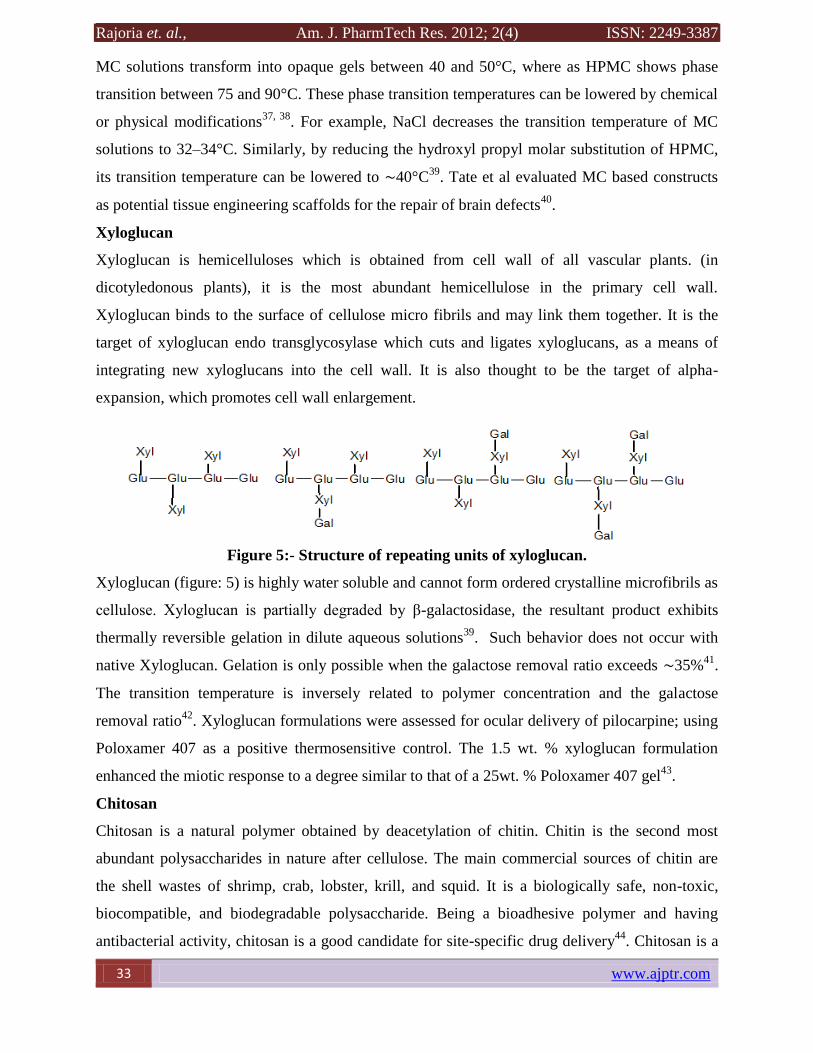
Xyloglucan
Xyloglucan is hemicelluloses which is obtained from cell wall of all vascular plants. (in
dicotyledonous plants), it is the most abundant hemicellulose in the primary cell wall.
Xyloglucan binds to the surface of cellulose micro fibrils and may link them together. It is the
target of xyloglucan endo transglycosylase which cuts and ligates xyloglucans, as a means of
integrating new xyloglucans into the cell wall. It is also thought to be the target of alpha-
expansion, which promotes cell wall enlargement.
Figure 5:- Structure of repeating units of xyloglucan.
Xyloglucan (figure: 5) is highly water soluble and cannot form ordered crystalline microfibrils as
cellulose. Xyloglucan is partially degraded by β-galactosidase, the resultant product exhibits
thermally reversible gelation in dilute aqueous solutions39
. Such behavior does not occur with
native Xyloglucan. Gelation is only possible when the galactose removal ratio exceeds ∼35%41
.
The transition temperature is inversely related to polymer concentration and the galactose
removal ratio42
. Xyloglucan formulations were assessed for ocular delivery of pilocarpine; using
Poloxamer 407 as a positive thermosensitive control. The 1.5 wt. % xyloglucan formulation
enhanced the miotic response to a degree similar to that of a 25wt. % Poloxamer 407 gel43
.
Chitosan
Chitosan is a natural polymer obtained by deacetylation of chitin. Chitin is the second most
abundant polysaccharides in nature after cellulose. The main commercial sources of chitin are
the shell wastes of shrimp, crab, lobster, krill, and squid. It is a biologically safe, non-toxic,
biocompatible, and biodegradable polysaccharide. Being a bioadhesive polymer and having
antibacterial activity, chitosan is a good candidate for site-specific drug delivery44
. Chitosan is a
Page 11
Rajoria et. al., Am. J. PharmTech Res. 2012; 2(4) ISSN: 2249-3387
www.ajptr.com 34
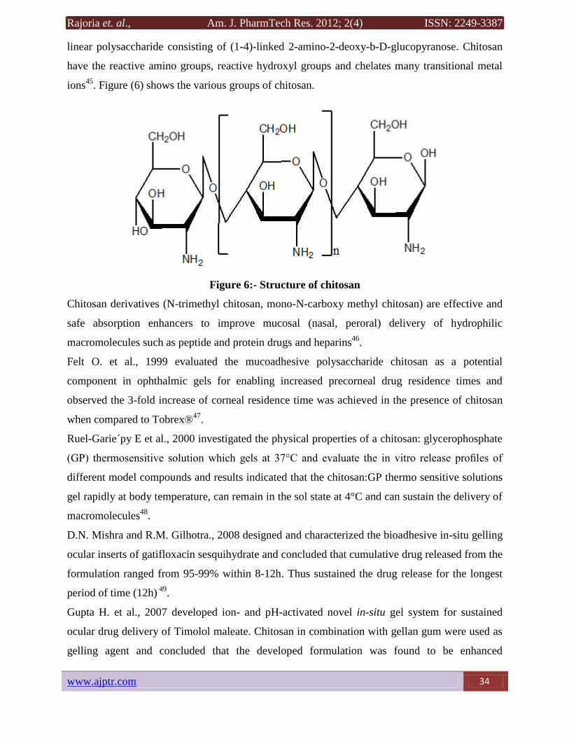
linear polysaccharide consisting of (1-4)-linked 2-amino-2-deoxy-b-D-glucopyranose. Chitosan
have the reactive amino groups, reactive hydroxyl groups and chelates many transitional metal
ions45
. Figure (6) shows the various groups of chitosan.
Figure 6:- Structure of chitosan
Chitosan derivatives (N-trimethyl chitosan, mono-N-carboxy methyl chitosan) are effective and
safe absorption enhancers to improve mucosal (nasal, peroral) delivery of hydrophilic
macromolecules such as peptide and protein drugs and heparins46
.
Felt O. et al., 1999 evaluated the mucoadhesive polysaccharide chitosan as a potential
component in ophthalmic gels for enabling increased precorneal drug residence times and
observed the 3-fold increase of corneal residence time was achieved in the presence of chitosan
when compared to Tobrex®47
.
Ruel-Garie´py E et al., 2000 investigated the physical properties of a chitosan: glycerophosphate
(GP) thermosensitive solution which gels at 37°C and evaluate the in vitro release profiles of
different model compounds and results indicated that the chitosan:GP thermo sensitive solutions
gel rapidly at body temperature, can remain in the sol state at 4°C and can sustain the delivery of
macromolecules48
.
D.N. Mishra and R.M. Gilhotra., 2008 designed and characterized the bioadhesive in-situ gelling
ocular inserts of gatifloxacin sesquihydrate and concluded that cumulative drug released from the
formulation ranged from 95-99% within 8-12h. Thus sustained the drug release for the longest
period of time (12h) 49
.
Gupta H. et al., 2007 developed ion- and pH-activated novel in-situ gel system for sustained
ocular drug delivery of Timolol maleate. Chitosan in combination with gellan gum were used as
gelling agent and concluded that the developed formulation was found to be enhanced
Page 12
Rajoria et. al., Am. J. PharmTech Res. 2012; 2(4) ISSN: 2249-3387
35 www.ajptr.com
transcorneal drug permeation, and prolonged the retention at corneal site. It was also found
suitable for sustained topical drug delivery to eyes and can prove as better alternative to
conventional eye drops for the better drug therapy of glaucoma and other ocular disorders25.
Mehra GR. et al., 2010 studied in situ gelling solution of pilocarpine based on alginate along
with novel bioadhesive tamarind gum and widely used bioadhesive, chitosan and found that the
tamarind gum based formulation released about 25 % drug in initial hour and about 80% of the
drug was released during the study of 12 h50
.
pH Sensitive Hydrogels
Gelling of the solution is triggered by a change in the pH. Cellulose acetate phthalate (CAP)
latex, cross linked acrylic, and derivatives such as Carbomer are used for this method51
.
Cellulose acetate derivatives are the only polymer known to have a buffer capacity that is low
enough to gel effectively in the cul-de-sac of the eye. The pH change of about 2.8 units after
instillation of the native formulation (pH 4.4) into the tear film leads to an almost instantaneous
transformation of the highly fluid latex into viscous gel6. Gel is formed at eye pH i.e,
(7.4).different polymers are used in the production of pH sensitive formulation like carbopol.
Cellulose acetate phthalate latex, poly acrylic acid.
Principle mechanism for pH sensitive gel:-
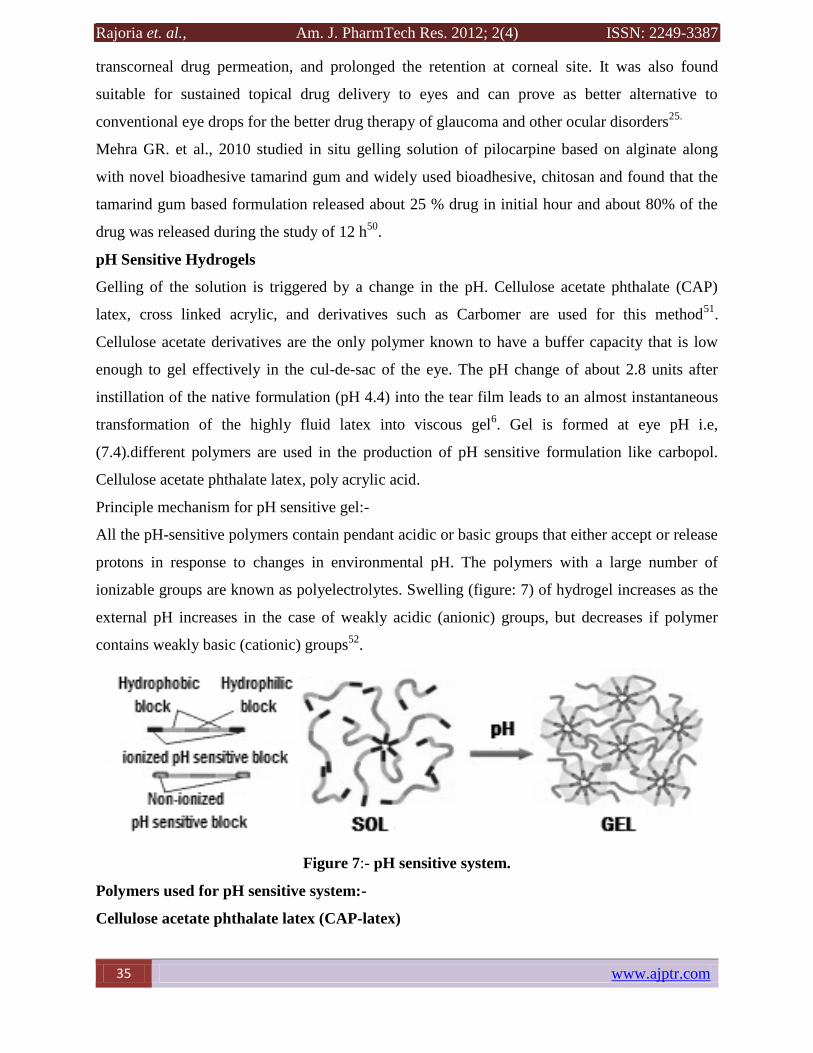
All the pH-sensitive polymers contain pendant acidic or basic groups that either accept or release
protons in response to changes in environmental pH. The polymers with a large number of
ionizable groups are known as polyelectrolytes. Swelling (figure: 7) of hydrogel increases as the
external pH increases in the case of weakly acidic (anionic) groups, but decreases if polymer
contains weakly basic (cationic) groups52
.
Figure 7:- pH sensitive system.
Polymers used for pH sensitive system:-
Cellulose acetate phthalate latex (CAP-latex)
Page 13
Rajoria et. al., Am. J. PharmTech Res. 2012; 2(4) ISSN: 2249-3387
www.ajptr.com 36
First preliminary investigations of pH sensitive nanoparticulate systems (latex) for ophthalmic
administration began in the early 1980s. The choice of this polymer was determined by the
compatibility of the polymer with the active compound, the ability of the CAP latex to be a free
running solution at pH 4.2 and a gel at 7.2, and finally52
, but the low pH of the preparation can
elicit discomfort in some patients. The gelation capacity of CAP latexes has been visualized in
vitro by scanning electron microscopy and in vivo in rabbits by incorporating methylene blue in
ophthalmic formulations. The efficacy of a preparation based on pseudolatex has been evaluated
by measuring pharmacological responses and precorneal residence time by γ scintigraphy. This
technique has clearly demonstrated the superiority of CAP latex over a solution to prolong the
corneal residence time of pilocarpine6. Figure (8) represent the structure of the cap.
Figure 8:- Structure of cellulose acetate phthalate.
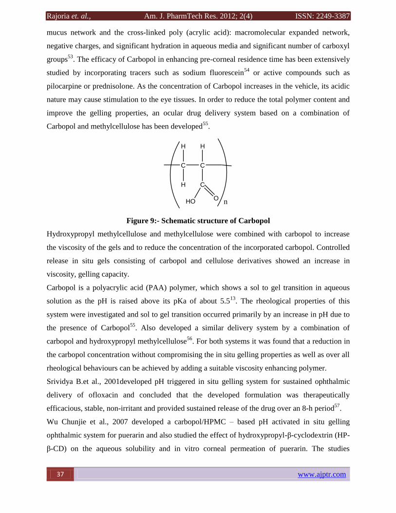
Carbomer
Cross-linked poly (acrylic acid) of high molecular weight, commercially available as Carbopol®
(figure: 9), is widely used in ophthalmology to enhance pre-corneal retention to the eye47
.
Carbopol® 934 is a synthetic polymer composed of 62% of carboxyl groups with a high
molecular weight (approximately 3×10⁶) formed by repeating units of acrylic acid, cross-linked
with either allyl sucrose or allyl ethers of pentaerythritol. Carbopol offers the advantage of
exhibiting excellent mucoadhesive properties when compared with other polymers (e.g. cellulose
derivatives, and polyvinylalcohol). The mechanisms involved in the mucoadhesion ability of
Carbopol have been investigated previously. Four mechanisms of interaction between mucin and
poly (acrylic acid) have been described: electrostatic interaction, hydrogen bonding, hydrophobic
interaction, and inter diffusion. These mechanisms can be explained by the similar features of the
Page 14
Rajoria et. al., Am. J. PharmTech Res. 2012; 2(4) ISSN: 2249-3387
37 www.ajptr.com
mucus network and the cross-linked poly (acrylic acid): macromolecular expanded network,
negative charges, and significant hydration in aqueous media and significant number of carboxyl
groups53
. The efficacy of Carbopol in enhancing pre-corneal residence time has been extensively
studied by incorporating tracers such as sodium fluorescein54
or active compounds such as
pilocarpine or prednisolone. As the concentration of Carbopol increases in the vehicle, its acidic
nature may cause stimulation to the eye tissues. In order to reduce the total polymer content and
improve the gelling properties, an ocular drug delivery system based on a combination of
Carbopol and methylcellulose has been developed55
.
C C
H
H
H
C
HOO
n
Figure 9:- Schematic structure of Carbopol
Hydroxypropyl methylcellulose and methylcellulose were combined with carbopol to increase
the viscosity of the gels and to reduce the concentration of the incorporated carbopol. Controlled
release in situ gels consisting of carbopol and cellulose derivatives showed an increase in
viscosity, gelling capacity.
Carbopol is a polyacrylic acid (PAA) polymer, which shows a sol to gel transition in aqueous
solution as the pH is raised above its pKa of about 5.513
. The rheological properties of this
system were investigated and sol to gel transition occurred primarily by an increase in pH due to
the presence of Carbopol55
. Also developed a similar delivery system by a combination of
carbopol and hydroxypropyl methylcellulose56
. For both systems it was found that a reduction in
the carbopol concentration without compromising the in situ gelling properties as well as over all
rheological behaviours can be achieved by adding a suitable viscosity enhancing polymer.
Srividya B.et al., 2001developed pH triggered in situ gelling system for sustained ophthalmic
delivery of ofloxacin and concluded that the developed formulation was therapeutically
efficacious, stable, non-irritant and provided sustained release of the drug over an 8-h period57
.
Wu Chunjie et al., 2007 developed a carbopol/HPMC – based pH activated in situ gelling
ophthalmic system for puerarin and also studied the effect of hydroxypropyl-β-cyclodextrin (HP-
β-CD) on the aqueous solubility and in vitro corneal permeation of puerarin. The studies
Page 15
Rajoria et. al., Am. J. PharmTech Res. 2012; 2(4) ISSN: 2249-3387
www.ajptr.com 38
concluded that combined polymer systems performed better in retaining puerarin than puerarin
eye drops58
.
Al-Kassas R.S. and El-Khatib M.M, 2009 designed controlled release ophthalmic delivery
systems for ciprofloxacin. The antimicrobial efficiency of the selected formulation against gram-
positive and gram-negative organisms including Echerichia coli, Staphylococcus strains and
Pseudomonas aeruginosa confirmed that the designed formulation has prolonged the
antimicrobial effect of ciprofoxacin and retained its properties against bacteria59
.
Nanjwade B.K. et al., 2009 developed a novel pH-triggered in situ gel for sustained ophthalmic
delivery of ketorolac tromethamine. The developed formulation is a viable alternative to
conventional eye drops by virtue of its ability to enhance bioavailability through its longer
precorneal residence time and ability to produce sustained drug release60
.
Gupta H. et al., 2010 developed ion- and pH-activated novel in-situ gel system for sustained
ocular drug delivery of Timolol maleate. Chitosan in combination with gellan gum were used as
gelling agent and concluded that the developed formulation was found to be non-irritant,
enhanced transcorneal drug permeation, and prolonged the retention at corneal site25
.
Pandey A, et al, 2010 Development and Optimization of Levobunolol Hydrochloride In-situ Gel
for Glaucoma Treatment. Carbopol provide phase change from liquid to gel and HMPC provide
Mucoadhesive strength and increase viscosity of formulation61
.
Mohanambal E, et al, 2011 Formulation and Evaluation of pH-triggered in-situ Gelling System
of Levofloxacin. The levofloxain in-situ gelling system formulated by using poly acrylic acid
(Carbopol 940) in combination with hydroxyl propyl methyl cellulose (HPMC) which acted as
viscosity enhancing agent. The developed formulation was stable, non-irritant and provided
sustained release over 8-hour period and it is a viable alternative to conventional eye drops62
.
Ion-Sensitive Hydrogels
In ion activated system gelling of solution is triggered by cations present in the present in the eye
tear fluid like Na+, Ca++ and Mg++. Generally anionic polymers are used in the formation of
ion sensitive drug delivery system52, 63
. Polymers like sodium alginate, gelrite, tamarind gum,
gellen gum are used in these formulations.
Various other polymers like methylcellulose (MC), hydroxyl propyl methyl cellulose (HPMC)
are used in combination of these polymers to increase the effect. They provide sustain release of
drug by providing mucoadhesivenes.
Page 16
Rajoria et. al., Am. J. PharmTech Res. 2012; 2(4) ISSN: 2249-3387
39 www.ajptr.com
Figure 10:- Mechanism showing Ion activated system.
This system based on the mechanism (figure 10) of ionic interaction of ions of polymer and
divalent ions of tear fluid. When anionic polymers come in contact with cationic ions they
convert into gel viscosity of solution increases to an extent64
.
Gelrite
Gellan gum (figure: 12) is a linear, anionic hetero polysaccharide secreted by the microbe
Sphingomonaselodea (formerly known as Pseudomonaselodea). Gelrite is deacetylated gellan
gum which gels upon instillation in the eye due to the presence of cations65
. The polysaccharide
can be produced by aerobic fermentation and then isolated from the fermentation broth by
alcohol precipitation. The polymer back bone consists of glucose, glucuronic acid, and rhamnose
in the molar ratio 2:1:166, 67
. It has the tendency of gelation which is temperature dependent or
cations induced .This gelation involves the formation of double helical junction zones followed
by aggregation of the double helical segments to form a three-dimensional network by
complexation with cations and hydrogen bonding with water68
.
Gelrite has been granted regulatory approval as pharmaceutical excipient and is marketed by
Merck in a controlled release glaucoma formulation called Blocarden® Depot (Timoptic®).
Formulations with the Gelrite can be administered to ocular mucosa as a low viscosity solution.
On contact with cations in tear fluid the formulation will form a clear gel. This is caused by cross
linking of the negatively charged polysaccharide helices by monovalent and divalent cations
(Na+, K+, Ca+). In anion free aqueous medium, The divalent ions such as magnesium or calcium
were superior to monovalent cations in promoting the gelation of the polysaccharide. However
the concentration of sodium in tears (2.6g/L) is quite sufficient to induce the gelation. Gelrite has
Page 17
Rajoria et. al., Am. J. PharmTech Res. 2012; 2(4) ISSN: 2249-3387
www.ajptr.com 40
also provided corneal residence time’s superior to those of other hydrogel preparations based on
polymers such as cellulosic derivatives or xanthan gum. The rheological properties of gellan gum
such as thixotropy, pseudo plasticity, and thermo plasticity are further advantages for its use in
ophthalmology13
. The most important gel-promoting ion in vivo is Na+69
.
Figure 11:- Schematic structure of gellan gum.
Balasubramaniam J. and Pandit J.K. 2003 developed ion - activated in situ gelling systems for
sustained ophthalmic delivery of ciprofloxacin Hcl and concluded that the formulated systems
provided sustained release of the drug over an 8-hr period in vitro71
.
Sultana Y et al., 2006 ion-activated, Gelrite based in Situ Ophthalmic Gels of Pefloxacin
Mesylate and compared with conventional eye drops and concluded that the system was capable
for effective and controlled management of conjunctivitis for 12hr72
.
Liu Y. et al., 2010 investigated in Situ Gelling Gelrite/Alginate Formulations as Vehicles for
ophthalmic drug delivery and found that the optimum concentration of Gelrite solution for the in
situ gel forming delivery systems was 0.3% (w/w) and that for alginate solution was 1.4% (w/w).
The mixture of 0.2% Gelrite and 0.6% alginate solutions showed a significant enhancement in
gel strength at physiological condition73
.
Vodithala S. et al., 2010 developed ion activated ocular in situ gels of ketorolac tromethamine
using Gelrite as a polymer and concluded that the developed formulations showed sustained
release of drug for upto 6hrs. The formulations were found to be non‐irritating with no ocular
dam-age74
.
Rajas N, J et al., 2011. In situ ophthalmic gels a developing trend. Levofloxacin hemihydrate
which is a broad spectrum anti bacterial agent used in the treatment of ocular infections was
successfully formulated as in situ gel using Gelrite as polymer. The formulated systems provided
sustained release of the drug for more than 8hr period. The developed formulation is a viable
alternative to conventional eye drop due to its ability to enhance bioavailability through its
longer pre-corneal residence time and ability to sustain release of the drug53
.
Page 18
Rajoria et. al., Am. J. PharmTech Res. 2012; 2(4) ISSN: 2249-3387
41 www.ajptr.com
Geethalakshmi A, et al., 2012. Sustained ocular delivery of brimonidine tartrate using ion
activated in situ gelling system. Gelrite is used as gelling agent in the preparation of brimonidine
tartrate to enhance the viscosity of formulation. Also provide sustain release of drug in the eye74
.
Rupenthal ID, et al., 2011 Jun. Comparison of ion-activated in situ gelling systems for ocular
drug delivery. Part 2: Precorneal retention and in vivo pharmacodynamic study. Various
polymers used like gellan gum, xanthan gum and carrageenan in the formulation of ion activated
system which provide sustained release of drug75
.
Alginates
Alginates (figure: 13) consist of (1→4) linked β-D-mannuronic acid (M) and α-L-guluronic acid
(G) residues of widely varying composition and sequence. By partial acid hydrolysis, alginate
was separated into three fractions. Alginate with a high guluronic acid content will improve the
gelling properties and reduce the total polymer to be introduced into the eye. The alginate forms
3-dimensional ionotropic hydrogel matrices, generally by the preferential interaction of calcium
ions with the G moieties resulting in the formation of in homogeneous gel52
. The characteristic
properties of these hydrogels, such as mechanical strength and porosity, are dependent upon the
G: M ratios, type of ionic cross linker (bio or polyvalent cations), concentration and viscosity of
the initial alginate solution. Alginates were approved by the regulatory authorities such as the
Food and Drug Administration, for human use as wound dressing material and as food
additives13
.
O
COO-
OH OH
OH
OH
O
COO-
OH OH
HO
OH
a.
-OOC
O
OH
O
OHO
OH
OH
COO-
O
O
-OOC HO
HO
O
O
HO
OH-OOC
OO
OH
-OOC
OH
O
b.
M G
Figure 12:- schematic structure of (a) Alginate monomers (b) Chain conformation
Cohen S. et al., 1997 developed a novel in situ forming ophthalmic drug delivery system from
alginates undergoing gelation in the eye and concluded that Pilocarpine is released slowly from
Page 19
Rajoria et. al., Am. J. PharmTech Res. 2012; 2(4) ISSN: 2249-3387
www.ajptr.com 42
alginate gels, over a period of 24h, and the release occurs mostly via diffusion from the gels.
Dissolution of the hydrogels in the releasing media was negligible for the first 12h of incubation
at 370C
76.
D.N. Mishra and R.M. Gilhotra. 2008 designed and characterized the bioadhesive in-situ gelling
ocular inserts of gatifloxacin sesquihydrate and concluded that cumulative drug released from the
formulation ranged from 95-99% within 8-12h. The formulation with 2% sodium alginate and
1% chitosan, sustained the drug release for the longest period of time (12h). Zero-order release
of the drug was from optimized the formulation with 2% sodium alginate and 1% chitosan77
.
Abraham S. et al., 2009 developed sustained ophthalmic delivery of ofloxacin from an ion-
activated in situ gelling system and concluded that the alginate/HPC solution retained the drug
better than the alginate or HPC solutions alone and drug release over a period of 8 hours78
.
Mehra G.R. et al., 2010 developed Pilocarpine in-situ gelling solution based on alginate along
with novel bioadhesive tamarind gum, widely used bioadhesive, chitosan and alginate as a
polymer and concluded that the formulation showed release about 25 % drug in initial hour and
about 80 % of the drug was released during the study of 12 h79
.
Preetha JP et al., 2010 developed an in situ gelling ophthalmic formulation of diclofenac sodium
and concluded that the formulation having 1.5% sodium alginate and 0.75% of HEC shows
better drug release when contacted with STF solution at 8 hrs study period and shows
antimicrobial, antibacterial and antifungal efficacy with selected microorganisms80
.
Liu Z. et al., 2006. Developed alginate/HPMC –based in situ gelling ophthalmic delivery system
for gatifloxacin and demonstrated that the alginate/HPMC mixture can be used as an in situ
gelling vehicle to enhance ocular bioavailability and patient compliance81
.
Gaonkar GV et al., Oct-Nov. 2010.Formulation Development And Evaluation Of Long Acting
Ophthalmic In-Situ Gelling Systems Of Ketorolac Tromethamine And Ofloxacin: A Research.
Aqueous solutions of Sodium Alginate, Xanthan Gum and Carbopol 971P coupled with added
viscolizers are attractive in-situ gel forming systems, promising controlled ocular drug delivery
of Ketorolac Tromethamine and Ofloxacin82
.
Liu Y. et al., 2010 investigated in Situ Gelling Gelrite/Alginate Formulations as Vehicles for
ophthalmic drug delivery and found that the optimum concentration of Gelrite solution for the in
situ gel forming delivery systems was 0.3% (w/w) and that for alginate solution was 1.4% (w/w).
The mixture of 0.2% Gelrite and 0.6% alginate solutions showed a significant enhancement in
gel strength at physiological condition72
.
Page 20
Rajoria et. al., Am. J. PharmTech Res. 2012; 2(4) ISSN: 2249-3387
43 www.ajptr.com
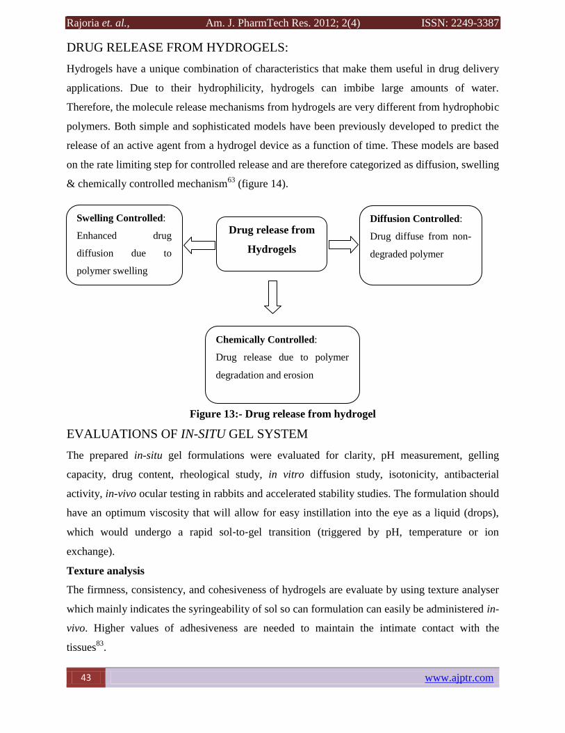
DRUG RELEASE FROM HYDROGELS:
Hydrogels have a unique combination of characteristics that make them useful in drug delivery
applications. Due to their hydrophilicity, hydrogels can imbibe large amounts of water.
Therefore, the molecule release mechanisms from hydrogels are very different from hydrophobic
polymers. Both simple and sophisticated models have been previously developed to predict the
release of an active agent from a hydrogel device as a function of time. These models are based
on the rate limiting step for controlled release and are therefore categorized as diffusion, swelling
& chemically controlled mechanism63
(figure 14).
Figure 13:- Drug release from hydrogel
EVALUATIONS OF IN-SITU GEL SYSTEM
The prepared in-situ gel formulations were evaluated for clarity, pH measurement, gelling
capacity, drug content, rheological study, in vitro diffusion study, isotonicity, antibacterial
activity, in-vivo ocular testing in rabbits and accelerated stability studies. The formulation should
have an optimum viscosity that will allow for easy instillation into the eye as a liquid (drops),
which would undergo a rapid sol‐to‐gel transition (triggered by pH, temperature or ion
exchange).
Texture analysis
The firmness, consistency, and cohesiveness of hydrogels are evaluate by using texture analyser
which mainly indicates the syringeability of sol so can formulation can easily be administered in-
vivo. Higher values of adhesiveness are needed to maintain the intimate contact with the
tissues83
.
Diffusion Controlled:
Drug diffuse from non-
degraded polymer
Swelling Controlled:
Enhanced drug
diffusion due to
polymer swelling
Drug release from
Hydrogels
Chemically Controlled:
Drug release due to polymer
degradation and erosion
Page 21
Rajoria et. al., Am. J. PharmTech Res. 2012; 2(4) ISSN: 2249-3387
www.ajptr.com 44
Physical parameters
The formulated in-situ gel solution is tested for clarity, pH, gelling capacity, and drug content
estimation.
Gelling capacity
The gelling capacity of the prepared formulation is determined by placing a drop of the
formulation in a vial containing 2.0 ml of freshly prepared simulated tear fluid and visually
observed. The time taken for its gelling is noted84, 85, 86
.
Rheological studies
The viscosity measurements can be calculated using Brookfield viscometer, Cone and Plate
viscometer. The in-situ gel formulations were placed in the sampler tube. From the literature it
was evident that, the formulation before gelling should have a viscosity of 5 to 1000 mPas. And
after ion gel activation by the eye, will have a viscosity of from about 50‐ 50,000 mPas. The
samples are analyzed both at room temperature at 25°C and thermo stated at 37°C ± 0.5°C by a
circulating bath connected to the viscometer adaptor prior to each measurement. The angular
velocity of the spindle was increased 20, 30, 50, 60, 100, 200 and the viscosity of the formulation
is measured. All the formulations exhibited Newtonian and pseudoplastic flow characteristics
before and after gelling in the simulated tear fluid respectively83, 87, 88
.
In vitro drug release studies
In vitro release study of in-situ gel solution was carried out by using Franz diffusion cell. The
formulation placed in donor compartment and freshly prepared simulated tear fluid in receptor
compartment. Between donor and receptor compartment dialysis membrane is placed (0.22μm
pore size). The whole assembly was placed on the thermostatically controlled magnetic stirrer.
The temperature of the medium was maintained at 37°C ± 0.5°C. 1ml of sample is withdrawn at
predetermined time interval of 1hr for 6 hrs and same volume of fresh medium is replaced. The
withdrawn samples are diluted to 10ml in a volumetric flask with respective solvent and
analyzed by UV spectrophotometer at respective nm using reagent blank. The drug content
calculated using the equation generated from standard calibration curve. The % cumulative drug
release (%CDR) calculated. The data obtained is further subjected to curve fitting for drug
release data. The best fit model is checked for Krosmeyers Peppas and Fickinian diffusion
mechanism for their kinetic89, 90
.
Isotonicity Evaluation
Isotonicity is important characteristic of the ophthalmic preparations. Isotonicity has to be
maintained to prevent tissue damage or irritation of eye. All ophthalmic preparations are
Page 22
Rajoria et. al., Am. J. PharmTech Res. 2012; 2(4) ISSN: 2249-3387
45 www.ajptr.com
subjected to isotonicity testing, since they exhibited good release characteristics and gelling
capacity and the requisite viscosity. Formulations are mixed with few drops of blood and
observed under microscope at 45X magnification and compared with standard marketed
ophthalmic formulation1, 91
.
Antibacterial activity
The microbiological growth of bacteria is measured by concentration of antibiotics and this has
to be compared with that produced by known concentration of standard preparation of antibiotic.
To carryout microbiological assay serial dilution method is employed1, 92
.
Ocular irritancy test
The Draize irritancy test was designed for the ocular irritation potential of the ophthalmic
product prior to marketing. According to the Draize test, the amount of substance applied to the
eye is normally 100μl placed into the lower cul-de-sac with observation of the various criteria
made at a designed required time interval of 1hr, 24hrs, 48 hrs, 72hrs, and 1week after
administration. Three rabbits (male) weighing 1.5 to 2kg are used for the study. The sterile
formulation is instilled twice a day for a period of 7 days, and a cross‐over study is carried out (a
3 day washing period with saline was carried out before the cross‐over study). Rabbits are
observed periodically for redness, swelling, watering of the eye92, 93, 94
.
Accelerated stability studies
Formulations are placed in ambient colour vials and sealed with aluminium foil for a short term
accelerated stability study at 40±2 °C and 75±5% RH as per International Conference on
Harmonization (ICH) states Guidelines. Samples are analyzed every month for clarity, pH,
gelling capacity, drug content, rheological evaluation, and in vitro dissolution91
.
CONCLUSION
The complications in eye formulation are mainly due to specific anatomical and physiological
features of eye. The development of in-situ stimuli activated gel-forming systems for ophthalmic
drug delivery provides simplest and best gel-forming systems. It is an ideal system that maintains
effective level of drug for the longer duration following a single application and offers the
primary requirement of a successful controlled release product that increases patient compliance.
Moreover, various polymers used in this system provide advantage over conventional drug
delivery system. This system is preferred over other systems for ocular delivery because it can be
administered in drop form and creates significantly fewer problems with vision as well as have
Page 23
Rajoria et. al., Am. J. PharmTech Res. 2012; 2(4) ISSN: 2249-3387
www.ajptr.com 46
sustained release. In the recent era of technology, combinatorial approach seems to be a focus of
research in the development of safe and efficient ophthalmic drug delivery systems.
REFERENCES
1. Rathore KS. In-situ Gelling Ophthalmic Drug Delivery System: An Overview Int J
Pharm Pharma Sci 2010; 2(4).
2. Gaudana R, Jwala J, Boddu HS, Sai K, Mitra AK. Recent Perspectives in Ocular Drug
Delivery. Pharma Res 2009; 26(5).
3. Shastri DH, Patel LD. A Novel Alternative to Ocular Drug Delivery System: Hydrogel.
Int J Pharma Res 2010; 2(1).
4. Jigar V. A Review on Novel in Situ Polymeric Drug Delivery System. Int J Pharma Res
Dev 2011; 3 (5):53-59.
5. Pandya et al., Ophthalmic In-Situ Gelling System. Int J Pharm Life Sci 2011; 2 (5): 730-
738.
6. Shastri, DH, Patel LD. A Novel Alternative to Ocular Drug Delivery System: Hydrogel.
Int J Pharm Res 2010; 2(1): 1-13.
7. Miller S, Donovan M. Effect of Poloxamer 407 Gel on the Miotic Activity of Pilocarpine
Nitrate in Rabbits. Int J Pharm 1982; 12:147-152.
8. Indu PK, Manjit S, Meenakshi K. Formulation and Evaluation of Ophthalmic
Preparations of Acetazolamide. Int J Pharm 2000; 199: 119–127.
9. Wei G, Xu H, Ding PT, Li S M, Zheng J M J, Thermosetting Gels With Modulated
Gelation Temperature For Ophthalmic Use. The Rheological and Gamma Scintigraphic
Studies. J Controlled Rel 2002; 83:64-65.
10. Qiu Y, Park K, Environment-Sensitive Hydrogels for Drug Delivery. Advanced Drug
Delivery Rev 2001; 53:321-39.
11. Bromberg LE, Ron ES, Temperature Responsive Gels and Thermo Gelling Polymer
Matrices for Protein and Peptide Delivery. Advanced Drug Delivery Rev 1998; 31:197-
221.
12. Kono K. Thermosensitive Polymer-Modified Liposomes. Advanced Drug Delivery Rev
2001; 53:307.
13. Nanjwade BK, Manvi FV, Manjappa AS. In Situ -Forming Hydrogels for Sustained
Ophthalmic Drug Delivery. J Controlled Rel 2007; 122: 119-134.
Page 24
Rajoria et. al., Am. J. PharmTech Res. 2012; 2(4) ISSN: 2249-3387
47 www.ajptr.com
14. Chen-Chow PC, Frank SG. In Vitro Release of Lidocaine from Pluronic F 127. Int J
Pharma Sci 1981; 8: 89-99.
15. Shastri DH, Patel LD. Novel Alternative to Ocular Drug Delivery System: Hydrogel. Int
J Pharma Res 2010; 2(1).
16. Desai SD, Blanchard J. Pluronic F127 - Based Ocular Delivery System Containing
Biodegradable Polyisobutylcyanoacrylate Nanocapsules Of Pilocarpine. Drug Delivery
2000; 7: 201-207.
17. Bochot A, Fattal E, Gulik A, Couarraze G, Couvreur P. Liposomes Dispersed Within A
Thermosensitive Gel: A New Dosage Form For Ocular Delivery Of Oligonucleotides.
Pharm Res 1998; 15(9):1364-9.
18. Hong-Ru Lin, Sung KC. Carbopol/pluronic phase change solutions for ophthalmic drug
delivery Journal of Controlled Release 2000; 69(3): 379–388.
19. Desai SD, Blanchard J, Pluronic F127 - Based Ocular Delivery System Containing
Biodegradable Polyisobutylcyanoacrylate Nanocapsules Of Pilocarpine. Drug Delivery
2000; 7: 201-207.
20. Carmignani C, Rossi S, Saettone MF, Burgalassi S. Ophthalmic Vehicles Containing
Polymer Solubilized Tropicamide: ‘‘In Vitro/In Vivo’’ Evaluation. Drug Dev Ind Pharm
2002; 28(1): 101–105.
21. El-Kamel AH. In Vitro and In Vivo Evaluation of Pluronic F127 - Based Ocular Delivery
System for Timolol Maleate. Int J Pharm 2002; 241: 47-55.
22. Yoo MK, Cho KY, Song HH, Cho YJ, Kwon JW, Kim MK, Lee JH, Wee WR, Cho CS.
Release Of Ciprofloxacin From Chondroitin 6-Sulfate - Graft - Poloxamer Hydrogel In
Vitro For Ophthalmic Drug Delivery. Drug Dev Ind Pharm 2005; 31: 455–463.
23. Qi H, Li, Li., Huang C, Li W, Wu C. Optimization And Physicochemical
Characterization Of Thermosensitive Poloxamer Gel Containing Puerarin For
Ophthalmic Use. Chem Pharm Bull 2006; 54(11): 1500-1507.
24. Dumortier G, El Kateb N, Sahli M, Kedjar S, Boulliat A, Chaumeil J C. Development Of
A Thermo gelling Ophthalmic Formulation Of Cysteine. Drug Dev Ind Pharm 2006;
32:63–72.
25. Gupta H, Jain S, Mathur R, Mishra P, Mishra AK. Sustained Ocular Drug Delivery from
A Temperature and PH Triggered Novel In-Situ Gel System. Drug Delivery 2007;
14:507–515.
Page 25
Rajoria et. al., Am. J. PharmTech Res. 2012; 2(4) ISSN: 2249-3387
www.ajptr.com 48
26. Wen-Di Ma, Xu H, Nie, Shu-Fang, Pan, Wei-San., Temperature-Responsive, Pluronic-G-
Poly(Acrylic Acid) Copolymers In Situ Gels For Ophthalmic Drug Delivery: Rheology,
In Vitro Drug Release, And In Vivo Resident Property. Drug Dev Ind Pharm 2008;
34:258–266.
27. Vehanen K, Hornof M, Urtti A, Uusitalo H. Peribulbar Poloxamer for Ocular Drug
Delivery. Acta Ophthalmologica 2008; 86:91–96.
28. Mayol L, Quaglia F, Borzacchiello A, Ambrosio L, Rotonda MI. A Novel
Poloxamers/Hyaluronic Acid In Situ Forming Hydrogel For Drug Delivery: Rheological,
Mucoadhesive And In Vitro Release Properties. Eur J Pharma Biopharm 2008; 70:199-
206.
29. Mansour M, Mansour S, Nahed D. Ocular Poloxamer-Based Ciprofloxacin
Hydrochloride In Situ Forming Gels. Drug Dev Ind Pharm 2008; 34:744–752.
30. Cao F, Zhang X, Ping Q. New Method for Ophthalmic Delivery of Azithromycin by
Poloxamer/Carbopol-Based In Situ Gelling System. Drug Delivery 2010; 17(7): 500–
507.
31. Gratieri T, Gelfuso GM, Rocha EM, Sarmento VH, Freitas O De, Lopez R F V. A
Poloxamer/Chitosan in Situ Forming Gel with Prolonged Retention Time for Ocular
Delivery. Eur J Pharm Biopharm 2010; 75: 186–193.
32. Ammar HO, Salama HA, Ghorab M, Mahmoud AA, Development of Dorzolamide
Hydrochloride In-Situ Gel Nanoemulsion for Ocular Delivery. Drug Dev Ind Pharm
2010; 1–10.
33. Asasutjarita R, Thanasanchokpibullb S, Fuongfuchatc, A, Veeranondhad S, Optimization
and Evaluation of Thermoresponsive Diclofenac Sodium Ophthalmic In-Situ Gels Int J
Pharm 2011; 411. 128–135.
34. Varshosaz J, Tabbakhian M, Salmani Z, Designing Of A Thermosensitive
Chitosan/Poloxamer In-Situ Gel For Ocular Delivery Of Ciprofloxacin The Open Drug
Delivery J 2008; 2, 61-70.
35. Qian Y, Et Al., Preparation And Evaluation Of In-Situ Gelling Ophthalmic Drug
Delivery System For Methazolamide. Drug Dev Ind Pharm 2010; 36(11): 1340-1347.
36. Ruel-Gariepy E, Leroux JC. In-Situ Forming Hydrogels — Review Of Temperature
Sensitive Systems. Eur J Pharm Biopharm 2004; 58: 409-26.
37. Sarkar N. Thermal Gelation Properties of Methyl and Hydroxypropyl Methylcellulose. J
Appl Poly Sci 1979; 24: 1073–87.
Page 26
Rajoria et. al., Am. J. PharmTech Res. 2012; 2(4) ISSN: 2249-3387
49 www.ajptr.com
38. Heymann E. Studies on Sol–Gel Transformations, I. The Inverse Sol–Gel
Transformation of Methylcellulose in Water, Trans. Faraday Soc 1935; 31:846–64.
39. Nanjwade BK, Manvi FV, Manjappa AS. In-Situ Forming Hydrogels for Sustained
Ophthalmic Drug Delivery. J Controlled Rel 2007; 122:119-34.
40. Tate MC, Shear DA, Homan SW. Biocompatibility Of Methylcellulose-Based Constructs
Designed For Intracerebral Gelation Following Experimental Traumatic Brain Injury.
Biomaterials 2001; 22:1113-23.
41. Shirakawa M, Yamatoya K, Nishinari K. Tailoring Of Xyloglucan Properties Using an
Enzyme. Food Hydrocolloids 1998; 12:25-28.
42. Shastri D, Pandya H, Parikh RK, Patel CN, Smart Hydrogels in Controlled Drug
Delivery. Pharma Times 2006; 38 (11): 13-18.
43. Miyazaki S, Suzuki S, Kawasaki N, Endo K, Takahashi A, Attwood D. In-Situ Gelling
Xyloglucan Formulations For Sustained Release Ocular Delivery Of Pilocarpine
Hydrochloride. Int J Pharm 2001; 229:29–36.
44. Patel V, Patel M, Patel R, Chitosan: A Unique Pharmaceutical Excipient. Drug Delivery
Technology 2005; 5(6).
45. Dutta PK, Dutta J, Tripathi, Chitin and Chitosan: Chemistry, Properties and Applications.
J Scientific Ind Res 2004; 63: 20-31.
46. Van Der Lubben, IM, Verhoef, JC, Borchard G, Junginger, HE. Chitosan and Its
Derivatives in Mucosal Drug and Vaccine Delivery. Eur J Pharm Sci 2001; 14: 201–207.
47. O Felt, Baeyens V, Zignani M, Buri P, Gurny R. Mucosal Drug Delivery Ocular—
Encyclopedia Of Controlled Drug Delivery, Vol.2, University Of Geneva, Geneva,
Switzerland 1999; 605–622
48. Ruel-Garie´Py, E, Cheniteb, A, Chaput, C, Guirguisa, S, Leroux, JC. Characterization of
Thermosensitive Chitosan Gels for the Sustained Delivery of Drugs. Int J Pharm 2000;
203: 89–98.
49. Mishra DN, Gilhotra RM. Design And Characterization Of Bioadhesive In-Situ Gelling
Ocular Inserts Of Gatifloxacin Sesquihydrate. Daru 2008; 16(1): 1-8.
50. Mehra, GR, Manish, Rashi MS, Neeraj G, Mishra DN. Enhancement Of Miotic Potential
Of Pilocarpine By Tamarind Gum Based In-Situ Gelling Ocular Dosage Form. Acta
Pharmaceutica Sci 2010; 52: 145-154.
51. Lin HR Sung KC. Carbopol/Pluronic Phase Change Solutions for Ophthalmic Drug
Delivery. J Controlled Rel 2000; 69: 379–388.
Page 27
Rajoria et. al., Am. J. PharmTech Res. 2012; 2(4) ISSN: 2249-3387
www.ajptr.com 50
52. Rajas NJ, Kavitha K, Gounder T, Mani T. In-Situ Ophthalmic Gels A Developing Trend.
Int J Pharm Sci Rev and Res 2011; Volume 7, Issue 1.
53. Leung SS, Robinson JR. The Contribution Of Anionic Polymer Structural Features To
Mucoadhesion. J Controlled Rel 1988; 5:223–231.
54. Chrai SS, Robinson JR. Ocular Evaluation of Methylcellulose Vehicle in Albino Rabbits.
J Pharm Sci. 1974; 63: 1218–1222.
55. Kumar S, Haglund BO, Himmelstein KJ. In-Situ Forming Gels For Ophthalmic Drug
Delivery, J Ocular Pharmacology 1994; 10(1): 47–56.
56. Kumar S, Himmelstein KJ, Modification Of In-Situ Gelling Behavior Of Carbopol
Solutions By Hydroxypropyl Methylcellulose, J Pharm Sci 1995; 84(3): 344–348.
57. Srividya. B, Cardoza, Rita M, Amin, PD. Sustained Ophthalmic Delivery of Ofloxacin
from a pH Triggered In-Situ Gelling System. J Controlled Release 2001; 73: 205–211.
58. Wu, Chunjie, QI, Hongyi, Chen, Wenwen, Huang, Chunyan, Su, Cheng, LI, Wenmin.,
Hou, Shixiyang. Preparationn and Evaluation of a Carbopol/HPMC- Based In-Situ
Gelling Ophthalmic System For Puerarin. Yakugaku Zasshi 2007; 127(1): 183-191.
59. Al-Kassas RS, And El-Khatib MM. Ophthalmic Controlled Release In-Situ Gelling
Systems For Ciprofoxacin Based On Polymeric Carriers. Drug Delivery 2009; 16(3):
145–152.
60. Nanjwade BK, Manjappa AS, Murthy RSR, Yuvaraj DPA Novel Ph-Triggered In-Situ
Gel For Sustained Ophthalmic Delivery of Ketorolac Tromethamine. Asian J Pharm Sci
2009; 4(3): 189-199.
61. Pandey A, Mali. PY, Patel DK, Ramesh R. Development and Optimization of
Levobunolol Hydrochloride In-Situ Gel for Glaucoma Treatment. Int J Pharm & Bio
Arch 2010; 1(2): 134 – 139.
62. Mohanambal E, Arun K, Abdul Hasan Sathali A. Formulation and Evaluation of pH-
Triggered In-Situ Gelling System Of Levofloxacin. Ind J Pharm Edu Res 2011; Vol 45/
Issue 1.
63. Pandya, TP, Modasiya MK, Patel. VM. Ophthalmic In-Situ Gelling System. Int J Pharm
& Life Sci 2011; 2(5).
64. Bhaskaran S, Lakshmi PK, Harish CG. Topical Ocular Drug Delivery: A Review. Ind J
Pharm Sci 2005; 64(4):404-408.
65. Carlfors J, Edsman K, Petersson R, Jornving, K. Rheological Evaluation Of Gelrite In-
Situ Gels For Ophthalmic Use. Eur J Pharm Sci 1998; 6: 113-119
Page 28
Rajoria et. al., Am. J. PharmTech Res. 2012; 2(4) ISSN: 2249-3387
51 www.ajptr.com
66. Miyanzaki S, Hirotatsu A, Kawasaki N, Wataru K, Attwood D. In-situ Gelling Gellan
Formulations as Vehicles For Ocular Drug Delivery J Control Rel 1999; 60. 287.
67. Crescenzi V, Dewtini M, Coviello T. Solution and Gelling Properties of Microbial
Polysaccharides of Industrial Interest. The Case of Gellan in Dawes EA, Editor Nowel
Biodegradable Microbial Polymers, Dordrecht: Kluwer Academic Publishers 1990; 227-
84.
68. Nirmal HB, Bakliwal SR, Pawar SP. In-Situ Gel: New Trends in Controlled and
Sustained Drug Delivery System. Int J Pharmtech Res 2010; Vol.2, No.2, Pp 1398-1408.
69. Paulsson, M, Hagerstrom, H, Edsman, K. Rheological Studies of the gelation of
Deacetylated Gellan Gum (Gelrite) In Physiological Conditions. Eur J Pharm Sci 1999;
9: 99–105.
70. Balasubramaniam J, Kant S, Pandit JK. In Vitro And In Vivo Evaluation Of The
Gelrite® Gellan Gum-Based Ocular Delivery System For Indomethacin. Acta Pharm
2003; 53:251–261.
71. Sultana Y, Aqil M, Ali A. Ion-Activated, Gelrite Based In-Situ Ophthalmic Gels of
Pefloxacin Mesylate: Comparison with Conventional Eye Drops. Drug Delivery 2006;
13:215–219.
72. Liu Y, Liu J., Zhang X, Zhang R, Huang Y, Wu C. 2010; In-Situ Gelling Gelrite/Alginate
Formulations As Vehicles For Ophthalmic Drug Delivery. Amer Assoc Pharm Sci 11(2):
610-620.
73. Vodithala, S, Khatry S, Shastri N, Sadanandam M. 2010; Formulation and evaluation of
ion activated ocular gels of Ketorolac Tromethamine. Int J Cur Pharm Res 2(3): 33-38.
74. Geethalakshmi A, Karki R, Jha SK, Venkatesh DP, Nikunj B. Sustained Ocular Delivery
Of Brimonidine Tartrate Using Ion Activated In Situ Gelling System. Current Drug
Delivery 2012; 9(2):197-204
75. Rupenthal ID, Green CR, Alany RG. Comparison of Ion-Activated In-Situ Gelling
Systems for Ocular Drug Delivery. Part 2: Precorneal Retention and In Vivo
Pharmacodynamic Study. Int J Pharm 2011; 15; 411(1-2):78-85.
76. Cohen S, Lobel E, Trevgoda A, Peled Y. A Novel In-Situ Forming Ophthalmic Drug
Delivery System from Alginates Undergoing Gelation in the Eye. J Controlled Rel 1997;
44:201–208.
77. Mishra DN, Gilhotra R.M. Design and Characterization of Bioadhesive In-Situ Gelling
Ocular Inserts Of Gatifloxacin Sesquihydrate. Daru 2008; 16(1):1-8.
Page 29
Rajoria et. al., Am. J. PharmTech Res. 2012; 2(4) ISSN: 2249-3387
www.ajptr.com 52
78. Abraham S, Furtado S, Bharath S, , Basavaraj BV, Deveswaran R, Madhavan V.
Sustained Ophthalmic Delivery Of Ofloxacin From An Ion-Activated In-Situ Gelling
System. Pak J Pharm Sci 2009; 22(2), 175-179.
79. Mehra, GR, Manish M, Rashi S, Neeraj G, DN Mishra. Enhancement of Miotic Potential
of Pilocarpine by Tamarind Gum Based In-Situ Gelling Ocular Dosage Form. Acta
Pharma Sci 2010; 52: 145-154.
80. Preetha JP, Karthika K, Rekha NR, Elshafie K. Formulation and Evaluation of In-Situ
Ophthalmic Gels of Diclofenac Sodium. J Chem Pharm Res 2010; 2(3):528-535.
81. Liu Z, Li J, Nie S, Liu H, Ding P, Pan W. Study Of An Alginate/HPMC-Based In-Situ
Gelling Ophthalmic Delivery System For Gatifloxacin. Int J Pharm 2006; 315 12–17.
82. Gaonkar GV, Gude RS. Formulation Development And Evaluation Of Long Acting
Ophthalmic In-Situ Gelling Systems Of Ketorolac Tromethamine And Ofloxacin: A
Research Int J Drug Formul & Res 2010; 1(Ii):185-200.
83. Sautou‐Miranda V, Labret F, Grand‐Boyer A, Gellis C, Chopineau J. Impact Of
Deep‐Freezing On The Stability Of 25 Mg/Ml Vancomycin Ophthalmic Solutions. Int J
Pharm 2002; 234: 205– 207.
84. Mitan R, Gokulgandhi Jolly R, Parikh, Megha B, Dharmesh MM. A pH Triggered In-situ
Gel Forming Ophthalmic Drug Delivery System for Tropicamide. Drug Deliv Technol
2007; 5: 44–49.
85. Pandit D, Bharathi, A, Srinatha, Ridhurkar, Singh S. Long Acting Ophthalmic
Formulation of Indomethacin: Evaluation Of Alginate Gel Systems. Indian J Pharm Sci
2007; 69: 37–40.
86. Rathore KS. In-situ Gelling Ophthalmic Drug Delivery System: An Overview. Int J
Pharm Pharm Sci 2010; 2(4).
87. Indu PK, Manjit S, Meenakshi K. Formulation and Evaluation of Ophthalmic
Preparations of Acetazolamide. Int J Pharm 2000; 199:119–127.
88. Kashyap N, Vishwnath B, Sharma G. Design and Evaluation Of Biodegradable,
Biosensitive In-Situ Gelling System For Pulsatile Delivery Of Insulin. Biomaterials 2007;
28: 2051‐60.
89. Gupta S, Vyas SP. Carbopol/Chitosan Based Ph Triggered In-Situ Gelling System For
Ocular Delivery Of Timolol Maleate. Scientia Pharmaceutica 2010; 78: 959-976.
90. Edsman K, Carlfors J, Petersson R. Rheological Evaluation Of Poloxamer As An In-Situ
Gel For Ophthalmic Use. Eur J Pharma Sci 1997; 6:105–112.
Page 30
Rajoria et. al., Am. J. PharmTech Res. 2012; 2(4) ISSN: 2249-3387
53 www.ajptr.com
91. Doijad RC, Manvi FV, Malleswara Rao VSN, Prajakta, Alsae. Sustained Ophthalmic
Delivery of Gatifloxacin from In-situ Gelling System. Indian J Pharm Sci 2006; 68: 814–
818.
92. Draize J, Woodward G, Calvery O. Methods For The Study Of Irritation And Toxicity Of
Substance Applied Topically To The Skin And Mucous Membrane. J Pharmacol Exp
Ther 1994; 82: 377–390.
93. Rathore KS, Nema RK. An Insight in to Ophthalmic Drug Delivery System, Int J Pharma
Sci Drug Res 2009; 1(1): 1‐5.
94. Rathore KS, Nema RK. Management of Glaucoma: A Review. Int J PharmTech Res
2009; 1(3): 863‐869.


![Liposome in situ gelling system: Novel carrier based ... · Recently a new concept of drug delivery “in situ gelling system”[8a] (i.e. polymer based formulation of drug that will](https://static.documents.pub/doc/80x56/5fabc894a41fa75d4e2ed45a/liposome-in-situ-gelling-system-novel-carrier-based-recently-a-new-concept.jpg)


