In Situ Probing of the Active Site Geometry of Ultrathin Nanowiresfor the Oxygen Reduction ReactionHaiqing Liu,† Wei An,‡ Yuanyuan Li,§ Anatoly I. Frenkel,§ Kotaro Sasaki,‡ Christopher Koenigsmann,†
Dong Su,∥ Rachel M. Anderson,⊥ Richard M. Crooks,⊥ Radoslav R. Adzic,‡ Ping Liu,‡
and Stanislaus S. Wong*,†,#
†Department of Chemistry, State University of New York at Stony Brook, Stony Brook, New York 11794-3400, United States‡Chemistry Department, Brookhaven National Laboratory, Building 555, Upton, New York 11973, United States§Department of Physics, Yeshiva University, New York, New York 10016, United States∥Center for Functional Nanomaterials, Brookhaven National Laboratory, Building 735, Upton, New York 11973, United States⊥Department of Chemistry, The University of Texas at Austin, Austin, Texas 78712-1224, United States#Condensed Matter Physics and Materials Science Department, Brookhaven National Laboratory, Building 480, Upton, New York11973, United States
*S Supporting Information
ABSTRACT: To create truly effective electrocatalysts for thecathodic reaction governing proton exchange membrane fuel cells(PEMFC), namely the oxygen reduction reaction (ORR),necessitates an accurate and detailed structural understanding ofthese electrocatalysts, especially at the nanoscale, and to preciselycorrelate that structure with demonstrable performance enhance-ment. To address this key issue, we have combined and interwoventheoretical calculations with experimental, spectroscopic observa-tions in order to acquire useful structural insights into the activesite geometry with implications for designing optimized nanoscaleelectrocatalysts with rationally predicted properties. Specifically, wehave probed ultrathin (∼2 nm) core−shell Pt∼Pd9Au nanowires,which have been previously shown to be excellent candidates forORR in terms of both activity and long-term stability, from thecomplementary perspectives of both DFT calculations and X-ray absorption spectroscopy (XAS). The combination andcorrelation of data from both experimental and theoretical studies has revealed for the first time that the catalytically activestructure of our ternary nanowires can actually be ascribed to a PtAu∼Pd configuration, comprising a PtAu binary shell and apure inner Pd core. Moreover, we have plausibly attributed the resulting structure to a specific synthesis step, namely the Cuunderpotential deposition (UPD) followed by galvanic replacement with Pt. Hence, the fundamental insights gained into theperformance of our ultrathin nanowires from our demonstrated approach will likely guide future directed efforts aimed at broadlyimproving upon the durability and stability of nanoscale electrocatalysts in general.
1. INTRODUCTION
Platinum is known to be the most active catalyst governing thecathodic reaction, namely the oxygen reduction reaction(ORR), in polymer electrolyte membrane fuel cells (PEMFCs)and related direct methanol fuel cells (DMFCs). However, theperformance and ubiquity of state-of-the-art Pt-based DMFCanode electrocatalysts, as an example, are limited by issuesassociated with their relative scarcity, high cost, as well as withtheir relatively poor performance due to issues associated withhigh overpotential and slow reaction kinetics. Moreover, thesematerials can be easily contaminated by oxygen-containingcarbon species, such as CO.1,2
Therefore, many studies have sought to develop viablealternative architectures in order to improve upon the
performance of conventional zero-dimensional (0-D) Ptnanoparticles.3 Not surprisingly, these efforts have culminatedin the rational design and generation of motifs such as but notlimited to Pt-based core−shell materials, ternary alloys, as wellas hierarchical ternary or quaternary core−shell structures, so asto reduce the overall usage of Pt.1,4−12 Our group has focusedquite generally on the development of one-dimensional (1-D)core−shell materials, which specifically offer a number ofprimary benefits toward enhancing the intrinsic activity ofelectrocatalysts. First, crystalline 1-D nanostructures possess (a)high aspect ratios, (b) fewer lattice boundaries, (c) long
Received: July 8, 2015Published: September 24, 2015
segments of smooth crystal planes, and (d) a relatively lownumber of surface defect sites, all of which are desirableattributes for fuel cell catalysts.13−17 Second, we take advantageof the so-called “ligand effect”, induced by the dopant metalupon the overall catalytic performance, in which the transitionmetal “M” core will couple with the external Pt shell, therebyresulting in a beneficial coupled electronic and structural effect,which should facilitate charge transfer and increase reactionkinetics. Third, core−shell motifs optimize the use of Pt, thusminimizing Pt loading, and allow for every surface Pt atom tobe catalytically accessible. Fourth, the addition of the transitionmetal “M” will lower the energy of the Pt d-band and create d-band vacancies, thereby enabling a lowered binding energy ofoxygen-containing species,18 which in turn facilitates thecompletion of ORR.More specifically, our group has been probing the effect of
systematic variations in size as well as chemical composition invarious types of Pt-based 1-D catalysts.19−23 In terms of trends,we noted that as the nanowire diameter decreases, thecorresponding ORR performance increases dramatically,presumably due to lattice contraction of surface atomsoriginating from surface strain effects.24,25 Furthermore, ourgroup examined the composition and size-dependent perform-ance in Pd1−xAux nanowires (NWs) encapsulated with aconformal Pt monolayer shell (Pt∼Pd1−xAux). We noted avolcano-type composition-dependence in the ORR activity ofultrathin Pt∼Pd1−xAux NWs as the Au content was increasedfrom 0 to 30% with the activity of the Pt∼Pd9Au NWs (i.e.,0.98 mA/cm2, 2.54 A/mgPt, measured at 0.9 V vs RHE),representing the optimum performance.21 Although significantenhancements in ORR activity were observed as a function ofNW composition, the precise origin of the enhancedperformance as a function of NW composition and sizeremains unclear, since these variables can yield complex andoften unforeseen effects on the electronic and structuralproperties of the analyzed nanostructures and the correspond-ing catalytic performance.As a logical extension of that prior finding, in this current
report, we probe the intimate physical and electronic structureof these “optimized” Pt∼Pd1−xAux core−shell type nanostruc-tures used as the starting point with the objective of deducingthe nature of the accompanying catalytic interface therein.Synchrotron X-ray absorption spectroscopy (XAS)-basedtechniques represent an exciting platform with which toexamine the nature of the electronic properties and bondingof 1D catalysts under standard operating electrochemicalconditions.26−29 Extended X-ray absorption fine structure(EXAFS) spectroscopy is known to be as valuable if notsuperior to other conventional structural analysis techniques,such as TEM or XRD, for obtaining relevant structuralinformation for small, catalytically relevant particles of lessthan 5 nm.30−35 Indeed, by analyzing the EXAFS spectrum ofeach metal within the context of either binary or ternarymetallic nanomaterials, information about local structuralparameters (i.e., metal−metal coordination number, bondlength, and the extent of disorder) can be obtained.30,36−43
By tuning the X-ray energy to the absorbing edge of each metal,the local environment surrounding atoms of each resonantelement can be separately analyzed and compared. For instance,EXAFS has been utilized in not only verifying chemicalcomposition but also probing the detailed formation mecha-nism of ultrathin Pd−Au alloy nanowires, fabricated throughgalvanic displacement.44
Therefore, the novelty and significance of this current studyis that we have not only utilized EXAFS to (i) compare and (ii)differentiate among different plausible structural models (i.e.,the presence of islands, partial monolayers, or localized alloys)of our as-synthesized Pt∼Pd9Au ultrathin core−shell nanowiresunder in situ electrochemical conditions but also corroboratedthese data with the net results of well-designed DFT-basedcomputational models. Therefore, the judicious combinationand subsequent correlation of data from both experimental andtheoretical studies has revealed for the first time that thecatalytically active structure of our ternary nanowires canactually be ascribed to a PtAu∼Pd configuration, possessing aPtAu outer binary shell and a pure inner Pd core. Moreover, wehave plausibly attributed the resulting structure to a specificsynthesis step, namely the Cu underpotential deposition(UPD) followed by galvanic replacement with Pt. Hence, it islikely that the fundamental insights gained into the perform-ance of our 1-D electrocatalysts using these complementarytools will likely guide future research in terms of defining newdirections for substantially improving upon durability andstability.
2. EXPERIMENTAL SECTION2.1. Computational. DFT calculations were performed by using
the Vienna ab initio simulation package (VASP).45,46 The spin-restricted GGA-PW91 functional,47 a plane-wave basis set with anenergy cutoff of 400 eV, and the projector augmented wave (PAW)method48 were adopted. The Brillouin zone of the supercell wassampled by 1 × 1 × 5 k-points using the Monkhorst−Pack scheme.49The conjugate gradient algorithm was used in optimization, allowingfor the convergence of 10−4 eV in total energy and 0.02 eV/Å in theHellmann−Feynman force on each atom.
To model nanowires (NW) using DFT, we constructed cylinder-like Pd1−xAux@Pt core−shell NWs measuring 2.2 nm in diameter,based upon the experimental observations,21,23,50 showing that the(111) and (200) surfaces were the dominant facets. Three differentchemical compositions of the PdAu core were considered, namelyPd9Au, Pd8Au2, and Pd7Au3. We associated these structures with theexperimentally measured lattice parameters of 3.923, 3.934, and 3.948Å, respectively, which we used to define the periodic boundaryconditions along the wire direction. Two unit cells along the wiredirection were included in a cubic supercell whose size was sufficientlylarge enough such that the separation between the NW sidewalls andtheir images in the other two directions was >15.0 Å. Furthermore,Pd@Pt and Pt NWs possessing the same size and shape were alsoincluded by means of comparison.
The relative stability of various core−shell nanowires (NW) wasevaluated using the formation energy in eV/atom, which is defined ineq 1 as
= − − −
+ +
E E j E m E n E
j m n
[ (NW) (Pt) (Pd) (Au)]
/( )
Form Pt Pd Au
Pt Pd Au (1)
where E(NW), E(Pt), E(Pd), and E(Au) represent the total energy ofthe NW, gas-phase Pt, Pd, and Au atom, respectively, while jPt, mPd,and nAu designate the number of Pt, Pd, and Au atoms in the NW.
Atomic O was used as a probing adsorbate for the ORR activity.The O-binding energy (denoted as BE-O) has been used as a good“descriptor” for probing the ORR,51 where the calculated BE-O wascorrelated with theoretically predicted and experimentally measuredORR activity.52,53 BE-O is defined in eq 2 as
‐ = − + −E E E EBE O (O/NW) (H O) (H ) (NW)2 2 (2)
where E(O/NW), E(H2O), and E(H2) correspond to the total energyof O-adsorbed NW, water, and hydrogen in the gas phase, respectively.Hence, a more positive BE-O implies weaker O-binding. To calculateBE-O, we chose fcc and hcp 3-fold hollow sites on (111) terraces far
away and nearly equidistant away from the edge, as the (111) terracesof a nanoparticle were found to contribute the most to the observedORR.54,55 No geometric constraints were applied during theoptimization process, which was found to be essential in order toexplicitly capture the subtle and actual size- and shape-dependenteffects associated with real nanocatalysts.56
2.2. Synthesis of Carbon Supported Pd9Au UltrathinNanowires. Ultrathin Pd−Au nanowires have been prepared, utilizinga method that has been adopted by our group but has been modifiedto efficiently produce gram-scale quantities of catalyst.21 Briefly, in atypical experiment, palladium nitrate (0.0504 mmol, 11.7 mg, AlfaAesar, 99.9%), tetrachloroauric acid hydrate (0.0056 mmol, 1.7 mg,HAuCl4·xH2O, Alfa Aesar, 99.999%), octadecylamine (400 mg, AcrosOrganics, 90%), and dodecyltrimethylammonium bromide (60 mg,TCI > 99%) were dissolved in 7 mL of toluene under vigorousmagnetic stirring. The entire mixture was left to react under an argonatmosphere, utilizing standard air-sensitive Schlenk-line procedures,and was subsequently sonicated for 20 min. Separately, solid sodiumborohydride (13 mg, Alfa Aesar, 98%) was dissolved into 2 mL ofdeoxygenated distilled water, and the solution was added dropwiseinto the precursor mixture, while stirring.After 1 h of reaction, the mixture was diluted with 2 mL aliquots of
distilled water and chloroform, thereby resulting in the separation ofthe organic and aqueous phases. The black organic phase was thenisolated, diluted with 10 mL of absolute ethanol, and eventuallycentrifuged for 10 min, ultimately resulting in the precipitation of ablack solid. The black solid was subsequently washed several timeswith ethanol, and allowed to dry in air. Adsorption of these as-prepared nanowires onto a conductive carbon support (Vulcan XC-72,Cabot) was achieved by first dispersing the isolated black solid,containing a mixture of Pd nanowires and residual surfactant into6 mL of chloroform, until a homogeneous black mixture was formed.An equal mass of Vulcan carbon (i.e., ∼6 mg) was then added to thismixture, and this was subsequently sonicated for 30 min in a bathsonicator. As-prepared composites were further isolated by centrifu-gation, and fixed onto the carbon substrate by immersing in hexanesfor 12 h. Multiple successive reactions were performed; accumulatedproducts were isolated in order to achieve a sufficiently large quantityfor EXAFS experiments.2.3. Synthesis of Pt∼Pd9Au Core−Shell Ultrathin Nanowires.
The preparation of the desired core−shell structure was achieved byutilizing a modified “bulk” gram-scale synthesis approach, initiallyreported by Sasaki et al.37 We should note that this protocol has neverbeen previously applied to the synthesis of ultrathin nanowires. Ineffect, the process consists of (a) a UV/ozone treatment followed by(b) CO stripping and (c) Pt deposition. Specifically, Pd9Au/C was firstdispersed into ethanol by sonication after which it was placed onto awatch glass and subsequently dried. The watch glass was placed into aUV ozone generator (UVOCS model no. T10X10−0ES) and treatedfor 15 min. As-treated nanowires were then isolated from theunderlying watch glass by sonication. The “reactor” environment,where the CO stripping and Pt deposition processes were allowed tooccur, consisted of a graphite sheet working electrode, a platinumblack (porous Pt film) counter electrode, and an Ag/AgCl (BASi)leak-free reference electrode.The UV/ozone treated sample was pre-dispersed in 50 mM H2SO4,
and then added to the “reactor”, prior to the “CO stripping” step.Adsorption of CO was achieved by bubbling in CO gas into theelectrolyte for 30 min, thereby forming a CO-saturated solution. Thenthe adsorbed CO was subsequently stripped from the surface by apotential sweep, running up to 1.1 V at 20 mV/s. After pre-treatment,a de-aerated aqueous CuSO4 solution in 50 mM H2SO4 was added tothe “reactor” in order to obtain a Cu2+ concentration of 50 mM. Afteraddition of CuSO4, the potential was held constant at approximately400 mV (vs RHE) in order to deposit a monolayer of Cu onto thePd9Au nanowires. A de-aerated aqueous solution of K2PtCl4 in 50 mMH2SO4 was immediately added dropwise to the “reactor” in order toinitiate galvanic replacement of the Cu by the Pt. After 5−10 min, thereaction was complete. The catalysts were subsequently washed withMilli-Q UV-plus water, and centrifuged. The resulting catalyst powder,
denoted as Pt∼Pd9Au/C, was dried in a vacuum at 80 °C prior tofurther analysis.
2.4. Materials Characterization. The as-prepared Pt∼Pd9Aunanowires were structurally probed using transmission electronmicroscopy (TEM). Energy dispersive X-ray analysis (EDS) wasperformed to obtain relevant data aimed at ascertaining the overallnanowire chemical composition, using a JEOL-1400 TEM instrument.High angle annular darkfield (HAADF) STEM images and electronenergy loss spectroscopy (EELS) data were collected using a Hitachiaberration-corrected scanning transmission electron microscope (HD-2700C). Specimens for all of these microscopy experiments wereprepared by dispersing the as-prepared product in ethanol, sonicatingfor 2 min to ensure an adequate dispersion of the nanostructures, andevaporating one drop of the solution onto a 300 mesh Cu grid, coatedwith a lacey carbon film.
2.5. Electrochemical Setup. The special homemade electro-chemical cell57 we used for our experiments is composed of severalTeflon pieces, clamped together to form an electrolyte reservoir,whose maximum inner volume measured around 30 mL. The center ofthe cell is carved and sealed with Kapton tape to ensure a clear path forthe X-ray beam. Other components of the setup include an Ag/AgCl(BASi) leak-free reference electrode and a glassy carbon counterelectrode. As-synthesized Pt∼Pd9Au/C nanowires were depositedonto a piece of carbon paper so as to form a homogeneous layer. Thenthe carbon paper was placed into the cell as a working electrode. Theelectrolyte utilized herein was 0.1 M HClO4.
Prior to measurement, oxygen gas was purged through theelectrolyte for 10−15 min so as to ensure an O2-saturated solution.For the cycling process which simulated a normal ORR environment,the potential was cycled from 0.6 to 1.0 V. EXAFS measurements andCV spectra were obtained after 0, 10, 50, 250, 500, 750, and 1000cycles, respectively. EXAFS spectra were collected while maintainingan oxygen-free environment by purging Ar throughout the entireprocess of data collection. The applied potential was held at a relativelylow value (i.e., 0.5 V vs RHE), in order to prevent possible surfaceoxide formation, which would have resulted in likely complicationsassociated with the fitting of the EXAFS spectra. We note that cyclicvoltammetry was performed in a deoxygenated solution (by purgingAr for 10−15 min) with a scan rate of 20 mV/s.
2.6. EXAFS. At the National Synchrotron Light Source, Pt L3-edgeand Au L3-edge data were respectively collected at the X18B and X19Abeamlines with both sets of measurements obtained in thefluorescence mode. Different parameters contributing to thetheoretical EXAFS equation (e.g., bond distances and energy origincorrection or coordination numbers and the bond length disorderparameters) were incorporated into the resulting fit. Constrainingthem during the fit improves the stability and viability of the results. Inour analysis, we modeled the Pt and Au L3-edge data concurrently byapplying multiple constraints.34 Only Pt−Au/Pt as well as Au−Pt/Au,and Au−Pd data contributed to the Pt and Au EXAFS results,respectively. Details of the data analysis and modeling are given in theSupporting Information section.
3. RESULTS AND DISCUSSION
3.1. Theoretical Calculations. DFT calculations wereperformed to determine the distribution of Au in our NWs. It isknown58 that Au-metal alloy systems appear to undergo somelevel of Au surface segregation. This phenomenon is even moreapparent and noticeable when either (a) the metal content orparticle size is increased or when (b) the temperature iselevated.59 In our DFT calculations, we first mapped out theadsorption preference of a single Au atom on a Pd NW (Figure1). Similar to the case of nanoparticles,60,61 a decreasingsequence was observed: edge (111)/(111) (A, 0.00 eV/atom,Figure 1) > edge (111)/(100) (B, 0.02 eV/atom) > Terrace(100) (C, 0.05 eV/atom) > Terrace (111) (D, 0.09 eV/atom).That is, the more active the Pd on the NW surface, the moreenergetically favorable it is to be replaced by Au. Moreover, we
also noticed that adsorption of oxygen species during the ORRcould induce either a surface restructuring or reconstructionprocess in Pt core−shell nanoparticles, which favored surfacesegregation of oxophilic core elements;62 yet for an PtAusystem, at least up to 0.25 monolayer (ML) of O was requiredin a vacuum to allow for the segregation of Pt back to theshell.63
Accordingly, for a PdAu@Pt NW, we simulated threepossible variations in structural configurations in order toobtain details concerning Au distribution within these ultrathinNWs, as shown in Figure 2: (i) Pd(1−x)Aux@Pt NWs with allAu atoms staying within the core; (ii) Pd(1−x)Au(x−y)Ptz@AuyPt(1−z) NWs with a portion of Au atoms segregated to theedge of the Pt shell; and (iii) Pd(1−x)Ptz@AuxPt(1−z) NWs withall Au atoms segregated to the Pt shell. Herein, (x, y) and zdenote the mole ratios of Au/(Au+Pd) and Pt, respectively.Pd(1−x)Aux@Pt was used to model the as-prepared sample,while Pd(1−x)Au(x−y)Ptz@AuyPt(1−z) and Pd(1−x)Ptz@AuxPt(1−z)were included in consideration of either partial or fullsegregation of Au to the shell during the ORR. Wesymmetrically arranged Au atoms in a way such that Aupreferentially replaced the active Pd sites on the surface asdemonstrated above, but avoided the formation of Au−Aubonds within the core, according to experiment (Figure 2).21,23
Such an arrangement was aimed at lowering the total energy of
each type of NWs, while making the calculated BE-Ocomparable among three types of NWs.It is believed that the electrochemically active sites in our
system consist effectively of the outermost surface Pt atoms.Essentially, due to the presence of a filled d-band, Au itself isinactive toward ORR.64 Moreover, it has been demonstratedthat Au surfaces do not adsorb oxygen in the relevant potentialwindow for ORR (i.e., ∼0.7−1.0 V vs RHE) in acidic media.65
Therefore, it is reasonable to conclude that the segregated Auatoms are largely inactive for ORR and that they are essentially“spectators”, with respect to the actual catalytic reaction.Figure 3 highlights the calculated formation of energy values
(EForm) for all NWs studied. When the chemical composition
was kept constant, one can see that the as-prepared sample, i.e.,Pd(1−x)Aux@Pt NWs, is the least stable, but that the stabilitycan be gradually increased by either partial, i .e.,Pd(1−x)Au(x−y)Ptz@AuyPt(1−z), or full, i.e., Pd(1−x)Ptz@AuxPt(1−z), segregation of Au to the shell. That is, Au within
Figure 1. (a) Single Au atom sitting at various sites of a Pd NW. A:Edge (111)/(111); B: Edge (111)/(100); C: Terrace (100); D:Terrace (111); (b) Pt NW; and (c) Pd@Pt NW.
Figure 2. Hexagonal 2.2 nm-diameter [(111)4,(200)2] nanowire models with various Au distributions. (a) Pd9Au@Pt NWs, (b) Pd8Au2@Pt NWs,and (c) Pd7Au3@Pt NWs. Upper panel: (i) Pd(1−x)Aux@Pt NWs. Middle panel: (ii) Pd(1−x)Au(x−y)Ptz@AuyPt(1−z) NWs. Lower panel: (iii)Pd(1−x)Ptz@AuxPt(1−z) NWs (see text). For clarity, eight supercells along the axis and bases at both ends were displayed.
Figure 3. Formation energy of 2.2 nm-diameter NWs as a function ofvarying Au chemical compositions, as shown in Figure 2. Square: (i)Pd(1−x)Aux@Pt; Dot: (ii) Pd(1−x)Au(x−y)Ptz@AuyPt(1−z); Triangle: (iii)Pd(1−x)Ptz@AuxPt(1−z). By means of comparison, pure Pt NWs andPd@Pt NWs were also included.
the core prefers to segregate to the shell and occupy both theactive edge sites as well as the (100) terrace sites. This is in linewith the strong tendency of Au atoms to segregate in the hostof most metals.66,67 When varying the composition, the stabilityof alloy NWs in term of EForm decreases with increasing Au.The maximum of EForm is observed for pure Pt NWs (EForm =−5.27 eV/atom) followed by Pd@Pt (EForm = −4.17 eV/atom),while that associated with Pd@AuPt is −4.14, −4.12, and −4.09eV/atom, corresponding to Au/(Au+Pd) ratios of 0.1, 0.2, and0.3, respectively.At the temperature for the ORR (i.e., below 100 °C), the
thermal stability of NWs is less critical than their electro-chemical stability, because metal catalysts can be oxidized anddissolved into electrolyte. Since Au (1.52 V) possesses a higherreduction potential than both Pt (1.19 V) and Pd (0.92 V),alloying Au into NW structures is expected to contribute totheir resistance to oxidation, though their thermal stability canbe slightly compromised. This finding agrees well with theexperimental observations that Au-alloyed NWs demonstrateda greatly improved electrochemical stability as compared withPd@Pt NWs.21,23,50
To scale the ORR activity, BE-O values were calculated andplotted in Figure 4. According to the previous study,51 theremoval of oxygen-containing species via either protonation orthe formation of *O, *OH, or *O2 species was found to slowdown the ORR on Pt, depending on the precise experimentalconditions. Therefore, it is reasonable to assume that a higherBE-O (more positive) should lead to a weaker interactionbetween Pt atoms and oxygen-containing species, therebyensuring a greater overall ORR performance.52,53 Of course, anexcessive weakening of O-binding will hinder the ORR, whereinthe adsorption of oxygen-containing species becomes problem-atic. One can see that Pd@Pt NWs (BE-O = 1.46 eV at the fccsite and 1.76 eV at the hcp site) can bind oxygen more weaklythan Pt NWs (BE-O = 1.44 eV at the fcc site and 1.82 eV at thehcp site). The effect of adding Au on BE-O depends on thespatial distribution of Au, while BE-O is not necessarilysensitive to variation in the amount of Au.In the case of Au with an Au/(Au+Pd) ratio of 0.12 for
instance, the BE-O for Pd@Pt is decreased by 0.2−0.3 eV whenadding Au into the Pd core (Figure 4i), while it is decreased by0.1−0.2 eV (Figure 4ii) and increased by 0.1−0.2 eV (Figure
4iii) by partial and full segregation of these Au atoms from thecore to the shell, respectively. By comparison, we note that thecorresponding variation in BE-O values for AuPd@Pt NWs as afunction of the Au/(Au+Pd) ratio is smaller (within 0.1 eV),when the same spatial distribution of Au is analyzed.The variation trend in BE-O correlates well with the surface
strain before oxygen adsorption, SPt−Pt= [dPt−Pt(NW)/dPt−Pt(Ptbulk) − 1] × 100 (Figure 5). Herein, only the active (111)
terrace of either the Pt shell or the Pt(111) shell wasconsidered. The lattice constants used herein for Pt, Pd, andAu, respectively, are 3.924, 3.890 and 4.080 Å. As comparedwith Pt bulk, the formation of NWs introduces significantsurface contraction on the Pt(111) shell (i.e., SPt−Pt = −2.2%,displayed as dots in Figure 5a). Because of the similarunderlying lattice between Pt and Pd, using Pd as the coreonly leads to small changes in the surface strain and therefore,the BE-O value. Adding Au into the core, thereby forming aPdAu alloy, diminishes the perturbative effect of contraction,and therefore, BE-O is lowered (square, Figure 5a), inagreement with a previous study.68 In principle, the more Au
Figure 4. Calculated O-binding energy values at 3-fold hollow sites on Pt(111) terraces. (i) Pd(1−x)Aux@Pt NWs, (ii) Pd(1−x)Au(x−y)Ptz@AuyPt(1−z)NWs, and (iii) Pd(1−x)Ptz@AuxPt(1−z) NWs.
Figure 5. (a) Calculated O-binding energy as a function of surfacestrain (SPt−Pt, only Pt−Pt bond length of the Pt triangle where O isbound was taken into account); (b) Surface strain (SPt−Pt) after O-binding as a function of surface contraction before O-binding; dot: Ptand Pd@Pt NWs; square: Pd(1−x)Aux@Pt NWs; triangle: Pd(1−x)Ptz@AuxPt(1−z) NWs.
is added into the core, the lower the surface strain will be, andtherefore, the more strongly the oxygen will be bound. Thecaveat is that at high Au concentrations, the nanowire surfacenot only adopts a tensile strain but also can be fully covered byAu sites due to the segregation, all of which can potentiallyinhibit ORR activity.With all Au segregated into the shell, the Pt(111) shell
becomes more contracted as compared with Pt NWs, i.e., from0.1% to 0.6% with increasing amounts of Au (triangles, Figure5a). However, the corresponding BE-O therefore stays more orless around 1.55 eV. The low sensitivity of BE-O to the surfacestrain is associated with the local structural flexibility of NWs.The variation in the BE-O values as a function of SPt−Pt prior tooxygen adsorption (Figure 5a) can be compared and correlated
with that of SPt−Pt after oxygen adsorption (Figure 5b). ForPd(1−x)Aux@Pt NWs, the relatively small contraction in thePt(111) shell promotes surface flexibility. With increasingamounts of Au in the core, the adsorption of oxygen canintroduce additional surface distortion. It has been demon-strated that lowering the degree of contraction on theneighboring Pt can perceptibly introduce tensile strain withSPt−Pt values, after oxygen absorption, ranging from 5.5% to6.5%. As a result, the Pt−O interaction is strengthened (Figure5a). By contrast, for Pd(1−x)Ptz@AuxPt(1−z) NWs, the strongdegree of contraction renders the NW more rigid. Hence, theresponse to oxygen adsorption is much less (SPt−Pt after oxygenabsorption < 4.6%, Figure 5b) and the variation in SPt−Pt withthe amount of Au in the shell is less significant. As a result, BE-
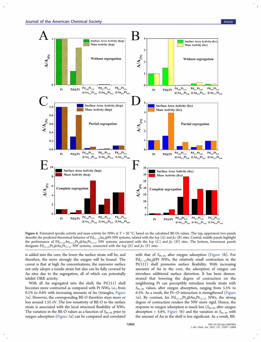
Figure 6. Estimated specific activity and mass activity for NWs at T = 20 °C, based on the calculated BE-Os values. The top, uppermost two panelsdescribe the predicted theoretical behavior of Pd(1−x)Aux@Pt NW systems, related with the hcp (A) and fcc (B) sites. Central, middle panels highlightthe performance of Pd(1−x)Au(x−y)Ptz@AuyPt(1−z) NW systems, associated with the hcp (C) and fcc (D) sites. The bottom, lowermost panelsdesignate Pd(1−x)Ptz@AuxPt(1−z) NW systems, connected with the hcp (E) and fcc (F) sites.
O is more positive and is not sensitive to the amount of Au inthe shell (Figure 5a).Our results indicate that the distribution of Au plays an
important role in tuning the surface strain of AuPd@Pt NWsand therefore of BE-O. In particular, the segregation of Au tothe shell implies two specific consequences: (1) it is able tostabilize the NWs (Figure 3), and (2) it increases the rigidity ofthe NW, which prevents the structural distortion upon oxygenadsorption and thereby weakens the binding energy (Figure 4).The importance of structural rigidity in influencing the
observed oxygen binding energy has been highlighted in ourprevious theoretical analysis of ORR on Pt@Pd nanoparticles,possessing a tetrahedral shape.56 Specifically, we determined inthat prior work that for nanostructures with a relativelysignificant surface contraction, i.e., such as the ∼2.3% noted forthe PdAu@Pt NWs studied herein, the presence of oxygenadsorption can thereby lead to a more significant distortion ofthe localized structure of the (111) facet as compared with thecorresponding (111) bulk surface. That is, nanostructures areintrinsically more flexible in their local structure as comparedwith bulk surfaces. This surface distortion which accommodatesfor the adsorption of oxygen can thereby lead to an increasedbinding energy of oxygen. It is noteworthy that the correlationbetween the metal d-band structure before adsorption and theoxygen binding energy, which is often used to explain the trendin binding energy on metal surfaces, does not necessarily applyfor such nanostructures, since the resulting d-band structure ofdistorted metal sites at the nanoscale limit can be very differentafter oxygen adsorption.Hence, the rigidity of the local structure plays a more
important role. That is, for the same kind of adsorption site, themore flexible the structure is, the more easily the interactedmetal atoms can distort upon adsorption, all of which canthereby lead to a stronger oxygen binding. Indeed, only whenthe NW adopts the conformation of Pd(1−x)Ptz@AuxPt(1−z)does the BE-O of AuPd@Pt NW become weaker than that ofPd@Pt (Figure 5(iii)). Therefore, a higher ORR activity isexpected. However, the degree of weakening is small, andhence, it is difficult to draw any clear and unequivocalconclusions that would correlate exactly with the experimen-tally measured ORR activity values, obtained by simplyexamining the trends in BE-O.21,23,50
Considering the gap between the BE-O and the ORRactivity, we have taken on the additional step of estimating themass and specific ORR activity values, based on the calculatedBE-Os. Considering that the promotion of ORR activity on Ptwas predicted by weakening the Pt−O bond,51 we haveestimated the mass activity as ΔAM ∝ exp(−0.5*BE-O/kBT)*NPt(111)/NPt(total), wherein NPt(total) and NPt(111)represents the number of Pt atoms in the NWs and on the(111) terrace, respectively. The charge transfer coefficient waschosen as “0.5” on the basis of previous experimental andtheoretical studies.69,70 The specific activity was estimated asΔAS ∝ exp(−0.5*BE-O/kBT)*NPt(111)/NPt(shell), whereinNPt(shell) represents the number of Pt atoms in the shell of theNR. The edge and (100) terrace sites, which account for 38%of total Pt surface sites, were assumed to be inactive and notinvolved in the ORR process.Figure 6 plots the variation of relative activity at T = 20 °C,
i.e., mass activity and specific activity, with respect to Pt NWs.We first want to clarify that all the activities reported herecorrespond to a potential of 0.9 V (vs RHE), unless otherwisespecified. We note that both mass and specific activities for pure
Pt NWs are set to 1 in the following Figures. A significantvariation of ORR activity with respect to both the amount anddistribution of Au within the AuPd@Pt NWs was observed. Fora NW with a certain quantity of Au, Pd(1−x)Ptz@AuxPt(1−z)NWs possessing full segregation of Au within the shell (panelsE and F) displayed the largest improvements in both mass andspecific activities with respect to pure Pt NWs among all threetypes of NWs we studied. By comparison, the ORR activities ofPd(1−x)Au(x−y)Ptz@AuyPt(1−z) NWs with partial Au segregation(panels C and D) and of as-prepared Pd(1−x)Aux@Pt NWs withAu localized in the core (panels A and B) are far lower. That is,the small variation in BE-O obtained by alloying Au (Figure 5a)can have a dramatic effect upon the ORR activity.For a certain Au distribution, the observed enhancements of
both mass and specific activities do depend on the amount ofAu. Specifically, the experimentally observed trend in bothtypes of activities in evolving from Pt and Pd@Pt to Au-containing analogues such as Pd9Au@Pt, Pd8Au2@Pt, andPd7Au3@Pt21 can be well captured and described by thecorresponding trend in the theoretically estimated activity onlywhen the Pd(1−x)Ptz@AuxPt(1−z) conformation is adopted andanalyzed. A direct comparison between the “volcano” trendobserved in our theoretical estimations and that found in ourcorresponding experimental data is shown in Figure 7. It isworth emphasizing that both sets of data point to Pd9Au@Pt asthe optimal chemical composition of the cathode for theORR.21 According to our DFT calculations, either the partial or
Figure 7. Comparison between (A) the theoretical estimated surfacearea activity (black) and mass activity (red) for different chemicalcompositions in a range of Pd(1−x)Ptz@AuxPt(1−z) NWs, specifically onthe fcc 3-fold sites, and (B) corresponding experimental results of ORRactivities for a series of as-prepared Pt∼Pd1−xAux nanowires. Datawithin panel B are taken from ref 21.
full segregation of Au is best able to stabilize the as-preparedPd(1−x)Aux@Pt NWs, which is likely to occur during the ORRprocess, whereas the structures of other types of as-preparedNWs may not be able to capture the behavior of the catalystunder operational ORR conditions.Overall, our DFT calculations show that the ORR activity of
AuPd@Pt NWs can be well tuned by purposefully varying theamount and distribution of Au. In effect, the segregation of Aufrom the core to the edge and (100) terrace sites of the shell islikely to occur during the ORR process and can promote theORR activity significantly. We note that the more Au remainsin the core, the lower the ORR activity of the correspondingAuPd@Pt NW. In addition, the amount of Au should bemoderate, i.e., large enough in order to render the NW rigidand therefore weaken the O−Pt interaction, while small enoughto prevent both the decrease of active Pt sites in the shell aswell as the overall confinement of Au to the core.In essence, our theoretical computations have attributed the
possibility of the formation of this hierarchical architecture asemanating from the in situ machinations of the oxygenreduction reaction process itself, thereby resulting in thedynamic surface segregation of certain metallic elements withinnanoparticles consisting of an outer Pt-rich shell coupled with acore comprised of either a single metal or a metal alloy.However, because prior experimental reports have beeneffectively bulk measurements, these have neither specificallyestablished nor determined the validity of this hypothesis.Hence, in order to better understand and verify the plausiblelocalized restructuring within our Pt∼PdAu ultrathin nanowiresystems, we have collected EXAFS data.3.2. Experimentally Probing Core−Shell Nanowires
with STEM, EELS, and EXAFS. We have previously reportedon the synthesis of as-prepared homogeneous Pd9Au ultrathinnanowires, which represent the precursor to the focal core−shell structure of this study.62 According to the structuralcharacterization results obtained from both X-ray diffraction(XRD) and TEM-based selected area electron diffraction(SAED), the measured lattice d-spacings in the as-preparedseries of Pd1−xAux nanowires match perfectly with the predictedvalues calculated based on the overall chemical composition,thereby verifying the existence of a homogeneous alloy-typematerial as opposed to either a core−shell or even a partial alloymotif.To justify the choice of this very particular nanoscale system
for such an elaborate study, we have provided a detailedrationale, based on previous theoretical and experimentalresults. Specifically, according to the “volcano” plot put forthby Nørskov et al., the challenge for optimizing Pt-basedelectrocatalysts for ORR has mainly been to overcome the factthat the adsorption energy of oxygen onto Pt is very strong.51
Hence, in Pt∼Pd core−shell structures in particular, the outerPd atoms not only act as a support but also contribute to thetuning of the electronic properties of the underlying Pt atoms.71
However, the presence of a Pt monolayer on a bare Pdsubstrate may not necessarily represent the optimal structuralmotif. As discussed in the manuscript, the lattice constants ofPt, Pd, and Au are 3.924, 3.890, and 4.080 Å, respectively.Therefore, when deposited onto either bare Pd or Au, the Ptmonolayer may experience either a compressive or a tensilestrain, respectively, thereby potentially altering the observedPt−Pt interatomic distances. Prior reports have demonstratedthat the ideal Pt−Pt distance can be achieved by utilizing as theunderlying “substrate”, Pd−Au alloy nanoparticles possessing a
Pd:Au molar ratio, that is close to 9:1.72 This supposition hasactually been validated previously in our work with theanalogous ultrathin nanowire system.21
Hence, in this paper, we chose our most active ORR sample,namely Pd9Au, as the “foundational” model material with whichto pursue our subsequent studies. Upon deposition of Pt, themorphology and chemical composition of as-prepared carbon-supported Pt∼Pd9Au nanowires were characterized by electronmicroscopy, as shown in Figure 8. Specifically, we noted that as-
generated Pt∼Pd9Au ultrathin nanowires gave rise to aninterconnected network morphology, with average diameters ofindividual wires measuring 2.5 ± 0.3 nm. As a consequence ofthe formation mechanism of the Pd9Au nanowires, there was ajuxtaposition of a small quantity of incompletely grown, shortnanorods73 coupled with a majority of longer nanowirespossessing average lengths of several tens of nanometers.The higher resolution STEM image coupled with the cross-
sectional EELS analysis indicated that the nanowires most likelypossessed a core−shell structure. What is important to note isthat with STEM, the Pt and Au elements could not be readilydifferentiated as a result of their extremely close and potentiallyoverlapping signature bands. However, it is clear from Figure
Figure 8. TEM results of as-synthesized Pt∼Pd9Au ultrathinnanowires. (A) Bright-field imaging, (B) high angle annular darkfield imaging (HAADF), (C) high-resolution STEM imaging, (D)cross-sectional EELS analysis, and (E) representative EDS spectra areseparately shown. The red line in panel C denotes the region wherecross-sectional EELS has been taken.
8D that both of these two elements were richer in quantity anddistribution at the outer, external circumference of the wires ascompared with the region within the inner central core. Theoverall elemental composition derived from EDS analysis wasdetermined to be Pt:Pd:Au = 7:81:12, with small uncertaintiesof roughly 2−3% in terms of the atomic ratio reported. ThePd:Au ratio was very close to the expected ratio of 90:10inherent to a Pd9Au nanowire. Therefore, our Pt∼PdAu samplecould be actually ascribed to a Pt7Pd81Au12 composition.Nevertheless, upon the deposition of that Pt outer shell, what
has been missing to date has been a precise study and inherentunderstanding of the actual spatial distribution of all threeelements, namely Pt, Pd, and Au, within the nominal Pt∼PdAucore−shell motif in order to fully corroborate our computa-tional findings. Indeed, we ourselves have previously shown21
that even though there is a systematic trend in the oxidereduction peaks in cyclic voltammograms (CV) for the series ofPd1−xAux nanowires as a function of varying “x” values, as weproceeded to deposit an outer Pt monolayer, the aforemen-tioned trend in CVs becomes much less defined and couldtherefore not serve as a means of precisely examining surfacestoichiometry. We hypothesize that this is due to the fact thesurface is now almost exclusively covered by Pt atoms whoseelectronic structure is only slightly modified by the underlyingPdAu core giving rise to a subtle if almost imperceptiblealteration in the accompanying CV profiles.21
As a means of overcoming these inherent sensitivitylimitations as discussed in the Introduction, EXAFS is knownas an excellent tool for probing the local atomic environmentwithin both bulk and nanomaterial systems. Therefore, weconducted a series of time-dependent in situ EXAFS experi-ments which enabled us to examine the effect of the ORRreaction on possible restructuring of our core−shell Pt∼PdAunanowires, since we assert that surface catalysis of O2 specieslies at the origin of our observed structural modification.Specifically, we deposited Pt∼Pd9Au nanowires onto con-ductive carbon paper, which was then placed into anelectrochemical cell, which had been specially designed forXAFS experiments. By doing so, we were able to perform theoxygen reduction reaction on our catalysts while obtainingspectra at designated time points of interest, as the reactionsystematically progressed in a range spanning 0 to 1000 cycles.It should be noted at the outset that the EXAFS data support allof our previous complementary spectroscopy and microscopydata indicating the formation of a core−shell motif.In terms of actual EXAFS data, since Pt and Au atoms are
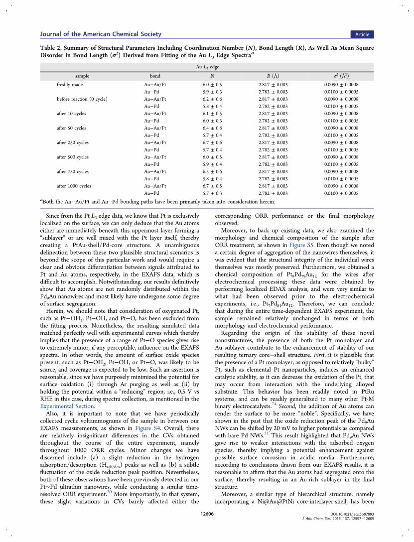
similar in terms of their photoelectron scattering properties, itwas nearly impossible to distinguish between these two types ofatoms as neighbors to either Pt or Au. Hence they areconsidered as equivalent in the fit. The relevant analysis detailsof each individual spectrum can be found in the SupportingInformation section (Figure S1 and S2). A summary of as-obtained structural parameters, including coordination num-bers, bond lengths, and their mean square disorders, isdisplayed in Tables 1 and 2.From these collected data, we should note that the atomic
ratio of Au:Pt can be directly calculated from the edge step oftheir respective regions within the EXAFS spectra. Thecorresponding data are displayed in Figure S3. As a result,the Au:Pt ratio was computed to be 2.5 ± 0.2, whereas theanalogous ratio value derived from EDAX data was 1.7 ± 0.7.We should clarify that both Au and Pt elements constitute aminority composition of the overall core−shell Pt∼Pd9Au
structure (i.e., close to 10%), thereby leading to a relativelylarge error (i.e., 2−3%) associated with the actual experimentalpercentage values themselves, i.e., 7% for Pt and 12% for Au,respectively, for our reported values. Therefore, it is reasonableto assume that the experimental ratios derived from EDAX andEXAFS, respectively, would be significantly different. Nonethe-less, we noted that in fact, the two sets of data arecomparatively close to each other in magnitude, therebyconfirming the validity and reliability of the Pt:Pd:Au = 7:81:12EDAX composition ratio we had noted earlier in the paper.When the Pt L3 edge was analyzed, the coordination number
of the first nearest neighboring Pt−Au/Pt was determined to be9−10, suggesting that the Pt atoms herein are predominantlysurrounded by either Pt or Au atoms. According to thechemical composition determined by EDS analysis, the sum ofPt and Au was less than 20% in terms of atomic ratio. In otherwords, if Pt atoms were to form a random alloy with the twoother elements, it is very likely that this occurred with Pdatoms, since Pd constitutes the majority of the wirecomposition, as opposed to either Pt or Au. Such a conclusionimplies that Pt atoms are essentially exclusively localized on thesurfaces of the nanowires themselves. In effect, the EXAFS andEELS data have collectively suggested that as a result of the Cuunderpotential deposition (UPD) process followed by galvanicreplacement with Pt, the resulting core−shell structure likelypossesses a thin outer Pt shell as opposed to a morerandomized inner Pt-rich alloy core.Regarding the Au L3 edge, the results are far more novel and
intriguing. Specifically, the coordination numbers for both Au−Au/Pt and Au−Pd are quite close to 6, with a reasonably smallerror bar. In essence, this implies that every Au atom issurrounded by 6 Pd atoms and either 6 Pt or Au atoms. Fromthe known actual chemical composition of Pt7 ∼ Pd81Au12, asdetermined from EDS, such a conclusion is inconsistent with apicture in which Au atoms are mixing homogeneously with Pdatoms within the core. The latter model would envision Au tobe almost exclusively surrounded by Pd atoms, because Pt wasfound (vide supra) to be localized on the surface of thenanowires.
Table 1. Summary of Structural Parameters, IncludingCoordination Number (N), Bond Length (R), As Well AsMean Square Disorder in Bond Length (σ2) Derived fromFitting of the Pt L3 Edge Spectraa
Since from the Pt L3 edge data, we know that Pt is exclusivelylocalized on the surface, we can only deduce that the Au atomseither are immediately beneath this uppermost layer forming a“sublayer” or are well mixed with the Pt layer itself, therebycreating a PtAu-shell/Pd-core structure. A unambiguousdelineation between these two plausible structural scenarios isbeyond the scope of this particular work and would require aclear and obvious differentiation between signals attributed toPt and Au atoms, respectively, in the EXAFS data, which isdifficult to accomplish. Notwithstanding, our results definitivelyshow that Au atoms are not randomly distributed within thePd9Au nanowires and most likely have undergone some degreeof surface segregation.Herein, we should note that consideration of oxygenated Pt,
such as Pt−OH2, Pt−OH, and Pt−O, has been excluded fromthe fitting process. Nonetheless, the resulting simulated datamatched perfectly well with experimental curves which therebyimplies that the presence of a range of Pt−O species gives riseto extremely minor, if any perceptible, influence on the EXAFSspectra. In other words, the amount of surface oxide speciespresent, such as Pt−OH2, Pt−OH, or Pt−O, was likely to bescarce, and coverage is expected to be low. Such an assertion isreasonable, since we have purposely minimized the potential forsurface oxidation (i) through Ar purging as well as (ii) byholding the potential within a “reducing” region, i.e., 0.5 V vsRHE in this case, during spectra collection, as mentioned in theExperimental Section.Also, it is important to note that we have periodically
collected cyclic voltammograms of the sample in between ourEXAFS measurements, as shown in Figure S4. Overall, thereare relatively insignificant differences in the CVs obtainedthroughout the course of the entire experiment, namelythroughout 1000 ORR cycles. Minor changes we havediscerned include (a) a slight reduction in the hydrogenadsorption/desorption (Hads/des) peaks as well as (b) a subtlefluctuation of the oxide reduction peak position. Nevertheless,both of these observations have been previously detected in ourPt∼Pd ultrathin nanowires, while conducting a similar time-resolved ORR experiment.20 More importantly, in that system,these slight variations in CVs barely affected either the
corresponding ORR performance or the final morphologyobserved.Moreover, to back up existing data, we also examined the
morphology and chemical composition of the sample afterORR treatment, as shown in Figure S5. Even though we noteda certain degree of aggregation of the nanowires themselves, itwas evident that the structural integrity of the individual wiresthemselves was mostly preserved. Furthermore, we obtained achemical composition of Pt9Pd79Au12 for the wires afterelectrochemical processing; these data were obtained byperforming localized EDAX analysis, and were very similar towhat had been observed prior to the electrochemicalexperiments, i.e., Pt7Pd81Au12. Therefore, we can concludethat during the entire time-dependent EXAFS experiment, thesample remained relatively unchanged in terms of bothmorphology and electrochemical performance.Regarding the origin of the stability of these novel
nanostructures, the presence of both the Pt monolayer andAu sublayer contribute to the enhancement of stability of ourresulting ternary core−shell structure. First, it is plausible thatthe presence of a Pt monolayer, as opposed to relatively “bulky”Pt, such as elemental Pt nanoparticles, induces an enhancedcatalytic stability, as it can decrease the oxidation of the Pt, thatmay occur from interaction with the underlying alloyedsubstrate. This behavior has been readily noted in PtRusystems, and can be readily generalized to many other Pt-Mbinary electrocatalysts.74 Second, the addition of Au atoms canrender the surface to be more “noble”. Specifically, we haveshown in the past that the oxide reduction peak of the Pd9AuNWs can be shifted by 20 mV to higher potentials as comparedwith bare Pd NWs.21 This result highlighted that Pd9Au NWsgave rise to weaker interactions with the adsorbed oxygenspecies, thereby implying a potential enhancement againstpossible surface corrosion in acidic media. Furthermore,according to conclusions drawn from our EXAFS results, it isreasonable to affirm that the Au atoms had segregated onto thesurface, thereby resulting in an Au-rich sublayer in the finalstructure.Moreover, a similar type of hierarchical structure, namely
incorporating a Ni@Au@PtNi core-interlayer-shell, has been
Table 2. Summary of Structural Parameters Including Coordination Number (N), Bond Length (R), As Well As Mean SquareDisorder in Bond Length (σ2) Derived from Fitting of the Au L3 Edge Spectraa
designed by Kang et al., wherein the presence of an Auinterlayer was found to have effectively inhibited surface oxideformation, thereby improving the overall long-term durability.75
These observations and examples support our motivation forincorporating Au into our nanowire system. To further confirmand corroborate the observed surface segregation within ourhierarchical nanowires, we also conducted EXAFS experimentson so-called “freshly-made” Pt∼Pd9Au samples, prepared as apowder as opposed to being deposited on carbon paper andimmersed into electrolyte. According to Tables 1 and 2, thesesamples exhibited identical local structure and behavior to thesample that had undergone electrochemical testing. Suchconsistency in our data implies that the Au surface segregationoriginated from the intrinsic synthesis and treatment processprior to the exposure of the sample to electrochemicalprocessing, as opposed to the very ORR reaction itself. Inother words, by a process of elimination, we propose that themost probable initiator, i.e., instigating event, of the observedsurface segregation can be ascribed to Cu underpotentialdeposition followed by galvanic replacement with Pt.To justify the validity of our conclusions, it is worth noting
that similar types of behavior have been reported in cases wherethe presence of different absorbates can result in surfacesegregation in metal alloy systems. Specifically, Pt atoms inPt3Co nanoparticles can undergo surface segregation during aCO annealing process at 200°C, thereby leading to theformation of a Pt-shell, PtCo-core structure.76 Moreover, high-temperature is not a necessary prerequisite for such surfacerestructuring to occur. For instance, Volker et al. have observedroom-temperature O2-induced Cu surface segregation inpolycrystalline bulk Cu−Au alloys using time-resolved XPSexperiments and rationalized these observations based on DFTcalculations.77 Similarly, Jirkovsky et al. discovered a reversible,external potential-dependent exchange process involving Pdmigrating between the core and the shell, within Pd−Au alloy(Pd-rich) nanoparticles.78 In other words, the chemicalcompositions of the surfaces of these Pd−Au nanoparticlesappeared to depend on the magnitude of the applied potential.For example, at low potentials (i.e., below 0.8 V vs RHE), thesurface tended to be Au-rich, whereas at correspondingly higherpotentials (i.e., higher than 1.0 V vs RHE), the surface wasfound to be Pd-rich.This latter paper serves as corroboration of our hypothesis,
since our entire Cu UPD process described herein wasconducted under a potential range of 0.5−0.8 V (vs RHE).Hence it is reasonable of us to propose Au segregation in theouter shell. Furthermore, it is worth noting that the relevantoperating ORR potential is merely within the 0.6−1.0 V range,which might not be sufficiently high enough to initiate thegeneration of an alternative scenario, i.e., a Pd-rich surface.
4. CONCLUSIONSIn this work, we have probed the local structure of ourPt∼Pd9Au ultrathin nanowires by a holistic approach consistingof a unique combination of both theoretical calculations as wellas X-ray Absorption Fine Structure Spectroscopy (XAFS).Specifically, DFT calculations of the binding energy (BE-O)values of oxygen species coupled with the correspondingcalculated ORR activities have allowed us to revisit our assumed“Pt-shell, PdAu random-alloy-core” model we had proposed inour previous work as a definitive description of Pt∼PdxAu1−x.In particular, the time-dependent EXAFS data on our Pt−Pd9Au nanowires have confirmed that (a) Au atoms will likely
undergo surface segregation and that (b) such segregationprocess likely happens during the synthesis of the core−shellstructure itself, i.e., in the midst of the Cu UPD processfollowed by galvanic replacement with Pt, implying that the“restructuring” occurs primarily upon polarization.By contrast with many other studies which have utilized DFT
calculations and/or XAFS techniques to probe structuralparameters, we have used our DFT results herein as a guidetoward thoughtfully designing EXAFS experiments. In otherwords, theoretical predictions have directed our experiments aswell as simplified the resulting analysis by providing forplausible possible structural models with which to differentiate,discriminate, and ultimately verify using EXAFS. Such asynergetic, feedback-based effort will likely be important andrelevant for future applications of XAFS.Meanwhile, the discovery of structural alterations ascribed to
the idiosyncrasies of specific synthesis approaches may lead tonew perspectives in terms of understanding the structural basisfor electrocatalysis, especially with a host of different andcomplementary nanoscale Pt-based hierarchical nanomaterials.To our knowledge, this is the first time that Au segregation hasbeen reported as a result of a seemingly unrelated Cu UPD/Ptgalvanic displacement process. In the future, it will be worthfurther differentiating between the specific significance ofchemical composition versus that of the ultrathin size in termsof explaining the nature of the phenomena observed.In summary, the novelty and importance of this report arises
from combining disparate, complementary, but nonethelessmutually supportive results from (a) in situ spectroscopy, (b)DFT calculations, and (c) electrochemical data into acomprehensive and substantive analysis for understanding thebehavior of functionally relevant nanostructures. Very fewanalogous reports exist.37,79,80 From a broader perspective, wewish to emphasize the value and wisdom of this approach forgaining key insights into nanoscale structure−propertycorrelations. In particular, the precise examination of localstructure and bonding can facilitate a proper understanding ofexisting nanomaterials and hence an ability to rationally designtruly distinctive classes of nanostructures. Hence, the strategyemployed herein can be readily generalized to other classes ofmaterials as well to the study of phenomena that havesignificance beyond ORR and/or electrochemistry.
■ ASSOCIATED CONTENT*S Supporting InformationThe Supporting Information is available free of charge on theACS Publications website at DOI: 10.1021/jacs.5b07093.
Additional structural characterization and electrochem-ical data of our samples are presented. (PDF)
■ ACKNOWLEDGMENTSFunds for research work (including support for HQ and SSW)at Brookhaven National Laboratory (BNL) were provided bythe U.S. Department of Energy, Office of Basic EnergySciences, Materials Sciences and Engineering Division underContract No. DE-AC02-98CH10886 and DE-SC-00112704.
AIF acknowledges support from the Department of EnergyGrant No. DE-FG02-03ER15476. Computational research(including support for WA and PL) was carried out atBrookhaven National Laboratory under contract DE-SC0012704 with the US Department of Energy, Division ofChemical Sciences. DFT calculations were performed usingcomputational resources at the Center for Functional Nano-materials, a user facility funded at Brookhaven NationalLaboratory under Contract No. DE-AC02-98CH10886, andat the National Energy Research Scientific Computing Center(NERSC), the latter of which is supported by the Office ofScience of the U.S. DOE under Contract No. DE-AC02-05CH11231. Beamlines X18B and X19A at the NSLS weresupported in part by the Synchrotron Catalysis Consortium,U.S. Department of Energy Grant No. DE-FG02-05ER15688.High-resolution electron microscopy data in this manuscriptwere collected in part at BNL’s Center for FunctionalNanomaterials, which is also supported by the U.S. Departmentof Energy under Contract No. DE-AC02-98CH10886 and DE-SC-00112704. RMC and RMA gratefully acknowledge supportfrom the Chemical Sciences, Geosciences, and BiosciencesDivision, Office of Basic Energy Sciences, Office of Science,U.S. Department of Energy (Contract No. DE-FG02-13ER16428). Finally, we thank Dr. Nebojsa Marinkovic(Synchrotron Catalysis Consortium at BNL) for helpfuldiscussions and advice.
■ REFERENCES(1) Morozan, A.; Jousselme, B.; Palacin, S. Energy Environ. Sci. 2011,4, 1238.(2) Chen, Z. W.; Higgins, D.; Yu, A. P.; Zhang, L.; Zhang, J. J. EnergyEnviron. Sci. 2011, 4, 3167.(3) Stephens, I. E. L.; Bondarenko, A. S.; Gronbjerg, U.; Rossmeisl,J.; Chorkendorff, I. Energy Environ. Sci. 2012, 5, 6744.(4) Nilekar, A. U.; Xu, Y.; Zhang, J. L.; Vukmirovic, M. B.; Sasaki, K.;Adzic, R. R.; Mavrikakis, M. Top. Catal. 2007, 46, 276.(5) Bing, Y. H.; Liu, H. S.; Zhang, L.; Ghosh, D.; Zhang, J. J. Chem.Soc. Rev. 2010, 39, 2184.(6) Mazumder, V.; Lee, Y.; Sun, S. H. Adv. Funct. Mater. 2010, 20,1224.(7) Zhang, H.; Jin, M. S.; Xia, Y. N. Chem. Soc. Rev. 2012, 41, 8035.(8) Wang, C.; Markovic, N. M.; Stamenkovic, V. R. ACS Catal. 2012,2, 891.(9) Oezaslan, M.; Hasche, F.; Strasser, P. J. Phys. Chem. Lett. 2013, 4,3273.(10) Guo, S. J.; Zhang, S.; Sun, S. H. Angew. Chem., Int. Ed. 2013, 52,8526.(11) Wu, J. B.; Yang, H. Acc. Chem. Res. 2013, 46, 1848.(12) Xu, Y.; Zhang, B. Chem. Soc. Rev. 2014, 43, 2439.(13) Xia, Y.; Yang, P.; Sun, Y.; Wu, Y.; Mayers, B.; Gates, G.; Yin, Y.;Kim, F.; Yan, H. Adv. Mater. 2003, 15, 353.(14) Weber, J.; Singhal, R.; Zekri, S.; Kumar, A. Int. Mater. Rev. 2008,53, 235.(15) Wang, N.; Cai, Y.; Zhang, R. Q. Mater. Sci. Eng., R 2008, 60, 1.(16) Cademartiri, L.; Ozin, G. A. Adv. Mater. 2009, 21, 1013.(17) Lim, B.; Jiang, M.; Camargo, P. H. C.; Cho, E. C.; Tao, J.; Lu,X.; Zhu, Y.; Xia, Y. Science 2009, 324, 1302.(18) Kitchin, J. R.; Nørskov, J. K.; Barteau, M. A.; Chen, J. G. Phys.Rev. Lett. 2004, DOI: 10.1103/PhysRevLett.93.156801.(19) Koenigsmann, C.; Zhou, W. P.; Adzic, R. R.; Sutter, E.; Wong, S.S. Nano Lett. 2010, 10, 2806.(20) Koenigsmann, C.; Santulli, A. C.; Gong, K. P.; Vukmirovic, M.B.; Zhou, W. P.; Sutter, E.; Wong, S. S.; Adzic, R. R. J. Am. Chem. Soc.2011, 133, 9783.(21) Koenigsmann, C.; Sutter, E.; Adzic, R. R.; Wong, S. S. J. Phys.Chem. C 2012, 116, 15297.
(22) Koenigsmann, C.; Tan, Z.; Peng, H.; Sutter, E.; Jacobskind, J.;Wong, S. S. Isr. J. Chem. 2012, 52, 1090.(23) Koenigsmann, C.; Scofield, M. E.; Liu, H. Q.; Wong, S. S. J.Phys. Chem. Lett. 2012, 3, 3385.(24) Strasser, P.; Koh, S.; Anniyev, T.; Greeley, J.; More, K.; Yu, C.F.; Liu, Z. C.; Kaya, S.; Nordlund, D.; Ogasawara, H.; Toney, M. F.;Nilsson, A. Nat. Chem. 2010, 2, 454.(25) Matanovic, I.; Kent, P. R. C.; Garzon, F. H.; Henson, N. J. J.Electrochem. Soc. 2013, 160, F548.(26) Russell, A. E.; Tessier, B. C.; Wise, A. M.; Rose, A.; Price, S. W.T.; Richardson, P. W.; Ball, S. C.; Theobald, B.; Thompsett, D.; Crabb,E. M. ECS Trans. 2011, 41, 55.(27) Rettew, R. E.; Allam, N. K.; Alamgir, F. M. ACS Appl. Mater.Interfaces 2011, 3, 147.(28) Cheon, J. Y.; Kim, T.; Choi, Y.; Jeong, H. Y.; Kim, M. G.; Sa, Y.J.; Kim, J.; Lee, Z.; Yang, T. H.; Kwon, K.; Terasaki, O.; Park, G. G.;Adzic, R. R.; Joo, S. H. Sci. Rep. 2013, 3, 2715.(29) Gao, J.; Zhong, J.; Bai, L. L.; Liu, J. Y.; Zhao, G. Q.; Sun, X. H.Sci. Rep. 2014, 4, 3606.(30) Nashner, M. S.; Frenkel, A. I.; Adler, D. L.; Shapley, J. R.;Nuzzo, R. G. J. Am. Chem. Soc. 1997, 119, 7760.(31) Frenkel, A. I. J. Synchrotron Radiat. 1999, 6, 293.(32) Frenkel, A. I.; Hills, C. W.; Nuzzo, R. G. J. Phys. Chem. B 2001,105, 12689.(33) Alayoglu, S.; Zavalij, P.; Eichhorn, B.; Wang, Q.; Frenkel, A. I.;Chupas, P. ACS Nano 2009, 3, 3127.(34) Frenkel, A. I. Chem. Soc. Rev. 2012, 41, 8163.(35) Jung, U.; Elsen, A.; Li, Y.; Smith, J. G.; Small, M. W.; Stach, E.A.; Frenkel, A. I.; Nuzzo, R. G. ACS Catal. 2015, 5, 1539.(36) Dutta, I.; Carpenter, M. K.; Balogh, M. P.; Ziegelbauer, J. M.;Moylan, T. E.; Atwan, M. H.; Irish, N. P. J. Phys. Chem. C 2010, 114,16309.(37) Sasaki, K.; Naohara, H.; Choi, Y. M.; Cai, Y.; Chen, W. F.; Liu,P.; Adzic, R. R. Nat. Commun. 2012, 3, 1115.(38) Kuttiyiel, K. A.; Sasaki, K.; Su, D.; Vukmirovic, M. B.;Marinkovic, N. S.; Adzic, R. R. Electrochim. Acta 2013, 110, 267.(39) Anderson, R. M.; Zhang, L.; Loussaert, J. A.; Frenkel, A. I.;Henkelman, G.; Crooks, R. M. ACS Nano 2013, 7, 9345.(40) Nashner, M. S.; Frenkel, A. I.; Somerville, D.; Hills, C. W.;Shapley, J. R.; Nuzzo, R. G. J. Am. Chem. Soc. 1998, 120, 8093.(41) Knecht, M. R.; Weir, M. G.; Frenkel, A. I.; Crooks, R. M. Chem.Mater. 2007, 20, 1019.(42) Shu, Y. Y.; Murillo, L. E.; Bosco, J. P.; Huang, W.; Frenkel, A. I.;Chen, J. G. Appl. Catal., A 2008, 339, 169.(43) Teng, X. W.; Han, W. Q.; Wang, Q.; Li, L.; Frenkel, A. I.; Yang,J. C. J. Phys. Chem. C 2008, 112, 14696.(44) Teng, X. W.; Wang, Q.; Liu, P.; Han, W.; Frenkel, A.; Wen, W.;Marinkovic, N.; Hanson, J. C.; Rodriguez, J. A. J. Am. Chem. Soc. 2008,130, 1093.(45) Kresse, G.; Furthmuller, J. Phys. Rev. B: Condens. Matter Mater.Phys. 1996, 54, 11169.(46) Kresse, G.; Hafner, J. Phys. Rev. B: Condens. Matter Mater. Phys.1993, 47, 558.(47) Perdew, J. P.; Chevary, J. A.; Vosko, S. H.; Jackson, K. A.;Pederson, M. R.; Singh, D. J.; Fiolhais, C. Phys. Rev. B: Condens. MatterMater. Phys. 1992, 46, 6671.(48) Blochl, P. E. Phys. Rev. B: Condens. Matter Mater. Phys. 1994, 50,17953.(49) Monkhorst, H. J.; Pack, J. D. Phys. Rev. B 1976, 13, 5188.(50) Koenigsmann, C.; Sutter, E.; Chiesa, T. A.; Adzic, R. R.; Wong,S. S. Nano Lett. 2012, 12, 2013.(51) Nørskov, J. K.; Rossmeisl, J.; Logadottir, A.; Lindqvist, L.;Kitchin, J. R.; Bligaard, T.; Jonsson, H. J. Phys. Chem. B 2004, 108,17886.(52) Stamenkovic, V.; Mun, B. S.; Mayrhofer, K. J. J.; Ross, P. N.;Markovic, N. M.; Rossmeisl, J.; Greeley, J.; Nørskov, J. K. Angew.Chem., Int. Ed. 2006, 45, 2897.(53) Shao, M.; Liu, P.; Adzic, R. R. J. Phys. Chem. B 2007, 111, 6772.
(54) Wang, J. X.; Inada, H.; Wu, L. J.; Zhu, Y. M.; Choi, Y. M.; Liu,P.; Zhou, W. P.; Adzic, R. R. J. Am. Chem. Soc. 2009, 131, 17298.(55) Zhang, J.; Yang, H. Z.; Fang, J. Y.; Zou, S. Z. Nano Lett. 2010,10, 638.(56) An, W.; Liu, P. J. Phys. Chem. C 2013, 117, 16144.(57) Weir, M. G.; Myers, V. S.; Frenkel, A. I.; Crooks, R. M.ChemPhysChem 2010, 11, 2942.(58) Dowben, P.; Miller, A.; Vook, R. Gold Bull. 1987, 20, 54.(59) Deng, L.; Hu, W. Y.; Deng, H. Q.; Xiao, S. F.; Tang, J. F. J. Phys.Chem. C 2011, 115, 11355.(60) Zhang, Y.; Hsieh, Y. C.; Volkov, V.; Su, D.; An, W.; Si, R.; Zhu,Y. M.; Liu, P.; Wang, J. X.; Adzic, R. R. ACS Catal. 2014, 4, 738.(61) Carino, E. V.; Kim, H. Y.; Henkelman, G.; Crooks, R. M. J. Am.Chem. Soc. 2012, 134, 4153.(62) Ramirez-Caballero, G. E.; Ma, Y. G.; Callejas-Tovar, R.;Balbuena, P. B. Phys. Chem. Chem. Phys. 2010, 12, 2209.(63) Herron, J. A.; Mavrikakis, M. Catal. Commun. 2014, 52, 65.(64) Hammer, B.; Nørskov, J. K. Nature 1995, 376, 238.(65) Rodriguez, P.; Garcia-Araez, N.; Koper, M. T. M. Phys. Chem.Chem. Phys. 2010, 12, 9373.(66) Ruban, A. V.; Skriver, H. L. Comput. Mater. Sci. 1999, 15, 119.(67) Ruban, A. V.; Skriver, H. L.; Nørskov, J. K. Phys. Rev. B: Condens.Matter Mater. Phys. 1999, 59, 15990.(68) Mavrikakis, M.; Hammer, B.; Nørskov, J. K. Phys. Rev. Lett.1998, 81, 2819.(69) Tripkovic, V.; Skulason, E.; Siahrostami, S.; Nørskov, J. K.;Rossmeisl, J. Electrochim. Acta 2010, 55, 7975.(70) Grgur, B. N.; Markovic, N. M.; Ross, P. N. Can. J. Chem. 1997,75, 1465.(71) Antolini, E. Energy Environ. Sci. 2009, 2, 915.(72) Xing, Y. C.; Cai, Y.; Vukmirovic, M. B.; Zhou, W. P.; Karan, H.;Wang, J. X.; Adzic, R. R. J. Phys. Chem. Lett. 2010, 1, 3238.(73) Greeley, J.; Nørskov, J. K. Surf. Sci. 2005, 592, 104.(74) Adzic, R. R.; Zhang, J.; Sasaki, K.; Vukmirovic, M. B.; Shao, M.;Wang, J. X.; Nilekar, A. U.; Mavrikakis, M.; Valerio, J. A.; Uribe, F.Top. Catal. 2007, 46, 249.(75) Kang, Y. J.; Snyder, J.; Chi, M. F.; Li, D. G.; More, K. L.;Markovic, N. M.; Stamenkovic, V. R. Nano Lett. 2014, 14, 6361.(76) Mayrhofer, K. J. J.; Juhart, V.; Hartl, K.; Hanzlik, M.; Arenz, M.Angew. Chem., Int. Ed. 2009, 48, 3529.(77) Volker, E.; Williams, F. J.; Calvo, E. J.; Jacob, T.; Schiffrin, D. J.Phys. Chem. Chem. Phys. 2012, 14, 7448.(78) Jirkovsky, J. S.; Panas, I.; Romani, S.; Ahlberg, E.; Schiffrin, D. J.J. Phys. Chem. Lett. 2012, 3, 315.(79) Kwon, G.; Ferguson, G. A.; Heard, C. J.; Tyo, E. C.; Yin, C. R.;DeBartolo, J.; Seifert, S.; Winans, R. E.; Kropf, A. J.; Greeley, J.;Johnston, R. L.; Curtiss, L. A.; Pellin, M. J.; Vajda, S. ACS Nano 2013,7, 5808.(80) Shao, M. H.; Huang, T.; Liu, P.; Zhang, J.; Sasaki, K.;Vukmirovic, M. B.; Adzic, R. R. Langmuir 2006, 22, 10409.


![119 Nanowires 4. Nanowires - UFAMhome.ufam.edu.br/berti/nanomateriais/Nanowires.pdf · 119 Nanowires 4. Nanowires ... written about carbon nanotubes [4.57–59], which can be ...](https://static.documents.pub/doc/80x56/5abfd11e7f8b9a5d718eba2b/119-nanowires-4-nanowires-nanowires-4-nanowires-written-about-carbon-nanotubes.jpg)


