Materials Science and Engineering A 456 (2007) 350–357
Influence of heat treatment on degradation behavior of bio-degradabledie-cast AZ63 magnesium alloy in simulated body fluid
Chenglong Liu a, Yunchang Xin a,b, Guoyi Tang b, Paul K. Chu a,∗a Department of Physics and Materials Science, City University of Hong Kong, Tat Chee Avenue, Kowloon, Hong Kong
b Tsinghua University, Shenzhen Graduate School, Shenzhen 518055, China
Received 4 September 2006; received in revised form 25 November 2006; accepted 7 December 2006
Abstract
The effects of hybrid aging and solution treatments on the degradation of bio-degradable die-cast AZ63 magnesium alloy in 37 ± 1 ◦C Tyrode’ssimulated body fluid have been investigated. The heat treatment is observed to alter the microstructure of the alloy. The amount of �-Mg17Al2
Traditional biomedical metal materials such as stainless steelsand Ti and Ti alloys have played an essential role in load-bearing implants for the repair or replacement of diseased ordamaged bone tissues. Durable implants represent a foreignbody and bear the risk of local inflammation. Furthermore, thestress-shielding effect may obstruct the stabilization of the bonetissues that need mechanical loads to obtain and maintain theirrigidity [1]. Degradable biocompatible implants, which dissolvein a biological environment, constitute an appropriate solu-tion. Unfortunately, currently used absorbable materials suchas poly-lactides have unsatisfactory mechanical properties. Incomparison, magnesium-based alloys possess a more attractivecombination of desirable properties including low density, goodelastic modulus, and compressive yield strength closer to thoseof natural bones, in addition to their high strength to weightratio [2]. Magnesium also has good biocompatibility and isthus a potential candidate in bone implants. However, the major
drawback of magnesium alloys is that they tend to corrode veryquickly in the physiological pH (7.4–7.6) environment therebylosing their mechanical integrity before the tissues have suffi-cient time to heal. Furthermore, rapid production of hydrogengas during the corrosion process may not be tolerated by thehost tissues [3]. Nevertheless, the use of magnesium alloys asbio-degradable implants is still promising, but their corrosionresistance must be enhanced [4,5].
New casting techniques can produce magnesium alloys witha minimal amount of impurities and help to control in vivocorrosion. However, the corrosion process does not dependon the elemental composition and processing history only [4].Effective means to mitigate corrosion include the applicationof a surface coating in order to separate the metal surfacefrom its surroundings or incorporation of corrosion inhibitingchemicals. Different surface modification techniques such aselectrochemical plating, conversion coatings, anodization, gas-phase deposition, laser surface alloying/cladding, and organiccoatings have been proposed for this purpose [6]. To be effective,the coating must be uniform, sticking, pore-free, and self-healing. Unfortunately, magnesium alloys have high chemicalreactivity, and once they come in contact with air or water, anoxide/hydroxide layer forms readily on the surface which can
C. Liu et al. / Materials Science and Engineering A 456 (2007) 350–357 351
Table 1Heat treatment parameters
#1 #2 #3 #4
Solution treatment Untreated 413 ◦C, 24 h 413 ◦C, 24 h 413 ◦C, 24 hAged treatment Untreated 216 ◦C, 1 h 216 ◦C, 5.5 h 216 ◦C, 12 h
have a detrimental effect on adhesion and uniformity. Thereis a close relationship between the coating and magnesiumalloy substrate. In fact, chemical heterogeneity caused by crys-tallization, precipitation, and segregation during the castingprocess can have a negative impact on the surface corrosionresistance.
Heat treatments can cause micro-structural changes andredistribution of metal elements. A homogeneous structure canbe obtained by a hybrid solution and aging process [7]. Wanget al. have studied the changes in the microstructures of AZ91Dmagnesium alloy during T4 or T6 treatments [7]. A solutiontreatment at 413 ◦C causes micro-structural evolution involv-ing four processes: dissolution of the � phase, formation of afine-grain structure, morphological change of the globules intoa traditional grain shape, and grain coarsening. During agingat 216 ◦C, discrete precipitates are preferentially initiated atsome of the grain boundaries and then interconnected precip-itates emerge inside the grains with accelerated age-hardeningkinetics. These micro-structural changes can affect selectedproperties of the magnesium alloys. Zhang et al. have foundthat a solution treatment decreases the amount of second-phaseparticles inside the grains and weakens strong pinning on dis-locations. The T4 treatment (solution) improves the ductilitysubstantially but reduces strength, whereas the T6 (solutionplus aging) process increases the ultimate tensile strength butcompromises the yield strength and ductility [8].
It has been reported that the secondary phase has an effecton the corrosion resistance of aluminum-containing magnesiumalloys [9–12]. The � phase plays dual roles that depend on theamount and distribution of this phase. A fine and homogeneousphase appears to be a better anti-corrosion barrier. Otherwise, thepresence of the � phase in the alloys could deteriorate the corro-sion performance as it can act as an effective galvanic cathode.The beneficial effect of the � phase on the corrosion proper-ties may be enhanced by grain refinement. Song et al. [11] havepointed out that moderate temperature (160 ◦C) aging of die-cast AZ91D changes the corrosion resistance in 5 wt.%NaClsaturated with Mg(OH)2. Aging at temperatures up to 230 ◦Cfor 36 h has only a slight effect on the corrosion resistance, buta temperature above 230 ◦C significantly reduces the corrosionresistance of die-cast AZ91 under salt spraying [13]. The influ-ence of the hybrid solution and aging treatment on the corrosionresistance of biomedical Mg alloys in a simulated body environ-ment has hitherto been seldom probed. From a practical point ofview, it is important to know the effects of the suitable heat treat-ment on the degradation rate in an in vitro environment. Betterunderstanding of the process and mechanism can improve thedegradation rate of magnesium alloys in a biological mediumthereby expediting the acceptance and use of the bio-degradablematerials in biomedical implants.
2. Experimental details
The composition of the die-cast AZ63 alloy used in thisstudy is as follows: Mg–5.9 wt.%Al–3.5 wt.%Zn–0.18 wt.%Mn–0.04 wt.%Si–0.004 wt.%Fe–0.003 wt.%Ni. The materialsthat were purchased from YiHo Corporation, Shenzhen, Chinawere produced by die-casting. Circular disks 2 mm thick werecut from the alloy rod with a diameter of 10 mm. Samples withdimensions of 10 mm × 10 mm × 2 mm were used in the in vitrodegradation tests.
2.1. Heat treatment
The AZ63 samples were subjected to heat treatmentsdescribed in Table 1 [7]. The solution treatment was carried outat 413 ◦C for 24 h (T4) in air with the sample surface protectedby carbon powders. The samples were cooled in air and thenaged for different periods of time. The micro-structural evolu-tion during heat treatment was examined by an Olympus opticalmicroscope (OM). The specimens for OM were prepared bya standard technique of grinding with SiC waterproof abrasivepaper, polishing with an Al3O2 suspension solution, and etchingin a 2% nital solution.
2.2. In vitro corrosion measurements
All the corrosion tests were carried out at 37 ± 1 ◦C inTyrode’s simulated body fluid (SBF) with the following com-position (mmol/l)—NaCl: 100.0, KCl: 10.0, KH2PO4: 1.2,MgSO4: 5.0, glucose: 20.0, taurine: 10.0 and MOPS: 10.0. Thesolution was maintained at the normal physiological pH at 7.2.A 0.9 wt.% physiological saline was also used.
2.2.1. In situ corrosion measurementThe specimens were etched in a 2% nital solution initially to
unravel their microstructures. They were then immersed hori-zontally about 1 mm below the surface of the test solution. Anoptical microscope was mounted above the solution to moni-tor the corrosion process with time. Afterwards, the specimenswere quickly washed with alcohol, dried, and examined underan optical microscope again.
2.2.2. Immersion testThe immersion test was carried out at 37 ± 1 ◦C. Each sample
was ground by #2400 waterproof abrasive paper and then ultra-sonically cleaned in alcohol for 5 min before testing. The Mgion concentration of each sample after different immersion timewas determined thrice using a SHIMADZU atomic absorptionflame emission spectrophotometer (AA6501S) at an excitationwavelength of 285.2 nm.
352 C. Liu et al. / Materials Science and Engineering A 456 (2007) 350–357
Fig. 1. Typical optical microstructure of the die-cast AZ63 alloy solution at 413 ◦C for 24 h followed by another aging step at 216 ◦C for different periods of time:(a) as-received sample, (b) 1 h, (c) 5.5 h, and (d) 12 h.
2.2.3. Corrosion rate measurementAfter the immersion tests, the corroded specimens were
removed from the solution, cleaned with distilled water,and dried. They were then immersed in chromate acid(200 g/l CrO3 + 10 g/l AgNO3) for 5–10 min to remove thecorrosion products. Afterwards, the specimens were washedwith distilled water and dried again. The dried specimenswere weighed and the corrosion rate was calculated asfollows:
CR = W
At, (1)
where CR is the corrosion rate, W the weight loss, A the originalsurface area exposed to the test solution, and t is the exposuretime [5].
2.2.4. Free corrosion potential evaluationThe free corrosion potential was measured using an M273
EG&G potentiostat. The experimental setup consisted of a con-ventional three-electrode cell comprising a working electrode,saturated calomel electrode (SCE), and pure carbon stick as thecounter electrode. The specimens were prepared by connectinga copper plate to one side of the sample covered by cold set-ting resin. The opposite surface of the specimen was exposed tothe solution. The exposed area was about 1 cm2. The changesin the free corrosion potential (Ecorr) were monitored as a func-tion of time under open circuit conditions for approximately4000 s.
3. Results
The typical optical micrographs of the as-received (sample1) and heat-treated (samples 2–4) AZ63 alloys are depicted inFig. 1. The die-cast AZ63 Mg alloy exhibits a microstructurewith the primary � phase grains and �-Mg17Al12 phase dis-persed heterogeneously in the matrix. Fig. 1a shows the partialcontinuous network of the � phase. After 1 h aging, more homo-geneous microstructures are formed and the � phase dissolvesin the � phase as shown in Fig. 1b. With increasing aging time(Fig. 1c), the � phase precipitates from the super-saturated �-Mg solid solution [7]. Discontinuous precipitation of the � phaseis typically initiated preferentially at some of the grain bound-aries, as indicated by coarse lamellar growth into the grains.After 12 h aging, the microstructure is observed to be differ-ent from that aged for 5.5 h. Both the amount and continuity ofthe � phase increase. Fig. 1d shows the continuous precipita-tion lamellae from the grain boundary into the grain and finecontinuous precipitation lamellae form inside the �-Mg grains[7].
The micro-corrosion morphologies of AZ63 in Tyrode’s sim-ulated body fluid as a function of immersion time are shown inFigs. 2 and 3. Based on the optical micrograph acquired fromsample 1 (untreated), intensive hydrogen evolution takes placemainly at the center of the � phase grain immediately after thesample is dipped in the test solution. In contrary, few hydro-gen bubbles form on the aged sample surfaces. After 10 minexposure, heavy localized corrosion attack appears on sample 1whereas only few localized corrosion holes can be seen on the
C. Liu et al. / Materials Science and Engineering A 456 (2007) 350–357 353
Fig. 2. Optical micrographs of the AZ63 surfaces after 10 min exposure in Tyrode’s simulated body fluid for samples aged for different periods of time: (a) sample1 (untreated), (b) 1 h, (c) 5.5 h, and (d) 12 h (corrosion holes indicated by arrows).
surfaces of the aged samples. Compared to samples 2 and 3, sam-ple 4 suffers the slightest corrosion attack. The � phase grains arecorroded but this does not affect the � phase. A similar behaviorhas been reported for AZ91 alloy [11]. With increasing immer-sion time, the developing rate and amount of hydrogen bubbles
increase rapidly for sample 1 while hardly any changes can beobserved on the aged samples. After 12 h immersion, sample1 is badly corroded as manifested by abundant clusters formedby localized corrosion (Fig. 3a). Compared to the surface mor-phology after 10 min, the number of localized corrosion holes
Fig. 3. Optical micrographs of the AZ63 surfaces after 12 h exposure in Tyrode’s simulated body fluid for samples aged for different periods of time: (a) sample 1(untreated), (b) 1 h, (c) 5.5 h, and (d) 12 h (corrosion holes indicated by arrows).
354 C. Liu et al. / Materials Science and Engineering A 456 (2007) 350–357
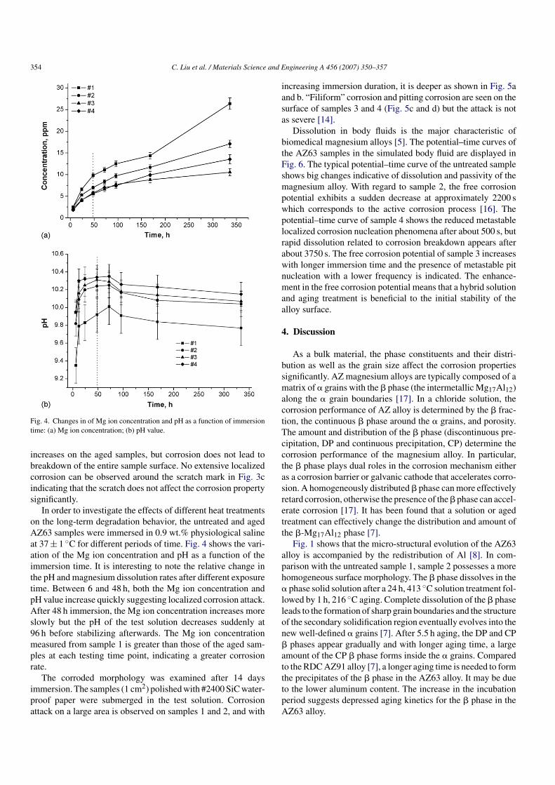
Fig. 4. Changes in of Mg ion concentration and pH as a function of immersiontime: (a) Mg ion concentration; (b) pH value.
increases on the aged samples, but corrosion does not lead tobreakdown of the entire sample surface. No extensive localizedcorrosion can be observed around the scratch mark in Fig. 3cindicating that the scratch does not affect the corrosion propertysignificantly.
In order to investigate the effects of different heat treatmentson the long-term degradation behavior, the untreated and agedAZ63 samples were immersed in 0.9 wt.% physiological salineat 37 ± 1 ◦C for different periods of time. Fig. 4 shows the vari-ation of the Mg ion concentration and pH as a function of theimmersion time. It is interesting to note the relative change inthe pH and magnesium dissolution rates after different exposuretime. Between 6 and 48 h, both the Mg ion concentration andpH value increase quickly suggesting localized corrosion attack.After 48 h immersion, the Mg ion concentration increases moreslowly but the pH of the test solution decreases suddenly at96 h before stabilizing afterwards. The Mg ion concentrationmeasured from sample 1 is greater than those of the aged sam-ples at each testing time point, indicating a greater corrosionrate.
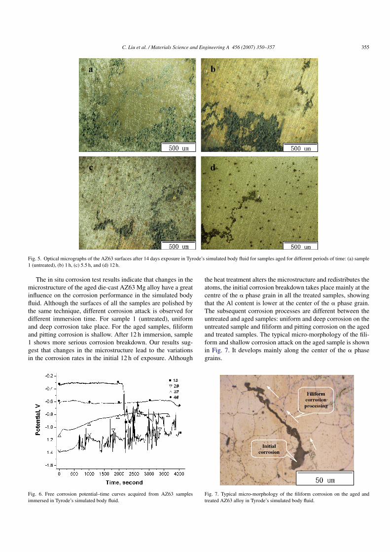
The corroded morphology was examined after 14 daysimmersion. The samples (1 cm2) polished with #2400 SiC water-proof paper were submerged in the test solution. Corrosionattack on a large area is observed on samples 1 and 2, and with
increasing immersion duration, it is deeper as shown in Fig. 5aand b. “Filiform” corrosion and pitting corrosion are seen on thesurface of samples 3 and 4 (Fig. 5c and d) but the attack is notas severe [14].
Dissolution in body fluids is the major characteristic ofbiomedical magnesium alloys [5]. The potential–time curves ofthe AZ63 samples in the simulated body fluid are displayed inFig. 6. The typical potential–time curve of the untreated sampleshows big changes indicative of dissolution and passivity of themagnesium alloy. With regard to sample 2, the free corrosionpotential exhibits a sudden decrease at approximately 2200 swhich corresponds to the active corrosion process [16]. Thepotential–time curve of sample 4 shows the reduced metastablelocalized corrosion nucleation phenomena after about 500 s, butrapid dissolution related to corrosion breakdown appears afterabout 3750 s. The free corrosion potential of sample 3 increaseswith longer immersion time and the presence of metastable pitnucleation with a lower frequency is indicated. The enhance-ment in the free corrosion potential means that a hybrid solutionand aging treatment is beneficial to the initial stability of thealloy surface.
4. Discussion
As a bulk material, the phase constituents and their distri-bution as well as the grain size affect the corrosion propertiessignificantly. AZ magnesium alloys are typically composed of amatrix of � grains with the � phase (the intermetallic Mg17Al12)along the � grain boundaries [17]. In a chloride solution, thecorrosion performance of AZ alloy is determined by the � frac-tion, the continuous � phase around the � grains, and porosity.The amount and distribution of the � phase (discontinuous pre-cipitation, DP and continuous precipitation, CP) determine thecorrosion performance of the magnesium alloy. In particular,the � phase plays dual roles in the corrosion mechanism eitheras a corrosion barrier or galvanic cathode that accelerates corro-sion. A homogeneously distributed � phase can more effectivelyretard corrosion, otherwise the presence of the � phase can accel-erate corrosion [17]. It has been found that a solution or agedtreatment can effectively change the distribution and amount ofthe �-Mg17Al12 phase [7].
Fig. 1 shows that the micro-structural evolution of the AZ63alloy is accompanied by the redistribution of Al [8]. In com-parison with the untreated sample 1, sample 2 possesses a morehomogeneous surface morphology. The � phase dissolves in the� phase solid solution after a 24 h, 413 ◦C solution treatment fol-lowed by 1 h, 216 ◦C aging. Complete dissolution of the � phaseleads to the formation of sharp grain boundaries and the structureof the secondary solidification region eventually evolves into thenew well-defined � grains [7]. After 5.5 h aging, the DP and CP� phases appear gradually and with longer aging time, a largeamount of the CP � phase forms inside the � grains. Comparedto the RDC AZ91 alloy [7], a longer aging time is needed to formthe precipitates of the � phase in the AZ63 alloy. It may be dueto the lower aluminum content. The increase in the incubationperiod suggests depressed aging kinetics for the � phase in theAZ63 alloy.
C. Liu et al. / Materials Science and Engineering A 456 (2007) 350–357 355
Fig. 5. Optical micrographs of the AZ63 surfaces after 14 days exposure in Tyrode’s simulated body fluid for samples aged for different periods of time: (a) sample1 (untreated), (b) 1 h, (c) 5.5 h, and (d) 12 h.
The in situ corrosion test results indicate that changes in themicrostructure of the aged die-cast AZ63 Mg alloy have a greatinfluence on the corrosion performance in the simulated bodyfluid. Although the surfaces of all the samples are polished bythe same technique, different corrosion attack is observed fordifferent immersion time. For sample 1 (untreated), uniformand deep corrosion take place. For the aged samples, filiformand pitting corrosion is shallow. After 12 h immersion, sample1 shows more serious corrosion breakdown. Our results sug-gest that changes in the microstructure lead to the variationsin the corrosion rates in the initial 12 h of exposure. Although
Fig. 6. Free corrosion potential–time curves acquired from AZ63 samplesimmersed in Tyrode’s simulated body fluid.
the heat treatment alters the microstructure and redistributes theatoms, the initial corrosion breakdown takes place mainly at thecentre of the � phase grain in all the treated samples, showingthat the Al content is lower at the center of the � phase grain.The subsequent corrosion processes are different between theuntreated and aged samples: uniform and deep corrosion on theuntreated sample and filiform and pitting corrosion on the agedand treated samples. The typical micro-morphology of the fili-form and shallow corrosion attack on the aged sample is shownin Fig. 7. It develops mainly along the center of the � phasegrains.
Fig. 7. Typical micro-morphology of the filiform corrosion on the aged andtreated AZ63 alloy in Tyrode’s simulated body fluid.
356 C. Liu et al. / Materials Science and Engineering A 456 (2007) 350–357
Some previous studies have pointed out that the volume frac-tion and distribution of the � phase have a large influence onthe corrosion properties of AZ magnesium alloys [12,17,18].Fig. 1a shows that the � phase is not abundant and its distribu-tion is uneven. As a result, the inter-distance of the � phase canbe very large thereby making interconnection almost impossi-ble. Therefore, when corrosion commences from the center ofthe � phase grains, dissolution of the � phase is accelerated. Thatis to say, the � phase is a cathode to the � phase, and hydrogenevolution preferentially takes place from the � phase makingit an effective cathode [18]. Daloz et al. [19] have pointed outthat when the � phase fraction is large, the film formed on the �phase is more stable because of the presence of greater amountof aluminum that could act as a barrier to inhibit the dissolu-tion of the � phase. Since the � phase fraction is low in theuntreated AZ63 alloy, it is impossible to form a homogeneousprotective film, and this is believed to be the main reason for thesevere corrosion attack. In contrast, the aged AZ63 alloys havea more homogeneous microstructure (Fig. 1b–d) and more evendistribution of aluminum. When exposed to air, the aluminumoxide formed over the � phase is more compact and uniform. Adescription of the formation of the oxide film outside the metal-lic surface consisting of Mg(OH)2, MgO and Al2O3 has beenproposed [11]. The existence of the Al2O3 layer may to a cer-tain extent depress the capillary penetration of the test solution[20], thereby lengthening the degradation period in the simulatedbody fluid. The variation in the free corrosion potential mea-sured from the untreated sample is extensive, indicating the usualbehavior when subjected to a very high frequency of metastablepit nucleation [15]. In contrast, the free corrosion potentials ofsamples 3 and 4 continue to rise (Fig. 6) and it may be ascribedto the relatively compact and stable oxide film inhibiting capil-lary action of the test solution and blocking the corrosion attack.Compared to samples 3 and 4, a sudden decrease takes place atapproximately 2200 s in the free corrosion potential for sample1, indicating instability of the oxide film. After 14 days in thesimulated body fluid, the surface morphologies of samples 3 and4 are clearly different from those of the other two samples. Thecorrosion attack consists of not only filiform corrosion but alsopitting corrosion. The appearance of pitting corrosion may beascribed to the formation of the � phase. According to Fig. 1cand d, the � phase is formed along or inside the � phase grains.Moreover, the volume fraction of the � phase increases withlonger aging time. It has been pointed out that a high volumefraction of the � phase that is also aluminum rich may miti-gate the overall corrosion of the alloy [11–13]. Therefore, theexistence of the � phase depresses the corrosion attack. Fig. 8indicates the variation of the average corrosion rate for the differ-ent samples after 14 days immersion. The lowest corrosion rateis approximately 1/2 of that of the untreated sample. The changein the pH may also suppress the degradation rate of the AZ63alloy in the simulated body fluid consisting of 0.9 wt.%NaCl.The dissolution of magnesium alloy in an aqueous solution withchloride ions proceeds by the following reactions.
Mg → Mg2+ + 2e−, 2H2O + 2e− → H2 + 2(OH)−,
Mg2+ + 2(OH)− → Mg(OH)2 (2)
Fig. 8. Average corrosion rates of the untreated and aged AZ63 alloys afterimmersion in Tyrode’s simulated body fluid for 14 days.
Altun et al. have shown that highly acidic solutions attackboth magnesium and aluminum, and hence a very high cor-rosion rate and a pH value above ∼9 favor the formation ofMg(OH)2 [21]. Chemically, both the forward and reverse reac-tions exist. Fig. 4b indicates that the pH value rises initiallyand then decreases after 48 h immersion. Before 48 h exposure,the release rate of magnesium ions rises almost exponentially.According to reaction (2), the great number of electrons raisesthe pH. Meanwhile, it can be determined that the forward reac-tion by which Mg(OH)2 is formed may be promoted due tothe higher pH. Because the Mg(OH)2 film can inhibit corro-sion of the magnesium alloy to some extent [4], the pH valuedecreases. At the same time, the chloride ions can transformMg(OH)2 to the more soluble MgCl2 in order to attain thereaction balance, lots of (OH)− are needed to make Mg(OH)2causing re-dissolution of magnesium. At equilibrium, the pHonly changes within a small range.
5. Conclusion
We have investigated the effects of heat treatment on thedegradation of die-cast AZ63 Mg alloy in Tyrode’s simulatedbody fluid. The solution treatment at 413 ◦C for 24 h followed byaging at 216 ◦C for up to 1 h leads to complete dissolution of the �phase into the � phase grains causing the formation of new well-defined � grains with sharp grain boundaries. The �-Mg17Al2precipitates form after 5.5 h of aging, and the amount and distri-bution become greater and wider with longer aging time. Withincreasing aging time, the treated alloys show greater corrosionresistance. After 14 days immersion in 37 ± 1 ◦C Tyrode’s simu-lated body fluid, the lowest corrosion rate achieved is about 1/2of that of the untreated alloy. The change in the microstruc-ture is thus shown to impact the corrosion behavior of thealloys. For the aged materials, shallow filiform and pitting cor-rosion is observed. In comparison, deep and uniform corrosionis observed on the untreated AZ63 alloy.
Acknowledgment
The project was supported by City University of Hong KongApplied Research Grant (ARG) no. 9667002.
C. Liu et al. / Materials Science and Engineering A 456 (2007) 350–357 357
References
[1] A. Gefen, Med. Biol. Eng. Comput. 40 (2002) 311–319.[2] X. Cao, M. Jahazi, J.P. Immarigeon, W. Wallace, J. Mater. Process. Technol.
171 (2006) 188–204.[3] F. Witte, V. Kaese, H. Haferkamp, E. Switzer, H. Windhagen, Biomaterials
26 (2005) 3557–3563.[4] G.L. Song, Adv. Eng. Mater. 7 (2005) 563–586.[5] F. Witte, J. Fischer, J. Nellesen, H.A. Crostack, H. Windhagen, Biomaterials
27 (2006) 1013–1018.[6] M. Wislawska, M. Janick-Czachr, Corros. J. 20 (1985) 36–45.[7] Y. Wang, G. Liu, Z. Fan, Acta Mater. 54 (2006) 689–699.[8] Z.Y. Zhang, X.Q. Zeng, W.J. Ding, Mater. Sci. Eng. A 392 (2005) 150–158.[9] S. Mathieu, C. Rapin, J. Steinmetz, Corros. Sci. 45 (2003) 2741–2755.
[10] C. Scharf, A. Ditze, A. Shkurankov, E. Morales, K.U. Kainer, Adv. Eng.Mater. 7 (2005) 1134–1142.
[11] G.L. Song, A. Atrens, X.L. Wu, B. Zhang, Corros. Sci. 40 (1998)1769–1791.
[12] T. Zhang, Y. Li, F.H. Wang, Corros. Sci. 48 (2006) 1249–1264.[13] G.L. Song, A. Bowles, D. Stjohn, Mater. Sci. Eng. A 366 (2004) 74–
86.[14] M. Kuhlein, U. Galovsky, Mater. Corros. 55 (2004) 444–448.[15] A. Fossati, F. Borgioli, E. Galvanetto, T. Bacci, Corros. Sci. 48 (2006)
1513–1527.[16] J. Bolton, X. Hu, J. Mater. Sci. Mater. Med. 13 (2002) 567–574.[17] G.L. Song, A. Atrens, M. Dargusch, Corros. Sci. 41 (1999) 249–
273.[18] R. Ambat, N.N. Aung, W. Zhou, Corros. Sci. 42 (2000) 1433–1455.[19] D. Daloz, P. Steinmetz, G. Michot, Corrosion 83 (1997) 944–949.[20] J. Masalski, J. Gluszek, J. Zabrzeski, P. Gluszek, Thin Solid Films 349
(1999) 186–190.[21] H. Altun, S. Sen, Mater. Des. 25 (2004) 637–643.