RESEARCH ARTICLE
In vitro analysis of the segmental flexibility of
the thoracic spine
Hans-Joachim Wilke*, Andrea Herkommer, Karin Werner, Christian Liebsch
Institute of Orthopaedic Research and Biomechanics, Trauma Research Centre Ulm, University of Ulm, Ulm,
Germany
* [email protected]
Abstract
Basic knowledge about the thoracic spinal flexibility is limited and to the authors’ knowledge,
no in vitro studies have examined the flexibility of every thoracic spinal segment under stan-
dardized experimental conditions using pure moments. In our in vitro study, 68 human tho-
racic functional spinal units including the costovertebral joints (at least n = 6 functional spinal
units per segment from T1-T2 to T11-T12) were loaded with pure moments of ±7.5 Nm in
flexion/extension, lateral bending, and axial rotation in a custom-built spine tester to analyze
range of motion (ROM) and neutral zone (NZ). ROM and NZ showed symmetric motion
behavior in all loading planes. In each loading direction, the segment T1-T2 exhibited the
highest ROM. In flexion/extension, the whole thoracic region, with exception of T1-T2 (14˚),
had an average ROM between 6˚ and 8˚. In lateral bending, the upper thoracic region (T1-
T7) was, with an average ROM between 10˚ and 12˚, more flexible than the lower thoracic
region (T7-T12) with an average ROM between 8˚ and 9˚. In axial rotation, the thoracic
region offered the highest overall flexibility with an average ROM between 10˚ and 12˚ in the
upper and middle thoracic spine (T1-T10) and between 7˚ and 8˚ in the lower thoracic spine
(T10-T12), while a trend of continuous decrease of ROM could be observed in the lower tho-
racic region (T7-T12). Comparing these ROM values with those in literature, they agree that
ROM is lowest in flexion/extension and highest in axial rotation, as well as decreasing in the
lower segments in axial rotation. Differences were found in flexion/extension and lateral
bending in the lower segments, where, in contrast to the literature, no increase of the ROM
from superior to inferior segments was found. The data of this in vitro study could be used
for the validation of numerical models and the design of further in vitro studies of the thoracic
spine without the rib cage, the verification of animal models, as well as the interpretation of
already published human in vitro data.
Introduction
Few studies have focused on the biomechanics of the thoracic spine, because prior research
mainly focused on the lumbar [1–4] and the cervical spine [5–7]. This is due to the fact that
chronic degenerative diseases of the thoracic spine are reported to have a lower incidence and
therefore seem to be clinically of lower importance [8]. However, pathologies of the thoracic
PLOS ONE | https://doi.org/10.1371/journal.pone.0177823 May 16, 2017 1 / 16
a1111111111
a1111111111
a1111111111
a1111111111
a1111111111
OPENACCESS
Citation: Wilke H-J, Herkommer A, Werner K,
Liebsch C (2017) In vitro analysis of the segmental
flexibility of the thoracic spine. PLoS ONE 12(5):
e0177823. https://doi.org/10.1371/journal.
pone.0177823
Editor: Alejandro A. Espinoza Orıas, Rush
University Medical Center, UNITED STATES
Received: January 31, 2017
Accepted: May 3, 2017
Published: May 16, 2017
Copyright: © 2017 Wilke et al. This is an open
access article distributed under the terms of the
Creative Commons Attribution License, which
permits unrestricted use, distribution, and
reproduction in any medium, provided the original
author and source are credited.
Data Availability Statement: All relevant data are
within the paper and its Supporting Information
files.
Funding: This study was supported by the German
Research Council (DFG), Project WI 1352/20-1 to
(HJW).
Competing interests: The authors have declared
that no competing interests exist.
spine should not be neglected. The overall number of vertebral fractures, for instance, is, in
absolute terms, just slightly higher in the lumbar spine than in the thoracic spine [9]. Further-
more, the growing number of traffic and sports accidents lead to an increasing number of seri-
ous injuries, particularly in the lower thoracic spinal segment [10]. These fractures are very
likely associated with neurological complications, since the thoracic spinal canal is quite nar-
row and high forces are required for the formation of vertebral fractures due to the stabilizing
effect of the rib cage. Also complex osteoporotic vertebral fractures and spinal metastases are
indications for reconstructive and stabilizing surgeries in the thoracic spine; their number has
steadily increased in the recent past due to demographic changes [11, 12]. To achieve optimum
restoration of injured thoracic spinal structures, basic knowledge of the biomechanics of the
intact thoracic spine is mandatory.
Finite element models provide a useful tool for the analysis of several spinal diseases, since
they offer the possibility of performing detailed studies of various biomechanical variables and
allow individualized simulations of surgical correction procedures [13–17]. For the calibration
and validation of finite element models of the healthy spine, a comprehensive database includ-
ing experimental in vivo and in vitro biomechanical data of asymptomatic thoracic spines is
required [18, 19]. However, the limited available data of previous studies with partially contra-
dictory data and the variety of measurement methods illustrate the need for current basic
research using new standardized measurement techniques.
Biomechanical investigations regarding the thoracic spinal flexibility are scarce. The first
comprehensive in vitro study on thoracic spine flexibility was carried out by White in 1969
[20]. In this biomechanical study, a two-dimensional analysis of monosegmental specimens
and a three-dimensional analysis of polysegmental specimens were performed. So far, this
study represents the basic knowledge of thoracic spine flexibility. However, the loads were not
applied by means of pure moments in this study, while the application of pure moments is the
gold standard in spine biomechanics today [21, 22]. A subsequent in vitro study of Panjabi
et al. used pure moments to determine the mechanical properties of the thoracic spine by
using load-deformation curves and calculating flexibility coefficients. They performed exem-
plary measurements, each with n = 1 monosegmental specimen for the whole thoracic region
from T1-T2 to T11-T12 [23]. Single functional spinal units of the thoracolumbar junction
were investigated by Markolf et al. and Oxland et al. [24, 25]. In all these in vitro studies pure
moments were applied, but different measurement equipment and torque levels were used. In
1990, White and Panjabi gave an overview of the ranges of motion for all functional spinal
units of the human spine. Multiple in vivo and in vitro studies, performed by different authors,
were combined in this overview, leading to high variations within the data [26].
The aim of the present study was therefore to investigate the segmental range of motion
and neutral zone of the healthy human thoracic spine by applying pure moments, with a suffi-
cient number of specimens, under controlled, standardized testing conditions.
Materials and methods
A total of 68 thoracic functional spinal units (FSUs) from 29 human donors were tested. For
each of the eleven segmental levels of T1-T2 to T11-T12, n = 6 specimens were used for testing,
except for the levels T4-T5 and T7-T8, of which each n = 7 specimens were available. The aver-
age age of the donors was 57 years (40–80 years), whereby 13 of the donors were male and 16
female (Table 1). None of the specimens showed any visible ligamentous, discogenic, or bony
damage relevant to biomechanical testing. Tumorous or fracture related damages were
excluded prior to preparation using conventional X-ray images (Faxitron 43805N, Hewlett
Packard, Palo Alto, USA). The spines were stored at -20˚C. Prior to testing, the specimens
Segmental flexibility of the thoracic spine
PLOS ONE | https://doi.org/10.1371/journal.pone.0177823 May 16, 2017 2 / 16
were thawed at 4˚C and prepared at room temperature. Muscles, nerves, fat, and other soft tis-
sues were carefully dissected while preserving the ligaments, joint capsules, intervertebral
discs, and costovertebral joints. The ribs were shortened to a length of about 1.5 cm using a
saw.
The upper half of the cranial vertebra and the lower half of the caudal vertebra were embed-
ded in polymethylmethacrylate (PMMA, Technovit 3040, Heraeus Kulzer, Wehrheim, Ger-
many). Before embedding, screws were fixed in the cranial and caudal endplates of each FSU
to ensure a firm connection between the vertebral bodies and the subsequent plastic cast. The
disc was adjusted horizontally and the costovertebral joints, the facet joints, as well as the
inter- and supraspinous ligaments were covered with modelling clay during embedding to
preserve the full mobility of the FSUs. After embedding, flanges were coaxially fixed to the
PMMA blocks. During preparation and testing, the specimens were kept moist with physiolog-
ical saline (0.9%) [21].
After manual alignment regarding the anatomical planes, the 68 FSUs were each loaded in
a custom-built spine tester (Fig 1) by applying pure moments of ±7.5 Nm in lateral bending
(±Mx), flexion/extension (±My), and axial rotation (±Mz) [27]. While the monitored rotation
axis was engaged, the specimens were allowed to move almost unconstrained in the remaining
five degrees of freedom due to a traveling gantry and balancing weights (RMS errors in maxi-
mum off axis torques: Mx = 0.2 Nm, My = 0.2 Nm, Mz = 0.1 Nm, RMS errors in maximum off
axis forces: Fx = 6.2 N, Fy = 7.0 N, Fz = 20.5 N). The pure moments were applied continuously
for 3.5 cycles with an angular velocity of 1˚/s in flexion/extension and lateral bending, as well
as 0.5˚/s in axial rotation using three stepper motors (Isel 3450, Isert-electronic, Eiterfeld, Ger-
many) with a torque of 55 Ncm and 1.8˚ per step. The moments were measured by a 6-DOF
load cell (FT 1500/40, Schunk GmbH & Co. KG, Lauffen/Neckar, Germany), which has a mea-
suring range of ±40 Nm, a resolution of 0.02 Nm and a measuring error of<1%. The first two
cycles served for preconditioning of the specimen, while the third cycle was used for data
evaluation.
The resulting load-deformation curves represent the typical stiffness properties of the single
motion segments (Fig 2) and were used for the determination of the biomechanical parameters
range of motion (ROM) and neutral zone (NZ). The ROM describes the deformation of the
specimen at the maximum load in the respective loading direction, while the NZ is the motion
range of the specimen in the unloading phase (at 0 Nm) and is therefore a measure for the lax-
ity of the motion segment [21, 22]. ROM and NZ were automatically determined using a self-
Table 1. Donor age (in years) and sex (m = male, f = female) of the thoracic FSUs.
Segmental level #1 #2 #3 #4 #5 #6 #7 Mean ± SD
T1-T2 53, f 58, f 46, f 40, m 43, f 60, m - 50 ± 8
T2-T3 57, f 56, m 79, m 66, f 46, m 58, m - 60 ± 10
T3-T4 54, m 45, m 53, f 59, f 58, f 60, m - 55 ± 5
T4-T5 54, f 57, f 44, f 66, f 46, f 63, f 56, m 55 ± 7
T5-T6 54, m 71, f 53, f 58, f 60, m 80, f - 63 ± 10
T6-T7 54, f 44, f 59, m 43, f 46, f 56, m - 50 ± 6
T7-T8 76, f 71, f 53, f 58, f 60, m 65, m 66, f 64 ± 7
T8-T9 56, m 54, f 46, f 46, m 51, m 54, m - 51 ± 4
T9-T10 50, m 57, f 71, f 62, f 75, f 65, m - 63 ± 8
T10-T11 54, m 69, m 60, m 51, m 63, f 44, f - 57 ± 8
T11-T12 49, f 71, f 62, f 59, m 65, m 57, f - 61 ± 7
https://doi.org/10.1371/journal.pone.0177823.t001
Segmental flexibility of the thoracic spine
PLOS ONE | https://doi.org/10.1371/journal.pone.0177823 May 16, 2017 3 / 16
programmed MATLAB routine (MathWorks Inc., Natick, USA) which fitted a polynomial
function to the curve to define the midpoint of the hysteresis curve regarding the angle and
measured the displacements at ±7.5 Nm (ROM) as well as at the midpoint of the connecting
line between the two turning points of the polynomial function (NZ). The evaluated hysteresis
curves of all experiments are depicted in the S5–S7 Dataset files.
The present in vitro study and related use of human specimens were approved by the ethical
committee board of the University of Ulm, Germany (No. 302/14).
Fig 1. Experimental setup. A typical thoracic spinal motion segment before load application in the spine tester.
https://doi.org/10.1371/journal.pone.0177823.g001
Segmental flexibility of the thoracic spine
PLOS ONE | https://doi.org/10.1371/journal.pone.0177823 May 16, 2017 4 / 16
Results
The motion segment T1-T2 was found to have the highest ROM in all six loading directions,
of which flexion was identified as the loading direction with the highest ROM in this specific
motion segment (Figs 3–5). The upper half of the thoracic spine from T1-T2 to T6-T7 showed
a higher range of motion than the lower thoracic spine from T7-T8 to T11-T12 in all loading
planes. The lowest ROMs were detected in extension for all motion segments between T2-T3
and T11-T12, followed by flexion, both having ROMs equal or less than 4˚, whereas the highest
ROMs were generally found in axial rotation for all motion segments from T2-T3 to T10-T11
and in lateral bending for T11-T12, respectively. In axial rotation, the NZ to ROM ratio was
the lowest of all three loading planes, whereas the highest NZ to ROM ratio was found in lat-
eral bending.
In flexion/extension, the level T1-T2 showed the greatest ROM of all motion segments with
a mean value of 7.1˚ in flexion and 6.7˚ in extension (Fig 3). In T2-T3, the ROM reduced to
Fig 2. Load-deformation curves. Characteristic hysteresis curves of representative thoracic spinal motion segments of the upper,
middle, and lower thoracic spine in flexion/extension, lateral bending, and axial rotation.
https://doi.org/10.1371/journal.pone.0177823.g002
Segmental flexibility of the thoracic spine
PLOS ONE | https://doi.org/10.1371/journal.pone.0177823 May 16, 2017 5 / 16
4.1˚ in both flexion and extension and was lowest in the segment T7-T8 with 2.8˚ for both
loading directions. In the upper part of the thoracic spine from T3-T4 to T6-T7 and the lower
part T8-T9 to T11-T12, the mean values of the ROM ranged from 3.1˚ to 3.8˚. The standard
deviations accounted in most segments for 20–36% of the ROM, while T2-T3 showed a large
standard deviation of 49% and T1-T2 a small standard deviation of 6%. The NZ was between
0.6˚ and 2.1˚, which makes up 14–30% of the mean ROMs (S1 Dataset).
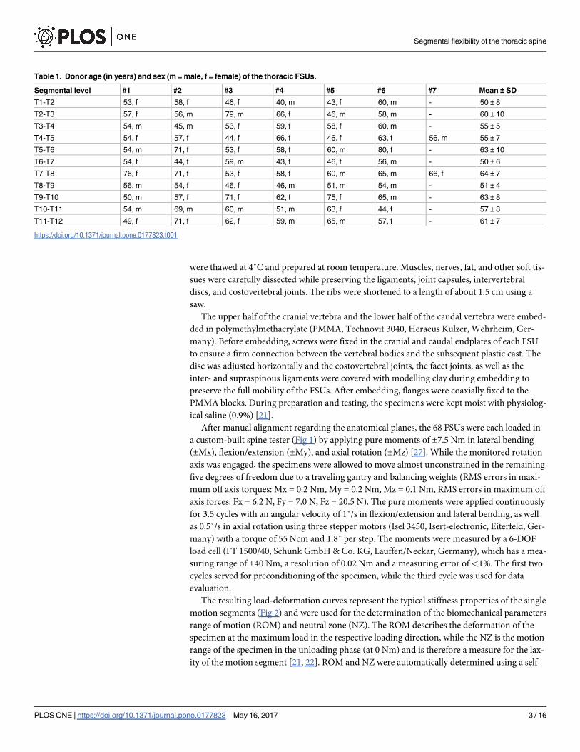
In lateral bending, the segment T1-T2 had the highest range of motion, with a mean value
of 5.9˚ in both directions (Fig 4). The ROM of the upper and middle segments from T2-T3 to
T6-T7 ranged between 4.9˚ and 5.6˚, whereas the inferior segments exhibited a smaller range
of motion between 3.8˚ and 4.5˚. The lowest ROM was again detected in the segment T7-T8
with 3.7˚. The standard deviations ranged between 17% and 34% of the mean ROMs, with the
largest standard deviation in the segment T3-T4. Generally, ROM and NZ had almost symmet-
rical motion behavior. The NZ was between 0.7˚ and 2.0˚, which makes up 16–35% of the
mean ROMs (S1 Dataset).
In axial rotation, almost symmetrical motion behavior for the rotation to each the left and
right direction was detected (Fig 5). The ROM was again highest in the first segment T1-T2
with 6.2˚, followed by segment T6-T7 with 5.9˚. In the upper thoracic spine from T2-T3 to
T5-T6, values between 5.2˚ and 5.7˚ were measured. In the segments from T7-T8 to T9-T10
the mean ROM was about 5˚, while a continuous decrease of range of motion could be
detected from T9-T10 to T11-T12. The smallest ROM with a mean of 3.3˚ was measured in
Fig 3. Flexion/extension. ROM and NZ at ±7.5 Nm pure moment in flexion/extension for all thoracic spinal motion
segments (n = 6, except n = 7 for T4-T5 and T7-T8), represented as mean values with standard deviations.
https://doi.org/10.1371/journal.pone.0177823.g003
Segmental flexibility of the thoracic spine
PLOS ONE | https://doi.org/10.1371/journal.pone.0177823 May 16, 2017 6 / 16
the last segment T11-T12. The standard deviations were between 18% and 31% of the mean
ROMs. The NZ, with values between 0.4˚ and 1.2˚, makes up 10–20% of the mean ROMs and
was lower than in the other both motion planes (S1 Dataset).
Discussion
The aim of the present study was to investigate the segmental range of motion and neutral
zone of the thoracic spine by applying pure moments under controlled, standardized testing
conditions, since the literature regarding the biomechanical properties of the single thoracic
spinal motion segments is scarce and the segmental flexibility of the thoracic spine has to be
determined to understand the biomechanics, to solve clinical problems, and to calibrate and
validate numerical models. The present in vitro study therefore provides monosegmental
ROM data for all thoracic spinal segments.
Specimens
68 FSUs from 29 different human donors were used in this study. Complete or almost com-
plete thoracic spines were available from nine donors and between three and five segments
could be used per donor. The missing segments were replaced by individual segments from 20
other donors. At least six monosegmental specimens were tested per segment, although a
higher number would have been preferable. However, due to the limited availability of human
Fig 4. Lateral bending. ROM and NZ at ±7.5 Nm pure moment in lateral bending for all thoracic spinal motion segments
(n = 6, except n = 7 for T4-T5 and T7-T8), represented as mean values with standard deviations.
https://doi.org/10.1371/journal.pone.0177823.g004
Segmental flexibility of the thoracic spine
PLOS ONE | https://doi.org/10.1371/journal.pone.0177823 May 16, 2017 7 / 16
specimens, as well as ethical and financial issues, n = 6 specimens per segment is generally
accepted for in vitro studies.
Specimens of young healthy donors are most appropriate to exclude variations in the bio-
mechanical properties which are caused by degenerative remodelling processes occurring in
middle age. Kettler et al. and Mimura et al. determined in their in vitro studies with lumbar
specimens a decrease of ROM in flexion/extension and lateral bending with increasing degree
of degeneration. A slight increase of ROM was found in axial rotation [28, 29]. Due to the
unavailability of younger donors, the average age of specimens in this study was 58 years (40–
80 years). Variations of donor age and thus the stage of disc degeneration could be responsible,
besides specific differences in specimen morphology, for the variations within the results and
could therefore affect their comparability. Since the specimens had to be randomly distributed
on the single segmental level groups due to the limited availability, donor age or sex had to be
disregarded. However, mean donor age and sex of the eleven segmental level groups were not
substantially different between the groups, varying between 50 and 64 years and a sex ratio
between 3:3 and 6:1 (Table 1).
During preparation, care was taken to preserve the costovertebral joints, since they stabilize
the thoracic spine in all three loading directions [30–32]. The anterior part of the rib cage, includ-
ing the sternal complex and the ribs, was excluded from the test setup to reach a high comparabil-
ity between the single motion segments, although it was also found to influence the stability of
the thoracic spine in former in vitro studies [33–35]. The effect of the ribs and its sternal portion
Fig 5. Axial rotation. ROM and NZ at ±7.5 Nm pure moment in axial rotation for all thoracic spinal motion segments (n = 6,
except n = 7 for T4-T5 and T7-T8), represented as mean values with standard deviations.
https://doi.org/10.1371/journal.pone.0177823.g005
Segmental flexibility of the thoracic spine
PLOS ONE | https://doi.org/10.1371/journal.pone.0177823 May 16, 2017 8 / 16
on the flexibility of the single motion segments should be evaluated separately. The ligaments
were left intact, since damaged or missing ligament structures were found to influence the ROM,
as well as the intradiscal pressure and the relative position of the vertebral bodies [36, 37].
The specimens were checked for damage prior to preparation using conventional X-ray.
CT or MRI scans, however, were not performed. The present study therefore solely provides
kinematic and stiffness properties for the mechanical validation of finite element models of the
thoracic spinal motion segments.
Testing conditions
According to current recommendations for in vitro experiments, the specimens were tested
with 3.5 loading cycles, of which the third loading cycle was evaluated [21, 22]. This procedure
has become the state of the art for biomechanical flexibility testing, because the first two load-
ing cycles are used for preconditioning to reduce viscoelastic effects [22, 26]. Moreover, the
monosegmental ROMs were determined by using pure moments of ±7.5 Nm to allow direct
comparison to ROM data in the literature for the lumbar spine, although ±5 Nm are recom-
mended for thoracic spinal flexibility testing [38]. Since there was no visible change in range of
motion, hysteresis, and elastic stiffness during all loading cycles, pure moments of ±7.5 Nm
were considered as acceptable for testing (S1 Dataset).
An axial preload was omitted in the present study, although it is recommended for spinal in
vitro testing [39, 40]. Axial preload may reduce the segmental mobility and effect the kinematic
response, wherefore it was used in previous studies to simulate the experimental motion of the
spine as physiologically as possible [41–43].
Another limitation of the present study represents the manual alignment of the specimens
in the spine tester, which could have led to slight off axis loads in flexion/extension and lateral
bending. However, it was tried to compensate the possible angle offsets by automatic determi-
nation of each hysteresis curve midpoint.
Care was also taken not to exceed the testing period of 20 hours during preparation and
testing for each specimen, since the biomechanical properties of the tested specimens will
change and autolytic processes will start [21].
Biomechanical interpretation of the results
Specific anatomical properties influence the range of motion of the single spinal regions. The
cross-sectional areas of the discs of the upper thoracic spine, for example, are relatively small
compared to those in the lower thoracic spine and increase inferiorly, whereas the disc heights
are approximately the same in the upper and lower thoracic spine [44], leading to a higher
ROM, given the same amount of pure moments for all segmental levels in our in vitro study,
in the upper thoracic spinal motion segments because of the lower moment of inertia of area.
In the present study, the first segment T1-T2 exhibited the highest flexibility of all segments in
all loading directions, indicating similar range of motion characteristics as the cervicothoracic
transition, whereas the range of motion tended to decrease in inferior direction towards the
lumbar spine in all six loading directions (Figs 3–5).
Another anatomical characteristic of the thoracic spine is provided by the different posi-
tions of the costovertebral joints. While these joints are positioned each between the single
motion segments at the level of the discs for all segments from T1-T2 to T9-T10, potentially
having a stabilizing effect together with the costotransverse joints and their ligament struc-
tures, the costovertebral joints of the two inferior pairs of ribs are located on the middle of T11
and T12, respectively. The costovertebral joints therefore have no potential stabilizing effect
on the motion segments T10-T11 and T11-T12. However, these two motion segments showed
Segmental flexibility of the thoracic spine
PLOS ONE | https://doi.org/10.1371/journal.pone.0177823 May 16, 2017 9 / 16
an equal or lower ROM compared to the other motion segments in the present study, indicat-
ing a larger stabilizing effect of the discs, ligaments, facet joint capsules, and facet joint orienta-
tions in these motion segments. Moreover, a stabilizing effect of the costovertebral joints was
only determined in combination with a radical discectomy in previous in vitro studies [30, 45].
The facet joints of the thoracic spine show a specific morphology because of their three-
dimensional orientation, which leads to a different motion behavior compared to the cervical
and the lumbar spine. In the inferior direction towards the lumbar spine, the facet joints
exhibit increasing tilt angles in the sagittal plane and slightly in the transversal plane [46],
limiting the range of motion, especially in flexion/extension and slightly in axial rotation. The
variation of the facet joint orientations within the thoracic spine can be high due to interindi-
vidual differences, but in general, the tilt angles increase gradually, whereby the tilt angles in
the transversal plane end in the thoracolumbar transition zone [26, 47]. Also the thoracic spi-
nal ligaments have a strong stabilizing effect, since they are thicker than those in the cervical
and lumbar spine [26, 48]. Together with the anterior part of the rib cage, the anterior and pos-
terior longitudinal ligaments prevent hyperflexion and -extension [49].
Literature comparison
Few in vitro studies have investigated the flexibility of the thoracic spine. Furthermore, the pre-
viously existing results are only partially comparable due to varying experimental setups and
loading conditions. By means of new basic in vitro data of the thoracic spinal flexibility, evalu-
ated using now widely accepted recommendations for in vitro testing of spinal segments [21,
22], a better interpretation of the already published data should be possible. In addition, a
comprehensive database could provide a basis for the development of new finite element mod-
els or multi-body systems of the healthy human thoracic spine.
The subsequent literature overview compares the results of the present study and previously
published results of in vitro and in vivo studies (Fig 6). The flexibility tests of the present study
were performed in a well-established spine tester [27]. One specific feature of this device is the
application of pure moments, which ensures that the load is applied precisely and reproducibly
on the whole tested spinal segment in each motion plane. Similar methods for in vitro testing of
spinal segments with pure moments were found in the literature for the experiments of Panjabi
et al., Markolf et al., and Oxland et al. [23–25]. The study, which is closest to the present study
because of its comprehensiveness, has been described by White [20], who applied eccentric
loads in his two-dimensional and three-dimensional analysis of flexion/extension and lateral
bending motions. It should be noted, however, that White and Markolf et al. used an experi-
mental setup, in which the upper vertebra exhibited a limited mobility in the remaining five
motion directions [20, 24], which could have limited flexibility in the main loading direction.
A literature overview was published by White and Panjabi [26], where ROM data of several
in vitro and in vivo studies, including different test setups and load applications, were summa-
rized. Therefore, these data allow comparisons with other data only in a limited extent.
Moreover, the in vivo study of Willems et al. was performed using the polysegmental sec-
tions T1-T4, T4-T8 and T8-T12 [50]; for better comparability, the values have been divided
according to the number of segmental portions. However, the flexibility of the individual spi-
nal segments is not known. When comparing in vitro and in vivo data, it should also be noted
that no standardized torque limits exist during in vivo measurements. Furthermore, other
effects, such as the rib cage, the muscles, the intraabdominal and intrathoracic pressure, as well
as individual pain sensations, have an influence on the flexibility of the spinal segments.
In general, differences regarding segmental flexibility between the three loading planes
were not as significant as those described in literature (Fig 6). In flexion/extension, the data of
Segmental flexibility of the thoracic spine
PLOS ONE | https://doi.org/10.1371/journal.pone.0177823 May 16, 2017 10 / 16
Segmental flexibility of the thoracic spine
PLOS ONE | https://doi.org/10.1371/journal.pone.0177823 May 16, 2017 11 / 16
the present study show the highest ROM in the first segment T1-T2. However, there is an
inverse trend of the results compared to the summary from White and Panjabi [26] with the
highest ROM at T11-T12, whereas the results of Markolf et al., Oxland et al., and Willems et al.
show a similar motion behavior for the lower thoracic segments [24, 25, 50]. Besides, the data
of White and Panjabi exhibited high value ranges in these two lower segments, which can
probably be explained by the high interindividual variations regarding the facet joint orienta-
tions in the thoracolumbar transition zone from T11-T12 to L1-L2 [47]. Morita et al. [51] eval-
uated similarities in their in vivo analysis of flexion/extension ROM, but just about half as high
ROM values as Willems et al. [50], which is probably due to their test setup, where the ROMs
were measured by CT scans in lying position.
In lateral bending, the ROM data of the present study are quite comparable with literature
data, especially regarding the upper half of the thoracic spine from T1-T2 to T6-T7. However,
the increase of ROM in T10-T11 and T11-T12 in the summary of White and Panjabi [26],
including the high value ranges, should be mentioned.
In axial rotation, the ROM values of the present study and their progression are quite compa-
rable with the data in the literature, where in general the same progression of ROM values was
detected. In the in vivo study of Gregersen and Lucas [52], using Steinmann pins, average ROMs
of 7˚ for all thoracic spinal motion segments were detected in each standing and sitting position,
with a substantially higher ROM in the segment T1-T2. In contrast, Fujimori et al. [53] evaluated
in their in vivo study ROMs between 2˚ and 5˚ for all thoracic spinal motion segments and
increasing ROMs from T1-T2 to T9-T10, which is also probably due to their test setup, where
they analyzed the thoracic spinal range of motion using CT Scans in lying position.
Conclusions
The literature regarding the biomechanical properties of the single thoracic spinal motion seg-
ments is scarce. Due to the limited available data of the few previous studies with partially con-
tradictory data and different measurement methods, the segmental flexibility of the whole
thoracic spine has to be determined to understand the biomechanics, to solve clinical prob-
lems, and to calibrate and validate numerical models of the thoracic spine.
The present study showed that in flexion/extension, the thoracic spinal segments have the
lowest range of motion in the spinal section from T2-T3 to T11-T12, but the highest range of
motion in T1-T2, of all three loading planes. In lateral bending, the upper half of the thoracic
spine from T1-T2 to T6-T7 showed generally a higher range of motion than the lower half
from T7-T8 to T11-T12. The highest range of motion was observed in the upper and middle
segments from T2-T3 to T9-T10 in axial rotation, where also a decrease in range of motion in
the lower thoracic segments was observed.
In flexion/extension as well as in lateral bending, no increase of range of motion in the
lower thoracic segments could be detected in the present study, which partially contrasts with
former in vitro studies. It is also remarkable that a higher range of motion was determined for
the upper thoracic spinal segments in all loading directions.
The data of the present study could be used for the validation of numerical models and the
design of further in vitro studies of the thoracic spine, the verification of applicability of animal
models, as well as the interpretation of already published human in vitro data.
Fig 6. Literature comparison. Comparison of the ROM data evaluated in the present study, represented as mean values with
standard deviations of the full ROM in each loading plane, with data extracted from the literature. The data of White and Panjabi [26]
are represented as mean values with value ranges.
https://doi.org/10.1371/journal.pone.0177823.g006
Segmental flexibility of the thoracic spine
PLOS ONE | https://doi.org/10.1371/journal.pone.0177823 May 16, 2017 12 / 16
Supporting information
S1 Dataset. Raw data. The data including all ROM and NZ values in all six loading directions
of the present in vitro study are summarized. Additionally, neutral zone stiffness (NZS), elastic
zone stiffness (EZS), and hysteresis area are listed.
(XLSX)
S2 Dataset. Load-displacement raw data T1-T5. The load-displacement raw data of all exper-
iments regarding the segmental levels T1-T2, T2-T3, T3-T4, and T4-T5 including all 3.5 load-
ing cycles are listed.
(XLSX)
S3 Dataset. Load-displacement raw data T5-T9. The load-displacement raw data of all exper-
iments regarding the segmental levels T5-T6, T6-T7, T7-T8, and T8-T9 including all 3.5 load-
ing cycles are listed.
(XLSX)
S4 Dataset. Load-displacement raw data T9-T12. The load-displacement raw data of all
experiments regarding the segmental levels T9-T10, T10-T11, and T11-T12 including all 3.5
loading cycles are listed.
(XLSX)
S5 Dataset. Hysteresis curves T1-T5. The hysteresis curves of the third loading cycle of all
experiments regarding the segmental levels T1-T2, T2-T3, T3-T4, and T4-T5 are depicted.
(XLSX)
S6 Dataset. Hysteresis curves T5-T9. The hysteresis curves of the third loading cycle of all
experiments regarding the segmental levels T5-T6, T6-T7, T7-T8, and T8-T9 are depicted.
(XLSX)
S7 Dataset. Hysteresis curves T9-T12. The hysteresis curves of the third loading cycle of all
experiments regarding the segmental levels T9-T10, T10-T11, and T11-T12 are depicted.
(XLSX)
Acknowledgments
The authors thank Tobias Bockers, Ulrich Fassnacht, Ernst Voigt, and Michael Reinehr from
the Institute of Anatomy and Cell Biology, School of Medicine/University of Ulm, for their
support and Kelly Wade for carefully editing the manuscript.
Author Contributions
Conceptualization: HJW.
Data curation: AH HJW.
Formal analysis: AH.
Funding acquisition: HJW.
Investigation: AH KW.
Methodology: HJW.
Project administration: HJW.
Resources: HJW.
Segmental flexibility of the thoracic spine
PLOS ONE | https://doi.org/10.1371/journal.pone.0177823 May 16, 2017 13 / 16
Supervision: HJW.
Validation: AH KW HJW.
Visualization: AH KW.
Writing – original draft: HJW KW CL.
Writing – review & editing: HJW KW CL.
References
1. Goel VK, Goyal S, Clark C, Nishiyama K, Nye T. Kinematics of the Whole Lumbar Spine: Effect of Dis-
cectomy. Spine. 1985; 10(6),543–54. PMID: 4081869
2. Panjabi MM, Oxland TR, Yamamoto I, Crisco JJ. Mechanical behavior of the human lumbar and lumbo-
sacral spine as shown by three-dimensional load-displacement curves. J Bone Joint Surg. 1994; 76
(3),413–24. PMID: 8126047
3. Wilke HJ, Wolf S, Claes LE, Arand M, Wiesend A. Stability Increase of the Lumbar Spine With Different
Muscle Groups: A Biomechanical In Vitro Study. Spine. 1995; 20(2),192–7. PMID: 7716624
4. Yamamoto I, Panjabi MM, Crisco T, Oxland T. Three-dimensional movements of the whole lumbar
spine and lumbosacral joint. Spine. 1989; 14(11),1256–60. PMID: 2603060
5. Goel VK, Clark CR, Harris KG, Schulte KR. Kinematics of the cervical spine: effects of multiple total
laminectomy and facet wiring. J Orthop Res. 1988; 6(4),611–19. https://doi.org/10.1002/jor.
1100060419 PMID: 3379514
6. Schulte K, Clark CR, Goel VK. Kinematics of the cervical spine following discectomy and stabilization.
Spine. 1989; 14(10),1116–21. PMID: 2588062
7. Wen N, Lavaste F, Santin JJ, Lassau JP. Three-dimensional biomechanical properties of the human
cervical spine in vitro. Eur Spine J. 1993; 2(1),2–11. PMID: 20058441
8. Wood KB, Garvey TA, Gundry C, Heithoff KB. Magnetic resonance imaging of the thoracic spine. Evalu-
ation of asymptomatic individuals. J Bone Joint Surg Am. 1995; 77(11),1631–8. PMID: 7593072
9. Magerl F, Aebi M, Gertzbein SD, Harms J, Nazarian S. A comprehensive classification of thoracic and
lumbar injuries. Eur Spine J. 1994; 3:184–201. PMID: 7866834
10. Meyer PR. Surgery of spine trauma. Edinburgh: Churchill Livingstone; 1989.
11. Aebi M. Spinal metastasis in the elderly. Eur Spine J. 2003; 12(2),202–13.
12. Cooper C, Melton LJ. Epidemiology of osteoporosis. Trends Endocrinol Metab. 1992; 3(6),224–9.
PMID: 18407104
13. Clin J, Aubin CE, Parent S. Biomechanical simulation and analysis of scoliosis correction using a fusion-
less intravertebral epiphyseal device. Spine. 2015; 40(6),369–76. https://doi.org/10.1097/BRS.
0000000000000777 PMID: 25584943
14. Little JP, Izatt MT, Labrom RD, Askin GN, Adam CJ. An FE investigation simulating intra-operative cor-
rective forces applied to correct scoliosis deformity. Scoliosis. 2013; 8(1),1–13.
15. Wang X, Aubin CE, Robitaille I, Labelle H. Biomechanical comparison of alternative densities of pedicle
screws for the treatment of adolescent idiopathic scoliosis. Eur Spine J. 2012; 21(6),1082–90. https://
doi.org/10.1007/s00586-011-2089-7 PMID: 22120199
16. Majdouline Y, Aubin CE, Sangole A, Labelle H. Computer simulation for the optimization of instrumenta-
tion strategies in adolescent idiopathic scoliosis. Med Biol Eng Comput. 2009; 47(11),1143–54. https://
doi.org/10.1007/s11517-009-0509-1 PMID: 19669822
17. Aubin CE, Labelle H, Chevrefils C, Desroches G, Clin J, Eng ABM. Preoperative planning simulator for
spinal deformity surgeries. Spine. 2008; 33(20),2143–52. https://doi.org/10.1097/BRS.
0b013e31817bd89f PMID: 18794755
18. Andriacchi T, Schultz A, Belytschko T, Galante J. A model for studies of mechanical interactions
between the human spine and rib cage. J Biomech. 1974; 7(6),497–507. PMID: 4452675
19. Sham ML, Zander T, Rohlmann A, Bergmann G. Effects of the Rib Cage on Thoracic Spine Flexibility.
Biomed Tech (Berl). 2005; 50(11),361–5.
20. White AA III. Analysis of the mechanics of the thoracic spine in man: an experimental study of autopsy
specimens. Acta Orthop Scand. 1969; 40(127),1–105.
Segmental flexibility of the thoracic spine
PLOS ONE | https://doi.org/10.1371/journal.pone.0177823 May 16, 2017 14 / 16
21. Wilke HJ, Jungkunz B, Wenger K, Claes LE. Spinal segment range of motion as a function of in vitro
test conditions: Effects of exposure period, accumulated cycles, angular-deformation rate, and moisture
condition. Anat Rec. 1998; 251(1),15–9. PMID: 9605215
22. Wilke HJ, Wenger K, Claes L. Testing criteria for spinal implants: recommendations for the standardiza-
tion of in vitro stability testing of spinal implants. Eur Spine J. 1998; 7(2),148–54. https://doi.org/10.
1007/s005860050045 PMID: 9629939
23. Panjabi MM, Brand RA, White AA. Mechanical properties of the human thoracic spine as shown by
three-dimensional load-displacement curves. J Bone Joint Surg Am. 1976; 58(5),642–52. PMID:
932062
24. Markolf KL. Deformation of the thoracolumbar intervertebral joints in response to external loads. J Bone
Joint Surg Am. 1972; 54(3),511–33. PMID: 5055150
25. Oxland TR, Lin RM, Panjabi MM. Three-dimensional mechanical properties of the thoracolumbar junc-
tion. J Orthop Res. 1992; 10(4),573–80. https://doi.org/10.1002/jor.1100100412 PMID: 1613631
26. White AA, Panjabi MM. Clinical biomechanics of the spine. 2nd ed. Philadelphia: Lippincott; 1990.
27. Wilke HJ, Claes L, Schmitt H, Wolf S. A universal spine tester for in vitro experiments with muscle force
simulation. Eur Spine J. 1994; 3(2),91–7. PMID: 7874556
28. Kettler A, Rohlmann F, Ring C, Mack C, Wilke HJ. Do early stages of lumbar intervertebral disc degen-
eration really cause instability? Evaluation of an in vitro database. Eur Spine J. 2011; 20(4),578–84.
https://doi.org/10.1007/s00586-010-1635-z PMID: 21125299
29. Mimura M, Panjabi MM, Oxland TR, Crisco JJ, Yamamoto I, Vasavada A. Disc degeneration affects the
multidirectional flexibility of the lumbar spine. Spine. 1994; 19(12),1371–80. PMID: 8066518
30. Feiertag MA, Horton WC, Norman JT, Proctor FC, Hutton WC. The Effect of Different Surgical Releases
on Thoracic Spinal Motion: A Cadaveric Study. Spine. 1995; 20(14),1604–11. PMID: 7570176
31. Oda I, Abumi K, Cunningham BW, Kaneda K, McAfee PC. An in vitro human cadaveric study investigat-
ing the biomechanical properties of the thoracic spine. Spine. 2002; 27(3),E64–70. PMID: 11805710
32. Takeuchi T, Abumi K, Shono Y, Oda I, Kaneda K. Biomechanical role of the intervertebral disc and cost-
overtebral joint in stability of the thoracic spine: a canine model study. Spine. 1999; 24(14),1414–20.
PMID: 10423785
33. Watkins R IV, Watkins R III, Williams L, Ahlbrand S, Garcia R, Karamanian A, et al. Stability provided by
the sternum and rib cage in the thoracic spine. Spine. 2005; 30(11),1283–6. PMID: 15928553
34. Liebsch C, Graf N, Wilke HJ. Wire cerclage can restore the stability of the thoracic spine after median
sternotomy: An in vitro study with entire rib cage specimens. Eur Spine J. 2016; 1–7.
35. Brasiliense LB, Lazaro BC, Reyes PM, Dogan S, Theodore N, Crawford NR. Biomechanical contribu-
tion of the rib cage to thoracic stability. Spine. 2011; 36(26),E1686–93. https://doi.org/10.1097/BRS.
0b013e318219ce84 PMID: 22138782
36. Heuer F, Schmidt H, Claes L, Wilke HJ. Stepwise reduction of functional spinal structures increase ver-
tebral translation and intradiscal pressure. J Biomech. 2007; 40(4),795–803. https://doi.org/10.1016/j.
jbiomech.2006.03.016 PMID: 16712856
37. Heuer F, Schmidt H, Klezl Z, Claes L, Wilke HJ. Stepwise reduction of functional spinal structures
increase range of motion and change lordosis angle. J Biomech. 2007; 40(2),271–80. https://doi.org/
10.1016/j.jbiomech.2006.01.007 PMID: 16524582
38. Wilke HJ, Wenger K., Claes L. Testing criteria for spinal implants: recommendations for the standardi-
zation of in vitro stability testing of spinal implants. Eur Spine J. 1998; 7(2):148–54. https://doi.org/10.
1007/s005860050045 PMID: 9629939
39. Patwardhan AG, Havey RM, Carandang G, Simonds J, Voronov LI, Ghanayem AJ, Meade KP, Gavin
TM, Paxinos O. Effect of compressive follower preload on the flexion–extension response of the human
lumbar spine. J Orthop Res. 2003; 21(3),540–6. https://doi.org/10.1016/S0736-0266(02)00202-4
PMID: 12706029
40. Volkheimer D, Malakoutian M, Oxland TR, Wilke HJ. Limitations of current in vitro test protocols for
investigation of instrumented adjacent segment biomechanics: critical analysis of the literature. Eur
Spine J. 2015; 24(9):1882–92. https://doi.org/10.1007/s00586-015-4040-9 PMID: 26038156
41. Goodwin RR, James KS, Daniels AU, Dunn HK. Distraction and compression loads enhance spine tor-
sional stiffness. J Biomech. 1994; 27(8),1049–57. PMID: 8089159
42. Panjabi MM. Experimental determination of spinal motion segment behavior. Orthop Clin North Am.
1977; 8(1),169–80. PMID: 857224
43. Tawackoli W, Marco R, Liebschner MA. The effect of compressive axial preload on the flexibility of the
thoracolumbar spine. Spine. 2004; 29(9),988–93. PMID: 15105669
Segmental flexibility of the thoracic spine
PLOS ONE | https://doi.org/10.1371/journal.pone.0177823 May 16, 2017 15 / 16
44. Pooni JS, Hukins DWL, Harris PF, Hilton RC, Davies KE. Comparison of the structure of human inter-
vertebral discs in the cervical, thoracic and lumbar regions of the spine. Surg Rad Anat. 1986; 8
(3),175–82.
45. Horton WC, Kraiwattanapong C, Akamaru T, Minamide A, Park JS, Park MS, Hutton WC. The role of
the sternum, costosternal articulations, intervertebral disc, and facets in thoracic sagittal plane biome-
chanics: a comparison of three different sequences of surgical release. Spine. 2005; 30(18),2014–23.
PMID: 16166888
46. Panjabi MM, Oxland T, Takata K, Goel V, Duranceau J, Krag M. Articular Facets of the Human Spine
Quantitative Three-Dimensional Anatomy. Spine. 1993; 18(10),1298–310. PMID: 8211362
47. Singer KP, Breidahl PD, Day RE. Variations in zygapophyseal joint orientation and level of transition at
the thoracolumbar junction. Surg Radiol Anat. 1988; 10(4),291–5. PMID: 3145570
48. Panjabi MM, Hausfeld JN, White AA. A biomechanical study of the ligamentous stability of the thoracic
spine in man. Acta Orthop Scand. 1981; 52(3),315–26. PMID: 7282325
49. Maiman DJ, Pintar FA. Anatomy and clinical biomechanics of the thoracic spine. Clin Neurosurg. 1991;
38,296–324.
50. Willems JM, Jull GA, Ng JF. An in vivo study of the primary and coupled rotations of the thoracic spine.
Clin Biomech. 1996; 11(6),311–6.
51. Morita D, Yukawa Y, Nakashima H, Ito K, Yoshida G, Machino M, et al. Range of motion of thoracic
spine in sagittal plane. Eur Spine J. 2014; 23(3),673–8. https://doi.org/10.1007/s00586-013-3088-7
PMID: 24217984
52. Gregersen GG, Lucas DB. An in vivo study of the axial rotation of the human thoracolumbar spine. J
Bone Joint Surg Am. 1967; 49(2),247–62. PMID: 6018729
53. Fujimori T, Iwasaki M, Nagamoto Y, Ishii T, Kashii M, Murase T, et al. Kinematics of the thoracic spine
in trunk rotation: in vivo 3-dimensional analysis. Spine. 2012; 37(21),E1318–28. https://doi.org/10.
1097/BRS.0b013e318267254b PMID: 22772578
Segmental flexibility of the thoracic spine
PLOS ONE | https://doi.org/10.1371/journal.pone.0177823 May 16, 2017 16 / 16