53
IN VITRO AND IN VIVO RADIOPROTECTIVE ACTIVITIES OF Polyalthia longifolia AGAINST LETHAL IRRADIATION JO THY LACHUMY A/P SUBRAMANION UNIVERSITI SAINS MALAYSIA 2015
| Date post: | 11-Jun-2018 |
| Category: |
Documents |
| Upload: | nguyenminh |
| View: | 217 times |
| Download: | 0 times |

IN VITRO AND IN VIVO RADIOPROTECTIVE
ACTIVITIES OF Polyalthia longifolia AGAINST LETHAL
IRRADIATION
JO THY LACHUMY A/P SUBRAMANION
UNIVERSITI SAINS MALAYSIA
2015
IN VITRO AND IN VIVO RADIOPROTECTIVE ACTIVITIES OF
Polyalthia longifolia AGAINST LETHAL IRRADIATION
by
JO THY LACHUMY A/P SUBRAMANION
Thesis submitted in fulfillment of the requirements for the degree of
Doctor of Philosophy
July 2015

ii
ACKNOWLEDGEMENT
This thesis is the end of my long journey in obtaining my PhD degree in
Universiti Sains Malaysia. In the name of God foremost, I would like to express my
heartfelt thanks to almighty for giving me the most gracious and the most merciful
strength to complete my PhD of Science. This dissertation would not have been
possible to complete without His grace. Moreover, there are some people who made
this journey easier with words of encouragement and more intellectually satisfying
by offering different places to look to expand my theories and ideas. I take this
opportunity to express my profound gratitude and deep regards to all of them.
Foremost, I would like to express my sincere gratitude to my supervisor
Associate Professor Dr. Sasidharan Sreenivasan for the continuous support of my
PhD research study for his patience, motivation, immense knowledge,
encouragement and providing an excellent atmosphere to complete the research
work successfully. I would like to thank him for his exemplary assistance on editing
my thesis; also his comments and suggestions throughout my project were very
helpful to build an excellent research work. The blessing, help and guidance given
by him time to time shall carry me a long way in the journey of life on which I am
about to embark. Secondly, I would like to express my deepest sense of gratitude to
my Co-supervisor Professor Dr. Azlan Abdul Aziz for his cordial support and
valuable information, guidance which help me in completing this project through
various stages.
I am truly indebted and thankful to Dr Cheah and Dr Lim from Oncologist
and Radiotherapist departments of LohGuanLye Specialist Center, Pulau Pinang for
helping me in irradiating the animal with highly recommended lethal irradiation
iii
doses. I also thankful to all staffs from Animal House of Universiti Sains Malaysia
specially Dr Hismah and Encik Yussof for providing me space and animals during in
vivo study. I am thankful to Ms. Shantini for her assistance in the histology study. I
would like to express my deepest gratitude to Dr. Tamio Saito and lab colleagues
from chemical biology department of RIKEN, Tokyo Japan for assisting and
providing me with appropriate facilities for isolation and identification of bioactive
compounds during my short internship. I acknowledge my gratitude to Institute for
Research in Molecular Medicine, Universiti Sains Malaysia, Pulau Pinang for
providing me proper lab facilities with appropriate materials and technical support
for my research.
I would like to forward my appreciation to Dr Chen Yeng from Universiti
Malaya for her guidance and knowledge. To my fellow colleagues Dr Kwan Yuet
Ping, thank you for all the support and patience you have shown me through the
years.
Finally, I take this great opportunity to express my profound gratitude to
Mybrain15 (MyPhD) Fellowship from Ministry of Higher Education, Malaysia
which support me financially and help me to complete my research project without
financial constrain. I would like to thank Universiti Sains Malaysia who funded my
research project by providing Research University Post Graduate Research Grant
Scheme (RU-PRGS).
JO THY LACHUMY SUBRAMANION
INFORMM, Universiti Sains Malaysia,
Pulau Pinang,
July 2015

iv
TABLE CONTENTS
PAGE
ACKNOWLEDGEMENTS ii
TABLE OF CONTENTS iv
LIST OF TABLES xv
LIST OF FIGURES xvi
LIST OF ABBREVATIONS xxi
ABSTRAK xxiii
ABSTRACT xxvi
CHAPTER 1.0: GENERAL INTRODUCTION 1
1.1 Overview and rationale of the study 1
1.1.1 Research objectives 6
CHAPTER 2.0: LITERATURE REVIEW 7
2.1 Radiation 7
2.2 Ionization and formation of free radicals 10
2.2.1 Free radicals and cell death 12
2.2.2 Free radicals and cancer 13
2.3 Radioprotection mechanisms by plant extract or compounds 14
2.4 Plant as anti-radiation sources 16
2.4.1 Traditional usage of medicinal plant as radioprotective agent 16
2.4.2 Medicinal plant with radioprotective effects 17
2.5 Antiradiation compounds 18
2.5.1 Modern technique for evaluation of radioprotective activity of
medicinal plants
23
2.5.2 Extraction 23

v
2.5.3 In vitro test 25
2.5.3.1 Antioxidant 25
2.5.3.2 Comet assay 28
2.5.3.3 Plasmid relaxation assay 32
2.5.3.4 Allium cepa Assay 35
2.6 In vivo screening assays 38
2.6.1 Acute toxicity and Maximum Tolerable Dose (MTD) (LD50) 40
2.6.2 Whole- body survival, body weight and dose reduction factor
(DRF)
40
2.6.3 Endogenous Spleen Colony Forming Unit (CFU) assay 42
2.6.4 Haematological assays 43
2.6.5 Gastrointestinal damage assays 44
2.7 Polyalthia longifolia 45
2.7.1 Botany 46
2.7.1.1 Distribution 46
2.7.1.2 Botanical Description 46
2.7.1.3 Propagation 48
2.7.2 Ethnomedicinal Uses 48
2.7.3 Phytochemistry 50
2.7.4 Pharmacological Activities of Polyalthia longifolia 54
2.7.4.1 Antibacterial activity 54
2.7.4.2 Antioxidant activity 56
2.7.4.3 Anti-inflammatory activity 57
2.7.4.4 Anticancer activity 58
2.7.4.5 Hepatoprotective activity 59
vi
2.7.5 Toxicological Assessment 59
2.7.6 Precautions/Safety for Usage 60
CHAPTER 3.0: CHROMATOGRAPHIC AND SPECTRAL
FINGERPRINTING OF Polyalthia longifolia A SOURCE OF
PHYTOCHEMICALS
61
3.1 INTRODUCTION 61
3.2 MATERIALS AND METHODS 64
3.2.1 Chemicals and Reagents 64
3.2.2 Plant Sample Collection 64
3.2.3 Microscopic Studies and Powder Analysis 64
3.2.4 Solvent Extraction 65
3.2.5 Phytochemical Analysis 65
3.2.5.1 Saponins (Frothing/ Foam test) 65
3.2.5.2 Tannins (Braemer’s test) 66
3.2.5.3 Alkaloids (Wagner test) 66
3.2.5.4 Terpenoids (Salkowski test) 66
3.2.5.5 Anthraquinones (Borntrager’s test) 66
3.2.5.6 Carbohydrates (Barfoed's test) 66
3.2.5.7 Steroid (Liebermann-Burchard test) 67
3.2.5.8 Glycoside (Keller -Killiani test) 67
3.2.6 Standardization of P. longifolia leaf methanolic extract 67
3.2.6.1 Quantification of Rutin 67
3.2.7 Fourier Transform Infrared (FTIR) Analysis 69
3.2.8 HPTLC Finger Print profiles for P. longifolia Leaf Extract 69
3.2.8.1 TLC condition 69

vii
3.2.8.2 Procedures 69
3.2.9 Heavy Metal Analysis 70
3.3 RESULTS 71
3.3.1 Plants extract yield percentage 71
3.3.2 Microscopy of Leaf 71
3.3.2.1 Transverse section 71
3.3.2.2 Leaf powder 71
3.3.3 Phytochemicals Analysis 74
3.3.4 Standardization of P. longifolia leaf methanolic extract 74
3.3.4.1 Quantification of rutin 74
3.3.5 Heavy Metal Analysis 74
3.3.6 High-Performance Thin Layer Chromatography (HPTLC)
Fingerprinting
80
3.3.7 Fourier Transform Infrared (FTIR) Spectral Fingerprinting 80
3.4 DISCUSSION 84
3.4.1 Light microscopy of Leaf 84
3.4.1.1 Light microscopy Leaf powder 85
3.4.2 Phytochemicals Analysis 85
3.4.3 Standardization of P. longifolia leaf methanolic extract 86
3.4.4 Heavy Metal Analysis 87
3.4.5 High-Performance Thin Layer Chromatography (HPTLC)
Fingerprinting
87
3.4.6 Fourier Transform Infrared (FTIR) Spectral Fingerprinting 88
3.5 CONCLUSION 91
viii
CHAPTER 4.0: ANTIOXIDANT ACTIVITY AND
HEPATOPROTECTIVE POTENTIAL OF Polyalthia longifolia LEAF
AGAINST PARACETAMOL-INDUCED LIVER INJURY
92
4.1 INTRODUCTION 92
4.2 MATERIALS AND METHODS 95
4.2.1 Plant collection and plant extract preparation 95
4.2.2 Determination of total phenolic and flavonoid contents 95
4.2.3 In vitro antioxidant Assays 96
4.2.3.1 DPPH radical-scavenging assay 96
4.2.3.2 Reducing power assay 97
4.2.3.3 Hydroxyl radical scavenging assay 97
4.2.3.4 Nitric oxide scavenging assay 98
4.2.3.5 Ferrous ion chelating assay 99
4.2.3.6 Anti-lipidperoxidation (ALP) assays 99
4.2.4 In vivo Hepatoprotective activity of P. longifolia leaf extract 100
4.2.4.1 Animals 100
4.2.4.2 Paracetamol Dose Regimen 101
4.2.4.3 Grouping of Mice and Treatments 101
4.2.4.4 Sacrifice and Organ Harvesting 101
4.2.4.5 Biochemical Parameters 102
4.2.4.6 Statistical Analysis 102
4.3 RESULTS 103
4.3.1 Total phenolic and flavonoid contents 103
4.3.2 DPPH radical scavenging assay 103
4.3.3 Reducing Power assay 107
ix
4.3.4 Hydroxyl radical scavenging assay 107
4.3.5 Nitric oxide scavenging assay 107
4.3.6 Ferrous ion chelating assay 111
4.3.7 Anti lipid peroxidation activity 111
4.3.8 Biochemical Parameters 111
4.3.9 Histopathological Analysis 115
4.4 DISCUSSION 117
4.4.1 Antioxidant activity 117
4.4.2 Total phenolic and flavonoid contents 117
4.4.3 DPPH radical scavenging assay 119
4.4.4 Reducing Power assay 119
4.4.5 Hydroxyl radical scavenging assay 120
4.4.6 Nitric oxide scavenging assay 121
4.4.7 Ferrous ion chelating assay 122
4.4.8 Anti lipid peroxidation activity 122
4.4.9 Biochemical Parameters 123
4.4.10 Histopathological Analysis 123
4.5 CONCLUSION 125
CHAPTER 5.0: EVALUATION OF THE GENOTOXIC POTENTIAL
AGAINST H2O2-RADICAL MEDIATED DNA DAMAGE AND ACUTE
ORAL TOXICITY OF STANDARDIZED EXTRACT OF Polyalthia
longifolia LEAF
126
5.1 INTRODUCTION 126
5.2 MATERIALS AND METHODS 129
5.2.1 Plant collection and plant extract preparation 129

x
5.2.2 Acute Oral Toxicity Study 129
5.2.2.1 Target Animal 129
5.2.2.2 Acute Toxicity Assays 129
5.2.2.3 Organs and Body Weight Analysis 130
5.2.2.4 Histopathology of Heart, Kidney, Liver, Lung and
Spleen
130
5.2.2.5 Blood Biomarker Assays 131
5.2.2.6 Statistical Analysis 131
5.2.3 Plasmid relaxation assay 131
5.2.4 Cytotoxicity screening 131
5.2.4.1 Vero Cell line 131
5.2.4.2 Cytotoxicity assay 132
5.2.5 Comet assay 133
5.2.5.1 Cell culture and treatment 133
5.2.5.2 Assessment of cellular DNA damage 133
5.2.6 Allium cepa assay 134
5.2.6.1. Pre-treatment 134
5.2.6.2 Preparation of slides 134
5.3 RESULTS 136
5.3.1 General signs and behavioural observation 136
5.3.2 Organ and body weight analysis 136
5.3.3 Histopathological Analysis 138
5.3.4 Haematology and Biochemical Analysis 141
5.3.5 Plasmid relaxation assay 142
5.3.6 Determinations of CC50 concentration 148

xi
5.3.7 Comet assay 148
5.3.8 Allium cepa assay 148
5.4 DISCUSSION 156
5.4.1 General signs and behavioural observation 156
5.4.2 Organ and body weight analysis 156
5.4.3 Histopathological analysis 157
5.4.4 Haematology and biochemical analysis 157
5.4.5 Plasmid relaxation assay 158
5.4.6 Determinations of CC50 concentration 160
5.4.7 Comet assay 160
5.4.8 Allium cepa assay 161
5.5 CONCLUSION 163
CHAPTER 6.0: RADIO-MODULATORY EFFECTS OF Polyalthia
longifolia AGAINST X-RAY IRRADIATION INDUCED
HEMATOPOIETIC, ENDOGENOUS ANTIOXIDANT, LIVER AND
GASTROINTESTINAL DAMAGE IN SWISS ALBINO MICE
164
6.1 INTRODUCTION 164
6.2 MATERIALS AND METHODS 167
6.2.1 Plant collection and plant extract preparation 167
6.2.2 Administration of plant extract 167
6.2.3 Animals 167
6.2.4 Irradiation 169
6.2.5 Experimental design 169
6.2.5.1 Whole body survival studies 169
6.2.5.2 Clinical Signs observations 170

xii
6.2.5.3 Hematological Study 170
6.2.5.4 Spleen colony-forming units (CFU-S) assay 170
6.2.5.5 Biochemical Estimations of Endogenous antioxidant 170
6.2.5.5.1 Lipid peroxidation (LPx) assay 171
6.2.5.5.2 Superoxide dismutase (SOD) assay 171
6.2.5.5.3 Catalase assay 172
6.2.5.6 Macropathology and Histopathology 172
6.2.6 Statistical Analysis 173
6.3 RESULTS 174
6.3.1 Whole body survival studies 174
6.3.2 Clinical Signs observations 174
6.3.3 Hematological Study 177
6.3.3.1 Hemoglobin (Hb) 177
6.3.3.2 Red blood cell (RBC) 177
6.3.3.3 White blood cell (WBC) 179
6.3.3.4 Platelets 179
6.3.4 Spleen colony-forming units (CFU-S) assay 179
6.3.5 Biochemical Estimation of Endogenous antioxidant 181
6.3.5.1 Lipid peroxidation (LPx) 181
6.3.5.2 Superoxide dismutase (SOD) 181
6.3.5.3 Catalase activity 184
6.3.6 Macropathology and Histopathology 184
6.3.6.1 Spleen 184
6.3.6.2 Small intestine (Ileum) 188
xiii
6.3.6.3 Protective effects of P. longifolia leaf extract on X-
ray irradiation-induced liver damage in mice
190
6.4 DISCUSSION 194
6.5 CONCLUSION 206
CHAPTER 7.0: BIOASSAY GUIDED ISOLATION OF ANTIOXIDANT
COMPOUND FROM Polyalthia longifolia
207
7.1 INTRODUCTION 207
7.2 MATERIALS AND METHODS 209
7.2.1 Plant collection and plant extract preparation 209
7.2.2 Bioassay Guided Isolation of MPLC fractions from the
solvents partitions
209
7.2.3 Purification of sub fraction EtOAc_F007 by using preparative
HPLC
212
7.2.4 Identification of antioxidant compound using mass
spectrometry analysis
213
7.3 RESULTS 216
7.3.1 Bioassay guided fractionations of P. longifolia crude extract 216
7.3.2 Evaluation of antioxidant activity of sub fractions 216
7.3.3 Purification of sub fraction EtOAc_F007 222
7.3.4 Identification of antioxidant compound using mass
spectrometry analysis
222
7.4 DISCUSSION 230
7.5 CONCLUSION 233
CHAPTER 8.0 GENERAL CONCLUSIONS AND SUGGESTIONS FOR
FUTURE STUDIES
234
8.1 Future work 239

xiv
REFERENCES 241
APPENDICES
LIST OF PUBLICATIONS
xv
LIST OF TABLES
PAGE
Table 2.1 Plant with radioprotective activity or antioxidant activity 19
Table 2.2 In Vitro antioxidant assays 27
Table 3.1 Extraction yields in percentage for Polyalthia longifolia leaf 72
Table 3.2 Phytochemical Analysis of the methanolic extract of Polyalthia
longifolia leaf
75
Table 3.3 Quantification of rutin in P. longifolia leaf methanolic extract 78
Table 3.4 Heavy Metal Concentrations in Polyalthia longifolia Leaf Extract 79
Table 4.1 Total phenolic contents of leaf extract Polyalthia longifolia 105
Table 4.2 Total flavonoid contents of leaf extract of Polyalthia longifolia 105
Table 4.3 Effect of Polyalthia longifolia leaf extract on liver marker
enzymes and serum bilirubin content
114
Table 5.1 General appearance and behavioral observations for control and
treated groups
137
Table 5.2 Effect of single oral administration of Polyalthia longifolia leaf
extract on organ-to-body weight index in rat
139
Table 5.3 Effect of single oral administration of the extract on
hematological parameters in Sprague Dawley (SD) rat
145
Table 5.4 Effect of single oral administration of the extract on biochemical
parameter in Sprague Dawley (SD) Rat
146
Table 5.5 Cytogenetic analysis of Allium cepa root tips exposed to different
concentrations of Polyalthia longifolia leaf and Fenton reagents
153
Table 6.1 Effect of Polyalthia longifolia leaf extract on liver marker
enzymes and serum bilirubin content
193
Table 7.1 The yield of the each partition obtained 217
Table 7.2 The yield of Hexane fractions (F1-F8) 218
Table 7.3 The yield of Ethyl acetate fractions (F1-F8) 219
Table 7.4 The yield of Butanol fractions (F1-F8) 220
xvi
LIST OF FIGURES
PAGE
Figure 2.1 Production of free radical and related damages by lethal
irradiation
8
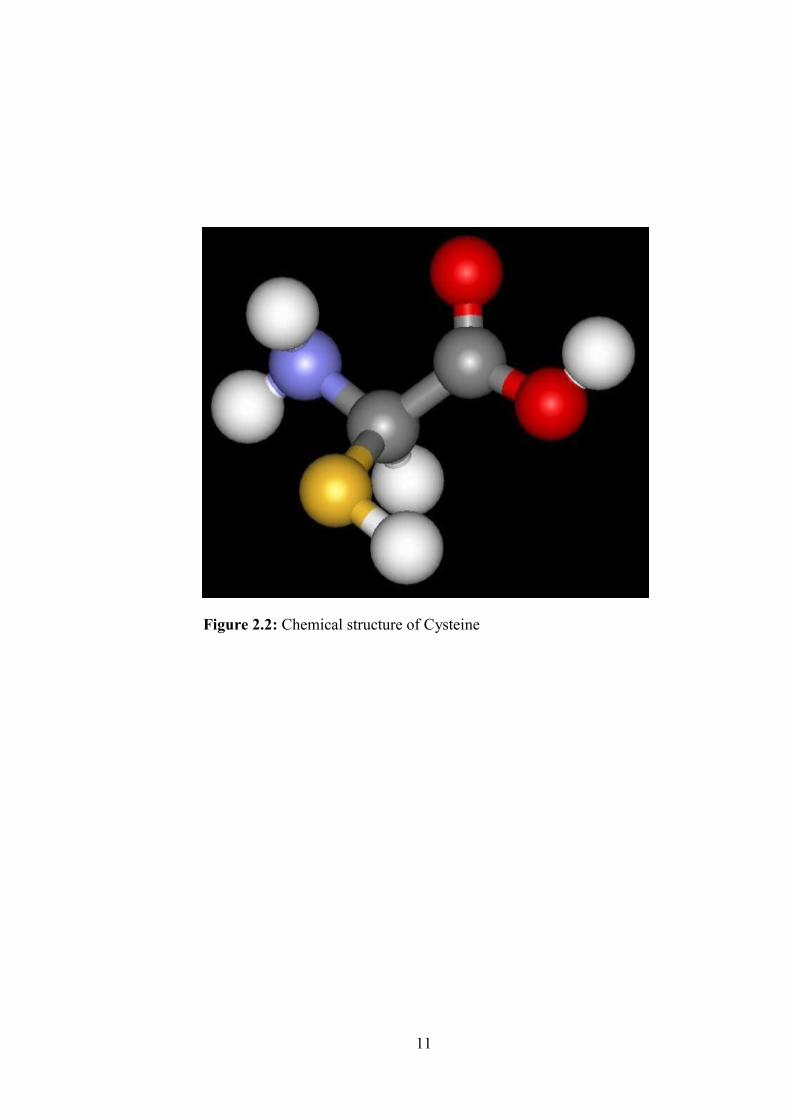
Figure 2.2: Chemical structure of Cysteine 11
Figure 2.3 Various steps involved in the development and evaluation of
radioprotective property of medicinal plants
24
Figure 2.4 Principle of comet assay in alkaline micro-gel electrophoresis
to cellular DNA damage.
30
Figure 2.5 Comet images with different level of DNA damage (A)
normal cell; (B) irradiated cell
31
Figure 2.6 (a): Genoprotective effect of the irradiated pUC18 plasmid
DNA in the presence of plant extract (b): various form of
plasmid
33
Figure 2.7 Stages of mitotic division in cells of Allium cepa exposed to
radiation and treated with plant extract
37
Figure 2.8 In vivo screening assays 39
Figure 2.9 Polyalthia longifolia 47
Figure 2.10 Various phytochemicals isolated from Polyalthia longifolia 51
Figure 3.1 Transverse section of the leaf of Polyalthia longifolia 72
Figure 3.2 Powder microscopic features of leaves of Polyalthia
longifolia
73
Figure 3.3 Extracted Ion Chromatogram of (A) P. longifolia leaf
methanolic extract and (B) standard rutin
76
Figure 3.4 A mass spectra of rutin in positive ion mode 77
Figure 3.5 Calibration curve of standard rutin 77
Figure 3.6 HPTLC profile of Polyathia longifolia leaf extract under
visible light (a) and 365 nm UV light (b)
81
Figure 3.7 HPTLC Chromatogram of Polyalthia longifolia leaf extract
showing the present of 10 peaks
82
Figure 3.8 FT-MIR spectrum of methanolic leaf extract of Polyalthia
longifolia
83

xvii
Figure 4.1 Standard curve for determination of Gallic acid Equivalents
for total phenolic content of leaf extract of P. longifolia
104
Figure 4.2 Standard curve for determination of catechin Equivalents for
total flavonoids content of leaf extract of P. longifolia
104
Figure 4.3 Scavenging effect of methanolic leaf extract of Polyalthia
longifolia on DPPH free radicals compared with butylated
hydroxytoluene (BHT).
106
Figure 4.4 Regression analyses for total phenolic content and 2-
diphenyl-1-picrylhydrazyl (DPPH) radical scavenging activity
determined in Polyalthia longifolia methanolic leaf extract
106
Figure 4.5 Reducing power of methanolic leaf extract of Polyalthia
longifolia compared to butylated hydroxytoluene (BHT)
108
Figure 4.6 Scavenging effect of methanolic leaf extract of Polyalthia
longifolia on hydroxyl radicals compared to ascorbic acid
109
Figure 4.7 Scavenging effect of methanolic leaf extract of Polyalthia
longifolia on nitric oxide radicals compared with Quercetin
110
Figure 4.8 Scavenging effect of methanolic leaf extract of Polyalthia
longifolia on ferrous ions compared to ascorbic acid
112
Figure 4.9 Anti-lipidperoxidation activity of methanolic leaf extract of
Polyalthia longifolia compared to ascorbic acid
113
Figure 4.10 Light microphotographs of liver cell of control (a), mice
exposed to paracetamol (b) and treated with Polyathia
longifolia extract (c)
116
Figure 5.1 Effect of single dose (5000 mg/kg) administration of the
Polyalthia longifolia leaf extract in rats
140
Figure 5.2 Representative histological photomicrographs of (A) spleen,
(B) lung, (C) liver of control and Polyalthia longifolia leaf
extract treated groups (at dose 5000 mg/kg).
143
Figure 5.2 Continued. (D) Kidney and (E) heart of control and
Polyalthia longifolia leaf extract treated groups (at dose 5000
mg/kg)
144
xviii
Figure 5.3 Agarose gel electrophoretic analysis of fenton-mediated DNA
oxidation
147
Figure 5.4 Effect of concentration on cytotoxicity of Polyalthia
longifolia leaf extract on Vero cells
149
Figure 5.5 The quantitation of DNA damage and repair in Vero cell line
represent the comet tail length
150
Figure 5.6 Protective effect of Polyalthia longifolia leaf extract against
H2O2-induced DNA damage and migration
151
Figure 5.7 Chromosome aberrations observed in Allium cepa
meristematic cells exposed to extracts of Polyalthia longifolia
leaf
154
Figure 6.1 (a) Survival rate of the X-ray irradiated mice treated with or
without Polyalthia longifolia (500 mg/kg and 250 mg/kg
b.w.). (b) Body weight response of mice pretreated with or
without Polyalthia longifolia for 30 days at exposure of 10
Gy whole body irradiation
175
Figure 6.2 General radiation Sickness and behavioral appearance of the
mice from control and experimental groups
176
Figure 6.3 Haematological alteration in blood of the mice post whole
body exposure to 10 Gy irradiation at with or without
Polyalthia longifolia at dose 500 mg/kg b.w. and 250 mg/kg
b.w. (a) Haemoglobin; (b) red blood cell; (c) white blood cell;
(d) platelets
178
Figure 6.4 Colony forming units (CFU) in spleen treated with Polyalthia
longifolia at dose 500 mg/kg b.w. and 250 mg/kg b.w.
180
Figure 6.5 Effect of Polyalthia longifolia administration on lipid
peroxidation (LPx) in Swiss albino mice
182
Figure 6.6 Effect of Polyalthia longifolia administration on superoxide
dismutase (SOD) activity in Swiss albino mice
183
Figure 6.7 Effect of Polyalthia longifolia administration on catalase
activity in Swiss albino mice
185
xix
Figure 6.8 Macropathology of (A) spleen; (B) Small intestine (ileum). 186
Figure 6.9 Histopathological demonstration of protective effect of
Polyalthia longifolia in the spleen of irradiated mice.
187
Figure 6.10 Histopathological demonstration of protective effect of
Polyalthia longifolia in the small intestine (Ileum) of
irradiated mice
189
Figure 6.11 Histopathological demonstration of protective effect of
Polyalthia longifolia in the Liver of irradiated mice
191
Figure 7.1 Schematic illustration of the solvent ratio for the partitions
obtained
210
Figure 7.2 Schematic diagram of solvent-solvent extraction 211
Figure 7.3 Flow chart of the isolation and identification of the bioactive
compound(s) from Polyalthia longifolia leaf extract
215
Figure 7.4 Four partitions were obtained from solvent-solvent extraction
method
217
Figure 7.5 Hexane fractions (Hex1-8) obtained from Medium Pressure
Liquid Chromatography (MPLC)
218
Figure 7.6 Ethyl acetate fractionS (EtOA1-8) obtained from Medium
Pressure Liquid Chromatography (MPLC)
219
Figure 7.7 Butanol fractionS (BuOH1-8) obtained from Medium
Pressure Liquid Chromatography (MPLC)
220
Figure 7.8 DPPH radical scavenging activity of (a) hexane, (b) ethyl
acetate and (c) butanol sub fractions of Polyalthia longifolia
obtained from MPLC
221
Figure 7.9 Pure compound (yellow) isolated from EtOAc_F007 using
HPLC
223
Figure 7.10 HPLC chromatograms of antioxidant compound isolated from
EtOAc_F007 fraction of Polyalthia longifolia leaf at 11.862
retention time
224
Figure 7.11 Positive full scan of EtOAc_F007 isolated from P. longifolia
leaf using LC-QTOF-MS
225
xx
Figure 7.12 MS/MS spectra and proposed fragmentation pathways for the
EtOAc_F007 isolated antioxidant compound from P.
longifolia leaf at the collision energy of 10 eV
226
Figure 7.13 Typical patterns and the percentage of abundance of rutin at
higher collision energy of 20 and 40 eV in ESI positive mode
228
Figure 7.14 Chemical structure of rutin 229
xxi
LIST ABBREVIATIONS
ANOVA BHT DMSO DPPH GAE LC-MS IC50
LD50 Rf HPTLC v/v w/v TLC SD ABS ROS AST ALP ALT CFU M.I. DRF P. longifolia
TBIL
Analysis of varians Buthylated hydroxytoluene Dimethyl sulfoxide 2, 2-diphenyl-1-picrylhydrazyl Gallic acid equivalents Liquid Chromatography-Mass Spectrometry Inhibitory Concentration at 50% Lethality Dosage at 50% Retention Factor High Performance Thin Layer Chromatography Volume per volume Weight per volume Thin Layer Chromatography Standard Deviation Absorbance Reactive Oxygen Species Aspartate aminotransferase Alkaline phosphate Alanine aminotransferase Colony Forming Units Mitotic Index Dose Reduction Factor
Polyalthia longifolia Total billirubin
xxii
MTT CV TBE MTD SOD LPX
HB WBC RBC HPLC MPLC DAD UV RRLC QTOF-MS ESI OECD
3-[4,5-dimethylthiazol-2-yl]-2,5 diphenyl tetrazolium bromide Central vein Tris-borate-EDTA Maximum Tolerable Dose Superoxide Dismutase Lipid Peroxidation Heamoglobin White Blood Cell Red Blood Cell High Performance Liquid Chromatography Medium Pressure Liquid Chromatography Diode Array Detector Ultraviolet Rapid Resolution liquid chromatography Accurate-Mass quadrupole time of flight mass spectrometer Electron spray ionization
Organization for Economic Co-operation and Development
xxiii
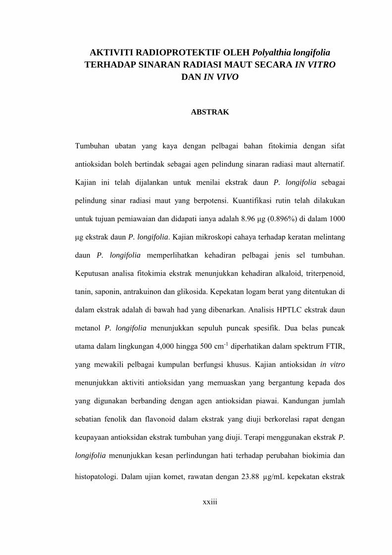
AKTIVITI RADIOPROTEKTIF OLEH Polyalthia longifolia
TERHADAP SINARAN RADIASI MAUT SECARA IN VITRO
DAN IN VIVO
ABSTRAK
Tumbuhan ubatan yang kaya dengan pelbagai bahan fitokimia dengan sifat
antioksidan boleh bertindak sebagai agen pelindung sinaran radiasi maut alternatif.
Kajian ini telah dijalankan untuk menilai ekstrak daun P. longifolia sebagai
pelindung sinar radiasi maut yang berpotensi. Kuantifikasi rutin telah dilakukan
untuk tujuan pemiawaian dan didapati ianya adalah 8.96 μg (0.896%) di dalam 1000
μg ekstrak daun P. longifolia. Kajian mikroskopi cahaya terhadap keratan melintang
daun P. longifolia memperlihatkan kehadiran pelbagai jenis sel tumbuhan.
Keputusan analisa fitokimia ekstrak menunjukkan kehadiran alkaloid, triterpenoid,
tanin, saponin, antrakuinon dan glikosida. Kepekatan logam berat yang ditentukan di
dalam ekstrak adalah di bawah had yang dibenarkan. Analisis HPTLC ekstrak daun
metanol P. longifolia menunjukkan sepuluh puncak spesifik. Dua belas puncak
utama dalam lingkungan 4,000 hingga 500 cm-1 diperhatikan dalam spektrum FTIR,
yang mewakili pelbagai kumpulan berfungsi khusus. Kajian antioksidan in vitro
menunjukkan aktiviti antioksidan yang memuaskan yang bergantung kepada dos
yang digunakan berbanding dengan agen antioksidan piawai. Kandungan jumlah
sebatian fenolik dan flavonoid dalam ekstrak yang diuji berkorelasi rapat dengan
keupayaan antioksidan ekstrak tumbuhan yang diuji. Terapi menggunakan ekstrak P.
longifolia menunjukkan kesan perlindungan hati terhadap perubahan biokimia dan
histopatologi. Dalam ujian komet, rawatan dengan 23.88 µg/mL kepekatan ekstrak

xxiv
P. longifolia selama 24 jam pada sel-sel Vero menyebabkan pengurangan kerosakan
DNA sebanyak 50.94% berbanding dengan kawalan yang tidak dirawat. Dalam ujian
relaksasi plasmid and komet ekstrak daun P. longifolia mempamerkan aktiviti
perencatan yang kuat terhadap kesan kerosakan yang diakibatkan oleh H2O2.
Peningkatan dalam nilai aberasi kromosom yang bersandarkan kepada kepekatan
ekstrak juga telah diperhatikan dalam ujian Allium cepa. Keabnormalan yang
diperhatikan adalah seperti kelekitan, c-mitosis, jambatan dan kromosom vagrant.
Sel mikronukleus juga telah diperhatikan dalam interfasa. Eksperimen ini adalah
laporan pertama tentang kesan perlindungan P. longifolia terhadap kerosakan DNA
yang disebabkan oleh radikal hidroksil. Tambahan pula, dalam ujian ketoksikan oral
akut, tikus betina telah dirawat dengan kepekatan ekstrak daun P. longifolia
sebanyak 5000 mg/kg berat badan tikus dan diperhatikan untuk tanda-tanda
ketoksikan selama 14 hari. Ekstrak daun P. longifolia tidak menunjukkan sebarang
tanda-tanda kesan toksik yang menunjukkan bahawa daun tersebut tidak toksik
berkaitan dengan rawatan tersebut. Seterusnya, kesan aktiviti radioprotektif P.
longifolia telah dikaji dengan menggunakan tikus. Rawatan P. longifolia pada tikus
menunjukkan peningkatan yang signifikan dalam jumlah hari tikus hidup (27 hari),
berbanding dengan 100% kematian dalam kumpulan tikus yang didedahkan pada
radiasi dalam tempoh 14 hari. Peningkatan ketara dalam kepekatan hemoglobin, sel
darah merah, sel darah putih dan jumlah platelet diperhatikan pada haiwan yang
menerima prarawatan ekstrak daun P. longifolia berbanding dengan kumpulan yang
didedahkan radiasi sahaja. Pemberian ekstrak daun P. longifolia sebelum
pendedahan radiasi juga telah meningkatkan jumlah CFU limpa dan saiz limpa
relatif. Penurunan yang bergantung kepada dos rawatan dalam nilai pengoksidaan
lipid diperhatikan dalam haiwan yang menerima prarawatan dengan P. longifolia.

xxv
Walau bagaimanapun, haiwan yang menerima prarawatan dengan P. longifolia
mempamerkan peningkatan yang ketara dalam aktiviti superoksida dismutase dan
katalase, tetapi nilai-nilai ini kekal di bawah nilai biasa di dalam hati dan usus.
Prarawatan dengan P. longifolia sebelum pendedahan pada radiasi juga
menyebabkan regenerasi semula krip mukosa dan vili usus. Tambahan pula, rawatan
awal ekstrak daun P. longifolia juga menunjukkan kesan perlindungan ke atas
kerosakan hati yang disebabkan oleh sinaran radiasi-X pada tikus dengan pemulihan
struktur sel hati yang normal dan pengurangan ketara aras ALT, AST dan tahap
bilirubin berbanding dengan tikus yang didedahkan dengan sinaran radiasi-X.
Keputusan ini menunjukkan keupayaan radioprotektif ekstrak daun P. longifolia
yang dimanifestasikan dalam beberapa sistem haiwan ujian. Untuk mengenal pasti
sebatian antioksidan, ekstrak daun P. longifolia telah dikenakan fraksinasi
berpandukan bioasai. Fraksi P. longifolia etil asetat, iaitu EtOAc_F007 menunjukkan
aktiviti antioksidan tertinggi di kalangan semua fraksi yang diuji. Analisis
selanjutnya dengan menggunakan kaedah LC-MS terhadap fraksi EtOAc_F007
membawa kepada pengenalan sebatian rutin sebagai agen antioksidan dalam ekstrak
P. longifolia. Kesimpulannya, hasil kajian ini menyokong potensi penggunaan
ekstrak daun P. longifolia sebagai produk semula jadi untuk diaplikasikan di masa
hadapan sebagai pembangunan agen pelindung radiasi maut baru yang bersifat
semula jadi.

xxvi
IN VITRO AND IN VIVO RADIOPROTECTIVE ACTIVITIES OF
Polyalthia longifolia AGAINST LETHAL IRRADIATION
ABSTRACT
Medicinal plants rich with various phytochemicals with antioxidant properties could
serve as an alternative radioprotective agent. The current study was designed to
evaluate the P. longifolia leaf extract as a potential radioprotector. Rutin
quantification was performed for standardization and was found to be 8.96 µg
(0.896%) in 1000 µg of P. longifolia leaf extract. Light microscopy of a transverse
section of the leaf of P. longifolia revealed the presence of various plant cells.
Phytochemical screening results of the extract revealed the presence of alkaloids,
triterpenoids, tannins, saponin, anthraquinones, and glycosides. The concentrations
of heavy metals determined in the extract were well below the permissible limit. The
HPTLC analysis of the methanolic extract of P. longifolia leaf showed ten specific
peaks. Twelve major peaks in the range of 4,000 to 500 cm-1 were observed in the
FTIR spectra, which represented various specific functional groups. The in
vitro antioxidant assays showed considerable in vitro antioxidant activities in a dose-
dependent manner when compared to the standard antioxidant Phenolic and
flavonoid content of these extracts is significantly correlated with antioxidant
capacity. Therapy of P. longifolia showed the liver protective effect on biochemical
and histopathological alterations. Moreover, histological studies also supported the
biochemical finding, that is, the maximum improvement in the histoarchitecture of
the liver. In the comet assay, the treatment of 23.88 µg/mL of P. longifolia extract
for 24 h on Vero cells caused decrease in DNA damage by approximately 50.94%
xxvii
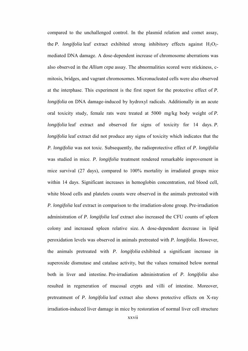
compared to the unchallenged control. In the plasmid relation and comet assay,
the P. longifolia leaf extract exhibited strong inhibitory effects against H2O2-
mediated DNA damage. A dose-dependent increase of chromosome aberrations was
also observed in the Allium cepa assay. The abnormalities scored were stickiness, c-
mitosis, bridges, and vagrant chromosomes. Micronucleated cells were also observed
at the interphase. This experiment is the first report for the protective effect of P.
longifolia on DNA damage-induced by hydroxyl radicals. Additionally in an acute
oral toxicity study, female rats were treated at 5000 mg/kg body weight of P.
longifolia leaf extract and observed for signs of toxicity for 14 days. P.
longifolia leaf extract did not produce any signs of toxicity which indicates that the
P. longifolia was not toxic. Subsequently, the radioprotective effect of P. longifolia
was studied in mice. P. longifolia treatment rendered remarkable improvement in
mice survival (27 days), compared to 100% mortality in irradiated groups mice
within 14 days. Significant increases in hemoglobin concentration, red blood cell,
white blood cells and platelets counts were observed in the animals pretreated with
P. longifolia leaf extract in comparison to the irradiation-alone group. Pre-irradiation
administration of P. longifolia leaf extract also increased the CFU counts of spleen
colony and increased spleen relative size. A dose-dependent decrease in lipid
peroxidation levels was observed in animals pretreated with P. longifolia. However,
the animals pretreated with P. longifolia exhibited a significant increase in
superoxide dismutase and catalase activity, but the values remained below normal
both in liver and intestine. Pre-irradiation administration of P. longifolia also
resulted in regeneration of mucosal crypts and villi of intestine. Moreover,
pretreatment of P. longifolia leaf extract also shows protective effects on X-ray
irradiation-induced liver damage in mice by restoration of normal liver cell structure
xxviii
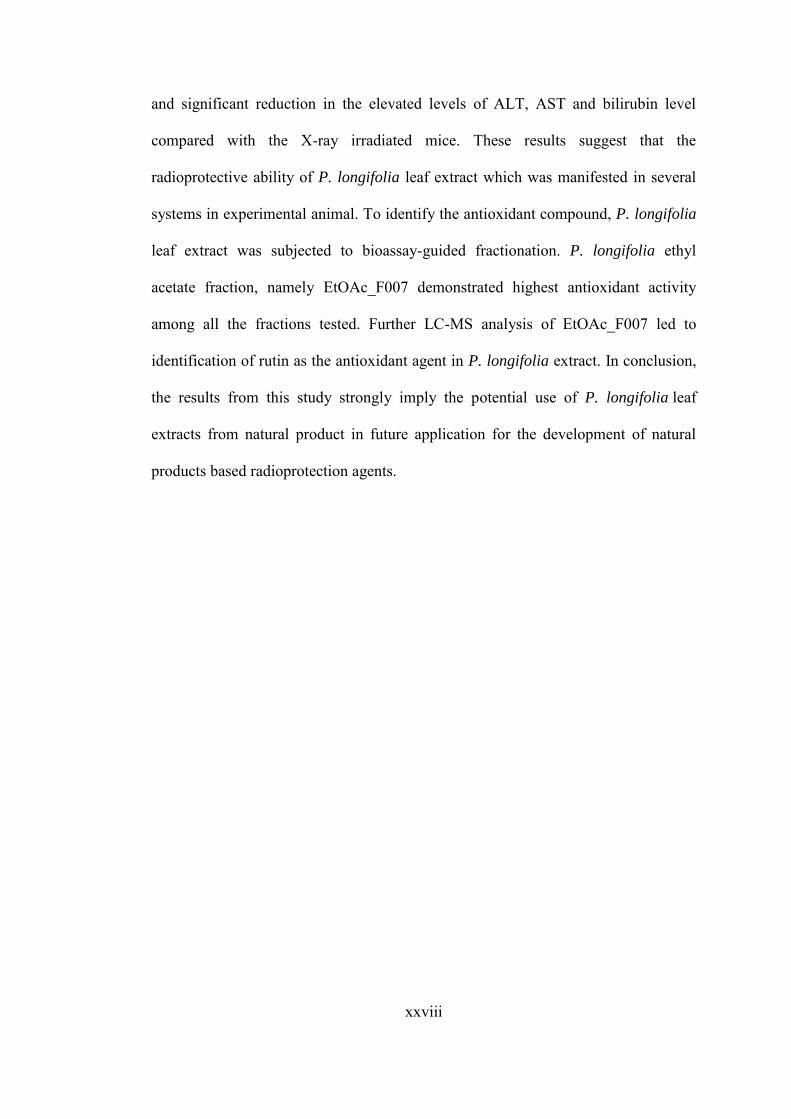
and significant reduction in the elevated levels of ALT, AST and bilirubin level
compared with the X-ray irradiated mice. These results suggest that the
radioprotective ability of P. longifolia leaf extract which was manifested in several
systems in experimental animal. To identify the antioxidant compound, P. longifolia
leaf extract was subjected to bioassay-guided fractionation. P. longifolia ethyl
acetate fraction, namely EtOAc_F007 demonstrated highest antioxidant activity
among all the fractions tested. Further LC-MS analysis of EtOAc_F007 led to
identification of rutin as the antioxidant agent in P. longifolia extract. In conclusion,
the results from this study strongly imply the potential use of P. longifolia leaf
extracts from natural product in future application for the development of natural
products based radioprotection agents.
1
CHAPTER 1.0: GENERAL INTRODUCTION
1.1 Overview and rationale of the study
The adverse effects of radiation has begun to realize immediately after the discovery
of X-ray by Roentgen (1896) and radioactivity by Becquerel (1896) and it was
considered as a remarkable turning point of human health care. Basically, humans
are constantly exposed to radiation either from planned included diagnostic,
therapeutic uses and industrial sources or unplanned included the nuclear explosion
such as atomic bomb blast, which brought tremendous damage at Hiroshima and
Nagasaki, Japan in 1945 and the natural background radiation emanating from the
earth or other radioactive sources (Jagetia, 2007). In general, the chances of radiation
exposure have been increased extensively and this further enhance by the rapid
advancement in technology which also leads to the additional radiation stresses.
Broadly, the radiation sources can be categorised into ionizing and non-
ionizing radiation. Ionizing radiation can be defined as any types of electromagnetic
or particles radiation with sufficient energy to knock electrons off of atoms or
molecules and such phenomenon known as ionization. Ionized molecules are
unstable and quickly undergo chemical changes. The amount of damage in the cell is
related to the dose of radiation it receives. The types of ionizing radiation included
X-ray and Gamma ray which widely used in cancer treatment. Meanwhile, non-
ionizing radiation has low energy that does not directly damage in molecules.
Common types of non-ionizing radiation include ultraviolet (UV) rays, visible light,
infrared rays, microwaves, and radiofrequency rays (radio waves), and cell phones
all emit (send out) non-ionizing radiation.
Ionizing radiation is an important modality in cancer treatment and almost
80% of cancer patients required radiotherapy during some point of their clinical

2
management either for curative or palliative purpose (Piya Paul et al., 2011). The
basic principle of radiotherapy is to destroy the cancer cells. However, the radiation
also induced damage to the normal tissues which results in adverse side effects after
months or years of therapy and this restricts the therapeutic doses of radiation, hence
limits the effectiveness of the treatments. Consequently, when the ionizing radiation
passing through living tissues it transfuses deleterious effects in biological system
through direct deposition of energy into crucial bio-macromolecules or by radiolysis
of water which leads to generation of reactive free radicals such as hydroxyl radicals
and hydrogen radicals. Thus the overproduction of these free radicals tend to become
reactive oxygen species and toxic which can interact with the critical bio-
macromolecules such as DNA, proteins, or membranes and induce cell damage
eventually leads to cell dysfunction and death (Hosseinimehr, 2007). In general, the
amount of reactive oxygen species increases in the biological system following
exposure to irradiation with sufficient dose and this directly correspond to the
intensity of cell damage.
The radiation has been considered an enigma to the general public and the
use of radiation for therapeutic purpose as well as spectacular advances made further
increases awareness of human health and such phenomenon been always associated
with some skepticism. The use of radioprotectors represents an obvious strategy to
improve the therapeutic index in radiotherapy. Therefore, the development of
effective radioprotectors is an area of great significance due to its wide applications
in planned as well as unplanned radiation exposure to reduce the risk of radiation
injury or severity of damage to normal tissues (Jagetia, 2007; Piya Paul et al., 2011).
Research in the development of radioprotectors world wide has focused on screening
a variety of chemical and biological compounds. Among the molecular
3
radioprotectors, WR-2721 [S-2-(3-aminopropyl-amino)] ethyl phosphorothioic acid
also known as amifostine, ethiophos (USA) or gammaphos (former USSR), is the
most thoroughly investigated radioprotective drug, developed at Walter Reed Army
Research Institute, under the Antiradiation Drug Development Program USA
(Schuchter and Glick, 1993; Sweeney, 1979). However, the strategy becomes
jeopardized when it comes to using synthetic chemical compound during
radiotherapy as it associated with undesirable side effects at clinically effective doses
and exorbitant cost the limitation greatly restricted in clinical use. In addition,
conflicting preclinical and clinical reports formulate it convoluted to accept the use
of synthetic compounds during radiotherapy in an unequivocal manner (Jena et al.,
2010). Therefore, the side effects profile of these compounds necessitated the search
for alternative drugs, which could be less toxic and highly effective at optimum dose
levels. Hence, such phenomenon diverts many researches attention towards the
plants and natural products focus on new drug discovery which would be safer than
available synthetic drugs.
Plant products have various pharmacological properties and have been used
to treat various diseases since ancient time based on traditional medicinal system.
The problem of safety of the pharmaceutical products with modern system of
medicine triggers increase in global interest in medicinal plants. Therefore,
medicinal plants have gained importance in the international market. Moreover,
more than 50% of drugs in markets are still based on natural products. In recent
years, herbal market mainly in the United States, Germany, France, India, Japan and
others have become huge potential market and has great room for scientific research
and technology. According to WHO (World Health Organization), more than 80% of
world population relies on traditional medicines, largely plant based for their

4
primary health care needs. Medicinal plants are currently in demand and their
popularity is increasing day by day in many parts of the world and has made a great
contribution towards maintaining human health. In addition, plant extracts eliciting
radioprotective efficacy contain a plethora of compounds including antioxidants,
immunostimulants, and cell proliferation stimulators, antiinflammatory and
antimicrobial agents, some will act in single compound as well as in combination
with other compounds from the same plant. Most studies using natural plant products
have focused on evaluation of radioprotective efficacy of whole extracts or
polyherbal formulations, and in some cases fractionated extracts and isolated
constituents (Arora et al., 2005). In general, biologically active compounds isolated
from plant largely contribute to medicinal field compare to the available synthetic
products. This may be due to the variability in chemical structures of secondary
metabolites which increasing the potential of new defense mechanisms against
various radiations induce damage.
A good radioprotector should be able to protect against the deleterious effects
of ionizing radiation either during therapeutic procedures as well as during nuclear
accidents or background irradiation. Apart from that, the agents should meets all the
prerequisites of an ideal radioprotectors including should be cheap, no cumulative or
irreversible toxicity in a wide dose range, provides effective long-term protection,
remains stable for a number of years without losing shelf life, and can be easily or
orally administered (Arora et al., 2006a; Arora et al., 2006b). Apart from that,
radioprotective activity of plant mediated through several mechanisms, including
radicals scavenging potential, detoxifying the radiation induced reactive species,
inducing cellular radioprotector such as superoxide dismutase (SOD) and
glutathione, enhancing the DNA repair by triggering one or more cellular DNA
5
pathways and also able to delay cell division and inducting hypoxia in the tissues
(Nair et al., 2001). Since, plant products possess complex mixture of active chemical
ingredients, therefore, it is able to contribute more on novel approaches of
radioprotection and mechanistic aspects as mention above, hence, it would be safe
and effective paradigm for radioprotection.
Malaysia being one of the 12 mega-biodiversity centers of the world is rich in
all three levels of biodiversity, namely, species diversity, genetic diversity and
habitat diversity with many plants used for medicinal and nutritional purposes. There
are more than 35,000 plant species being used in various medicinal purposes around
the world. In Peninsular Malaysia 1,200 species of higher plants and 2,000 species in
Sabah and Sarawak are reported to have medicinal value and have been used for
many generation in traditional health care system (Yoga Latha et al., 2011).
Therefore scientific investigation may be utilized to develop drugs for various
diseases which is easily accessible, available and affordable for the poor community
of bottom billion society.
Polyalthia longifolia leaf has been used as potential medicinal plants since
ancient time based on traditional medicine systems. The P. longifolia was widely
used in traditional medicine as febrifuge and tonic (The Wealth of India, 1969).
Fundamentally, the selected plant should be rich in antioxidants to minimize free
radical generation, a savor for macromolecules like lipids, proteins and DNA, should
be able to enhance internal defense mechanism, possess properties of potential
disinfectant and also a good immune rejuvenator. Hence, current study was designed
to evaluate the Polyalthia longifolia methanolic leaf extract as a potential
radioprotector.
6
1.1.1 Research objectives
The current study was undertaken with the following objectives:
1. To standardize the methanolic extract of P. longifolia leaf
2. To evaluate the in vitro antioxidant activity, in vivo hepatoprotective activity
and to determine total phenolic and flavonoid contents of methanolic extract
of P. longifolia leaf
3. To evaluate the potential genoprotective effect of the methanolic extract of P.
longifolia leaf
4. To evaluate in vivo radioprotective activities of the methanolic extract of P.
longifolia leaf using an animal model.
5. To isolate and identify of active compound/ fraction with antioxidant activity
from the methanolic extract of P. longifolia leaf
7
CHAPTER 2.0: LITERATURE REVIEW
2.1 Radiation
Human is constantly exposed to lethal radiation either from planned radiation, such
as during radiotherapy or unplanned radiation, such as the nuclear industry, sun’s
radiation and natural background radiation emanating from the earth or other
radioactive sources. Once exposed to this lethal radiation, it will cause various
adverse implications in our bodily system by the deposition of energy directly into
the bio macromolecules, which leads to the production of free radicals, as shown in
Figure 2.1. The free radicals are fundamental in modulating various biochemical
processes and represent an essential part of aerobic life and metabolism (Tiwari,
2001). The most common Reactive Oxygen Species (ROS) include superoxide
anion (O2-), hydrogen peroxide (H2O2) and hydroxyl radicals (OH-), which result
from the cellular redox processes. At low or moderate concentrations, ROS exert
beneficial effects on cellular response and the immune function, however, at high
levels, these radicals become toxic and disrupt the antioxidant defence system of the
body, which may lead to “oxidative stress” (Pham-Huy et al., 2008). These reactive
oxygen species, in turn, react with different bio-molecules viz., lipid, DNA, proteins
and inflict oxidative damage in them (Figure 2.1).
The mediated reactions of major reactive oxygen species (ROS) include lipid
peroxidation, removal of thiol group from cellular and membrane proteins, strand
breaks and base alterations leading to DNA damage (Shukla and Gupta, 2010). After
the widespread realization concerning the adverse effects from lethal irradiation
various safety measurements were introduced to overcome this problem. However,
the radioprotective system developed against the lethal irradiation, most of the time,
8
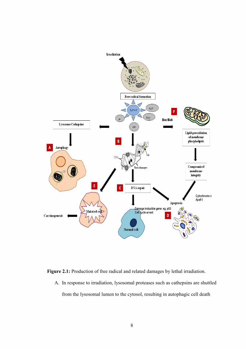
Figure 2.1: Production of free radical and related damages by lethal irradiation.
A. In response to irradiation, lysosomal proteases such as cathepsins are shuttled
from the lysosomal lumen to the cytosol, resulting in autophagic cell death
9
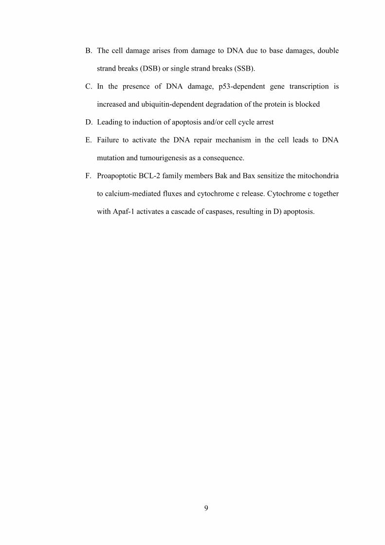
B. The cell damage arises from damage to DNA due to base damages, double
strand breaks (DSB) or single strand breaks (SSB).
C. In the presence of DNA damage, p53-dependent gene transcription is
increased and ubiquitin-dependent degradation of the protein is blocked
D. Leading to induction of apoptosis and/or cell cycle arrest
E. Failure to activate the DNA repair mechanism in the cell leads to DNA
mutation and tumourigenesis as a consequence.
F. Proapoptotic BCL-2 family members Bak and Bax sensitize the mitochondria
to calcium-mediated fluxes and cytochrome c release. Cytochrome c together
with Apaf-1 activates a cascade of caspases, resulting in D) apoptosis.
10
is burdensome to use or less practical in various situations, such as during space
travel. Therefore, medicinal plants rich with various phytochemicals with
antioxidant properties could serve as an alternative radioprotective agent and could
be the most practical strategy to protect from lethal irradiation, which leads to
various diseases including cancer. The development of safe, non-toxic, cheap,
reliable and accessible radioprotective agents is crucial to overcome radiation related
problems, especially for patients undergoing radiotherapy. Plants will be the ideal
source to achieve this noble intention. In 1948, for the first time, Patt et al. (1949)
discovered that cysteine (Figure 2.2) was a radioprotector agent and proved that it
protected mice against the harmful effects of X irradiation. Ever since then, a
number of compounds have been evaluated by various scientists from various
sources including plants for the development of a radioprotective agent. Medicinal
plants remain the exclusive choice for the development of a safe and effective non-
toxic radioprotector since most of the medicinal plants are rich with antioxidant
phytochemicals.
2.2 Ionization and formation of free radicals
When cells are exposed to radiation they interact with target atoms and deposit the
energy resulting in ionization or excitation. Subsequently, the absorbed energy starts
to damage the molecules directly or indirectly. The damage occurs directly through
the ionization of atoms in the key molecules in the biological system, which leads to
functional alteration of the molecule. Absorption of energy is enough to get rid of an
electron, which results in bond breaks in the molecules. Conversely, the indirect
mechanism involves ionization in the cytoplasm, which produces reactive free
radicals of which the toxicity to the essential molecules results in an adverse effect
and biological effects, as shown in Figure 2.1.
11
Figure 2.2: Chemical structure of Cysteine
12
2.2.1 Free radicals and cell death
DNA damage within the cell may occur as a result of a direct radiation hit or
indirectly from free radicals (ROS). Eukaryotic cells typically respond to radiation
by activating the DNA repair pathways and cell cycle checkpoints, followed by
either full biological recovery or cell death (Ozben, 2007) (Figure 2.1). Radiation-
induced ROS production can lead to cell death through several mechanisms
including apoptosis, necrosis and autophagy (Ozben, 2007; Azad et al., 2009;
Wochna et al., 2007).
Apoptosis is a type I programmed cell death that occurs through two main
pathways, triggered either by the release of apoptotic proteins from the mitochondria
(intrinsic pathway) or by death-receptor ligation (extrinsic pathway) (Edinger and
Thompson, 2004). Apoptosis is depicted by membrane blebbing, early collapse of
the cytoskeleton, externalisation of phosphatidylserine (PS) on the cell surface,
cytoplasmic shrinkage, chromatin condensation, and, subsequently, the formation of
apoptotic bodies. In contrast to apoptosis, necrosis is regarded as a passive form of
cell death. Necrotic cells swell and lose their membrane integrity, then lyse and
release their contents into the extracellular space, causing inflammation and damage
to the surrounding tissue (Edinger and Thompson, 2004). In many cases, apoptosis
and necrosis may occur sequentially or simultaneously within the same tissue due to
irradiation. Through a series of well-designed studies, Wochna et al. (2007)
hypothesised that the switch from apoptotic to necrotic cell death involves not only a
diminution in cellular adenosine triphosphate (ATP) during cellular dysfunction, but
also an explosion of intracellular ROS.
Mitochondria organelles are the energy powerhouse of the cell. Irradiation
causes lipid peroxidation of membrane phospholipids and compromised membrane
13
integrity resulting in the release of small molecules including cytochrome c (Liu et
al., 1996) from the intermembrane space and apoptosis-inducing factor AIF (Susin et
al., 1999), resulting in cell death. The pro-apoptotic BCL-2 family members are
mediators of cell death that reside upstream of the mitochondria (Tsujimoto, 2003).
In response to irradiation, the p53 tumour suppressor induces the expression of a
number of damage induced genes regulating apoptosis, including death receptors and
proapoptotic members of the Bcl-2 family, Bax and Bak (Chipuk et al., 2004). The
p53-induced apoptosis proceeds through a series of events from the liberation of
cytochrome c from the mitochondria to the activation of caspase cascades (Villunger
et al., 2003).
Autophagy or type II programmed cell death is caspase independent and does
not involve DNA fragmentation. In autophagic cell death, organelles in the
cytoplasm, including mitochondria, are sequestered in an autophagosome, which
then fuses with the lysosomes (Azad et al., 2009). Lysosomal proteases, cathepsins,
will be shuttled from the lysosomal lumen to the cytoplasm in response to ROS. The
hydroxide produced, as in mitochondria by ROS, diffuses into lipofuscin-loaded
lysosomes, and the hydroxide causes damage to the lysosomal membranes, which
causes the leak of lysosomal enzymes. The lysosomal enzymes permeabilise the
mitochondrial membranes, resulting in the release of cytochrome c, the apoptosis-
inducing factor (AIF), and the second mitochondria-derived activator of caspase
(Smac)/direct inhibitor of apoptosis protein binding protein with low pI (DIABLO),
hence triggering cell death (Ghavami et al., 2010; Szumiel, 2011).
2.2.2 Free radicals and cancer
However, the irradiated cells that escape cell death may undergo mutation, which
creates an error in the DNA blueprint leading to altered gene expression and protein

14
modification; peptide bond cleavage and cross linking, for example, may affect
protein localization, interactions and enzyme activity. Although ROS-mediated DNA
damage may enable cells to function partially and proliferate, they eventually
develop into cancer, especially if the regulation of the tumour suppressor genes is
impaired (Wu, 2006). The high levels of ROS in cancer cells can further contribute
to oxidative stress, which may further stimulate tumour growth, invasion,
angiogenesis and metastasis (Wu, 2006; Girdhani et al., 2005).
The level of ROS production and antioxidant signalling appear to be altered
in malignant cells, contributing to cancer progression. However, the results from
different studies have been paradoxical, for instance, superoxide dismutase (SOD)
expression has been shown to decrease cancer cell proliferation and tumorigenicity
in vitro (Oberley, 2005), albeit its expression was found to be associated with bad
prognosis in patients with gastric cancer (Kim, 2002).
2.3 Radioprotection mechanisms by plant extract or compounds
Numerous investigations on radioprotection mechanisms have been carried out in
several biological systems and the following radioprotection mechanisms have been
proposed from these studies: free radical scavenger, repair by hydrogen donation to
target molecules, formation of mixed sulphides, delaying of cellular division and
induction of hypoxia in the tissue (Varanda and Tavares, 1998). The mechanism of
free radical scavenger suggests that medicinal plants will donate electrons to the free
radicals and form a stable compound incapable of reacting with other cellular
components. This mechanism prevents the free radicals from reacting with the vital
cell components. Another mechanism that has been proposed is the repair by
hydrogen donation. If a R-H molecule is converted into a radical R by exposure to
radiation, the antioxidant plant extract or compound can donate a hydrogen atom to

15
this radical, restoring it to its original state (Biaglow, 1987), which is not vulnerable
to the vital components of our bodily system. In addition, the mechanism of the
formation of mixed sulphides suggests aminothiols, which involves radioprotector
binding to cellular components. According to this proposed mechanism, the
sulphydryl compound of medicinal plants form mixed disulphides with sulphydryl
compounds of cellular proteins. Once the free radicals generated by irradiation attack
the disulphides, the sulphur atoms will be reduced and the other sulphur atom will be
oxidized (Varanda and Tavares, 1998). This mechanism prevents the free radicals
from reacting with the vital cell components because if the sulphur atom of the
protein is reduced by the free radicals and the sulphur atom of the protective agent is
oxidized, the protein is not damaged.
Delaying of cellular division and granting additional time for repairing DNA
damage caused by irradiation has been considered a potentially important
mechanism in radioprotection activity. For this type of mechanism, Brown (1967)
proposed that the sulphydryl compounds of the radioprotective agents will bind to
the cellular DNA and inhibit its replication and provide additional time for repair of
the damaged DNA. Protection by the induction of hypoxia in the tissue has also been
considered a potentially important mechanism in radioprotection activity. Oxidation
of the radioprotective agents uses enough oxygen to reduce its tension, and it has
already been revealed that hypoxia is radioprotective. Moreover, the induction of
hypoxia in tissue in certain conditions may contribute to radioprotection.
Nevertheless, other mechanisms might be involved, since some compounds exhibit
radioprotective activity without altering the oxygen tension on the tissue (Varanda
and Tavares, 1998). There is evidence of the existence of more than one
radioprotective mechanism of a certain compound, and that one of the compounds

16
might be more or less important, depending on the irradiated system and on the
specific radiation conditions (Prasad, 1982).
2.4 Plant as anti-radiation sources
2.4.1 Traditional usage of medicinal plant as radioprotective agent
For eons, plants and plant products have been infused in human life, as palatable and
remedial sources. Traditional healers exploited plants to treat various maladies long
before the discovery of drugs (Cragg et al., 1997). Moreover, the conventional plant
preparations are also demonstrated to be non-toxic or less-toxic, considering their
derivation from natural resources.
Gingko biloba is one of the world’s ancient trees and is believed to have
survived an atomic bomb explosion dropped on Hiroshima on 6 August 1945 by the
Americans (Anonymous, 2013a). The surviving trees were found near the blast
centre and appeared to sprout without major deformations. The observation
substantiates the plant’s amazing resistance to mutagen agents like radiation
(Pickstone, 2010). On a different occasion, the Buddhist monks took delight in
tending to these trees by preserving them near the pagodas in China’s Imperial
Gardens and on sacred grounds to ward off fire. G. biloba is also denoted as a
symbol of longevity.
Although folklore does not directly imply that plants impart a radioprotective
effect, much evidence has been found of their incorporation in ceremonies and
rituals in which specific plants are utilized. The Tulsi or Ocimum sanctum, for
example, is worshipped along with milk, yogurt, honey and Ganga (river) water,
which are consumed by the devotees at the end of the ritual (McGuire, 2012). The
ancient Indian legend states that this Queen of Herbs came as an incarnation of the
Hindu goddess Tulsi and is favoured by the Lord Vishnu, Krishna and Ram (Miller
17
and Miller, 2003). A plant with radioprotective effect can also be identified with the
presence of other properties, such as anti-inflammatory, antioxidant, antimicrobial
and immune modulatory (Jagetia, 2007). Likewise, Tulsi, within the confinement of
Ayurveda, was used to regulate fever, relieve coughs and flu, and mobilize mucus in
bronchitis and asthma. The leaves especially were used to treat tuberculosis and
ringworm of the skin. The tulsi oil is rich in vitamin C, carotene, calcium and
phosphorus and is also believed to possess other properties including antibacterial,
antifungal and antiviral (Anonymous, 2013b).
Radiation interacts and distresses the atoms that compose the cells. The
affected atoms will subsequently form free radicals that disrupt molecules, cells,
tissues and organs that eventuate to the detriment of the organism (USNRC
Technical Training Center, 2013). Since free radicals are responsible for inducing
radiation-damage, the radioprotective property of Panax ginseng is associated either
directly or indirectly with its free radical scavenging capability (Lee et al., 2005).
Ginseng refers to the root and the rhizome of Panax ginseng C.A. Meyer
(Araliaceae), which have been conventionally utilised by the Chinese for more than
200 years. The Chinese believe that ginseng is a reservoir with a range of
pharmacological roles, such as restorative, tonic, nootropic and anti-aging (Lee et
al., 2005).
2.4.2 Medicinal plant with radioprotective effects
Naturally occurring herbs constitute a wide variety of antioxidants, such as alpha
carotene, ascorbic acids, flavones, flavanones, flavanols, stilbenoids, anthocyanins,
phenolic acids, etc., which are reported to have a broad spectrum of radiation
absorption properties (Bajpai et al., 2005; Ashawat et al., 2006; Nichols and Katiyar,
2010; Vaid and Katiyar, 2010). In addition, it has been shown that these
18
phytoconstituents have a synergistic photo-protective effect and can be used as
sunscreen to protect cellular damage of the skin from radiation light exposure
(Campos et al., 2006; Afaq et al., 2003). The radioprotective effect of
phytochemicals has gained popularity in skin care and attention has been focused in
developing topical formulations, which can be used as complementary as well as
alternate medicine to heal and rejuvenate skin from various disorders (Griffiths et
al., 2005; Kapoor et al., 2009; Saraf and Kaur, 2010; Svobova et al., 2003). Some of
the medicinal plants with radioprotective properties – antioxidant, anti-inflammatory
and immunomodulatory – are listed in Table 2.1.
2.5 Antiradiation compounds
Antiradiation compounds are studied by in vitro and in vivo tests that assess some of
these aspects. Assay of free radicals and antioxidant assay of pharmacological agents
are suggested as a good means for evaluating the radioprotective potential (Jagetia,
2007). The polyphenolic compounds, especially flavonoids, ubiquitously present in
plants, have been reported to possess various beneficial biological properties, most
of which are attributed to antioxidant activity. It is not surprising that radioprotective
potential has been reported for extracts of herbs containing flavonoids, as well as for
individually isolated flavonoids. The radioprotective effect of two extracts of
Caesalpinia digyna and the isolated compound bergenin were compared using in
vitro methods by Singh et al. (2009). The in vitro approach compared the protective
action against the damaging effect of protein carbonylation in bovine serum albumin,
lipid peroxidation in liposomes, and DNA breakage in pBR322 plasmid. The study
showed that the flavonoid, bergenin, from the plant is equally potent in inhibiting
DNA damage as the extracts, albeit the extracts were more potent in protecting the
proteins and lipids. The pBR322 model was also used in

19
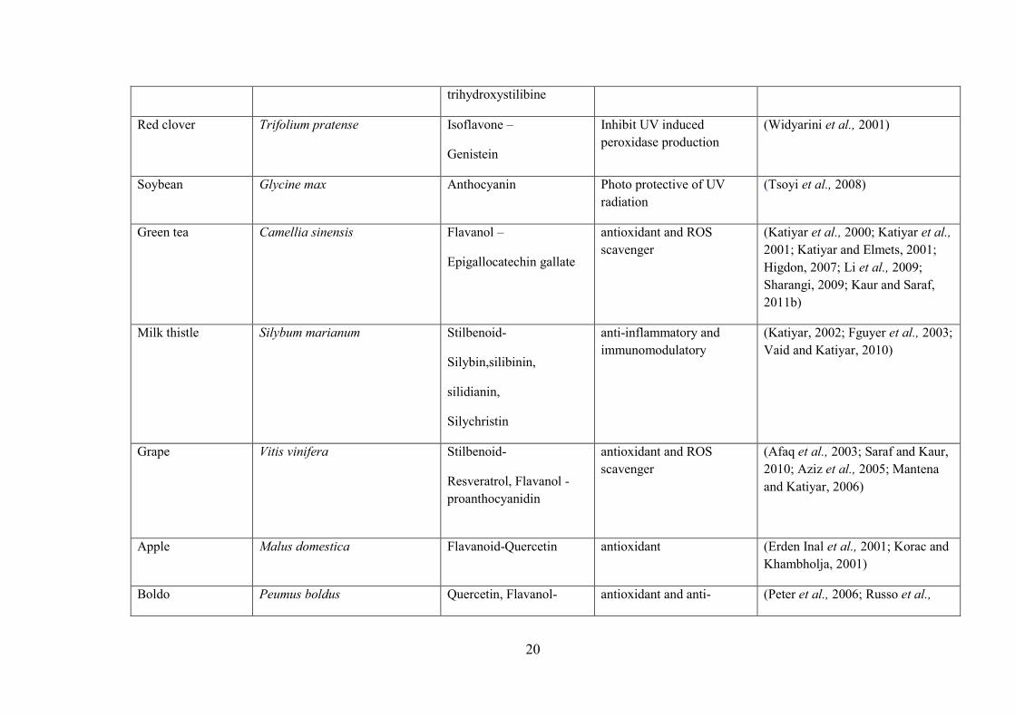
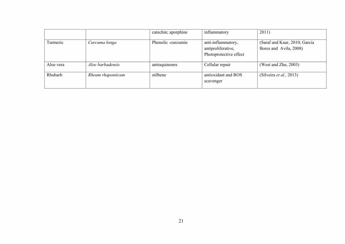
Table 2.1: Plant with radioprotective activity or antioxidant activity
Plant species Scientific names component activity Reference
Tomato Solanum Lycopersicum Carotenoids –
lycopenes
antioxidant (Griffiths et al., 2005; Saraf and Kaur, 2010; Ravichandran et al., 2005)
Carrot Daucus carota β-carotene antioxidant (Griffiths et al., 2005; Svobova et al., 2003)
Papaya Carica papaya L-ascorbic acid Antioxidant and photoprotective
(Vile , 1997)
orange Citrus sinensis L-ascorbic acid antioxidant
(Cimino et al., 2007)
Lemon Citrus limon L-ascorbic acid antioxidant (Apak et al., 2007)
Mango Mangifera indica L-ascorbic acid antioxidant
with anti-inflammatory and immunomodulatory activities.
(Song et al., 2013)
Pomegranate Punica granatum ascorbic acid antioxidant (Kumar et al., 2009)
Celery
Apium graveolens Flavones –
5,7,4’-
antioxidant and ROS scavenger
(Griffiths et al., 2005; Svobova et al., 2003)
20
trihydroxystilibine
Red clover Trifolium pratense Isoflavone –
Genistein
Inhibit UV induced peroxidase production
(Widyarini et al., 2001)
Soybean Glycine max Anthocyanin Photo protective of UV radiation
(Tsoyi et al., 2008)
Green tea Camellia sinensis Flavanol –
Epigallocatechin gallate
antioxidant and ROS scavenger
(Katiyar et al., 2000; Katiyar et al., 2001; Katiyar and Elmets, 2001; Higdon, 2007; Li et al., 2009; Sharangi, 2009; Kaur and Saraf, 2011b)
Milk thistle Silybum marianum Stilbenoid-
Silybin,silibinin,
silidianin,
Silychristin
anti-inflammatory and immunomodulatory
(Katiyar, 2002; Fguyer et al., 2003; Vaid and Katiyar, 2010)
Grape Vitis vinifera Stilbenoid-
Resveratrol, Flavanol -proanthocyanidin
antioxidant and ROS scavenger
(Afaq et al., 2003; Saraf and Kaur, 2010; Aziz et al., 2005; Mantena and Katiyar, 2006)
Apple Malus domestica Flavanoid-Quercetin antioxidant (Erden Inal et al., 2001; Korac and Khambholja, 2001)
Boldo Peumus boldus Quercetin, Flavanol- antioxidant and anti- (Peter et al., 2006; Russo et al.,
21
catechin; aporphine inflammatory 2011)
Turmeric Curcuma longa Phenolic -curcumin anti-inflammatory, antiproliferative, Photoprotective effect
(Saraf and Kaur, 2010; Garcia Bores and Avila, 2008)
Aloe vera Aloe barbadensis antraquinones Cellular repair (West and Zhu, 2003)
Rhubarb Rheum rhaponticum stilbene antioxidant and ROS scavenger
(Silveira et al., 2013)
22
assessing the protective effect of pure compounds isolated from Phyllanthus amarus
(Londhe et al., 2009). The flavonoids, quercetin 3-O-glucoside followed by rutin,
offered the greatest protection on DNA as seen by the decrease in the nicked circular
form of plasmid. However, the ellagitannins, namely amariin, 1-galloyl-2,3-
dehydrohexahydroxydiphenyl (DHHDP)-glucose, repandusinic acid, geraniin,
corilagin, phyllanthusiin D were also effective. The protective effects of these
compounds on protein and lipids damage by radiation were assessed by using rat liver
mitochondria. The compounds, rutin and repandusinic acid offered maximum
protection against lipid damage whereas protection against carbonyl formation in
proteins was highest in rutin, phyllanthusiin D, geraniin and quercetin 3-O-glucoside.
The effects of flavonoids have also been studied by using in vivo techniques.
For example, various doses of preparation containing 12 flavonoids (FAC) from seeds
of Astragalus complanatus protected mice from radiation damage (Qi et al., 2011).
Basically, FAC increased the survival rate of irradiated mice and had a protective
effect on haematopoietic tissue and the immune system. The alkaline comet assay,
which involves single cell electrophoresis was able to show the protective effect
against DNA damage in mouse liver cells by the FAC. Studies on radioprotection
have also taken advantage of the availability of synthetic drugs that have been used
clinically in humans. The protective effect of troxerution, a flavonoid derivative used
for treating venous disorders, was also ascertained by using the comet assay. In this
study, the method assessed the protection against DNA damage in mice blood, bone
marrow and tumour cells (Maurya et al., 2004).
23
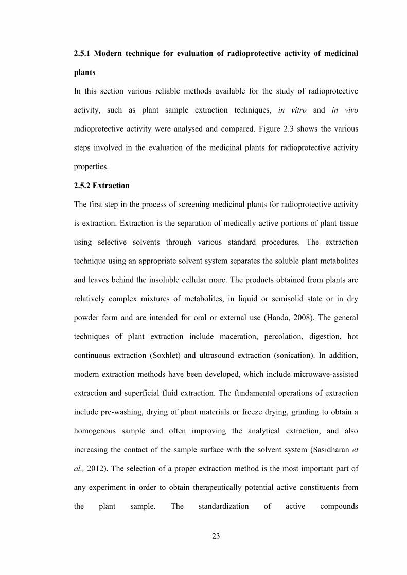
2.5.1 Modern technique for evaluation of radioprotective activity of medicinal
plants
In this section various reliable methods available for the study of radioprotective
activity, such as plant sample extraction techniques, in vitro and in vivo
radioprotective activity were analysed and compared. Figure 2.3 shows the various
steps involved in the evaluation of the medicinal plants for radioprotective activity
properties.
2.5.2 Extraction
The first step in the process of screening medicinal plants for radioprotective activity
is extraction. Extraction is the separation of medically active portions of plant tissue
using selective solvents through various standard procedures. The extraction
technique using an appropriate solvent system separates the soluble plant metabolites
and leaves behind the insoluble cellular marc. The products obtained from plants are
relatively complex mixtures of metabolites, in liquid or semisolid state or in dry
powder form and are intended for oral or external use (Handa, 2008). The general
techniques of plant extraction include maceration, percolation, digestion, hot
continuous extraction (Soxhlet) and ultrasound extraction (sonication). In addition,
modern extraction methods have been developed, which include microwave-assisted
extraction and superficial fluid extraction. The fundamental operations of extraction
include pre-washing, drying of plant materials or freeze drying, grinding to obtain a
homogenous sample and often improving the analytical extraction, and also
increasing the contact of the sample surface with the solvent system (Sasidharan et
al., 2012). The selection of a proper extraction method is the most important part of
any experiment in order to obtain therapeutically potential active constituents from
the plant sample. The standardization of active compounds

24
Plant • Extraction • Standardization
in vitro test
• Antioxidant assay• Comet assy• Plasmid relaxation assay• Allium cepa assay
in vivo test
• Endogenous spleen colony forming unit (CFU) assay
• Whole- body survival and body weight
• Gastrointestinal damage assay
Figure 2.3: Various steps involved in the development and evaluation of radioprotective property of medicinal plants
assay


