Mohd Hilmi et al. SpringerPlus 2013, 2:79http://www.springerplus.com/content/2/1/79
RESEARCH Open Access
In vitro characterization of a chitosan skinregenerating template as a scaffold for cellscultivationAbu Bakar Mohd Hilmi1, Ahmad Sukari Halim1*, Asma Hassan2, Chin Keong Lim1,3, Kartini Noorsal4
and Ismail Zainol5
Abstract
Chitosan is a marine-derived product that has been widely used in clinical applications, especially in skinreconstruction. The mammalian scaffolds derived from bovine and porcine material have many limitations, forexample, prion transmission and religious concerns. Therefore, we created a chitosan skin regenerating template(SRT) and investigated the behavior of fibroblast cell-scaffold constructs. Primary human dermal fibroblasts (HDF)were isolated and then characterized using vimentin and versican. HDF were seeded into chitosan SRT at a densityof 3×10⁶ cells/cm2 for fourteen days. Histological analysis and live cells imaging revealed that the cell-chitosanconstructs within interconnected porous chitosan showed significant interaction between the cells as well asbetween the cells and the chitosan. Scanning electron microscopy (SEM) analysis revealed cells spreading andcovering the pores. As the pore sizes of the chitosan SRT range between 40–140 μm, an average porosity is about93 ± 12.57% and water uptake ratio of chitosan SRT is 536.02 ± 14.29%, it is a supportive template for fibroblastattachment and has potential in applications as a dermal substitute.
Keywords: Chitosan SRT, Interconnected pores, Human dermal fibroblasts, Three dimensional
BackgroundChitosan is a biopolymer (β-1,4-D-glucosamine) deriva-tive of chitin (poly-N-acetylglucosamine) that has be-come an excellent biomedical resource in clinicalmedicine, especially in dermatology and plastic recon-structive surgery. Chitosan biomaterials are normallyused to cover full-thickness skin defects resulting fromtraumas (Halim et al. 1998; Pusateri et al. 2003), burns(Jin et al. 2007; Guo et al. 2011) and skin ulcers(Anandan et al. 2004; Park et al. 2009). The recent useof chitosan in regenerative medicine, particularly in skintissue engineering, has been beneficial for the treatmentof full-thickness skin defects (Tchemtchoua et al. 2011).Chitosan was reported to improve homeostasis (Kozen
et al. 2008) and promote granulation by enhancing thefunction of inflammatory cells such as leukocytes, fibro-blasts and macrophages (Ueno et al. 2001). Furthermore,chitosan has antimicrobial effects against bacteria, fungi
* Correspondence: [email protected] Sciences Unit, Universiti Sains Malaysia, Kelantan, MalaysiaFull list of author information is available at the end of the article
and viruses (Rabea et al. 2003). In addition to the skin,chitosan scaffolds also stimulate the regeneration ofspinal cord (Li et al. 2009), cornea (Auxenfans et al.2009) and liver (Wang et al. 2008) tissues in their re-spective three-dimensional (3-D) environments.As a marine product, chitosan has many advantages
compared with other collagen biomaterials that are pre-pared from bovine or porcine sources. These mamma-lian products have resulted in the transmission of prionsto humans. In contrast, marine-derived products are safefor human use due to the species barrier (Prusiner1998).Fibroblasts are mesenchymal cells that provide tissue
maintenance and support by secreting extracellularmatrix (ECM) in connective tissue. In the full-thicknessskin model, cultured allogenic fibroblasts in 3-D scaf-folds induce inflammation and scar formation, which areimportant events in wound healing (Lamme et al. 2002).This model has been reported to improve the regener-ation of dermis (Lamme et al. 2000) and epidermis (El-Ghalbzouri et al. 2002). Van den Bogaerd et al. (2002)
is is an Open Access article distributed under the terms of the Creativemmons.org/licenses/by/2.0), which permits unrestricted use, distribution, andinal work is properly cited.
Mohd Hilmi et al. SpringerPlus 2013, 2:79 Page 2 of 9http://www.springerplus.com/content/2/1/79
demonstrated that dermis is the best source for skin tis-sue engineering applications, based on fibroblast yieldsand the reduced contraction of the wound.Recently, a study conducted by Lim et al. (2010) showed
that chitosan porous skin regenerating templates (SRT)are biocompatible, as determined using primary humankeratinocytes with the direct-contact method. They foundthat chitosan porous SRT did not induce additionalinflammatory responses or DNA damage. Their datahighlighted the potential use of chitosan porous SRT inskin tissue engineering. However, its potential as a scaf-fold to support cell attachment and proliferation is stillunexplored.Therefore, this study investigated the ability of a 3-D
structure of chitosan porous SRT to support humanfibroblast spreading and viability. Chitosan SRT supportsfibroblast attachment and has potential for use in skintissue engineering applications especially in the de novofabrication of dermal substitutes.
Results and discussionIsolation and characterization of HDFOn day 2 after their isolation, primary HDF were ob-served to be attached to the culture flask and the
A
C
Figure 1 Characterization of the HDF using versican. Scale bar 100 μmgreen with FITC (B). The merged image of HDF in polygonal (asterisk) and
medium was changed to remove the floating cells anddebris. The HDF were spindle-shaped and had polygonalmorphology and were confluent on days 10 to 12.Characterization of the HDF was performed by examin-ing versican (Figure 1) a type-lll intermediate filament(IF) protein and vimentin (Figure 2) a chondroitin sul-fate proteoglycan. In both assays, the nuclei were stainedblue with DAPI and the cell membrane was stainedgreen with fluorescein isothiocyanate (FITC). Vimentinis a mesenchymal marker for which epithelial and endo-thelial cells are negative (Chang et al. 2002). Vimentinconstitute a major portion of the cytoskeleton, has animportant role in supporting organelle organization(Katsumoto et al. 1990) and contributes to the plasmamembrane fusion machinery in fibroblast (Faigle et al.2000). As an IF (10 nm filament), vimentin is involved inthe formation of membrane proteins such as cystic fi-brosis transmembrane conductance regulator (Johnstonet al. 1998). Versican is another important marker foundabundantly in dermal fibroblasts especially as versicanisoform V1 (Hattori et al. 2011). Apart from influencingthe dermal fibroblast phenotype, versican has an import-ant role in activating cells adhesion, preventing apoptosis(Wu et al. 2002) and extracellular matrix assembly
B
. Nuclei were stained blue with DAPI (A). Cell membrane was stainedelongated spindle-shape (arrow) (C).
BA
C
Figure 2 Characterization of the HDF using vimentin. Scale bar 100 μm. Nuclei were stained blue with DAPI (A). Cell membrane was stainedgreen with FITC (B). The merged Image (C).
Mohd Hilmi et al. SpringerPlus 2013, 2:79 Page 3 of 9http://www.springerplus.com/content/2/1/79
(Thomas NW, 2002). The characterization of these twomarkers is important for the future engineering of skin tis-sue. Dermal fibroblast vimentin+ for instance, is an indica-tor of the multipotency of adult stem cells (Chen et al.2007) and fibroblast versican+ mediates the mesenchymal-epithelial transition (Sheng et al. 2006). Both these charac-teristics are essential for 3-D tissue regeneration.
A
Figure 3 The macroscopic view of chitosan SRT of 5 mm diameter anbar 140 μm (B).
Characterization of chitosan SRT, cell attachment andproliferation in vitroThe macroscopic view of sponge chitosan SRT is shownin Figure 3A. Chitosan SRT is a graded porous scaffoldwith pore sizes ranging between 40 ± 15.11 μm to 140 ±18.21 μm in diameter (Figure 3B) and with other mech-anical properties described in Table 1. The viability
B
d 2 mm thickness (A). Chitosan SRT with interconnected pores. Scale
Mohd Hilmi et al. SpringerPlus 2013, 2:79 Page 4 of 9http://www.springerplus.com/content/2/1/79
analysis of HDF in 3-D cultures was described in Table 2.The HDF maintained its viability until 14 days of culturesin the chitosan scaffold. Scaffolds with interconnectedmacroscopic-microscopic pore structures allow the acceler-ation of tissue regeneration (Peter XM, 2008). Scaffoldswith macroscopic pores that are, at least 100 μm in diam-eter play a role in enhancing ingrowths of cells and bloodvessels (Chen and Ma, 2004) while microscopic porosityleads to high cell attachment, proliferation and cellular re-sponses (Ma and Choi, 2001). To allow 3-D tissue regener-ation, scaffolds should perform a few critical functions.Firstly, they should provide the cells with a proper surfacefor attachment and proliferation. Secondly, they shouldhave interconnected pores to allow uniform cells spreading.Lastly, they should provide a 3-D template for specific tis-sue reconstruction (Zeltinger et al. 2001). Using HDF, thechitosan SRT has performed these functions successfully.As a graded porous scaffold, chitosan SRT mimics the
natural porous structure more closely than a uniformlyporous scaffold. Uniformly porous scaffolds have manylimitations. They only allow one cell type to grow de-pending on the pore size, whereas graded porous scaf-fold regenerated multiple types of tissue simultaneously(Oh et al. 2007). Yannas et al. (1989) found that theoptimum pore size for skin regeneration templatesranged between 20 – 125 μm. Hence, chitosan SRT ful-fills this criterion and is suitable as a template for skintissue engineering.The ability of chitosan to adsorb nutrients involved in
wound healing and exudation of the wound bed is im-portant factors in skin regeneration. Therefore, the de-termination of water adsorption of chitosan is importantto enhance the biological activity of a dermal equivalent.The water uptake ratio of chitosan SRT (536.02 ±14.29%) was higher than reported previously by Shi et al.(2006). It can attribute to hydrophilicity and the main-tenance of 3-D structure. Scaffold with high water up-take ratio is suitable both in full thickness wound thatcontains excess exudates and chronic inflammation thatleading to impair wound healing. To enhance the dermal
Table 1 Tabulated data for the characterization ofchitosan SRT (mean ± SEM, n = 6)
Properties Units
Tensile strength 5.02 ± 0.73 N/m2
Average pore diameter
Small pore 39.41 ± 15.11 μm
Large pore 143.55 ± 18.21 μm
Average porosity 93.21 ± 12.57%
Moister flux 3.97 ± 0.34 mg/cm2 hr
Water uptake ratio 536.02 ± 14.29%
In vitro degradation 35 ± 5 days
regeneration and wound closure, it is important to main-tain a moist of wound bed. They can be achieved if scaf-fold has suitable water vapor permeability (WVP). TheWVP of chitosan SRT (3.97 ± 0.34 mg/cm2 hr) washigher than suggested by Lamke et al. (1977). It hap-pened because of chitosan SRT adsorbed more water.Therefore, more water was vaporized. The WVPdepended on scaffold thickness and the ratio of scaffoldarea to water surface area (Hu et al. 2001). The WVPmust be suitable for wound dressing. If the WVP is toohigh, the wound bed will dry and will increase the me-tabolism activity. Contrary, if the WVP is too low, theaccumulation of exudates will trigger the onset of bac-terial growth. The tensile strength of chitosan SRT wasdetermined as 5.02 ± 0.73 MPa. Jansen and Rottier(1958) reported that the tensile strength for male skinranged between 3 – 14 MPa. Meanwhile, for female skin,the ranged was 4 – 13 MPa. Therefore, chitosan SRT issuitable for both male and female skin regeneration.SEM imaging showed HDF attached to the chitosan
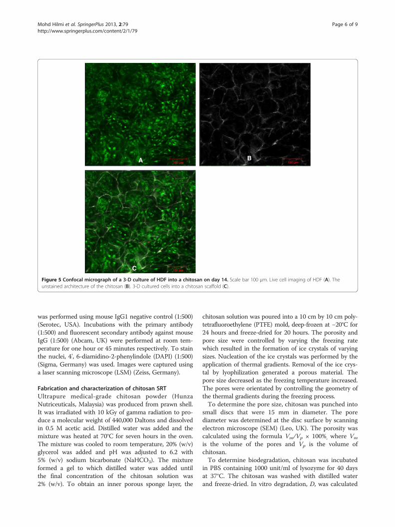
and covering most of the pores (Figure 4). Analysis athigher magnification revealed high cell adhesion andlayers formation (Figure 4B). Confocal microscopy ofthe cell-chitosan construct revealed that the live cellswere stained with green fluorescence in 3-D cultures(Figure 5). Histological analysis showed that the HDFmigrated over and penetrated the chitosan (Figure 6).The interconnected pores facilitate nutrient flux, cell mi-gration and metabolic waste exchange. Cell migration isa primary factor modulating cell behavior and phenotype(Frederick 2003; Even-Ram and Yamada 2005).
ConclusionsIn conclusion, we demonstrated that a chitosan SRT sup-ports HDF attachment. The presence of interconnectedpores of both macroscopic and microscopic size in thechitosan is considered as a factor in regulating the sur-vival and attachment of the cell-chitosan construct. Fur-thermore, the physical and mechanical properties ofchitosan SRT are useful for the cultivation of HDF fordermal regeneration. To the best of our knowledge, this isthe first microscopic study to analyze the integration ofHDF and chitosan SRT.
MethodsThis research was approved by the Research EthicsCommittee (Human) of Universiti Sains Malaysia (ap-proval code: USMKK/PPP/JEPeM [212.3(15)]).
Isolation and characterization of HDFSkin samples were obtained from ten consenting pa-tients who underwent plastic surgery (according to aprotocol approved by an ethics committee and in ac-cordance with the Declaration of Helsinki). To avoid
Table 2 OD570 value of fibroblasts into chitosan (mean ± SEM, n = 4)
2 days 4 days 6 days 8 days 10 days 12 days 14 days
Mohd Hilmi et al. SpringerPlus 2013, 2:79 Page 5 of 9http://www.springerplus.com/content/2/1/79
contamination and to maintain cellular integrity, the skinwas dissected within 24 hours. It was cut into smallpieces, each approximately 0.3 cm2 to 0.5 cm2, in a Petridish (Nunc, Denmark) containing phosphate-buffered sa-line (PBS) (Gibco, USA). PBS was used for washing offcell debris, including fat and coagulated blood. To sep-arate the epidermis and dermis, the skin pieces wereincubated in William’s E Medium (Sigma, Germany)supplemented with 0.1% (w/v) reconstituted dispase(Invitrogen, Japan) and 1% (v/v) antimycotic (Invitrogen,USA) overnight at 4°C or for two to three hours at 37°C.After the epidermis was discarded, the dermis wasincubated in William’s E Medium (Sigma, Germany)containing 0.1% (w/v) collagenase type I (Gibco, USA)overnight in a 37°C incubator-roller (Binder, Germany).After vigorous resuspension, the cell suspensions were fil-tered using 70-μm strainers (BD Falcon, USA) to separatethe hair shafts and agglutinated cells. The William’s E
A
C
Figure 4 SEM micrograph of a 3-D culture of HDF into chitosan on daproliferation into the chitosan. Scale bar 4 μm (B). Fibroblasts covering the
Medium was dispensed onto the strainer to increase theyield of cells. Human dermal fibroblasts (HDF) were col-lected by centrifugation (Hettichzentrifugen, Germany)at 420 × g for 5 minutes and cultured in 6-well plates(Nunc, Denmark) at a density of 1×10⁵ cells per well inDulbecco’s Modified Eagle Medium (DMEM) (Invitrogen,USA) supplemented with 1% (v/v) antimycotic and 10%(v/v) fetal bovine serum (FBS) (Invitrogen, USA). TheHDF cultures were monitored daily to confirm that therewas no contamination.For characterization experiments, HDF cells from pas-
sages two to five were used. The cells were seeded on a4-well chamber slide (Nunc, Denmark) at a density of1×103 cell/ml and fixed with cold methanol (Merck,Germany) for 10 minutes at −20°C. Indirect immuno-fluorescence staining was performed using mouse mono-clonal antibodies against human vimentin (Abcam, UK)and human versican (Abcam, UK). The negative control
B
D
y 14. Chitosan scaffold without cells. Scale bar 200 μm (A). Fibroblastpores. Scale bar 200 μm (C). Cross section. Scale bar 200 μm (D).
C
B
A
A
Figure 5 Confocal micrograph of a 3-D culture of HDF into a chitosan on day 14. Scale bar 100 μm. Live cell imaging of HDF (A). Theunstained architecture of the chitosan (B). 3-D cultured cells into a chitosan scaffold (C).
Mohd Hilmi et al. SpringerPlus 2013, 2:79 Page 6 of 9http://www.springerplus.com/content/2/1/79
was performed using mouse IgG1 negative control (1:500)(Serotec, USA). Incubations with the primary antibody(1:500) and fluorescent secondary antibody against mouseIgG (1:500) (Abcam, UK) were performed at room tem-perature for one hour or 45 minutes respectively. To stainthe nuclei, 4', 6-diamidino-2-phenylindole (DAPI) (1:500)(Sigma, Germany) was used. Images were captured usinga laser scanning microscope (LSM) (Zeiss, Germany).
Fabrication and characterization of chitosan SRTUltrapure medical-grade chitosan powder (HunzaNutriceuticals, Malaysia) was produced from prawn shell.It was irradiated with 10 kGy of gamma radiation to pro-duce a molecular weight of 440,000 Daltons and dissolvedin 0.5 M acetic acid. Distilled water was added and themixture was heated at 70°C for seven hours in the oven.The mixture was cooled to room temperature, 20% (w/v)glycerol was added and pH was adjusted to 6.2 with5% (w/v) sodium bicarbonate (NaHCO3). The mixtureformed a gel to which distilled water was added untilthe final concentration of the chitosan solution was2% (w/v). To obtain an inner porous sponge layer, the
chitosan solution was poured into a 10 cm by 10 cm poly-tetrafluoroethylene (PTFE) mold, deep-frozen at −20°C for24 hours and freeze-dried for 20 hours. The porosity andpore size were controlled by varying the freezing ratewhich resulted in the formation of ice crystals of varyingsizes. Nucleation of the ice crystals was performed by theapplication of thermal gradients. Removal of the ice crys-tal by lyophilization generated a porous material. Thepore size decreased as the freezing temperature increased.The pores were orientated by controlling the geometry ofthe thermal gradients during the freezing process.To determine the pore size, chitosan was punched into
small discs that were 15 mm in diameter. The porediameter was determined at the disc surface by scanningelectron microscope (SEM) (Leo, UK). The porosity wascalculated using the formula Vm/Vp × 100%, where Vm
is the volume of the pores and Vp is the volume ofchitosan.To determine biodegradation, chitosan was incubated
in PBS containing 1000 unit/ml of lysozyme for 40 daysat 37°C. The chitosan was washed with distilled waterand freeze-dried. In vitro degradation, D, was calculated
Figure 6 Histological analysis of HDF in 3-D culture. HDF integrated to each other (purple, asterisk) and into a chitosan scaffold (purple-red,arrow) on day 14. Scale bar 500 μm.
Mohd Hilmi et al. SpringerPlus 2013, 2:79 Page 7 of 9http://www.springerplus.com/content/2/1/79
using the formula D= (W0 – W1)/W0 × 100%, where W0
is the original weight and W1 is the weight at the time ofmeasurement.The tensile strength was measured by applying strain
at the rate of 0.01 m/min using a universal testing ma-chine (Tinius Olsen, USA). Chitosan of 2 mm thicknesswas cut into 15 mm × 50 mm sections. The tensilestrength, E was calculated using the following formula.
E ¼ FA� 1þ ΔL
L0
� �
where F is force, A is the cross-sectional area of the chi-tosan, ΔL is its total elongation (change in length) and Lis its original length.The water uptake ratio was determined using the for-
mula W1-Wo/Wo × 100%, where Wo is the initial weightand W1 is the wet weight after incubating the chitosanin PBS at room temperature for 24 hours.The water vapor permeability (WVP) of the chitosan
was determined using the flexible bottles permeationtest (Systech, UK). The bottles were stored at roomtemperature for 5 hours and the mass of water lost fromthe bottles was monitored as a function of time. WVP wascalculated at steady-state using the formula WVP = W/ATwhere W is the mass of water lost, A is the area (1.18 cm2)and T is the exposure time.
3-D cultivation of HDFFor 3-D cultivation, chitosan disc of 5 mm diameter and2 mm thickness were used. The chitosan discs were putinto a 96-well plate and initially seeded with 40 μl of cul-ture medium containing cells at a density of 3×10⁶/cm2.The first 20 μl of cells were dispensed using a micropip-ette onto the center of each chitosan disc. After thesecells were adsorbed into the chitosan, the remainingcells were dispensed at its edge. After 2 hours, another100 μL of culture medium was added to ensure adequatecells proliferation. After 6 hours, the chitosan discs weretransferred into 24-well plates to ensure that the cellshad enough growth medium for 3-D proliferation. Thecultured chitosan discs were incubated in 5% CO2 at37°C. The growth medium was changed every day. Onday 14, the chitosan discs with the 3-D cell cultures wereharvested for further experiments.
Attachment of HDF onto chitosan SRT and viabilityanalysisThe viability of cells grown into chitosan was analyzedusing a cell viability kit (Invitrogen, USA). The 3-D cellcultures grown into chitosan were incubated withcalcein and ethidium, (1:4) in 2 mL PBS to stain live ordead cells respectively. The incubation was performed in24-well culture plates (Orange Scientific, Belgium) atroom temperature for 30 minutes. Chitosan withoutcells was used as a control. The 3-D cultivated live cells
Mohd Hilmi et al. SpringerPlus 2013, 2:79 Page 8 of 9http://www.springerplus.com/content/2/1/79
were imaged using a laser scanning confocal microscope(LSCM) (Zeiss, Germany).To observe the interactions of HDF onto chitosan, the
3-D cells cultures were washed twice with PBS and fixedwith 2.5% glutaraldehyde for an hour at 4°C. Serial dehy-dration using a graded ethanol series of 30, 60 and 100%was performed for 5 minutes each followed by air dryingat 37°C for 4 hours. Prior to imaging, the surface ofchitosan was sputter-coated with gold (Leica, CzechRepublic). Images were captured using SEM (Quanta,Netherlands).The attachment of cells into 3-D chitosan scaffold was
further analyzed by hematoxylin and eosin staining.Briefly, 3-D cultured cells into the chitosan scaffold werefixed with 10% formalin, embedded in paraffin and sec-tioned at 5 μm thickness. The attachment of cells intothe chitosan was viewed using a Mirax Desk scanner(Zeiss, Germany).To evaluate the viability of cells in 3-D culture, HDF from
four independent samples were cultured in a 24-well plateat a density of 3×10⁶/cm2. Each well including one withchitosan but without cells as a negative control, was incu-bated with Presto Blue Cell Viability Reagent (Invitrogen,USA) (1:10) for 24 hours. The medium (100 μl) from eachwell was transferred into a 96-well plate (Orange, Belgium)and analyzed using a Nano Quant enzyme-linked immuno-sorbent assay reader (Tecan, Austria). The absorbancewas determined at 570 nm with a reference wavelengthof 600 nm.
StatisticsThe data are presented as the mean ± SEM.
Competing interestsThe authors declare they have no competing interests.
Authors’ contributionABMH involved acquisition, analyzed and interpretation of all data. ASHdesigned and executed this study. AH designed 3D cultures. CKL establishedfibroblasts culture. KN and IZ fabricated the chitosan. All authors read andapproved the final manuscript.
AcknowledgmentThis study was supported by the Universiti Sains Malaysia Research UniversityGrant (1001/PPSP/812037) and the Research University PostgraduateResearch Grant Scheme (1001/PPSP/8144012). We thank the CraniofacialLaboratory staff for their permission and guidance in using the latestmachines in PPSG. We are most grateful to Rageshwari Ramupillai from CarlZeiss (M) for her special technical assistance, support and commitment. Thegenerous support of Mohd Hafiz from SIRIM is gratefully acknowledged.
Author details1Reconstructive Sciences Unit, Universiti Sains Malaysia, Kelantan, Malaysia.2Department of Anatomy, Universiti Sains Malaysia, Kelantan, Malaysia.3Universiti Teknologi MARA, Selangor, Malaysia. 4Standard and IndustrialResearch Institute of Malaysia (SIRIM), Kedah, Malaysia. 5Universiti PendidikanSultan Idris, Perak, Malaysia.
Received: 23 December 2012 Accepted: 20 January 2013Published: 5 March 2013
ReferencesAnandan R, Nair PGV, Mathew S (2004) Anti-ulcerogenic effect of chitin and
chitosan on mucosal antioxidant defence system in hcl-ethanol-inducedulcer in rats. J Pharm Pharmacol 56:265–269
Auxenfans C, Builles N, Andre V, Lequeux C, Fievet A, Rose S, Braye FM, FradetteJ, Janin-Manificat H, Nataf S, Burillon C, Damour O (2009) Porous matrix andprimary-cell culture: A shared concept for skin and cornea tissueengineering. Pathol Biol (Paris) 57:290–298
Chang HY, Chi J-T, Dudoit S, Bondre C, van de Rijn M, Botstein D, Brown PO(2002) Diversity, topographic differentiation, and positional memory inhuman fibroblasts. Proceedings of the National Academy of Sciences99:12877–12882
Chen FG, Zhang WJ, Bi D, Liu W, Wei X, Chen FF, Zhu L, Cui L, Cao Y (2007)Clonal analysis of nestin- vimentin+ multipotent fibroblasts isolated fromhuman dermis. J Cell Sci 120:2875–2883
El-Ghalbzouri A, Gibbs S, Lamme E, Van Blitterswijk CA, Ponec M (2002)Effect of fibroblasts on epidermal regeneration. Br J Dermatol147:230–243
Even-Ram S, Yamada KM (2005) Cell migration in 3d matrix. Curr Opin Cell Biol17:524–532
Faigle W, Colucci-Guyon E, Louvard D, Amigorena S, Galli T (2000) Vimentinfilaments in fibroblasts are a reservoir for snap23, a component of themembrane fusion machinery. Mol Biol Cell 11:3485–3494
Frederick G (2003) Fibroblast biology in three-dimensional collagen matrices.Trends Cell Biol 13:264–269
Guo R, Xu S, Ma L, Huang A, Gao C (2011) The healing of full-thickness burnstreated by using plasmid DNA encoding vegf-165 activated collagen-chitosan dermal equivalents. Biomaterials 32:1019–1031
Halim AS, Stone CA, Devaraj VS (1998) The hyphecan cap: A biological fingertipdressing. Injury 29:261–263
Hattori N, Carrino DA, Lauer ME, Vasanji A, Wylie JD, Nelson CM, Apte SS (2011)Pericellular versican regulates the fibroblast-myofibroblast transition. J BiolChem 286:34298–34310
Hu Y, Topolkaraev V, Hiltner A, Baer E (2001) Measurement of water vaportransmission rate in highly permeable films. Journal of Applied PolymerScience 81:1624–1633
Jansen L, Rottier P (1958) Comparison of the mechanical properties of strips ofhuman abdominal skin excised from below and from above the umbilic.Dermatology 117:252–258
Jin Y, Ling P-X, He Y-L, Zhang T-M (2007) Effects of chitosan and heparin on earlyextension of burns. Burns 33:1027–1031
Johnston JA, Ward CL, Kopito RR (1998) Aggresomes: A cellular response tomisfolded proteins. The Journal of Cell Biology 143:1883–1898
Katsumoto T, Mitsushima A, Kurimura T (1990) The role of the vimentinintermediate filaments in rat 3y1 cells elucidated by immunoelectronmicroscopy and computer-graphic reconstruction. Biol Cell 68:139–146
Kozen BG, Kircher SJ, Henao J, Godinez FS, Johnson AS (2008) An alternativehemostatic dressing: Comparison of celox, hemcon, and quikclot. AcadEmerg Med 15:74–81
Lamke LO, Nilsson G, Reithner H (1977) The evaporative water loss from burnsand the water-vapour permeability of grafts and artificial membranes used inthe treatment of burns. Burns 3:159–165
Lamme EN, Van Leeuwen RTJ, Brandsma K, Van Marle J, Middelkoop E (2000)Higher numbers of autologous fibroblasts in an artificial dermal substituteimprove tissue regeneration and modulate scar tissue formation. The Journalof Pathology 190:595–603
Lamme EN, van Leeuwen RT, Mekkes JR, Middelkoop E (2002) Allogeneicfibroblasts in dermal substitutes induce inflammation and scar formation.Wound Repair Regen 10:60–152
Li X, Yang Z, Zhang A, Wang T, Chen W (2009) Repair of thoracic spinal cordinjury by chitosan tube implantation in adult rats. Biomaterials 30:1121–1132
Lim CK, Yaacob NS, Ismail Z, Halim AS (2010) In vitro biocompatibility of chitosanporous skin regenerating templates (psrts) using primary human skinkeratinocytes. Toxicology in Vitro 24:721–727
Ma PX, Choi J-W (2001) Biodegradable polymer scaffolds with well-definedinterconnected spherical pore network. Tissue Eng 7:23–33
Oh SH, Park IK, Kim JM, Lee JH (2007) In vitro and in vivo characteristics of pclscaffolds with pore size gradient fabricated by a centrifugation method.Biomaterials 28:1664–1671
Mohd Hilmi et al. SpringerPlus 2013, 2:79 Page 9 of 9http://www.springerplus.com/content/2/1/79
Park CJ, Clark SG, Lichtensteiger CA, Jamison RD, Johnson AJW (2009)Accelerated wound closure of pressure ulcers in aged mice by chitosanscaffolds with and without bfgf. Acta Biomaterialia 5:1926–1936
Peter XM (2008) Biomimetic materials for tissue engineering. Advanced DrugDelivery Reviews 60:184–198
Prusiner SB (1998) Prions. Proceedings of the National Academy of Sciences95:13363–13383
Pusateri AE, McCarthy SJ, Gregory KW, Harris RA, Cardenas L, McManus AT,Goodwin CWJ (2003) Effect of a chitosan-based hemostatic dressing onblood loss and survival in a model of severe venous hemorrhage andhepatic injury in swine. The Journal of Trauma 54:177–182
Rabea EI, Badawy MET, Stevens CV, Smagghe G, Steurbaut W (2003) Chitosan asantimicrobial agent: Applications and mode of action. Biomacromolecules4:1457–1465
Sheng W, Wang G, La Pierre DP, Wen J, Deng Z, Wong C-KA, Lee DY, Yang BB(2006) Versican mediates mesenchymal-epithelial transition. Mol Biol Cell17:2009–2020
Shi Y, Ma L, Zhou J, Mao Z, Gao C (2006) Collagen/chitosan-‐silicone membranebilayer scaffold as a dermal equivalent. Polymers for advanced technologies16:789–794
Tchemtchoua VT, Atanasova G, Aqil A, FileÌe P, Garbacki N, Vanhooteghem O,Deroanne C, As NÌ, JeÌrome C, Nusgens B, Poumay Y, Colige A (2011)Development of a chitosan nanofibrillar scaffold for skin repair andregeneration. Biomacromolecules 12:3194–3204
Thomas NW (2002) Versican: A versatile extracellular matrix proteoglycan in cellbiology. Curr Opin Cell Biol 14:617–623
Ueno H, Mori T, Fujinaga T (2001) Topical formulations and wound healingapplications of chitosan. Advanced Drug Delivery Reviews 52:105–115
Van den Bogaerdt AB, van Zuijlen PZ, van Galen MG, Lamme EL, Middelkoop EM(2002) The suitability of cells from different tissues for use in tissue-engineered skin substitutes. Arch Dermatol Res 294:135–142
Wang X, Yu X, Yan Y, Zhang R (2008) Liver tissue responses to gelatin andgelatin/chitosan gels. Journal of Biomedical Materials Research Part A87A:62–68
Wu Y, Chen L, Zheng P-S, Yang BB (2002) B1-integrin-mediated glioma celladhesion and free radical-induced apoptosis are regulated by binding to a c-terminal domain of pg-m/versican. J Biol Chem 277:12294–12301
Yannas IV, Lee E, Orgill DP, Skrabut EM, Murphy GF (1989) Synthesis andcharacterization of a model extracellular matrix that induces partialregeneration of adult mammalian skin. Proceedings of the National Academyof Sciences 86:933–937
Zeltinger J, Sherwood JK, Graham DA, Mueller R, Griffith LG (2001) Effect of poresize and void fraction on cellular adhesion, proliferation, and matrixdeposition. Tissue Eng 7:557–572
doi:10.1186/2193-1801-2-79Cite this article as: Mohd Hilmi et al.: In vitro characterization of achitosan skin regenerating template as a scaffold for cells cultivation.SpringerPlus 2013 2:79.
Submit your manuscript to a journal and benefi t from: