In vitro characterization of polyacrylamidehydrogels for application as implant coatingfor stimulus-responsive local drug deliveryIngo Minratha, Daniela Arbeitera, Klaus-Peter Schmitza, Katrin Sternberga
Keywords: antigen-responsive drug delivery; semi-interpenetrating hydrogel; polyacrylamide; bioconjugation; implant
INTRODUCTION
The interface between implants and the surrounded tissue is ofhigh research interest in modern regenerative medicine. Withthe purpose of influencing implant-cell/tissue interactions, a lotof surface modification strategies were investigated and estab-lished in the last 10 years. Many implants are already nowadaysequipped with drug-containing polymeric coatings in order toobtain local drug delivery (LDD) systems with diffusion-controlled and sustained rates of drug release.[1] Besides thebenefit that the drug is fixed on the implant’s surface and rela-tively protected against mechanical stress, a polymeric coatingallows a wide range of control over the release characteristicsof the drug.[2] Chemically controlled LDD systems offer anotherstrategy for drug release. In these systems, drug release isachieved by cleavage of chemically bound drugs or by degrada-tion or swelling of the drug carrier.[3] However, both systems,diffusion and chemically controlled, provide drug release inde-pendently of their need, possibly leading to ineffectiveness ofthe LDD system at time of interest. Having this in mind, it wouldbe highly beneficial if the drug is delivered by a system thatsenses the signal caused by disease, judges the magnitude ofthe signal and then acts to release the right amount of drug inresponse.[4] Here, stimulus-responsive hydrogels with swellingproperties dictated by changes in pH,[5,6] temperature[7–9] orthe electrical field of the surrounding,[10,11] which have already
been described years ago, come into discussion. The inductionof hydrogel swelling by such physical and chemical stimuliallows control over release of incorporated or underlying drugs.They are therefore discussed for future application as self-regulated drug delivery systems.[4] However, the cited stimuliare rather non-specific. An alternative are specific chemo-responsive hydrogels, which are designed to contain internalnon-covalent interactions based on pendant ligands andcomplementary receptors.[12] These cross-links can be brokenby soluble ligand or receptor competitors, which are specificfor certain diseases and can diffuse into the gel displacing theinternal affinity cross-links. Subsequently, the degree of cross-linking decreases and drugs can be released. Examples inliterature are glucose-responsive dextrane hydrogels with con-canavalin A-glucose affinity cross-links[13] and nicotinamideadenine dinucleotide-responsive dextrane hydrogels with cibacronblue/lysozyme affinity cross-links.[14,15] Likewise interesting areantigen-responsive hydrogels, where antibody–antigen pairs,
* Correspondence to: Dr. Svea Petersen, Institute for Biomedical Engineering,University of Rostock, Friedrich-Barnewitz-Straße 4, 18119 Rostock, Germany.E-mail: [email protected]
a I. Minrath, D. Arbeiter, K.-P. Schmitz, K. Sternberg, S. PetersenInstitute for Biomedical Engineering, University of Rostock, Friedrich-Barnewitz-Straße 4, 18119 Rostock, Germany
Special issue: Research article
Received: 31 January 2014, Accepted: 27 February 2014, Published online in Wiley Online Library
chemically grafted to the hydrogel network, serve as reversiblecross-linkers. Several model systems have been established,[12,16,17]
while the best studied is probably the one of Miyata et al.,[18–22]
which is based on immunoglobulin G (IgG) antigens and corre-sponding antibodies grafted onto a polyacrylamide- (PAAm-)hydrogel, forming affinity cross-links cleavable in the presenceof free antigen. Moreover, they could show that a hydrogelwith a semi-interpenetrating network architecture allowsreversibility of the stimulus-induced swelling and hence drugrelease, which is considered of high interest for stimulus-responsive, implant-associated LDD systems.[18] However, thechallenging implementation of such antigen-responsivehydrogels as implant coating and their applicability for implant-associated LDD systems has not been explored so far. Besidesthe afforded stable surface attachment, thorough in vitro investi-gations are necessary in order to define adequate gel propertieswith regard to mechanical stability and controlled drug releasefrom implant surfaces, which is likely controlled via size andshape of pores and general gel porosity. According to literature,these gel properties are mainly determined by the degree ofcross-linking.[12] In this context, we prepared a matrix at theexample of PAAm-hydrogels of different gel architectures, gelcontents and cross-linking degrees and investigated their impacton the permeation of three differently sized model proteins andthe mechanical stability of the polymeric network. The releasestudy setup in a specifically designed chamber simulating animplant-associated LDD system with hydrogels as coatings wasconsidered as highly important. Moreover, after definition ofpromising gel properties, the grafting of stimulus-responsivefunctions was additionally investigated.
EXPERIMENTAL
Materials
All chemicals for PAAm-hydrogel preparation, acrylamide (AAm,>99%), N,N-methylenebisacrylamide (MBAAm, >99%), N,N,N,N-tetramethylethylendiamine (TEMED) and ammoniumpersulfate(APS, >99%) were purchased from Sigma-Aldrich (Taufkirchen,Germany). Phosphate buffered saline (Dulbecco’s PBS, DPBS,pH 7.2, Thermo Scientific, Karlsruhe, Germany) was used forin vitro drug release studies and labeling of hydrogels for fluores-cence microscopy.
The modification of the used model antibody–antigen pair(rabbit anti mouse IgG (RAM IgG, Sigma-Aldrich)/mouse IgG(M IgG, Jackson ImmunoResarch Europe, Newmarket, UK) forgrafting on the hydrogel network was performed with acrylicacid N-hydroxysuccinimide (NSA, Sigma-Aldrich), and ZebaSpin Desalting Columns 7-k MWCO (Thermo Scientific) wereused for subsequent purification of the modified NSA-IgG.Detection of the coupled M IgG/RAM IgG was carried out by fluo-rescence microscopy with the following FITC-labeled secondaryantibodies: goat anti-mouse IgG (GAM IgG, Abcam, Cambridge,UK) and goat anti rabbit IgG (GAR IgG, Sigma-Aldrich). Bovineserum albumin (BSA, 66 kDa, SERVA Feinbiochemica, Heidelberg,German), fluorescein isothiocyanate- (FITC-) labeled BSA (BSA-FITC,Sigma-Aldrich), protein A (SPA, 44 kDa, ProSpec, Rehovot, Israel)and myoglobin (from equine heart, 17 kDa, Sigma-Aldrich) wereused as model proteins. Proteinase K (Sigma-Aldrich) was usedfor digestion of BSA-FITC for fluorescence enhancement afterin vitro release. Protein quantification was performed with thebicinchoninic acid protein assay reagent (BCA, Thermo Scientific),
and for the quantification of the water uptake of the differentPAAm-gels, Hydranal-Coulomat AG KF reagent (Sigma-Aldrich)was used.
PAAm-hydrogel preparation
For synthesis of conventional and semi-interpenetrating gels,two solutions (A and B) were prepared. While solution A(16.38w% AAm/DPBS) is the same for both applied cross-linkingratios (AAm/MBAAm 58/1 and 117/1 [w/w]), solution B (47.74w%AAm/DPBS) differs in MBAAm content (AAm/MBAAm 58/1 [w/w]:1.16w% MBAAm; AAm/MBAAm 117/1 [w/w]: 0.59w% MBAAm).
Conventional PAAm-hydrogels
For preparing conventional gels of different gel content, variousvolumes of solution A, solution B and DPBS were mixed (e.g. for a15.0w% gel: 183.1-μl solution A, 134-μl solution B and 326-μlDPBS). To the resulting solutions, 22.95μl of 0.1M APS in DPBSand 22.95μl of 0.8M TEMED in DPBS were added. Three hundredand thirty microliters of these solutions was either pipettedbetween two glass cover slips (diameter: 25mm) or in a formfor shouldered test bars (reclined to ISO 527-2) for tensile tests.After reaction for 50min at 23 ± 2°C, the resulting hydrogelswere transferred into DPBS for 16 h for removal of monomerresidues and swelling.
Semi-interpenetrating PAAm-hydrogels
The preparation of semi-interpenetrating hydrogels follows atwo-step protocol according to Miyata et al.[21] In a first step,solution A was combined with the corresponding volume ofDPBS (e.g. for a 15.0w% gel: 183.1-μl solution A and 186-μlDPBS). Then, to each of the solutions, 5.25-μl 0.1M APS in DPBSand 5.25-μl 0.8M TEMED in DPBS were added. After reaction for7min at 23 ± 2°C, 134-μl solution B, 137-μl DPBS, 17.7-μl 0.1MAPS in DPBS and 17.7-μl 0.8M TEMED in DPBS were added.Formation of hydrogels was performed in the same manner asdescribed for conventional hydrogels.
Bioconjugated PAAm-hydrogels
For afforded antibody or antigen modification with vinyl groups,120μl of a 2mg/ml antibody (antigen) solution was mixed with3.5-μl (8-μl) 0.5mg/ml (2mg/ml) NSA solution and incubatedunder vigorous shaking for 60min at 37°C. While a lower NSAconcentration was applied during antibody modification, sincethey are to be integrated within linear polymer fibers, antigenswere functionalized in a higher extent to guarantee attachmentwithin the 3D network. After reaction, the solutions were purifiedfrom residual NSA by filtration with Zeba Spin Desalting Columns.For generating a stimulus-responsive hydrogel, preparation nowfollows nearly the samemanner as semi-interpenetrating hydrogelpreparation. In a first step, solution A was combined with 120μl ofmodified antibody in DPBS, 66-μl DPBS, 5.25-μl 0.1M APS and5.25-μl 0.8M TEMED. After reaction for 7min at 23 ± 2°C, 134-μlsolution B, 120-μl modified antigen in DPBS, 17-μl DPBS, 17.7-μl0.1M APS and 17.7-μl 0.8M TEMED were added. For verificationof successful hydrogel bioconjugation, lower volumes of the2mg/ml antibody and antigen solutions were additionallyapplied. After mixing, 330μl was pipetted between two glasscover slips (diameter: 25mm) and let to react for 50min at23 ± 2°C. Finally, resulting hydrogels were transferred into DPBS
overnight for removal of monomer residues, unbound antibodiesand antigens as well as swelling.
PAAm-hydrogel characterization
Oven-based Karl Fischer titration
Hydrogel samples of 5mm in diameter, swollen for 16 h in DPBS,were carefully swabbed with a fuzz-free tissue in order to re-move all adherent water. Subsequently, samples were placedin the Drying Oven D0308 (Mettler Toledo, Gießen, Germany)of the Karl Fischer titrator and heated up to 230°C. The watercontent was finally determined using a Karl Fischer CoulometerC20 (Mettler Toledo, Gießen, Germany) with the Hydranal-Coulomat AG KF. The determined water content of at least fivereplicates was set in proportion to the dried PAAm mass, deter-mined prior to the measurements by means of a microbalance(UMX5, Mettler Toledo, Gießen, Germany).
Fluorescence microscopy
In order to evaluate the successful incorporation of the antibodyand the antigen within PAAm-hydrogels, hydrogel samples of8mm in diameter were incubated with 30μg/ml FITC-labeledsecondary antibodies against the corresponding coupled bio-molecule in DPBS for 1.5 h at 23 ± 2°C. Relative light intensityand micrographs were then recorded by fluorescence micros-copy (λexc = 488 nm; λem = 505–525 nm) with a FluView1000(Olympus, Hamburg, Germany) after washing with DPBS.
Tensile tests
Uniaxial tensile testing was accomplished on a Zwicki ZN 2.5(Zwick GmbH & Co. KG, Ulm, Germany) with reference to ISO527, section 4. The tests were performed with a 10-N load celland a crosshead speed of 12mm/min. Tensile force is measuredas a function of elongation. Tensile strength (σm), elongation atbreak (εB) and elastic modulus (E) were determined. To deter-mine E, linear regression was used.
In vitro release study
Setup
For investigation of the drug permeability of the different geltypes, a test chamber was designed (Fig. 1C), which guaranteedthe permeation of the model proteins (BSA, SPA, myoglobin)
through and not alongside the hydrogel samples (Fig. 1B). Afterswelling for 16 h in DPBS, hydrogel samples of 11mm in diameterwere prepared and placed on top of glass cover slips (9mmdiameter) coated via pipetting with 200μg of the model proteinspositioned on the bottom of the chamber (Fig. 1A). To start thein vitro release study, 500-μl DPBS were filled in the chambers ontop of the hydrogels and incubated at 23±2°C. For quantificationof permeated protein concentrations via BCA assay, 400μl of themedia was replaced by fresh DPBS after 24, 48 and 72h. For inves-tigation of stimulus-responsive drug release via bioconjugatedhydrogels, BSA-FITCwas used asmodel protein, allowing the directmeasurement of released protein in the presence of M IgG, used asstimulus in a concentration of 40μg/ml in DPBS.
Quantification of protein release via BCA-assay
The BCA protein assay is based on the reduction of Cu2+ to Cu+
(Biuret reaction) in the presence of proteins under alkaline condi-tions and the subsequent formation of a purple colored complexwith bicinchoninic acid. Therefore, 25μl of the model proteincontaining release media was incubated for 30min at 37°C with200μl of working reagent containing the following in a 50:1 ratio:a 0.1M NaOH solution containing sodium carbonate, sodiumbicarbonate, bicinchoninic acid, sodium tartrate and 4.0% cupricsulfate pentahydrate. Standards (750μg/ml to 25μg/ml BSA inDPBS) were treated identically. Finally, absorbance of both, stan-dards and samples, was assayed at 560 nm by means of the spec-trometer Fluostar Optima (BMG Labtech, Ortenberg Germany).
Quantification of BSA-FITC via fluorescence spectroscopy
To ensure reproducible determination of BSA-FITC, 250μl of releasemedia, ten times diluted with DPBS, was combined with 5-μl pro-teinase K (250μg/ml in DPBS) and incubated for 30min at 37°C fordigestion according to Wischke et al.[23] After cooling down to23±2°C, quantificationwith a Fluostar Optima (BMG Labtech) (λexc =485nm, λem=520nm) was performed. Standards (100μg/ml to2.5μg/ml BSA-FITC in DPBS) were treated identically.
RESULTS
Impact of gel content, cross-linking and architecture on in vitrodrug release
The impact of gel content, cross-linking and gel architecture ondrug release was tested with three differently sized proteins,
Figure 1. Setup for the in vitro drug release study including A) photograph of the test chamber bottom for location of drug-coated glass coverslips andhydrogels, B) schematic representation of the chamber design guaranteeing drug release through the hydrogel (dark gray: hydrogel, gray: modelprotein coating; light gray: model implant surface) and C) 3D construction of the whole test chamber.
STIMULUS-RESPONSIVE POLYACRYLAMIDE HYDROGELS AS IMPLANT COATING
namely BSA (66 kDa), SPA (42 kDa) and myoglobin (17 kDa). InFig. 2, all results are summed up. The results indicate thedistinct effects of gel content, cross-linking, gel architecture andprotein size. While increasing the gel content lowers drug releasefor all four illustrated gel types (independently of AAm/MBAAmratio and gel architecture) and for all used model protein drugs,neither the AAm/MBAAm ratio nor the gel architecture seemsto have a considerable effect on drug release. With regard tothe model proteins, performed experiments indicate higherrelease rates with decreasing size (myoglobin> SPA> BSA).
Impact of gel content, cross-linking and architecture on water uptake
Evaluation of the water uptake of PAAm-hydrogels prepared withdifferent gel content, AAm/MBAAm ratio and architecture wasperformed by means of Karl Fischer titration. The water uptake ofthe different hydrogel types is shown in Table 1. Again, whileincreasing the gel content lowers water uptake considerably fromapproximately 21mg water/mg hydrogel to 9.5mg water/mg hy-drogel (independently of AAm/MBAAm ratio and gel architecture),neither the AAm/MBAAm ratio nor the gel architecture seems tohave a considerable effect on water uptake. Based on the fact, thatwater uptake for the conventional hydrogels accord to semi-interpenetrating hydrogels and for better clarity of presentation,data for conventional PAAm-hydrogels are exclusively shown forhydrogels prepared with an AAm and MBAAm content of 15w%.
Impact of gel content, cross-linking and architecture on tensilestrength, elongation at break and E moduli
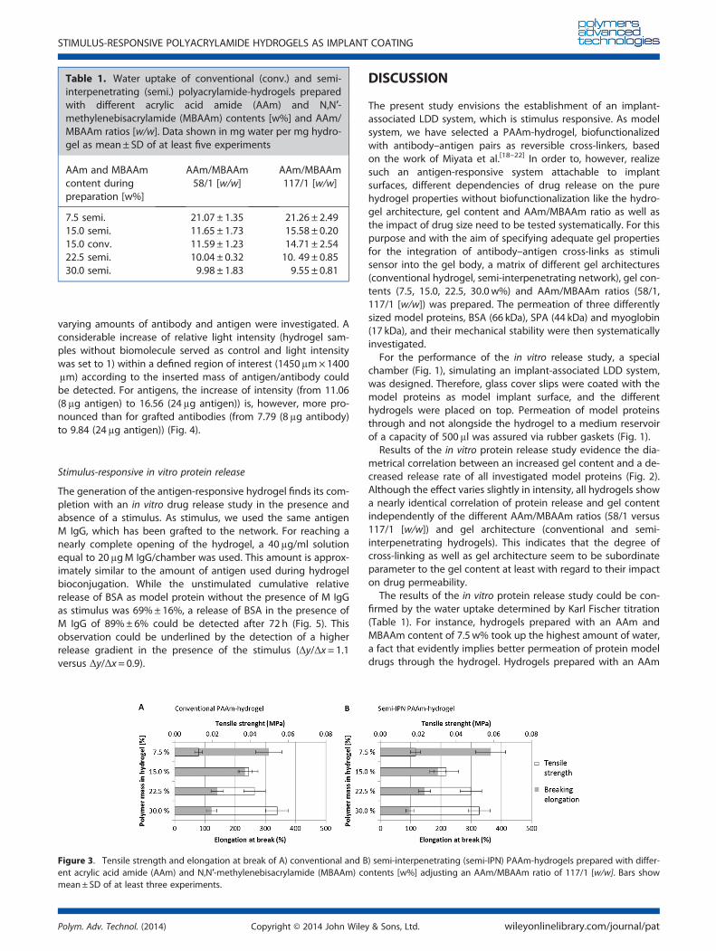
To evaluate the resistibility of hydrogels of different propertiesagainst mechanical impact, mechanical properties (tensile
strength σm, elongation at break εB and elastic modulus E) weredetermined. The results, given in Fig. 3 (σm, εB) and Table 2 (E),give an impression of the shifting of determined mechanicalvalues with regard to gel content and gel architecture. Data forthe impact of the cross-linking ratio are not shown as it evolvedas non-considerable in accordance to the drug release study(Fig. 2). For tensile strength, a general correlation between itselevation and increasing gel content was observed for conven-tional and semi-interpenetrating gels with an AAm/MBAAm ratioof 117/1 [w/w]. Results ranged from 0.013MPa± 0.002MPa(conv. 7.5w%) and 0.019MPa± 0.002MPa (semi. 7.5w%) to0.054MPa±0.006MPa (conv. 30.0w%) and 0.054MPa±0.006MPa(semi. 30.0w%). An opposing trend can be described for the elon-gation at break. εB dropped from 311%±43% (conv. 7.5w%) and365%±49% (semi. 7.5w%) to 121%±17% (conv. 30.0w%) and97%±12% (semi. 30.0w%). The elastic moduli (E) for themeasuredhydrogels are shown in Table 2. The data show a considerable in-crease of E with increasing gel content for both gel architectures.
Verification of successful hydrogel bioconjugation
The chemical grafting of antibody–antigens pairs to the PAAm-hydrogels as reversible cross-linkers is an absolute requirementfor the generation of an antigen-responsive hydrogel. As Miyataet al. reported the importance of a semi-interpenetrating hydro-gel architecture for reversibility of stimulus-induced hydrogelswelling,[18] bioconjugation was only performed with this gelarchitecture. Verification of successful biomolecule integrationwas performed by fluorescence microscopy after labeling thebioconjugated hydrogels with the corresponding FITC-labeledsecondary antibodies. Three different hydrogels prepared with
Figure 2. Cumulative relative release of model proteins (albumin, protein A and myoglobin) after 72 h in DPBS at 23 ± 2°C through A–B) con-ventional and C–D) semi-interpenetrating PAAm-hydrogels (semi-IPN) prepared with different acrylic acid amide (AAm) and N,N′-methylenebisacrylamide (MBAAm) contents [w%] adjusting an AAm/MBAAm ratio of 58/1 [w/w] (A, C) or 117/1 [w/w] (B, D). Bars show mean ± SDof at least four experiments.
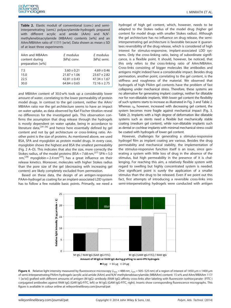
varying amounts of antibody and antigen were investigated. Aconsiderable increase of relative light intensity (hydrogel sam-ples without biomolecule served as control and light intensitywas set to 1) within a defined region of interest (1450μm×1400μm) according to the inserted mass of antigen/antibody couldbe detected. For antigens, the increase of intensity (from 11.06(8μg antigen) to 16.56 (24μg antigen)) is, however, more pro-nounced than for grafted antibodies (from 7.79 (8μg antibody)to 9.84 (24μg antigen)) (Fig. 4).
Stimulus-responsive in vitro protein release
The generation of the antigen-responsive hydrogel finds its com-pletion with an in vitro drug release study in the presence andabsence of a stimulus. As stimulus, we used the same antigenM IgG, which has been grafted to the network. For reaching anearly complete opening of the hydrogel, a 40μg/ml solutionequal to 20μgM IgG/chamber was used. This amount is approx-imately similar to the amount of antigen used during hydrogelbioconjugation. While the unstimulated cumulative relativerelease of BSA as model protein without the presence of M IgGas stimulus was 69%±16%, a release of BSA in the presence ofM IgG of 89%± 6% could be detected after 72 h (Fig. 5). Thisobservation could be underlined by the detection of a higherrelease gradient in the presence of the stimulus (Δy/Δx= 1.1versus Δy/Δx= 0.9).
DISCUSSION
The present study envisions the establishment of an implant-associated LDD system, which is stimulus responsive. As modelsystem, we have selected a PAAm-hydrogel, biofunctionalizedwith antibody–antigen pairs as reversible cross-linkers, basedon the work of Miyata et al.[18–22] In order to, however, realizesuch an antigen-responsive system attachable to implantsurfaces, different dependencies of drug release on the purehydrogel properties without biofunctionalization like the hydro-gel architecture, gel content and AAm/MBAAm ratio as well asthe impact of drug size need to be tested systematically. For thispurpose and with the aim of specifying adequate gel propertiesfor the integration of antibody–antigen cross-links as stimulisensor into the gel body, a matrix of different gel architectures(conventional hydrogel, semi-interpenetrating network), gel con-tents (7.5, 15.0, 22.5, 30.0w%) and AAm/MBAAm ratios (58/1,117/1 [w/w]) was prepared. The permeation of three differentlysized model proteins, BSA (66 kDa), SPA (44 kDa) and myoglobin(17 kDa), and their mechanical stability were then systematicallyinvestigated.
For the performance of the in vitro release study, a specialchamber (Fig. 1), simulating an implant-associated LDD system,was designed. Therefore, glass cover slips were coated with themodel proteins as model implant surface, and the differenthydrogels were placed on top. Permeation of model proteinsthrough and not alongside the hydrogel to a medium reservoirof a capacity of 500μl was assured via rubber gaskets (Fig. 1).
Results of the in vitro protein release study evidence the dia-metrical correlation between an increased gel content and a de-creased release rate of all investigated model proteins (Fig. 2).Although the effect varies slightly in intensity, all hydrogels showa nearly identical correlation of protein release and gel contentindependently of the different AAm/MBAAm ratios (58/1 versus117/1 [w/w]) and gel architecture (conventional and semi-interpenetrating hydrogels). This indicates that the degree ofcross-linking as well as gel architecture seem to be subordinateparameter to the gel content at least with regard to their impacton drug permeability.
The results of the in vitro protein release study could be con-firmed by the water uptake determined by Karl Fischer titration(Table 1). For instance, hydrogels prepared with an AAm andMBAAm content of 7.5w% took up the highest amount of water,a fact that evidently implies better permeation of protein modeldrugs through the hydrogel. Hydrogels prepared with an AAm
Figure 3. Tensile strength and elongation at break of A) conventional and B) semi-interpenetrating (semi-IPN) PAAm-hydrogels prepared with differ-ent acrylic acid amide (AAm) and N,N′-methylenebisacrylamide (MBAAm) contents [w%] adjusting an AAm/MBAAm ratio of 117/1 [w/w]. Bars showmean± SD of at least three experiments.
Table 1. Water uptake of conventional (conv.) and semi-interpenetrating (semi.) polyacrylamide-hydrogels preparedwith different acrylic acid amide (AAm) and N,N′-methylenebisacrylamide (MBAAm) contents [w%] and AAm/MBAAm ratios [w/w]. Data shown in mg water per mg hydro-gel as mean± SD of at least five experiments
and MBAAm content of 30.0w% took up a considerably loweramount of water, correlating to the lower permeability of proteinmodel drugs. In contrast to the gel content, neither the AAm/MBAAm ratio nor the gel architecture seems to have an impacton water uptake, as data observed by Karl Fischer titration revealno differences for the investigated gels. This observation con-firms the assumption that drug release through the hydrogelsis mostly dependent on water uptake, being in accordance toliterature data,[24–26] and hence here essentially defined by gelcontent and not by gel architecture or cross-linking ratio. An-other point is the size of proteins. As mentioned above, we usedBSA, SPA and myoglobin as protein model drugs. In every case,myoglobin shows the highest and BSA the smallest permeability(Fig. 2 A–D). This indicates that also the size, more correctly theStokes radius, of the model proteins (BSA = 7.68 nm,[27] SPA = 5.0nm,[28] myoglobin = 2.4 nm[29]) has a great influence on theirrelease kinetics. Moreover, molecules with higher Stokes radiusthan the pore size of the gel (decreasing with increasing gelcontent) are likely completely excluded from permeation.
Based on these data, the design of an antigen-responsivePAAm-hydrogel as coating for an implant-associated LDD systemhas to follow a few notable basic points. Primarily, we need a
hydrogel of high gel content, which, however, needs to beadapted to the Stokes radius of the model drug (higher gelcontent for model drugs with smaller Stokes radius). Althoughthe gel architecture has no influence on drug release, the semi-interpenetrating gel architecture is favorable because it guaran-tees reversibility of the drug release, which is considered of highinterest for stimulus-responsive, implant-associated LDD sys-tems. Only the cross-linking ratio, being of subordinate signifi-cance, is a flexible point. It should, however, be noticed, thatthis only refers to the cross-linking ratio of AAm/MBAAm.Cross-links consisting of bigger molecules like antibodies andantigens might indeed have a considerable impact. Besides drugpermeation, another point, correlating to the gel content, is thestiffness and roughness of the material. We observed thathydrogels of high PAAm gel contents have the problem of fastcollapsing under mechanical stress. Therefore, these systems areno alternative for generating implant coatings, neither for dilatablenor for non-dilatable implants. With lower gel content the flexibilityof such systems starts to increase as illustrated in Fig. 3 and Table 2.Whereas εB, however, increased with decreasing gel content, thesystem becomes more fragile against mechanical impact (Fig. 3,Table 2). Implants with a high degree of deformation like dilatablesystems such as stents need a flexible but mechanically stablecoating (medium gel content), while non-dilatable implants suchas dental or cochlear implants with minimal mechanical stress couldbe coated with hydrogels of lower gel content.However, challenges for generating a stimulus-responsive
hydrogel film as implant coating are various. Besides the drugpermeability and mechanical stability, the implementation ofthe stimulus-responsive function itself is an issue, since gen-erating a system with little loss of drug in the absence of thestimulus, but high permeability in the presence of it is chal-lenging. For reaching this aim, a relatively flexible system withregard to swelling but highly concentrated system is needed.One significant point is surely the application of a smallerstimulus than the drug to be released. Even if we point out thisfact, first attempts of introducing a reversible cross-links intosemi-interpenetrating hydrogels were conducted with antigen
Figure 4. Relative light intensity measured by fluorescence microscopy (λexc = 488 nm, λem= 505–525nm) of a region of interest of 1450μm×1400μmof semi-interpenetrating PAAm-hydrogels (acrylic acid amide (AAm) and N,N′-methylenebisacrylamide (MBAAm) content: 15w% and AAm/MBAAm 117/1 [w/w]) grafted with different amounts of antigen (M IgG)–antibody (RAM IgG) affinity cross-links after labeling with fluorescein isothiocyanate- (FITC-)conjugated antibodies against RAM IgG (GAR IgG-FITC, left) or M IgG (GAM IgG-FITC, right). Inserts show corresponding fluorescence micrographs. Thisfigure is available in colour online at wileyonlinelibrary.com/journal/pat
Table 2. Elastic moduli of conventional (conv.) and semi-interpenetrating (semi.) polyacrylamide-hydrogels preparedwith different acrylic acid amide (AAm) and N,N′-methylenebisacrylamide (MBAAm) contents [w%] and anAAm/MBAAm ratio of 117/1 [w/w]. Data shown as mean± SDof at least three experiments
(M IgG)/antibody (RAM IgG) coupled hydrogel with M IgG asstimulus and BSA as model protein. Gel properties were chosenaccording to generated data, searching for a system that onthe one hand does not release the model drug too fast and onthe other hand makes it possible to introduce a noticeableamount of antibody–antigen cross-links. Moreover, acceptablemechanical stability should be guaranteed. In this context, anAAm/MBAAm ratio of 117/1 [w/w] for enough flexibility and agel content of 15.0w% due to acceptable release kinetics (Fig. 2B)and mechanical properties (Fig. 3B) were chosen.The integration of the antibody–antigen cross-links followed a
two-step protocol. As exposed, we introduced several concentra-tions of antibody and antigen from 8μg/50mm2 (0.16μg/mm2)up to 24μg/50mm2 (0.48μg/mm2) (Fig. 4). The detection of in-corporated antigens via fluorescence microscopy after labelingwith corresponding FITC-labeled secondary antibodies reveals aconsiderable increase of intensity with the amount of insertedantigen, which could not be illustrated to the same extent forthe antibody incorporation.This seems astonishing, as the affinity of the FITC-labeled
secondary antibody to the antibody should be higher than tothe antigen because of the lower amount of residues modifiedvia NSA. An inferior integration of antibody or shielding of incor-porated antibodies by antigens integrated within the secondstep during gel preparation might be the reason.As integration of both, antibody and antigen, was successful,
we tried to demonstrate a stimulus-triggered release of BSA inthe following. Even if it is not the perfect system like describedbefore (antigen size vs. drug size) we could demonstrate anincrease of the cumulative release of BSA of about 20% after72 h (Fig. 5) in the presence of the stimulus. To the best of ourknowledge, this is the first evidence of an antigen-responsivedrug delivery from a model implant-associated LDD system.However, the fact that the amounts of released BSA were twiceas much as for non-functionalized semi-interpenetrating hydro-gel (AAm/MBAAm 117/1 [w/w], 15.0w%.) in the absence ofstimulus indicates continuative need for action. For results withhigher significance and lower drug permeation in the absenceof a stimulus, it is our aim in the future to change the type ofantigen to a smaller one. This should guarantee a lower drugpermeability of bioconjugated hydrogels in the absence of the
stimulus by reason of lower pore sized due to smaller anti-body–antigen cross-links.
CONCLUSION
Within this study, we presented a thorough in vitro characteriza-tion of PAAm-hydrogels for application as implant coating forstimulus-responsive LDD. In this context, we defined adequategel properties (gel content, cross-linking ratio and gel architec-ture) with regard to drug permeability using the different modelproteins BSA, SPA and myoglobin in a specially designed cham-ber simulating an implant-associated LDD system and mechani-cal stability. Within the investigated matrix, a medium gelcontent of 15.0w% AAm and MBAAm evidenced low drugpermeability, afforded in order to prevent drug release in theabsence of the stimulus, enough tensile strength and flexibilityfor integration of antibody–antigen cross-links. Interestingly,neither gel architecture nor cross-linking ratio of AAm/MBAAmseemed to have a considerable impact on drug permeationand mechanical stability, which we dedicated to the lackingimpact on hydrogel water uptake. By the successful integrationof M IgG/RAM IgG as antibody–antigen cross-links into aPAAm-hydrogel of chosen properties in the following, we wereable to demonstrate to the best of our knowledge for the firsttime an antigen-triggered release of BSA as model drug from amodel implant-associated LDD system. The continuation of workwill now focus on the real implementation of such stimulus-responsive hydrogels to implant surfaces. Besides stable attach-ment of the hydrogel to drug-coated implant surfaces, this willinclude the investigation of novel stimulus-drug combinationsin order to overcome observed challenges when using a biggerstimulus than the drug to be released.
Acknowledgements
The authors thank Gabriele Karsten and Maria Boeck for theirexpert technical assistance in fluorescence microscopy andprotein quantification, respectively. Furthermore, financial sup-port by Bundesministerium für Bildung und Forschung (BMBF)within REMEDIS “Höhere Lebensqualität durch neuartige Mikro-implantate” (FKZ: 03IS2081) is gratefully acknowledged.
REFERENCES[1] S. Garg, P. W. Serruys, J. Am. Coll. Cardiol. 2010, 56, S1.[2] G. Acharya, K. Park, Adv. Drug Deliv. Rev. 2006, 58, 387.[3] K. Sternberg, S. Petersen, N. Grabow, V. Senz, H. Meyer zu
Schwabedissen, H. K. Kroemer, K.-P. Schmitz, Biomed. Tech. (Berl)2013, 58, 417.
[4] Y. Qiu, K. Park, Adv. Drug Deliv. Rev. 2001, 53, 321.[5] R. A. Siegel, B. A. Firestone, Macromolecules 1988, 21, 3254.[6] T. Tanaka, Phys. Rev. Lett. 1978, 40, 820.[7] A. S. Hoffman, J. Control. Release 1987, 6, 297.[8] Y. H. Bae, T. Okano, R. Hsu, S. W. Kim, Makromol. Chem. Rapid Comm.
1987, 8, 481.[9] T. Aoki, M. Kawashima, H. Katono, K. Sanui, N. Ogata, T. Okano,
Y. Sakurai, Macromolecules 1994, 27, 947.[10] T. Tanaka, I. Nishio, S.-T. Sun, S. Ueno-Nishio, Science 1982, 218, 467.[11] I. C. Kwon, Y. H. Bae, S. W. Kim, Nature 1991, 354, 291.[12] R. Zhang, A. Bowyer, R. Eisenthal, J. Hubble, Biotechnol. Bioeng.
2007, 97, 976.[13] M. Tang, R. Zhang, A. Bowyer, R. Eisenthal, J. Hubble, Biotechnol.
Bioeng. 2003, 82, 47.[14] A. G. Mayes, J. D. Moore, R. Eisenthal, J. Hubble, Biotechnol. Bioeng.
1990, 36, 1090.
Figure 5. Cumulative relative release of albumin as a function of time inDPBS with and without the addition of 40μg/mlM IgG at 23 ± 2°Cthrough bioconjugated semi-interpenetrating (semi-IPN) PAAm-hydrogels prepared with 15w% of acrylic acid amide (AAm) and N,N′-methylenebisacrylamide (MBAAm) adjusting an AAm/MBAAm ratio of117/1 [w/w]. Data is indicated as mean± SD of at least four experiments.
STIMULUS-RESPONSIVE POLYACRYLAMIDE HYDROGELS AS IMPLANT COATING
[15] M. Tang, R. Zhang, A. Bowyer, R. Eisenthal, J. Hubble, [email protected], Biotechnol. Bioeng. 2004, 87, 791.
[16] Z. R. Lu, P. Kopecková, J. Kopecek, Nat. Biotechnol. 1999, 17, 1101.[17] Z.-R. Lu, P. Kopečková, J. Kopeček, Macromol. Biosci. 2003, 3, 296.[18] T. Miyata, N. Asami, T. Uragami, Nature 1999, 399, 766.[19] T. Miyata, N. Asami, T. Uragami, Macromolecules 1999, 32, 2082.[20] T. Miyata, T. Uragami, K. Nakamae, Adv. Drug Deliv. Rev. 2002, 54, 79.[21] T. Miyata, N. Asami, T. Uragami, J Polym Sci B 2009, 47, 2144.[22] T. Miyata, N. Asami, Y. Okita, T. Uragami, Polym. J. 2010, 42, 834.
[23] C. Wischke, H.-H. Borchert, Die Pharmazie Int. J. Pharmaceut. Sci.2006, 61, 770.
[24] A. S. Hoffman, Adv. Drug Deliv. Rev. 2012, 64, Supplement, 18.[25] J. L. Drury, D. J. Mooney, Biomaterials 2003, 24, 4337.[26] N. A. Peppas, P. Bures, W. Leobandung, H. Ichikawa, Eur. J. Pharm.
Biopharm. 2000, 50, 27.[27] T. Nishikawa, K. Akiyoshi, J. Sunamoto, J. Am. Chem. Soc. 1996, 118, 6110.[28] I. Björk, B.-Å. Petersson, J. Sjöquist, Eur. J. Biochem. 1972, 29, 579.[29] Y. Walbroehl, J. W. Jorgenson, J. Microcolumn. Sep. 1989, 1, 41.