In vitro co-culture model of the inflamed intestinal mucosa Berlin, December 13, 2011 Eva-Maria Collnot, [email protected]Helmholtz Institute for Pharmaceutical Research Saarland Departement of Drug Delivery (DDEL)
Transcript
In vitro co-culture model of the inflamed intestinal mucosa
Berlin, December 13, 2011Eva-Maria Collnot, [email protected] Institute for Pharmaceutical Research SaarlandDepartement of Drug Delivery (DDEL)
Seite 2 |
Inflammatory bowel disease
A group chronic or recurrent inflammatory conditions of the colon and small intestine (Crohn’s Disease and Ulcerative Colitis)
Symptoms: diarrhea, weight loss, pain
Treatment: induction and maintenance ofremission using immunosuppresents, glucocorticoids, monoclonal antibodies(anti TNF-α)
State of the art: animal models in drug/formulation development for IBD treatment
Seite 3 |
Rodent colitis models-Transgenic- Chemically induced, e.g. TNBS, DSS
Arita M et al. PNAS 2005;102:7671-7676
Symptoms:Diarrhea, rectal bleeding, weight loss, pain, colon perforation, sepsis, death
Evaluation of treatment: scoring system, histological stainings, weight and length of colon
Issues: unethical, differences in species and pathogenesis
In vitro test sytems for oral bioavailability
Seite 4 |
Caco-2 monolayer Intestinal mucosa
Adding complexity: immune cells
Seite 5 |
3D in vitro model of the inflamed intestinal mucosa
Seite 6 |
Co-culture of Caco-2 intestinal epithelial cells with blood derived dendritric cells and macrophages
Stimulation of inflammation by addition of lipopolysaccharides or pro-inflammatorycytokines (interleukin-1β) to the cell culture medium
Should reflect the relevant pathophysiological changes occuring in vivo: release of pro-inflammatory markers (IL-8, TNF-α), re-organisation of thight junctional proteins, reduced barrier function, increased mucus production
Pathophysiological changes in the 3D model
Seite 7 |
Collagen layer
Filter membrane
Infiltration of immunocompetent cells (macrophages + dendritic cells)
Leonard et al, Mol Pharm: 7(6), 2103-19 (2010)
Pathophysiological changes in the 3D model
Seite 8 |
Upregulation and release of pro-inflammatory markers, e.g. IL-8 or TNF-α
Leonard et al, Mol Pharm: 7(6), 2103-19 (2010)
Pathophysiological changes in the 3D model
Seite 9 |
Changes in tight junctional organization (ZO-1) …
Leonard et al, Mol Pharm: 7(6), 2103-19 (2010)
Pathophysiological changes in the 3D model
Seite 10 |
… and barrier function
Leonard et al, Mol Pharm: 7(6), 2103-19 (2010)
Pathophysiological changes in the 3D model
Seite 11 |
Increased mucus production
control inflamed
Leonard et al, Mol Pharm: 7(6), 2103-19 (2010)
Pathophysiological changes in the 3D model
Seite 12 |
control
inflamed
Leonard et al, Mol Pharm: 7(6), 2103-19 (2010)
Increased activity of immune cells
Pathophysiological changes in the inflamed mucosa:Threat or potential?
Seite 13 |
Healthy mucosal barrier Inflamed mucosal barrier
Macrophage
Intestinal epithelial cell
Tight junctions
MicroparticleNanoparticle
In vivo investigations in human IBD patients
Confocal laser endoscopy
Weiss et al, J Nanosci Nanotechnol: 6, 1-9 (2006)
Fluorescent PLGA nanoparticles
Seite 14 |
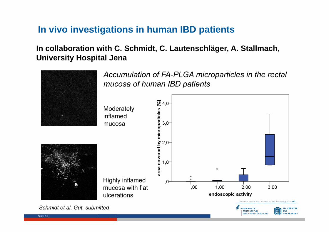
In vivo investigations in human IBD patients
Seite 15 |
Moderatelyinflamedmucosa
Highly inflamedmucosa with flat ulcerations
Accumulation of FA-PLGA microparticles in the rectal mucosa of human IBD patients
In collaboration with C. Schmidt, C. Lautenschläger, A. Stallmach, University Hospital Jena
It‘s only a matter of support: new directions for advanced intestinal cell models
Seite 22 |
1 µm10 µm
It‘s only a matter of support: new directions for advanced intestinal cell models
Seite 23 |
CSEM CorningBDCaco-2
Summary
Successful establishment of a novel cell culture model simulating theintestinal mucosa in the state of inflammation
Pathophysiological changes reflected in the model: release of pro-inflammatory markers, activation of immune cells, decreased barrierfunction, re-organization of tight junctions, increased mucusproduction
Applications of the model:
anti-inflammatory drug and formulation testing in pharmaceuticaldevelopment
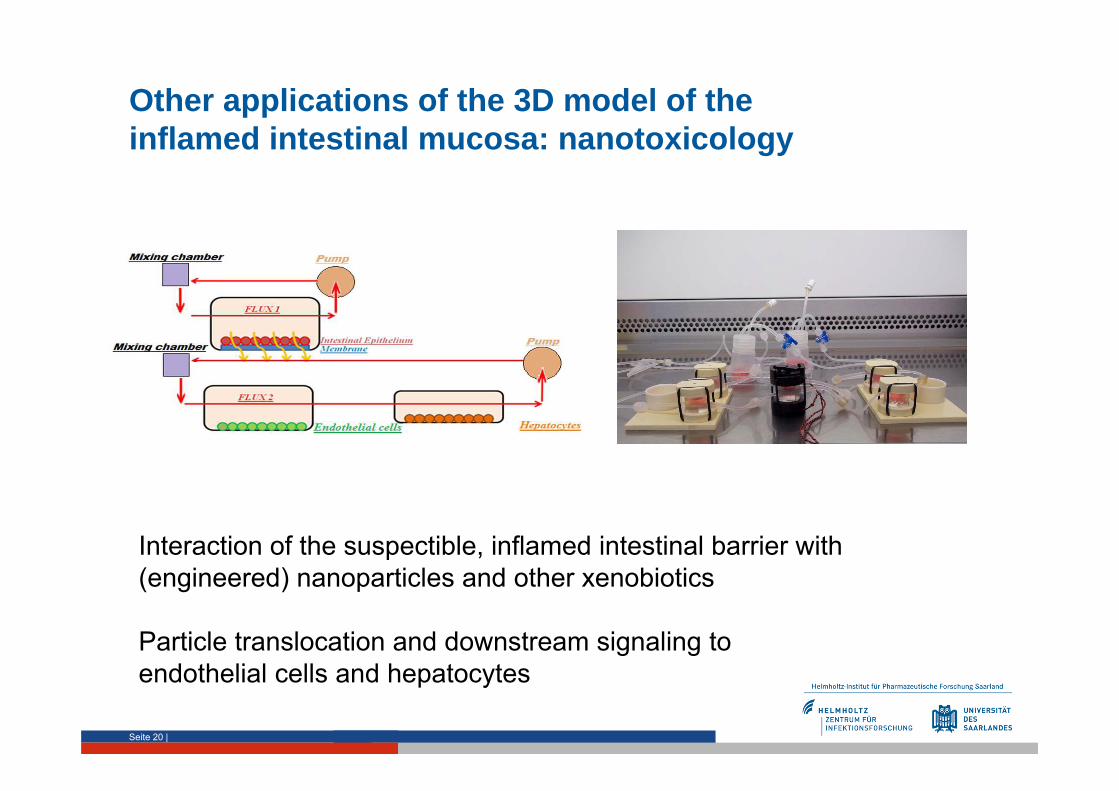
investigation of the interaction of (engineered) nanoparticles or otherxenobiotics with the suspectible barrier
Advantages over existing animal models: ethical aspect, no speciesdifferences, similar pathogenesis, mechanistical insight, cost and time reduction