It has been proposed that proteases secreted by cancer cells facilitatemetastasis by degrading extracellular matrix. Estrogen receptor-positivebreast cancer cells secrete a M, 52,000 pro-cath-D under estrogenstimulation, whereas this protease is produced constitutively by estrogenreceptor-negative cancer cells. We report on the degradation in vitro ofextracellular matrix by purified M, 52,000 cathepsin D (cath-D) and byconditioned media prepared from different cell lines. The purified M,52,000 pro-cath-D was autoactivated at pH 4.5 into a M, 51,000 cath-Dand found to digest the extracellular matrix of endothelial bovine cornealcells labeled with [3H]proline or [35SJmethionine.Culture medium conditioned by estrogen-treated MCF7 cells had a similar effect at pH 4.5 butnot at pH 7.4. Matrix degradation was totally inhibited by pepstatin.Other breast cancer cells (BT20, MDÂ-MB231, T47D cells, etc.) andother cancer cells also secreted a pepstatin-sensitive proteinase able todegrade extracellular matrix. By contrast, the U2 variant of MCF? cells,which lacks the M, 52,000 cath-D gene, and the nontumoral epithelialmammary cells secreted a negligible amount of this proteinase. In allconditioned media, the pepstatin-dependent extracellular matrix degrading activity was highly correlated to the M, 52,000 cath-D concentrationmeasured by immunoenzymatic assay. We conclude that the M, 52,000cath-D is the major acidic protease secreted by mammary cancer cells.We suggest that this protease may degrade basement membrane andconsequently facilitate tumor invasion when it is released in an acidicmicroenvironment.
INTRODUCTION
The mechanism involved in cancer cell metastasis is unknown. Its determination could help in developing treatmentsspecific for the metastatic process, which is responsible formost of the mortality by cancer. In breast cancer, estrogens areknown to facilitate the growth of primary or metastatic cellswhen these cells contain estrogen receptors (1). While studyingestrogen-induced proteins in breast cancer cells, we have purified and characterized a M, 52,000 protein, which is secretedinto medium and displays a mitogenic effect on dormant MCF7cells (for review, see Ref. 2). This protein has been identified asa precursor of cath-D5 which is processed intracellularly into
mature enzymes (M, 48,000 and 34,000 plus 14,000) (3) accumulated in lysosomes. The corresponding complementary DNAhas been cloned, and its sequence indicates high homology withcath-D of normal tissues apart from 5 nucleotide changes (4).
Received 11/6/87; revised 3/21/88; accepted 3/29/88.The costs of publication of this article were defrayed in part by the payment
of page charges. This article must therefore be hereby marked advertisement inaccordance with 18 U.S.C. Section 1734 solely to indicate this fact.
precursor, of M, 52,000; ECM, extracellular matrix; ER, estrogen receptor; DEM,Dulbecco's modified Eagle's medium; FCS, fetal calf serum; BCE, bovine corneal
endothelial cells; IEMA, immunoenzymatic assay; BSA, bovine serum albumin;SDS, sodium dodecyl sulfate; PAGE, polyacrylamide gel electrophoresis; estra-diol-CM, conditioned medium prepared from 17#-estradioI-treated cells; C-CM,conditioned medium prepared from control cells.
The optimal pH for proteolytic activity of this M, 52,000 pro-cath-D is very acidic (pH 3.5) using methemoglobin, and lessacidic using proteoglycans from human cartilage (pH 5) aspreviously reported (3). Moreover, as in the case of cath-Dfrom normal fibroblast (5), but in larger amounts, M, 52,000cath-D is secreted as an inactive precursor which can subsequently be autoactivated by partial proteolysis into a M, 51,000protein (3). Proteases have for long been suspected to facilitatetumor invasion (6-8). In breast cancer, plasminogen activator(6), which is also regulated by estrogens (9), cathepsin B (10-12), and collagenases (7), has mostly been studied. In contrast,cath-D has received much less attention, due to its activity beingrestricted to low pH. However, breast cancer cells produce andsecrete high proportions of this protease compared to nontumoral mammary cells both in vivo (13) and in vitro.6 In the
present study, we considered its potential role in tumor invasionby testing in vitro its ability to degrade extracellular matrix,whether it is purified to homogeneity or present in conditionedmedia secreted by different cancer cells.
MATERIALS AND METHODS
Cell Cultures. Breast cancer cell lines containing ER, i.e., MCF? cells(14) (two sublines, Rich and Lippman) and T47D cells (15), and threebreast cancer cell lines containing no ER, i.e., BT20, MDA-MB231(16), and U2 (17), were cultured as previously described (18). HeLacells (19) were cultured in monolayer in DEM with 5% FCS. TheIshikawa cmloinetri.il adenocarcinoma cell line (20) was cultured inDEM with 15% FCS. A431 human epidermoid carcinoma cells (21), agift of Dr. C. Cochet (U 244 INSERM, Grenoble, France), werecultured in DEM-Ham's F12 plus 10% FCS. Normal mammary epi
thelial glands were prepared as previously described (22, 23) fromreduction mammoplasties, following digestion with collagenase andfiltration. Primary cultures were performed in Ham's F12 supple
mented with 10% FCS.Preparation of ECM from Bovine Endothelial Cells. Primary cultures
of BCE cells were prepared from eyes of freshly slaughtered cows,according to Gospodarowicz (24). Stock cultures were maintained on60 mm tissue culture dishes coated with extracellular matrix (Clinisci-ences) in DEM supplemented with 20% (vol/vol) FCS, 25 lU/ml ofpenicillin, 25 ¿ig/mlof streptomycin (Flow), and 2.5 iig/ml of Fungi-zone (Gibco). Confluent cultures were passaged by trypsinization at aratio of 1 to 10. Matrices were obtained by plating BCE cells on 8-mm-diameter wells, in the presence of 50 ng/ml of bovine brain fibroblastgrowth factor (Clinisciences). One wk after seeding, matrices werelabeled with 1 to 5 ßCi/mlof L-[4,5-3H]proline (Commissariat al'Energie Atomigue; 32 /ttCi/mmol) or 25 pCi/ml of [35S]methionine
(Amersham; 1400 Ci/mmol), for 1 wk as described elsewhere (25, 26).Matrices were then prepared as described elsewhere (24, 26). Briefly,cells were lysed by 3 washes with bidistilled water, followed by 3 washeswith 20 mM NH4OH, 2 washes with bidistilled water, and 2 washeswith phosphate-buffered saline.
Preparation of Enzymatic Solutions and Conditioned Media. Type IAcollagenase from Clostridium histolyticum (Worthington Biochemicals)was dissolved in DEM to a stock concentration of 1 mg/ml. Prior to
6 F. Capony, C. Rougeot, P. Montcourrier, V. Cavailles, G. Salazar, and H.Rochefort. Increased secretion and altered processing of 52-kD pro-cathepsin Din breast cancer cells compared to normal epithelial mammary cells, submittedfor publication.
use, the enzyme was diluted to 10 Mg/ml in DEM. Secreted and cellularcath-D of MCF? cells was purified by immunoaffinity as previouslydescribed (27). Conditioned media were prepared from cells culturedwith 10% PCS to 90% confluence and then cultured in serum-freeconditions for 15 to 18 h as previously described (28). It was importantto remove contamination by PCS, which contains potent proteaseinhibitors such as a2-macroglobulin. This was done by three 30-minwashes of cells with DEM or RPMI before conditioning. Before a 2-day estradici (10 nM) treatment, cells were cultured in DEM withcharcoal-stripped PCS without phenol red (29). Enzymes and conditioned media were acidified or not with 200 HIMcitric acid prior to use.The protease inhibitors, leupeptin and pepstatin (Sigma, St. Louis,MO), were used at a final concentration of 1 to 10 MM-
Extracellular Matrix Degradation. Enzymes and conditioned mediato be tested were applied on 8-mm microwells coated with 2-wk-oldBCE-ECM in a final volume of SO to 200 ¿/I.Following incubations(routinely for 72 h at 37CC), the supernatants were removed, and the
radioactivity released from the ECM was counted; the remaining matrixwas obtained by lysis with 1% SDS, for 2 h and 2 N NaOH for 18 h(26) and directly counted for radioactivity to evaluate the percentage ofhydrolysis. Hydrolysis of ECM was expressed as the percentage ofradioactivity released with respect to total radioactivity present on thecoated well.
IEMA of M, 52,000 cath-D. We used a two-site solid-phase assay oftotal M, 52,000 cath-D (30, 31). Briefly, microtiter plates were coatedwith the D7E3 monoclonal antibody to M, 52,000 cath-D. After saturation of the remaining free adsorption sites with gelatin solution,plates were washed 3 times with buffer. Conditioned medium and asecond antibody conjugated with alkaline phosphatase (M1G8-PA)were incubated for 18 h at 4°Cin a buffer containing 1% BSA and
0.1% Tween 20. The plates were then washed 5 times with 0.9% NaCl-0.05% Tween 20 buffer (pH 8.6) and incubated in a 0.1 M diethanola-mine chromogen solution (pH 9.8) containing 1 mg/ml of paranitro-phenylphosphate for 30 to 120 min at room temperature (in darkness).Plates were read at 405 nm using a Titertek MC Multiskan photometer(Flow Laboratories) coupled to an Apple II microcomputer. The M,52,000 cath-D concentration was determined from the absorbance of 6dilutions (1/1 to 1/80) compared to a standard linear curve obtainedfrom 5 dilutions (0 to 40 ng/ml) of a standard conditioned medium(950 ng/ml). Protein concentration was determined according to Bradford (32) using bovine 7-globulin as the standard.
The sensitivity of the assay was 5 ng/ml, and its intra- and interassayreproducibility was 10% and 15%, respectively.
RESULTS
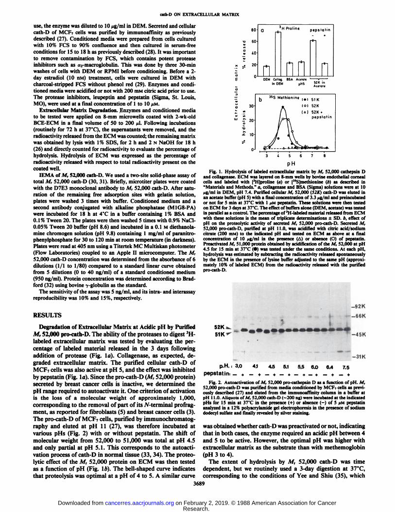
Degradation of Extracellular Matrix at Acidic pH by PurifiedM, 52,000 pro-cath-D. The ability of the proteases to digest 3H-
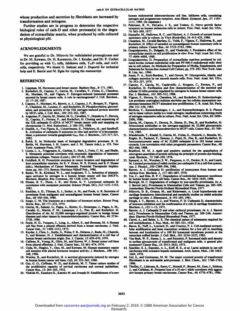
labeled extracellular matrix was tested by evaluating the percentage of labeled material released in the 3 days followingaddition of protease (Fig. la). Collagenase, as expected, degraded extracellular matrix. The purified cellular cath-D ofMCF7 cells was also active at pH 5, and the effect was inhibitedby pepstatin (Fig. la). Since the pro-cath-D (M, 52,000 protein)secreted by breast cancer cells is inactive, we determined thepH range required to autoactivate it. One criterion of activationis the loss of a molecular weight of approximately 1,000,corresponding to the removal of part of its W-terminal profragment, as reported for fibroblasts (5) and breast cancer cells (3).The pro-cath-D of MCP7 cells, purified by immunochromatog-raphy and eluted at pH 11 (27), was therefore incubated atvarious pHs (Fig. 2) with or without pepstatin. The shift ofmolecular weight from 52,000 to 51,000 was total at pH 4.5and only partial at pH 5.1. This corresponds to the autoacti-vation process of cath-D in normal tissue (33, 34). The proteo-lytic effect of the M, 52,000 protein on ECM was then testedas a function of pH (Fig. 16). The bell-shaped curve indicatesthat proteolysis was optimal at a pH of 4 to 5. A similar curve
80TJ
SOCKS
«o^
20-
n-Qn3H
Prolinepepstotmnn-i-n
DEM Collag BSA Aerial«in DEM pH5 j>ZK ln
Aceraie
30-
r 20-
10-
0J
loi 52K(M 52K •>
pepstatin
pHFig. 1. Hydrolysis of labeled extracellular matrix by M, 52,000 cathepsin D
and collagenase. ECM was layered on 8-mm wells by bovine endothelial cornealcells and labeled with [3H]proline (a) or ("S]methionine (b) as described in"Materials and Methods." a, collagenase and BSA (Sigma) solutions were at 10jig/ml in DEM, pH 7.4. Purified cellular M, 52,000 (S2K) cath-D was eluted inan acetate buffer (pH 5) with a final concentration of 3.3 wg/ml and preincubatedor not for 5 min at 37*C with l UMpepstatin. These solutions were then testedon ECM for 3 days at 37'C. The effect of buffers alone (DEM, acetate) was testedin parallel as a control. The percentage of 3H-labeled material released from ECM
with these solutions is the mean of triplicate determinations ±SD. b, effect ofpH on the proteolytic activity of secreted M, 52,000 pro-cath-D. Secreted M,52,000 pro-cath-D, purified at pH 11.0, was acidified with citric acid/sodiumcitrate (200 n>M) to the indicated pH and tested on ECM as above at a finalconcentration of 10 ¿in/inlin the presence (A) or absence (O) of pepstatin.I'reactivated M, 51,000 protein obtained by acidification of the M, 52,000 at pH4.5 for 15 min at 37'C (•)was tested under the same conditions. At each pH,
hydrolysis was estimated by subtracting the radioactivity released spontaneouslyby the ECM in the presence of lysine buffer adjusted to the same pH (approximately 10% of labeled ECM) from the radioactivity released with the purifiedpro-cath-D.
-92K
-66K
-45K
-31K
P.M.: 3.0 4.1 45 5.1 5.5 6.0 6.4 7.5
pepstatin: _+ _ + _ + _*_+_+ _+ _ +Fig. 2. Autoactivation of M, 52,000 pro-cathepsin D as a function of pH. M,
52,000 pro-cath-D was purified from media conditioned by NUT- cells as previously described (27) and eluted from the immunoaffinity column in a buffer atpH 11.0. Aliquots of M, 52,000 cath-D (-200 ng) were incubated at the indicatedpHs for 15 min at 37'C in the presence (+) or absence (-) of 5 /IM pepstatin
analyzed in a 12% polyacrylamide gel electrophoresis in the presence of sodiumdodecyl sulfate and finally revealed by silver staining.
was obtained whether cath-D was preactivated or not, indicatingthat in both cases, the enzyme required an acidic pH between 4and 5 to be active. However, the optimal pH was higher withextracellular matrix as the substrate than with methemoglobin(pH 3 to 4).
The extent of hydrolysis by M, 52,000 cath-D was timedependent, but we routinely used a 3-day digestion at 37°C,
corresponding to the conditions of Yee and Shiu (35), which3689
allowed a sufficient accumulation of reaction product in themedium even though the reaction rate was slower than in thefirst 12 h (not shown). Under these conditions, the degradationof ECM was proportional to the amount of M, 52,000 cath-Dtested up to 0.5 tig/ml. The radioactivity released from ECMby the M, 52,000 cath-D preparation was essentially due to
degradation and not to desorption of labeled precursor basedon two lines of evidence, (a) The extent of release induced bybuffer, or albumin solution, or in the presence of pepstatin,corresponding to a background level, was much lower (Fig. 1).(b) SDS-PAGE analysis of the supernatant following digestionof [35S]methionine-labeled ECM indicated that M, S 60,000
proteins and peptides were formed after incubation with M,52,000 cath-D but not when the enzyme was inhibited bypepstatin (not shown).
cath-D has a broad substrate specificity. The fact that [3H]-
proline was released from ECM suggests that the M, 52,000cath-D is able to degrade collagen, even though collagen is nota classical cath-D substrate (36). The Mr 52,000 cath-D canalso degrade proteoglycans from human cartilage (37) as previously described (3, 38). This was confirmed by showing thattheir molecular weight was much lower following M, 52,000cath-D treatment, as estimated by gel filtration chromatogra-Phy.7
Degradation of ECM by the M, 52,000 pro-cath-D of MediaConditioned by MCF7 Cells. Since purified M, 52,000 pro-cath-D can be autoactivated and degrade ECM in vitro, we thenconsidered its activity in conditioned media prepared fromMCF7 cells, where putative inhibitors could mask its effect orprevent its activation. There was no hydrolysis at pH 7.4,indicating an absence of active neutral proteases in these media(Fig. 3fl) which confirmed a previous report (35). At pH 5,however, ECM was hydrolyzed, and the estradiol-CM was muchmore active than the C-CM containing the same amount oftotal secreted proteins but much less M, 52,000 pro-cath-D(Fig. 3a). Similar results were obtained whether ECM waslabeled with [35S]methionine or with [3H]proline. The extent ofhydrolysis was higher with [3H]proline-labeled ECM than with[35S]methionine-labeled ECM in Fig. 3, but varied markedly
depending on the experiment and the different preparations oflabeled bovine ECM used. The M, 52,000 cath-D concentrationmeasured by IEMA in the MCF7 cell-conditioned media wasfound to vary from 0.1 to 0.8 ng/m\. The ECM degradingability of pro-cath-D in these media was similar to its activityfollowing purification to homogeneity as long as PCS components (which include protease inhibitors) were removed bywashing cells. This suggests that no or few endogenous inhibitors of cath-D are released by MCF7 cells, contrary to what isobserved with other proteases (8, 10).
We then tested protease inhibitors (Fig. 3Z>).Pepstatin inhibited all degrading ability of the estradiol-CM, whereas leupeptinwas almost inactive, suggesting that most of the hydrolysingability of estradiol-CM is due to an aspartyl protease, probablyA/r 52,000 cath-D, which is highly concentrated in these media.By contrast, cathepsin B is secreted in very small amounts byMCF7 cells8 and may be inhibited by endogenous inhibitor(s).
Considering that incubation at acidic pH of conditioned mediacontaining A/r 52,000 pro-cath-D did not allow processing ofthe precursor into mature enzymes (M, 48,000 and 34,000 plus14,000 forms), and that this processing is normally mediatedin cells by a cysteinyl protease (33), it would appear that nocysteinyl protease is active in these media.
90-50-10-0 3HProlinepH
7.4nnnpH 5.nfì-t
DEM C.CM E2.CM DEM C.CM E2.CM
¿020-nbri-,,
35.S.MethloninepH
4.5*
n HH_LDEM E2CM DEM E2.CM DEM E2.CM
>pcp&rarin ru pipi in
Fig. 3. Digestion of the ECM by media conditioned by MC F7 cells. Mediaconditioned by MCF7 cells treated for 4 days with 10 nM estradici (E¿-CM)orwithout estradici (C-CM) were prepared as described (28). They containedproteins released for 18 h under serum-free conditions at pH 7.4 in DEM withoutphenol red. Protein concentrations were equalized in estradiol-CM and C-CM(a), and the media were concentrated 10-fold by lyophilization, after which theywere acidified or not to pH S with 0.1 N HC1 (a) or 200 mM citric acid (b); 100t/1of each conditioned medium and 100 ¿tlof culture medium acidified or not asa control were incubated for 3 days with [3H]proline-labeled ECM (a) or with[3*S]methionine-labeled ECM (b). Pepstatin and leupeptin (10 pM each) weretested in parallel (b) on the estradiol-CM. Hydrolysis was estimated as thepercentage of radioactivity released into the medium. Columns, mean of triplicatedeterminations or of duplicate experiments (•);bars, SD.
Comparison of Conditioned Media of Several Breast CancerCells in Their Ability to Degrade ECM at Acidic pH. We thenevaluated the proteolytic activity at pH 4.5 on [35S]methionine-
labeled extracellular matrix of conditioned media preparedfrom a series of cell lines cultured at subconfluence. Fig. 4shows that breast cancer cells in general secrete more pepstatin-sensitive (i.e., aspartyl) protease(s) that can degrade ECM thanother cancer cells. Among the breast cancer cells tested, MCF7,BT20, and MDA-MB231 cells were the most active. T47D cellswere less active, and U2, which is an MCF7 cell variant lackingthe gene for cath-D, had almost no activity. Cancer cells fromskin epithelioma (A431), en do met riunì(Ishikawa), and uterinecervix (HeLa) also secreted some cath-D-like material. By contrast, normal mammary glands in primary culture displayedalmost no degrading activity on ECM. We found a good correlation between the pepstatin-sensitive degrading ability ofconditioned media and their secreted M, 52,000 cath-D content,as measured by double determinant immunoenzymatic assay(31) (Fig. 5). However, for concentrations of Mr 52,000 cath-Dhigher than 530 ng/ml (with MCF7 R variant), the degradationwas not further increased since the amount of the ECM substrate was limiting. We conclude that cath-D is the most abundant protease secreted by cancer cells able to degrade ECM ata distance and at acidic pH. Breast cancer cells secrete relativelylarge amounts of this protease compared to other cancer cellsand to normal mammary cells.
DISCUSSION
1 D. R. Mitrovic and M. Morisset, unpublished results." F. Capony, unpublished experiments.
We have shown that the Mr 52,000 cath-D secreted by breastcancer cells can degrade extracellular matrix following its au-
Fig. 4. Comparison of degrading ability of ECM by conditioned media prepared from different cells. Media conditioned by different types of cells all culturedto subconfluence on plastic were concentrated 5 times by lyophilization and testedon labeled extracellular matrix at pH 4.5 for 3 days at 37'C, as described in Fig.
1. Pepstatin (pepst.) and leupeptin (leup.) were tested in parallel at 10 MM.Controldigestion (medium) was that obtained with the respective culture media alone(DEM or RPMI for T47D and BT20 or F12 for normal mammary cells). ECMdegradation without (C.'.M.) and with protease inhibitors (pepst. or leup.) wasdetermined for each cell line and expressed as in Figs. 1 and 3. M( l:, R andMCF7 L are two sublines provided by Marvin Rich and M. Lippman, respectively.
ou
40-
30-
20-
10
O
r = 0.96MCF7
0.1 0.2 0.3 0.4 0.5
52K - calh D ( jjg / m I I
Fig. S. Correlation between the pepstatin-sensitive degrading ability of conditioned media and their M, 52,000 cathepsin D concentrations. The pepstatin-sensitive protease activities of 200 n\ of the different conditioned media in Fig. 5were plotted versus the At, 52,000 cath-D concentrations (in fig/ml) in the sameCM as assayed by IEMA (see "Materials and Methods"). The pepstatin-sensitive
protease activity of the CM was calculated by subtracting the radioactivity releasedfrom extracellular matrix with pepstatin, from that released without pepstatin.\.\fC. M DA. and Ish refer to normal mammary cells, MDA-MB23 1, and Ishikawacells. Linear regression was calculated by Pearson's least-squares method. Thecorrelation coefficient (r = 0.96) is statistically significant (/' = 0.001) accordingto Student's t test.
toactivation at pH 4 to 5. The optimal pH for autoactivationinto a Mr 51,000 protein, as determined with pure M, 52,000pro-cath-D, is similar to that of the normal cath-D in humanfibroblasts and rabbit heart cells (33, 34). Moreover, M, 52,000cath-D is also autoactivatable in media conditioned by MCI- -
cells treated with estradiol and cultured without serum. Thesubstrate specificity of normal cath-D is known to be broad,and it can degrade cartilage proteoglycans (3), whereas its effecton collagen I appears to be less (36). The low pH required forthe proteolytic effect on ECM indicates that an acidic environment is absolutely necessary for this M, 52,000 cath-D activity(39). Other groups have shown that breast cancer cells (35) andmalignant melanoma (10) can degrade basement membrane viaproteases, whereas media conditioned by these cells are unable
to mimic this effect. It has therefore been suggested that proteases are either membrane bound (and not released) or releasedwith inhibitors preventing their action. By contrast, we showhere that aspartyl protease may be secreted by cancer cells toact at a distance and may play an important role, since it ishighly active on ECM even without purification, provided thelocal pH is sufficiently low (a5.5). This low pH may be foundin a microenvironment generated by malignant cells close toextracellular matrix. Several mechanisms can be consideredsuch as localized acidic pH at the cell membrane (40) due to anincreased release of H+ from membrane cancer protein or H+
ATPase (41), or an increased sialic acid content in oligosac-charidic chains of cancer cells (42). Moreover, tumor acidification has been described as a consequence of anoxia (43).
The second major information provided by this study is thatM, 52,000 pro-cath-D is the most abundant and active acidic
protease to be secreted by breast cancer cells and to a lesserdegree by other cancer cells. Among the cells tested, M, 52,000cath-D was the only protease of the conditioned media thatcould degrade extracellular matrix. Practically, no leupeptin-sensitive protease activity was found. However, the cysteinylproteases, cathepsins L (44) and B (10), also act at acidic pH.It cannot be excluded that, under the conditions used, theseproteases are inactivated or inhibited by endogeneous inhibitors. The U2 cells were inactive, but they do not express theestrogen receptor and M, 52,000 cath-D (17), and the MM96melanoma cell line displayed more leupeptin-sensitive, cathepsin B-iike activity than pepstatin-sensitive activity (not shown).Normal mammary cells secrete very little protease activity thatcan degrade ECM and almost no cath-D-like proteases, whichis in agreement with their low production of M, 52,000 cath-D, as suggested by immunoperoxidase staining (13) and measured by biometabolic labeling and IEMA.6 The induction of M,
52,000 cath-D by estrogens in ER-positive cell lines suggests
that estrogens may also facilitate tumor invasion by increasingthe amount of secreted pro-cath-D autoactivatable at acidic pH.The negative results obtained with T47D cells by Yee and Shiu(35) are probably due to the fact that the conditioned mediawere only tested at neutral pH. These in vitro studies showingthat, in breast cancer, M, 52,000 cath-D can potentially degradeECM in vivo, as long as the pH is low, are supported by itsprognostic value in breast cancer patients. A retrospective clinical study, in which M, 52,000 cath-D was assayed in breastcancer cytosol, has shown that high concentrations of A/r 52,000cath-D are an independent prognostic factor correlated with ashorter relapse-free survival.9 In addition, the concentration
and secretion of A/r 52,000 cath-D in breast cancer cells arehigher than in normal mammary cells (Ref. 13; Footnote 6).These results together suggest that cath-D may play an impor
tant role in mammary carcinogenesis. Recent localization of itsgene by in situ hybridization (4) close to H-ras on the short armof chromosome 11 in a region whose deletion has been implicated in mammary carcinogenesis (45) also supports this hypothesis.
In addition to neutral proteases bound to or inserted intoplasma membrane, we therefore propose that lysosomal proteases may play a major role in degrading ECM and facilitatingtumor invasion. Another example of a cathepsin involved incarcinogenesis is cathepsin L, or major excreted protein (44),
'S. M. Thorpe, H. Rochefort, M. Garcia, G. Freiss, 1. J. Christensen, S.
Khalaf, F. Paolucci, B. Pau, B. B. Rasmussen, and R. Carsten. High concentrations of 52K cathepsin D predict poor prognosis in primary, postmenopausalbreast cancer, submitted for publication.
whose production and secretion by fibroblasts are increased bytransformation and mitogens.
Further studies are in progress to determine the respectivebiological roles of cath-D and other protease(s) in the degradation of extracellular matrix, when produced by cells culturedat physiological pH.
1. Lippman, M. Hormones and breast cancer. Banbury Rep., &•171, 1981.2. Rochefort, H., Capony, F., Garcia, M., Cavailles, V., Freiss, G., Chambón,
M., Morisse!, M., and Vignon, F. Estrogen-induced lysosomal proteasessecreted by breast cancer cells. A role in carcinogenesis? J. Cell. Biochem.,35: 17-29, 1987.
3. Capony, F., Morisset, M., Barrett, A. J., Capony, J. P., Broquel, P., Vignon,F., Chambón, M., Louisot, P., and Rochefort, H. Phosphorylation, glycosyl-ation, and proteolytic activity of the 52K estrogen-induced protein secretedby MCF, cells. J. Cell. Biol., 104: 253-262,1987.
4. Augereau, P., Garcia, M., Mattel, M. G., Cavailles, V., Depadova, F., Derocq,D., Capony, F., Ferrara, P., and Rochefort, H. Cloning and sequencing ofthe 52K cathepsin D cDNA of MCF7 breast cancer cells and mapping onchromosome 11. Mol. Endocrinol., 2: 186-192, 1988.
5. Hasilik, A., Von Figura, K., Conzerlmann, E., Nehrkorn, H., and Sandhoff,K. Activation of cathepsin D precursor in vitro and activity of j3-hexosamin-idase, a precursor towards ganglioside (<•,,-.Eur. J. Biochem., 125:317-321,1982.
6. Reich, E. In: Molecular basis of biological degradative processes. R. D.Berlin, M. Herrman, I. H. Lepow, and J. M. Tanzer (eds.), p. 155. NewYork: Academic Press, 1978.
7. 1 ¡olia,L. A., Tryggvason, K., Garbisa, S., Hart, I., Foltz, C. M., and Shafie,S. Metastatic potential correlates with enzymatic degradation of basementmembrane collagen. Nature (Lond.), 284:67-68, 1980.
8. Goldfarb, R. H. Proteolytic enzymes in tumor invasion and degradation ofhost extracellular matrices. In: K. V. Honn, W. E. Powers, and B. F. Sloane(eds.), Mechanisms of Cancer Metastasis. Potential Therapeutic Implications, pp. 341-373. Boston: Martinus Nijhoff Pub., 1986.
9. Butler, W. B., Kirkland, W. L., and Jorgensen, T. L. Induction of plasmin-ogen activator by estrogen in a human breast cancer cell line (MCF7).Biochem. Biophys. Res. Commun., 90:1328-1334, 1979.
10. Sloane, B. F., Dunn, J. R., and Honn, K. V. Lysosomal cathepsin B:correlation with metastatic potential. Science (Wash. DC), 212:1151-1153,1981.
11. Recklies, A. D., Tiltman, K. J., Stoker, A. M., and Poole, A. R. Secretion ofproteinase from malignant and nonmalignant human breast tissue. CancerRes., 40: 550-556, 1980.
12. Szego, C. M. The lysosome as a mediator of hormone action. Recent Prog.Horm. Res., 30:171-233, 1974.
14. Soule, H. D., Vazquez, J., Long, A., Albert, S.. and Brennan, M. A Humancell line from a pleural effusion derived from a breast carcinoma. J. Nati.Cancer Inst., 51: 1409-1413, 1973.
15. Keydar, I., Chen, L., Karby, S., Weiss, F. R., Delarea, J., Radu, M., Chaitcik,S., and Brenner, H. J. Establishment and characterization of a cell line ofhuman breast carcinoma origin. Eur. J. Cancer, 15:659-670, 1979.
16. Cailleau, R., Young, R., Olive, M., and Reeves, W. J. Breast tumor cell linesfrom pleural effusions. J. Nati. Cancer Inst., 53: 661-674, 1974.
17. Ueda, M., Hagino, Y., Ono, M., and Kuwano, M. Human mammary cancercell mutants with altered hormone receptor activity. J. Biochem., 700: 341-348, 1986.
18. Westley, B., and Rochefort, H. A secreted glycoprotein induced by estrogenin human breast cancer cell lines. Cell, 20: 352-362, 1980.
19. Gey, G. O., Coffman, W. D., and Kubicek, M. T. Tissue culture studies ofthe proliferative capacity of cervical carcinoma and normal epithelium.Cancer Res., 12: 264-265, 1952.
20. Nishida M., Kasahara K., Kaneko M. and Iwasaki H. Establishment of a new
human endometrial adenocarcinoma cell line, Ishikawa cells, containingestrogen and progesterone receptors. Acta Obstet. Gynaecol. Jpn., 37: 1103-1111, 1985. (In Japanese.)
21. Fabricant, R. N., DeLarco, J. E., and Todaro, G. Nerve growth factorreceptors on human melanoma cells in culture. Proc. Nati. Acad. Sci. USA,74: 565-569, 1977.
22. Stampfer, M., Hallowes, R. C., and Hackett, A. J. Growth of normal humanmammary cells in culture. In Vitro (Rockville), 16:415-425, 1980.
24. Gospodarowicz, D., Delgado, D., and Vlodavsky, 1. Permissive effect of theextracellular matrix on cell proliferation in vitro. Proc. Nati. Acad. Sci. USA,77:4094-4098, 1980.
25. Gospodarowicz, D. Preparation of extracellular matrices produced by cultured bovine corneal endothelial cells and PF-HR-9 endoderma! cells: theiruse in cell culture. In: Methods for Preparation of Media, Supplements, andSubstrata for Serum-free Animal Cell Culture, pp. 275-293. New York: AlanR. Liss, Inc., 1984.
26. Jones, P. A., Scott-Burdent, T., and Gevers, W. Glycoprotein, elastin, andcollagen secretion by rat smooth muscle cells. Proc. Nati. Acad. Sci. USA,76: 353-357, 1979.
27. Capony, F., Garcia, M., Capdevielle, J., Rougeot, C., Ferrara, P., andRochefort, H. Purification and tirsi characterization of the secreted andcellular 52-kDa proteins regulated by estrogens in human breast cancer cells.Eur. J. Biochem., 161: 505-512, 1986.
29. Berthois, Y., Katzenellenbogen, J. A., and Katzenellenbogen, B. S. Phenolred in tissue culture media is a weak estrogen: implications concerning studyof estrogen-responsive cells in culture. Proc. Nati. Acad. Sci. USA, 83:2496-2500, 1986.
30. Garcia, M., Capony, F., Derocq, D., Simon, D., Pau, B., and Rochefort, H.Monoclonal antibodies to the estrogen-regulated M, 52,000 glycoprotein:characterization and immunodetection in MCF7 cells. Cancer Res., 45:709-716, 1985.
32. Bradford, M. M. A rapid and sensitive method for the quantitation ofmicrogram quantities of protein utilizing the principle of protein-dye binding.Anal. Biochem., 72: 248-250, 1976.
33. Samare!, A. M., Worobec, S. W., Ferguson, A. G., Decker, R. S., and Lesch,M. Limited proteolysis of rabbit cardiac pro-cathepsin D in a cell-free system.Am. J. Physiol., 250: C589-596, 1986.
34. Barrett, A. J. Cathepsin D: purification of isoenzymes from human andchicken liver. Biochem. J., 117: 601-607, 1970.
35. Yee, C., and Shiu, R. P. C. Degradation of endothelial basement membraneby human breast cancer cell lines. Cancer Res., 46:1835-1839, 1986.
37. Mitrovic, D. R., Gruson, M., and Rickewaert, A. Local hyperthermia andcartilage breakdown: histochemical and metabolic studies on rabbit articularcartilage in vitro. J. Klieumami.. 8: 193-203, 1981.
38. Dingle, J. T., Barrett, A. J., and Weston, P. D. Cathepsin D, characteristicsof immuno-inhibition and the ion firmal ion of a role in cartilage breakdown.Biochem. J., 723:1-13, 1971.
42. Van Beek, W. P., Smets, L. A., and Emmelot, P. Increased sialic acid densityin surface glycoprotein of transformed and malignant cells. A general phenomenon? Cancer Res., 33:2913-2922, 1973.
44. Gal, S., and Gottesman, M. M. The major excreted protein of transformedfibroblasts is an activatable acid-protease. J. Biol. ("hem., 267: 1760-1765,
1986.45. Theillet, C., Lidereau, R., Escot, C., Hutzell, P., Brunei, M., Gest, J., Schlom,
J., and Callahan, R. Frequent loss of a H-ros-1 alÃelecorrelates with aggressive human primary breast carcinomas. Cancer Res., 46:4776-4781, 1986.