In Vitro Hemocompatibility and Cytotoxicity Study of Poly(2-methyl-2-oxazoline) for Biomedical Applications Marius Bauer, 1 Susann Schroeder, 1 Lutz Tauhardt, 2,3 Kristian Kempe, 2,3 Ulrich S. Schubert, 2,3 Dagmar Fischer 1,3 1 Department of Pharmaceutical Technology, Institute of Pharmacy, Friedrich-Schiller University Jena, Otto-Schott-Str. 41, 07745 Jena, Germany 2 Laboratory of Organic and Macromolecular Chemistry (IOMC), Friedrich-Schiller-University Jena, Humboldtstr. 10, 07743 Jena, Germany 3 Jena Center for Soft Matter (JCSM), Friedrich-Schiller-University Jena, Philosophenweg 7, 07743 Jena, Germany Correspondence to: D. Fischer (E-mail: [email protected]) Received 28 December 2012; accepted 11 January 2013; published online 7 February 2013 DOI: 10.1002/pola.26564 ABSTRACT: Since poly(2-methyl-2-oxazolines) (PMeOx) attract high attention for the potential use in drug delivery, cytotoxic- ity, and hemocompatibility of a set of PMeOxs with molar masses in the range from 2 to 20 kDa are systematically inves- tigated under standardized conditions in terms of molar mass, concentration and time dependency. PMeOx polymers are well tolerated in red blood cell aggregation and hemolysis assays without any damaging effects even at high concentrations up to 80 mg/mL. Only in long term cytotoxicity tests PMeOx poly- mers moderately influence cell viability in a time, concentra- tion, and molar mass dependent manner. Referring to these results it can be concluded that PEtOx could be promising non- ionic hydrophilic polymers for many biomedical applications without any cyto- and hemotoxic effects at typically used thera- peutic doses. V C 2013 Wiley Periodicals, Inc. J. Polym. Sci., Part A: Polym. Chem. 2013, 51, 1816–1821 KEYWORDS: biocompatibility; hemocompatibility; hydrophilic polymer; poly(2-methyl-2-oxazoline); ring-opening poly- merization INTRODUCTION The use of the hydrophilic, nonionic poly(2-methyl-2-oxazoline) (PMeOx) in biomedical and pharmaceutical applications has enormously evolved during the last years. 1,2 On the one hand, PMeOx can be considered chemically as a poly(ethylene imine) (bPEI) backbone with amide-bonded side groups. On the other hand, these polymers were also described as pseudopeptides or bioinspired poly- mers due to their structural relation to polypeptides. In contrast to peptides, the tertiary amine groups of PMeOx can- not be recognized by enzymes for hydrolytic cleavage which provides high stability in the biological environment. 3 Since the early 90s several studies are available reporting the use and importance of PMeOx as a component of drug delivery systems such as peptide, protein, and lipid conju- gates as well as for liposomes and micelles (for reviews, see e.g., refs. 1 and 2). Miyamoto et al. 4 demonstrated that the covalent binding of PMeOx to liver catalase influenced the enzyme activity dependent on polymer molar mass and degree of conjugation. Furthermore, it was possible to pre- serve the enzymatic activity in organic solvents due to an increase of solubility provided by the conjugated PMeOx. 4 Amphiphilic block copolymers of PMeOx and poly(2-butyl-2- oxazoline)s were tested as micellar carrier systems for the delivery of hydrophobic drugs such as paclitaxel. The results revealed a low intrinsic toxicity of the micelles and a high micellar drug uptake without loss of drug activity. 5 Similar results were received for micelles formed of block copoly- mers with hydrophobic poly(L-lactide) or poly(e-caprolac- tone) and PMeOx. 6,7 Many of the observed effects were found to be comparable to the beneficial properties of poly(ethylene glycol) (PEG) with regard to stealth behavior, biodistribution, and biocom- patibility. PMeOx can induce a stealth effect in a similar way as PEG when grafted on liposomes or surfaces. 8,9 Clearance from the blood was similar for PEG and PMeOx, whereas for instance the more lipophilic poly(2-ethyl-2-oxazoline) (PEtOx) was removed somewhat faster. 10 Liposomes deco- rated with PMeOx were characterized by a prolonged blood circulation time and a preferential organ distribution in liver, spleen, and kidney after 24 h. 8,10 Protein-repellent and non-fouling, antimicrobial effects after coating of PMeOx to surfaces, for example, of implants, biosensors or nanoparticles quantitatively reached the excellent properties of PEG. 9,11 Additional Supporting Information may be found in the online version of this article. V C 2013 Wiley Periodicals, Inc. 1816 JOURNAL OF POLYMER SCIENCE, PART A: POLYMER CHEMISTRY 2013, 51, 1816–1821 ARTICLE WWW.POLYMERCHEMISTRY.ORG JOURNAL OF POLYMER SCIENCE
Transcript
In Vitro Hemocompatibility and Cytotoxicity Study
of Poly(2-methyl-2-oxazoline) for Biomedical Applications
Marius Bauer,1 Susann Schroeder,1 Lutz Tauhardt,2,3 Kristian Kempe,2,3
Ulrich S. Schubert,2,3 Dagmar Fischer1,3
1Department of Pharmaceutical Technology, Institute of Pharmacy, Friedrich-Schiller University Jena, Otto-Schott-Str. 41, 07745
Jena, Germany2Laboratory of Organic and Macromolecular Chemistry (IOMC), Friedrich-Schiller-University Jena, Humboldtstr. 10,
07743 Jena, Germany3Jena Center for Soft Matter (JCSM), Friedrich-Schiller-University Jena, Philosophenweg 7, 07743 Jena, Germany
INTRODUCTION The use of the hydrophilic, nonionicpoly(2-methyl-2-oxazoline) (PMeOx) in biomedical andpharmaceutical applications has enormously evolved duringthe last years.1,2 On the one hand, PMeOx can be consideredchemically as a poly(ethylene imine) (bPEI) backbone withamide-bonded side groups. On the other hand, these polymerswere also described as pseudopeptides or bioinspired poly-mers due to their structural relation to polypeptides. Incontrast to peptides, the tertiary amine groups of PMeOx can-not be recognized by enzymes for hydrolytic cleavage whichprovides high stability in the biological environment.3
Since the early 90s several studies are available reportingthe use and importance of PMeOx as a component of drugdelivery systems such as peptide, protein, and lipid conju-gates as well as for liposomes and micelles (for reviews, seee.g., refs. 1 and 2). Miyamoto et al.4 demonstrated that thecovalent binding of PMeOx to liver catalase influenced theenzyme activity dependent on polymer molar mass anddegree of conjugation. Furthermore, it was possible to pre-serve the enzymatic activity in organic solvents due to anincrease of solubility provided by the conjugated PMeOx.4
Amphiphilic block copolymers of PMeOx and poly(2-butyl-2-
oxazoline)s were tested as micellar carrier systems for thedelivery of hydrophobic drugs such as paclitaxel. The resultsrevealed a low intrinsic toxicity of the micelles and a highmicellar drug uptake without loss of drug activity.5 Similarresults were received for micelles formed of block copoly-mers with hydrophobic poly(L-lactide) or poly(e-caprolac-tone) and PMeOx.6,7
Many of the observed effects were found to be comparableto the beneficial properties of poly(ethylene glycol) (PEG)with regard to stealth behavior, biodistribution, and biocom-patibility. PMeOx can induce a stealth effect in a similar wayas PEG when grafted on liposomes or surfaces.8,9 Clearancefrom the blood was similar for PEG and PMeOx, whereasfor instance the more lipophilic poly(2-ethyl-2-oxazoline)(PEtOx) was removed somewhat faster.10 Liposomes deco-rated with PMeOx were characterized by a prolonged bloodcirculation time and a preferential organ distribution inliver, spleen, and kidney after 24 h.8,10 Protein-repellentand non-fouling, antimicrobial effects after coating ofPMeOx to surfaces, for example, of implants, biosensors ornanoparticles quantitatively reached the excellent propertiesof PEG.9,11
Additional Supporting Information may be found in the online version of this article.
VC 2013 Wiley Periodicals, Inc.
1816 JOURNAL OF POLYMER SCIENCE, PART A: POLYMER CHEMISTRY 2013, 51, 1816–1821
ARTICLE WWW.POLYMERCHEMISTRY.ORGJOURNAL OF
POLYMER SCIENCE
A requirement for the administration of PMeOx in patients isthe biocompatibility of PMeOx which was first demonstratedin vivo by Goddard et al.12 after intravenous injection inmice. It was reported that PMeOx did not accumulate inorgans and can be rapidly cleared from the blood stream.13
Only some high molar mass PMeOx polymers were found inskin and muscle tissue.12 However, in vitro hemocompatibil-ity data are not available for PMeOx to the best of ourknowledge and in vitro cytotoxicity data are limited. Kroneket al.14 investigated only one PMeOx polymer with 8.5 kDain a [3-(4,5-dimethylthiazol-2-yl)-2,5-diphenyl tetrazoliumbromide] (MTT) assay and found no cytotoxic effects up to aconcentration of 5 mg/mL after 24 and 48 h. Luxenhoferet al.15 used only two PMeOx derivatives of about 4 kDa,however modified with piperazine and Boc-piperazine, todemonstrate the biocompatibility of PMeOx in a cytotoxicity(MTT) assay over up to 24 h. More information is only avail-able for copolymers with PMeOx.15
Consequently, in the present study in vitro cytotoxicity andhemocompatibility of pure PMeOx polymers with a widerange of molar masses from 2 to 20 kDa were systematicallyinvestigated under standardized conditions to close the gaps.In particular, the influence of polymer molar mass, polymerconcentration and incubation time was investigated.
RESULTS AND DISCUSSION
Four PMeOx polymers with pharmaceutical and biomedicalrelevant molar masses from 2 to 20 kDa were synthesized asdescribed in the Supporting Information. All PMeOxs wereobtained as white crystalline powders. Identity and purity ofthe polymers were proven by means of 1H NMR spectros-copy and size exclusion chromatography analysis which con-firmed that the PMeOx polymers were synthesized withdefined molar masses and acceptable molar mass distribu-tion [polydispersity index (PDI) values < 1.45] (Fig. 1 andSupporting Information Table S1).
Before administration of the PMeOxs to cells, potential non-specific effects of the polymer solutions falsifying the results
of the toxicity tests were excluded. Polymer loss due toincomplete passage of PMeOx through the sterile filter couldbe excluded by comparing the mass of unfiltered and filteredstock solutions after freeze drying (data not shown). pH val-ues and osmolarities of all stock solutions were routinelymeasured before application to the cells. The pH values of allpolymer solutions were comparable to the correspondingpure medium. Osmolarities of the stock solutions rangedfrom 335 to 420 mosmol/kg (Supporting Information TableS2). Differences were attributed to varying polymer molarmass and type of medium. A MTT assay using culturemedium of different osmolarities (280–650 mosmol/kg)demonstrated that osmolarities up to 490 mosmol/kg werewell tolerated by L929 cells without morphological changesor decrease in cell viability up to 24 h (Supporting Informa-tion Fig. S1). Only the highest osmolarity of 650 mosmol/kgshowed a reduction of the cell viability to 34% after 24 hwhich correlates well with reports for other cell types.16
Many of the polymers used in medical applications or asdrug delivery system come into close contact with bloodplasma and blood cells, in particular after systemic adminis-tration. Therefore, the influence of PMeOx polymers on theaggregation behavior and hemolysis of sheep erythrocyteswas investigated. Erythrocyte aggregation of PMeOx wasqualitatively visualized by microscopy and quantitativelymeasured by UV/Vis spectrophotometry. Quantitative meas-urements determined the changes in absorbance based ondifferent light scattering of aggregated and non-aggregatederythrocytes as described by Cardoso et al.17 who showedthat with increasing degree of aggregation the sampleabsorbance decreased. Serial dilutions of the four PMeOxpolymers (20–80 mg/mL) without addition of erythrocytesshowed absorbances (maximum A ¼ 0.083) comparable tophosphate buffered saline (PBS) blank values (A ¼ 0.039).Consequently, a polymer related error of the measurementcould be excluded. The selection of polymer concentrationswas based on the reported concentrations of previousstudies of other non-ionic hydrophilic polymers.18 These con-centrations represent the worst case scenario such as over-dosing or polymer accumulation in the body. As negativecontrol red blood cells were treated only with PBS showingno signs of aggregation. A 25 kDa branched bPEI solution(30 lg/mL) was used as positive control causing strongaggregation with formation of three-dimensional clusters dueto its high cationic charge as described before.19
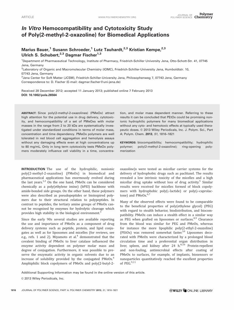
In quantitative measurements no signs of aggregation couldbe observed for any of the PMeOx polymers. Regardless ofthe used concentration or molar mass of all four polymersno erythrocyte aggregation dependent decline of absorptioncould be detected (Fig. 2). Values were found to be in therange of PBS as negative control with a mean value of 0.5896 0.081. All values of the polymer solutions were higherthan that of 25 kDa bPEI as positive control (0.228 6
0.123). The results were verified qualitatively by microscopicobservation. Erythrocyte aggregation was classified with athree stage classification system as described in the Support-ing Information. Stage 3 was determined with 25 kDa bPEI
FIGURE 1 SEC diagram, showing the refraction index (RI)
depending on eluted polymer volume (Vel) of the PMeOx
synthesized by microwave-assisted living cationic ring-opening
polymerization at 140 �C with methyl tosylate as initiator and
WWW.MATERIALSVIEWS.COM JOURNAL OF POLYMER SCIENCE, PART A: POLYMER CHEMISTRY 2013, 51, 1816–1821 1817
[Supporting Information Fig. S2(a)] whereas erythrocytessuspended in PBS were classified as stage 1 [SupportingInformation Fig. S2(b)]. Independent of polymer molar massand concentration all tested PMeOx solutions did not showany aggregation and were therefore classified as stage 1[Supporting Information Fig. S2(c–f)]. These data confirmedthe results of the spectrophotometric measurements.
Hemolysis caused by PMeOx polymers was quantified by therelease of hemoglobin as an indicator for erythrocyte mem-brane damage. Erythrocytes were incubated with PMeOxsolutions at 20–80 mg/mL for 1 h as well as with PBS bufferas negative control and 1% Triton X-100 as positive control(100%). Release of hemoglobin between 0 and 2% wasregarded as non-hemolytic as defined in the ASTM (Ameri-can Society for Testing and Materials) standard F756-08.20
All PMeOxs did not show values higher than 1.06% regard-less of their molar mass and concentration indicating thatthey did not disturb the erythrocyte membranes (data notshown). For comparison, the mean hemolysis of the negativecontrol was 0.31%.
Taken the hemocompatibility data together, the 2–20 kDaPMeOx polymers demonstrated an excellent blood compati-
bility comparable to other non-ionic hydrophilic polymerssuch as PEG,18 PEtOx,18,21 and polyglycerol.22
For further investigations of the influence of PMeOx poly-mers on the membrane integrity as an early indicator fortoxicity, short term incubations were performed with fibro-blast cell cultures using a lactate dehydrogenase (LDH)assay. The release of LDH into culture medium was detectedafter 0–4 h polymer treatment using concentrations of 40,60, and 80 mg/mL (Fig. 3). According to official guidelinesL929 mouse fibroblasts were used as cell line for the cyto-toxicity experiments.23,24 As described by Choksakulnimitret al.25 a LDH release > 10% was regarded as cytotoxic.Assays performed with polymer solutions up to 80 mg/mLwithout the addition of cells showed that the test polymersitself had no influence on the chemical reactions of the assayand the spectrophotometric measurement (data not shown).The membrane integrity of L929 mouse fibroblasts at thebeginning of the experiment was determined by drawingsamples directly after the addition of the test polymers. Upto 4 h treatment with PMeOx polymers up to 20 kDa LDHvalues ranged from 0.1 to 8.1% (Fig. 3), confirming theintactness of the cells even at the highest concentration of
FIGURE 2 Concentration dependent spectrophotometric quantification of erythrocyte aggregation after PMeOx polymer treatment
at 37 �C for 2 h. Absorbance was measured at 645 nm. Data are presented as mean 6 SD (n ¼ 3).
ARTICLE WWW.POLYMERCHEMISTRY.ORGJOURNAL OF
POLYMER SCIENCE
1818 JOURNAL OF POLYMER SCIENCE, PART A: POLYMER CHEMISTRY 2013, 51, 1816–1821
80 mg/mL. Spontaneous LDH release of the negative controlranged in comparison from 1.5 to 1.9%. Results of theexperiments correlated well with data of the hemolysis testsand demonstrated that under the chosen conditions thepresence of PMeOx polymers had no influence on the mem-brane integrity of sheep erythrocytes and L929 mouse fibro-blasts independent of the polymer molar mass.
Long-term treatments were investigated via MTT tests withincubation times of 3, 12, and 24 h, respectively (Fig. 4). Apreliminary experiment performed with the PMeOx solutionswithout cells confirmed that the presence of test polymersup to 80 mg/mL did not affect the chemical reaction of theassay and the spectrophotometric measurement (data notshown). Cell viability of 100% was obtained from cellstreated only with culture medium (negative control). As posi-tive control a 0.02% thiomersal solution which reduced theaverage cell viability to 0.23%, was used. According to thespecifications of ISO 10993-5, cell viability <70% wasregarded as cytotoxic.
As shown in Figure 4(a,b), after 3 and 12 h incubation 4,10, and 20 kDa PMeOx had no influence on the cell viabil-ity of L929 cells. Only 2 kDa PMeOx at the highest concen-tration of 80 mg/mL reduced the cell viability to about 60and 44%, after 3 and 12 h, respectively. After 24 h a con-centration, time, and molar mass dependent increase in cy-totoxicity could be detected. At the highest concentrationonly 20 kDa PMeOx showed a cell viability higher than
70%. From the results of the MTT assay it could by con-cluded that the molar mass of the polymers seems to playan important role for the biocompatibility of PMeOx. Micro-scopic observations made before and after the polymertreatment confirmed the results of the MTT assay. L929fibroblasts treated with culture medium or non-toxic poly-mer solutions were adhesive, grew as confluent monolayersand were typically spindle-shaped. Cells treated with thio-mersal solution and toxic polymer solutions showed areduction of the total cell number as well as changes inmorphology such as rounding and detachment from thedish bottom.
The high biocompatibility at concentrations < 40 mg/mL iscomparable with data from the literature. PMeOx with molarmasses of 4 and 8.5 kDa tested at maximum concentrationsof 20 and 5 mg/mL, respectively, showed no reduction ofcell viability.14,15
CONCLUSION
Concluding the in vitro toxicity studies, no toxic effects ofthe PMeOx polymers of molar masses up to 20 kDa could bedetected in red blood cells and short term L929 cell (LDH)assays. Only in long term treatments (MTT assay) at concen-trations higher than typically used in biomedical applica-tions, polymers were found to moderately influence theviability of cells in a time, concentration, and molar mass de-pendent manner. Unspecific effects such as changes of pH
FIGURE 3 Concentration dependent LDH release of PMeOx polymers after treatment for 0 h (a), 1 h (b), 2 h (c), 4 h (d). LDH levels
lower than 10% (plotted line) were regarded as non-toxic. Results are presented as mean 6 SD of three determinations.
WWW.MATERIALSVIEWS.COM JOURNAL OF POLYMER SCIENCE, PART A: POLYMER CHEMISTRY 2013, 51, 1816–1821 1819
value or osmolarity of the medium by the polymer solutionscould be excluded. Therefore, PMeOx represents a promisingpolymer for many biomedical applications, since it possessesnot only an easy synthetic access and beneficial physico-
chemical characteristics like low viscosity and high stability,but additionally excellent in vitro toxicological features.
EXPERIMENTAL
Experimental details including polymerization procedure,physicochemical properties, and biological investigations areprovided in the Supporting Information.
ACKNOWLEDGMENTS
The authors gratefully acknowledge the Thuringian Ministryfor Education, Science, and Culture (Grant #B514-09051, NanoConSens) for financial support. K. K. is grateful to the Landes-graduiertenfoerderung Thueringen for financial support.
REFERENCES AND NOTES
1 N. Adams, U. S. Schubert, Adv. Drug Deliv. Rev. 2007, 59,
1504–1520.
2 R. Hoogenboom, Angew. Chem. Int. Ed. Engl. 2009, 48,
7978–7994.
3 R. Hoogenboom, H. Schlaad, Polymers 2011, 3, 467–488.
4 M. Miyamoto, K. Naka, M. Shiozaki, Y. Chujo, T. Saegusa,
Macromolecules 1990, 23, 3201–3205.
5 R. Luxenhofer, A. Schulz, C. Roques, S. Li, T. K. Bronich, E.
V. Batrakova, R. Jordan, A. V. Kabanov, Biomaterials 2010, 31,
4972–4979.
6 G. H. Hsiue, C. H. Wang, C. L. Lo, C. H. Wang, J. P. Li, J. L.
Yang, Int. J. Pharm. 2006, 317, 69–75.
7 S. Cheon Lee, C. Kim, I. Chan Kwon, H. Chung, S. Young
Jeong, J. Controlled Release 2003, 89, 437–446.
8 M. C. Woodle, C. M. Engbers, S. Zalipsky, Bioconjugate
Chem. 1994, 5, 493–496.
9 R. Konradi, B. Pidhatika, A. Muhlebach, M. Textor, Langmuir
2008, 24, 613–616.
10 S. Zalipsky, C. B. Hansen, J. M. Oaks, T. M. Allen, J. Pharm.
Sci. 1996, 85, 133–137.
11 B. Pidhatika, J. M€oller, E. M. Benetti, R. Konradi, E. Rakhma-
tullina, A. Muhlebach, R. Zimmermann, C. Werner, V. Vogel,
M. Textor, Biomaterials 2010, 31, 9462–9472.
12 P. Goddard, L. E. Hutchinson, J. Brown, L. J. Brookman,
J. Controlled Release 1989, 10, 5–16.
13 F. C. Gaertner, R. Luxenhofer, B. Blechert, R. Jordan, M.
Essler, J. Controlled Release 2007, 119, 291–300.
14 J. Kronek, Z. Kronekov�a, J. Luston, E. Paulovicov�a, L.
Paulovicov�a, B. Mendrek, J. Mater. Sci. Mater. Med. 2011, 22,
1725–1734.
15 R. Luxenhofer, G. Sahay, A. Schulz, D. Alakhova, T. K. Bro-
nich, R. Jordan, A. V. Kabanov, J. Controlled Release 2011,
153, 73–82.
16 E. H. Luh, S. R. Shackford, M. A. Shatos, J. A. Pietropaoli, J.
Surg. Res. 1996, 60, 122–128.
17 A. V. Cardoso, M. H. Pereira, G. D. A. Marcondes, A. R.
Ferreira, P. R. D. Ara�ujo, Mater. Res. 2007, 10, 31–36.
18 M. Bauer, C. Lautenschlaeger, K. Kempe, L. Tauhardt, U.
S. Schubert, D. Fischer, Macromol. Biosci. 2012, 12,
986–998.
19 P. Petersen, P. M. Fechner, A. L. Martin, K. Kunath, S.
Stolnik, C. J. Roberts, D. Fischer, M. C. Davies, T. Kissel,
Bioconjugate Chem. 2002, 13, 845.
FIGURE 4 Time-, concentration, and molar mass dependent
cytoxicity of PMeOx polymers after 3 h (a), 12 h (b), and 24 h
(c) incubation of L929 cells. Cell viability was determined by
MTT assay and specified according to ISO 10993-5. Results are
presented as mean 6 SD of seven determinations.
ARTICLE WWW.POLYMERCHEMISTRY.ORGJOURNAL OF
POLYMER SCIENCE
1820 JOURNAL OF POLYMER SCIENCE, PART A: POLYMER CHEMISTRY 2013, 51, 1816–1821
20 ASTM F756-08, 2000 (2008). In Annual Book of ASTM
Standards. ASTM: Philadelphia; 2012; Vol. 13.01.
21 T. X. Viegas, M. D. Bentley, J. M. Harris, Z. Fang, K. Yoon,
B. Dizman, R. Weimer, A. Mero, G. Pasut, F. M. Veronese,
Bioconjugate Chem. 2011, 22, 976–986.
22 D. Motlagh, J. Yang, K. Y. Lui, A. R. Webb, G. A. Ameer,
Biomaterials 2006, 27, 4315–4324.
23 Biological evaluation of medical devices Part 5: Tests for in
vitro cytotoxicity. Geneva (Switzerland): International Organiza-
tion for Standardization/ANSI;ISO ISO 10993–5:2009 second
edition, Geneva, 2009.
24 ASTM F813-07, 1996 (2012). In Annual Book of ASTM
Standards; ASTM: Philadelphia; 2008; Vol. 13.01.
25 S. Choksakulnimitr, S. Masuda, H. Tokuda, Y. Takakura, M.