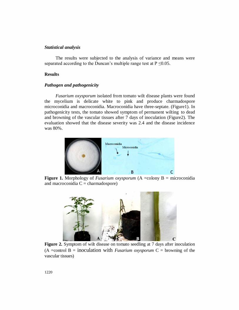
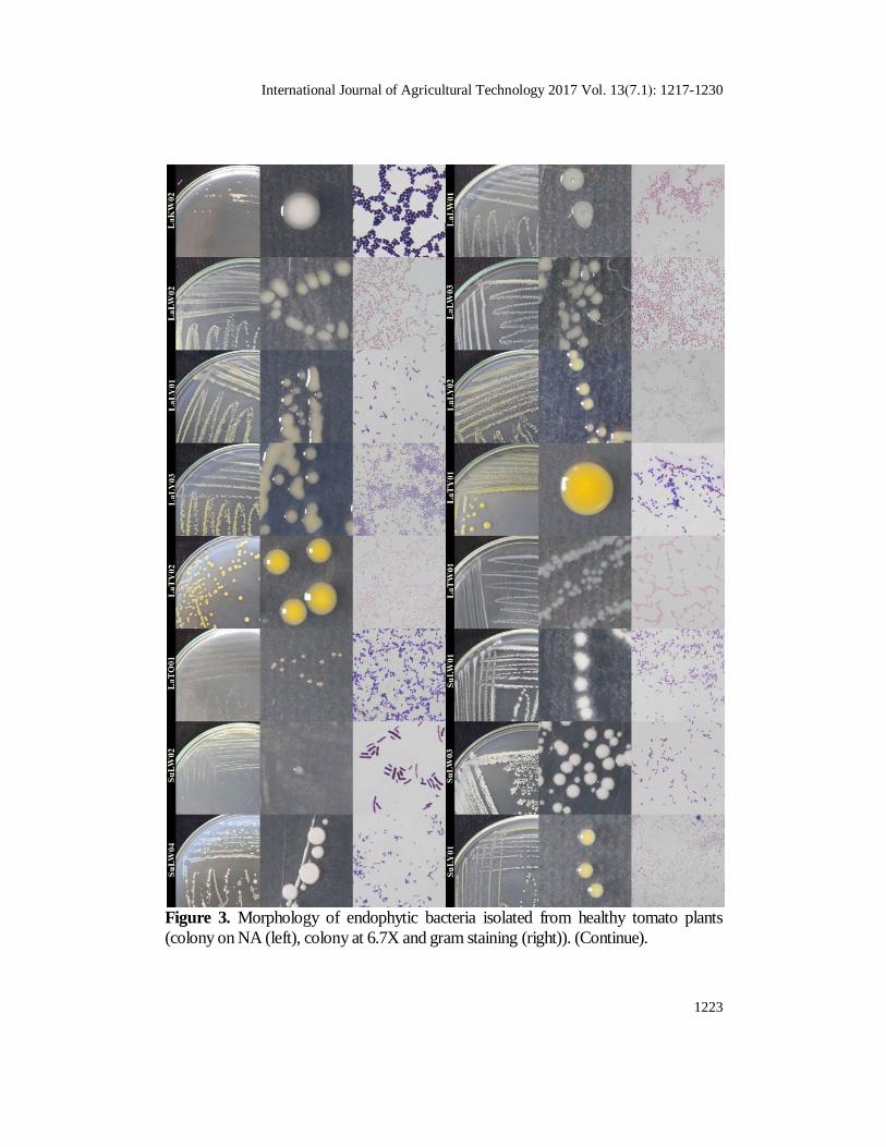
International Journal of Agricultural Technology 2017 Vol. 13(7.1):1217-1230 Available online http://www.ijat-aatsea.com ISSN 1686-9141 In Vitro Study of Endophytic Bacteria Isolated from Tomato Plant against Fusarium oxysporum Panisa Prasom, Potjana Sikhao and Prommart Koohakan * Faculty of Agricultural Technology, King Mongut's Institute of Technology Ladkrabang, Bangkok, Thailand. Panisa Prasom, Potjana Sikhao and Prommart Koohakan (2017). In Vitro Study of Endophytic Bacteria Isolated from Tomato Plant against Fusarium oxysporum. International Journal of Agricultural Technology 13(7.1): 1217-1230. In this study, 43 isolates of endophytic bacteria isolated from healthy tomato plants against Fusarium oxysporum, which causes Fusarium wilt disease of tomato, was studied. Initially effects of endophytic bacteria on the growth of tomato seedlings were tested. The results showed that most endophytic bacteria did not affect the growth of tomato seedlings. Characterization by gram staining revealed that most of them were gram-positive bacteria. Subsequently they were tested on the antagonistic activity against Fusarium oxysporum by dual culture technique. It was found that only seven isolates showed the ability to inhibit the pathogen more than 30 percent. The best isolates including SuRW02 SuRW01 and LbRW03 were highest inhibition percentage of 71.94, 68.33 and 68.19%, respectively. The potential isolates found in this study will be further study and develop for coating tomato seed which an alternative method to control Fusarium wilt disease in the future. Keywords: endophytic bacteria, Fusarium oxysporum, Tomato Introduction Fusarium oxysporum causes Fusarium wilt in tomato is a major pathogen affecting tomato production. The symptoms of this disease include wilting, chlorosis, and stunted seedling. As a result, the plants die or got lower yields (Hussain et al., 2016). Agriculturists had many controlled measures by using several methods, including cultural technique and chemical application. Especially the use of chemicals has been widely used. Although the use of chemicals is effective in controlling the disease, this medthod is harmful to organisms and the environment. Therefore, safe strategies would be used in the management of this disease. Biological control has been reported as a potential for the management of several disease. It consists of a variety of antagonistic microorganisms which have activity for controlling of various plant pathogens, including Fusarium * Corresponding Author: Prommart Koohakan; E-mail: [email protected]
Transcript
International Journal of Agricultural Technology 2017 Vol. 13(7.1):1217-1230
Available online http://www.ijat-aatsea.com
ISSN 1686-9141
In Vitro Study of Endophytic Bacteria Isolated from Tomato
Plant against Fusarium oxysporum
Panisa Prasom, Potjana Sikhao and Prommart Koohakan *
Faculty of Agricultural Technology, King Mongut's Institute of Technology Ladkrabang,
Bangkok, Thailand.
Panisa Prasom, Potjana Sikhao and Prommart Koohakan (2017). In Vitro Study of Endophytic
Bacteria Isolated from Tomato Plant against Fusarium oxysporum. International Journal of
Agricultural Technology 13(7.1): 1217-1230.
In this study, 43 isolates of endophytic bacteria isolated from healthy tomato plants against
Fusarium oxysporum, which causes Fusarium wilt disease of tomato, was studied. Initially
effects of endophytic bacteria on the growth of tomato seedlings were tested. The results
showed that most endophytic bacteria did not affect the growth of tomato seedlings.
Characterization by gram staining revealed that most of them were gram-positive bacteria.
Subsequently they were tested on the antagonistic activity against Fusarium oxysporum by dual
culture technique. It was found that only seven isolates showed the ability to inhibit the
pathogen more than 30 percent. The best isolates including SuRW02 SuRW01 and LbRW03
were highest inhibition percentage of 71.94, 68.33 and 68.19%, respectively. The potential isolates found in this study will be further study and develop for coating tomato seed which an
alternative method to control Fusarium wilt disease in the future.
include Fusarium oxysporum causes Fusarium wilt in tomatoes. (Nejad and
Johnson. 2000; Amaresan et al., 2012 and Nandhini et al., 2102).
This research showed that endophytic bacteria isolated from healthy
tomato plants tissue are capable of promoting the growth of tomato seedlings
and inhibit the growth of Fusarium oxysporum. The results of this study are the
guideline for further study on the control of Fusarium sp. by biological method.
Acknowledgement
This research was supported by Geduate Research Fund from Faculty of Agricultural
Technology, King Mongut's Institute of Technology Ladkrabang, Bangkok, Thailand.
References
Amaresan, N., Jayakumar, V., Kumar, K., and Thajuddin, N. (2012). Isolation and
characterization of plant growth promoting endophytic bacteria and their effect on
tomato (Lycopersicon esculentum) and chilli (Capsicum annuum) seedling
growth. Annals of microbiology, 62(2), 805-810.
Bacon, C.W. and White, J.F. (2000). Microbial endophytes, Marcel Dekker Inc., New York,
USA. Hussain, I., Alam, S. S., Khan, I., Shah, B., Naeem, A., Khan, N., and Shah, S. R. A. (2016).
Study on the biological control of fusarium wilt of tomato. Journal of Entomology and
Zoology Studies 4(2): 525-528.
Hundley NJ. (2005). Structure Elucidation of bioactive compounds isolated from endophytes of
alstonia scholaris and acmena graveolens. MS thesis. Univ. of Brigham Young.
Khan, A. L., Waqas, M., Kang, S. M., Al-Harrasi, A., Hussain, J., Al-Rawahi, A., and Lee, I. J.
(2014). Bacterial endophyte Sphingomonas sp. LK11 produces gibberellins and IAA and
promotes tomato plant growth. Journal of Microbiology, 52(8), 689-695.
Larkin, R. P., and Fravel, D. R. (1998). Efficacy of various fungal and bacterial biocontrol
organisms for control of Fusarium wilt of tomato. Plant disease, 82(9): 1022-1028.
Marlatt, M.L., Correll, J.C., Kaufmann, P. and Cooper, P.E. (0991). Two genetically distinct populations of Fusarium oxysporum f. sp. lycopersici race3 in the United States. Plant
Disease 01 )02:(0331-0332. .
Nandhini, S., Sendhilvel, V., and Babu, S. (2012). Endophytic bacteria from tomato and their
efficacy against Fusarium oxysporum f. sp. lycopersici, the wilt pathogen. Journal of
Biopesticides 5(2): 178.
Nejad, P., & Johnson, P. A. (2000). Endophytic bacteria induce growth promotion and wilt
disease suppression in oilseed rape and tomato. Biological control, 18(3): 208-215.
1230
Nirmaladevi, D., Venkataramana, M., Srivastava, R. K., Uppalapati, S. R., Gupta, V. K., Yli-
Mattila, T., and Chandra, N. S. (2016). Molecular phylogeny, pathogenicity and
toxigenicity of Fusarium oxysporum f. sp. lycopersici. Scientific reports, 6: 21367. Purnawati1, A., Sastrahidayat, I. R., Latief Abadi, A., and Hadiastono, T. (2014). Endophytic
bacteria as biocontrol agents of tomato bacterial wilt disease. Journal of tropical life
science. 4(1):33-36.
(Received: 22 October 2017; accepted: 25 November 2017)


![ISOLATION OF ENDOPHYTIC BACTERIA FROM …2].pdf · volume: 2: issue-3: july-sept -2011 issn 0976-4550 isolation of endophytic bacteria from green gram and study on](https://static.documents.pub/doc/80x56/5abcfea97f8b9af27d8ea50b/isolation-of-endophytic-bacteria-from-2pdfvolume-2-issue-3-july-sept-2011.jpg)


