In Vivo Efficacy of Anuran Trypsin Inhibitory Peptides againstStaphylococcal Skin Infection and the Impact of Peptide Cyclization
U. Malik,a O. N. Silva,b I. C. M. Fensterseifer,b L. Y. Chan,a R. J. Clark,a,c O. L. Franco,b,e N. L. Daly,a,d D. J. Craika
Institute for Molecular Bioscience, The University of Queensland, Brisbane, Australiaa; Centro de Análises Proteômicas e Bioquímicas, Universidade Católica de Brasília,Brasília, Brazilb; School of Biomedical Sciences, The University of Queensland, Brisbane, Australiac; Australian Institute of Tropical Health and Medicine, Centre forBiodiscovery and Molecular Development of Therapeutics, James Cook University, Cairns, Australiad; S-Inova, Universidade Catolica Dom Bosco, Campo Grande, MS,Brazile
Staphylococcus aureus is a virulent pathogen that is responsible for a wide range of superficial and invasive infections. Its resis-tance to existing antimicrobial drugs is a global problem, and the development of novel antimicrobial agents is crucial. Antimi-crobial peptides from natural resources offer potential as new treatments against staphylococcal infections. In the current study,we have examined the antimicrobial properties of peptides isolated from anuran skin secretions and cyclized synthetic analoguesof these peptides. The structures of the peptides were elucidated by nuclear magnetic resonance (NMR) spectroscopy, revealinghigh structural and sequence similarity with each other and with sunflower trypsin inhibitor 1 (SFTI-1). SFTI-1 is an ultrastablecyclic peptide isolated from sunflower seeds that has subnanomolar trypsin inhibitory activity, and this scaffold offers pharma-ceutically relevant characteristics. The five anuran peptides were nonhemolytic and noncytotoxic and had trypsin inhibitoryactivities similar to that of SFTI-1. They demonstrated weak in vitro inhibitory activities against S. aureus, but several hadstrong antibacterial activities against S. aureus in an in vivo murine wound infection model. pYR, an immunomodulatory pep-tide from Rana sevosa, was the most potent, with complete bacterial clearance at 3 mg · kg�1. Cyclization of the peptides im-proved their stability but was associated with a concomitant decrease in antimicrobial activity. In summary, these anuran pep-tides are promising as novel therapeutic agents for treating infections from a clinically resistant pathogen.
Staphylococcus aureus is one of the most virulent and opportu-nistic pathogens, causing increasing numbers of nosocomial
and community-acquired infections, and is a leading cause of skinand soft tissue infections (1–3). These Gram-positive cocci pro-duce a myriad of virulence factors that allow the bacteria to attachto host cells, to invade tissues, to evade the host immune system,and to release an array of exoproteins and toxins (4, 5). Conta-gious S. aureus skin infections can lead to severe muscle or boneinfections that ultimately can spread to the lungs or heart (6). Theprimary treatment involves prescription of �-lactam antibioticssuch as penicillins and cephalosporins, along with clinical wound-cleaning procedures (6, 7). Strains resistant to antibiotics havebeen emerging since the 1960s, however, especially methicillin-resistant S. aureus (MRSA), which is most common in nosoco-mial skin infections (8). Alarmingly, there have been reports ofS. aureus strains that are resistant to the drug of last resort,vancomycin (9).
Antimicrobial peptides are now recognized as novel alternativetherapeutic agents for infection control (10). Several hypotheseshave been examined regarding the mode of action of antimicro-bial peptides, including autolysin activation, lipopolysaccharide(LPS) permeabilization, fatal depolarization of the energized bac-terial membrane, formation of barrel-stave pores that cause leak-age of cellular contents, activation of processes that degrade thecell wall, membrane thinning/thickening, impairment of essentialintracellular targets after internalization, and disturbance of thedistribution of cellular membrane lipids (11–15). Attributes ofantimicrobial peptides that make them viable candidates for de-velopment as anti-infective therapeutic agents include broad-spectrum antimicrobial activity, novel mechanisms of action andease of manipulation, and synthesis and tailoring of peptide se-quences (16, 17). Conversely, major obstacles to the successful
development of antimicrobial peptides as therapies include theirpotential toxicity, high manufacturing costs, and degradation dueto proteases, heat, or extremes of pH (10). The design of peptidesthat are stable against these processes while maintaining their an-timicrobial properties would be a breakthrough in this field.
In amphibians, antimicrobial peptides play an important rolein defense against pathogenic microbes. The skin secretions ofanuran frogs contain a cocktail of compounds, with various bio-logical activities, that have potential for drug development (18).Among these secreted compounds are the anuran antimicrobialpeptides, which have a broad range of antibacterial and antifungalactivities (19). These ribosomally synthesized peptides of 8 to 63amino acids have high affinity for microbial cell membranes (20).They also have serine protease inhibitory activity (21–23).
The amphibian antimicrobial peptides shown in Table 1 con-tain a conserved Bowman-Birk inhibitor (BBI)-like (24) trypsininhibitory loop (CWTKSIPPKPC) and share high sequence ho-mology and structural similarities with sunflower trypsin inhibi-tor 1 (SFTI-1), a 14-amino-acid cyclic peptide discovered in sun-
Received 16 September 2014 Returned for modification 16 October 2014Accepted 21 January 2015
Accepted manuscript posted online 26 January 2015
Citation Malik U, Silva ON, Fensterseifer ICM, Chan LY, Clark RJ, Franco OL, Daly NL,Craik DJ. 2015. In vivo efficacy of anuran trypsin inhibitory peptides againststaphylococcal skin infection and the impact of peptide cyclization. AntimicrobAgents Chemother 59:2113–2121. doi:10.1128/AAC.04324-14.
flower seeds (Helianthus annuus) (25). SFTI-1 is the smallest andone of the most potent BBIs. The structure of SFTI-1 has an ex-tensive hydrogen bonding network and a disulfide bond connect-ing two �-strands, which form a tightly folded scaffold with twodistinct loops, namely, the trypsin binding loop (loop 1) and thesecondary loop (loop 2) (26). Loop 1 contains the active site resi-due for trypsin inhibitory activity, and loop 2 contains the site forbackbone cyclization in the natural biosynthetic process. The an-uran peptides studied have additional conserved residues in thebinding loop (W and P) and additional residues in the secondaryloop, where only the glycine preceding the first cysteine residue isconserved. Based on these sequence similarities, we have termedthese anuran peptides with similarity to SFTI-1 the SFTI-1-likefrog (SLF) peptides.
The cyclic nature of SFTI-1 contributes to its stability and itsresistance to degradation by proteases (26), which is a limitationof linear antimicrobial peptides. Cyclization has been used as oneof the strategies in drug design to improve stability; for example,cyclization of the antimicrobial peptide pyrrhocoricin improvedits in vivo pharmaceutical properties (27). The in vitro stability andantimalarial activity of the antimicrobial peptide gomesin, fromthe Brazilian spider Acanthoscurria gomesiana, were also im-proved after backbone cyclization (28).
In this study, a series of SLF peptides were evaluated for their invivo efficacy in a murine Staphylococcus aureus wound infectionmodel. Three of the active peptides were cyclized in an attempt toimprove their stability. The structures of the most active peptideand its cyclic analogue were determined to elucidate the effects ofcyclization on their antimicrobial and trypsin inhibitory activities.
MATERIALS AND METHODSPeptide synthesis and purification. 9-Fluorenylmethoxycarbonyl(Fmoc)-based chemistry was used to synthesize linear peptides on anautomated synthesizer (Symphony; Protein Technologies, Inc.). 2-Chlo-rotrityl resin (Novabiochem) was used to assemble the peptide chain, andamino acids were Fmoc deprotected using 30% piperidine (Auspep Pty.Ltd.) in dimethylformamide (DMF). Fmoc-protected amino acids (CSBio Co.) were coupled with HCTU [O-(6-chlorobenzotriazol-1-yl)-N,N,N=,N=-tetramethyluronium hexafluorophosphate] (Peptide Interna-tional) and DIPEA (N,N-diisopropylethylamine) (Auspep Pty. Ltd.) inDMF (RCI Labscan Ltd.) (28). The cyclic peptides were synthesized by
t-butyloxycarbonyl (Boc)-based solid-phase peptide synthesis using stan-dard protocols (29, 30). A thioester-based linker (�-mercaptopropionicacid) was used to facilitate cyclization by native chemical ligation (NCL)(31). Previously described standard protocols for hydrogen fluoridecleavage were used to cleave the peptides from the resin (32). The cleavedlinear reduced peptides were purified by C18 reverse-phase (RP)-high-performance liquid chromatography (HPLC) using a gradient of buffer B(90% acetonitrile in 0.05% aqueous trifluoroacetic acid [TFA]) and bufferA (0.05% aqueous TFA) of 1% per minute. Peptide cyclization wasachieved by native chemical ligation with a procedure involving two steps,i.e., cyclization in the presence of a reducing agent (TCEP [tris(2-carboxy-ethyl)phosphine]) followed by oxidation, with both steps involving over-night stirring at room temperature in 0.1 M ammonium bicarbonatebuffer (pH 8.5). Peptides were purified by RP-HPLC at each step. Themolecular weight and purity of the peptides were confirmed by liq-uid chromatography-electrospray ionization-mass spectrometry (LC-ESI-MS).
Structure determination by NMR spectroscopy. 1H nuclear mag-netic resonance (NMR) measurements were carried out with a BrukerAvance-600 spectrometer for all peptides. Each peptide (�3 mg) wasdissolved in 90% H2O-10% D2O (99.9 and 99.99%, respectively; Cam-bridge Isotope Laboratories, Woburn, MA) at pH 5, and DSS (4,4-di-methyl-4-silapentane-1-sulfonic acid) was added as a chemical shift ref-erence. Two-dimensional NMR data were recorded in phase-sensitivemode using time-proportional phase incrementation for quadrature de-tection in the t1 dimension (33). Total correlation spectroscopy (TOCSY)experiments used an MLEV-17 spin-lock sequence (34) with a mixingtime of 80 ms, and nuclear Overhauser effect spectroscopy (NOESY) ex-periments used mixing times of 100 to 200 ms (35). Amide proton tem-perature sensitivity and deuterium exchange experiments were conductedto determine hydrogen-bonding constraints. Talos� was used for predic-tion of the � and � backbone angles (36). Structural calculations werecarried out with CYANA 3.0, using torsion angle dynamics. The 20 low-est-energy structures with the best MolProbity scores were selected for thefinal ensemble (37).
Trypsin inhibition. Trypsin inhibition constants (Ki) of the peptideswere measured by a rapid microplate assay using a modification of themethod described by Erlanger et al. (38). Reactions were conducted in a96-well plate containing 15 �l of 0.45 mg/ml bovine pancreatic trypsin(Sigma-Aldrich), 5 �l of buffer (50 mM Tris, 20 mM CaCl2 [pH 8.2]), 125�l of 0.435 mg/ml L-BAPNA (N�-benzoyl-L-arginine-4-nitroanilide hy-drochloride) (Sigma-Aldrich), and peptides at final concentrations rang-ing from 6.67 mM to 0.065 mM. The reaction was quenched with 25 �l of
TABLE 1 Characteristics of SLF peptides from anuran skin secretions that were highly similar to SFTI-1
Peptide Origin Sequencea
Peptide property
NC aHI pI
Composition (%)
HY BA NE AC
ORB Odorrana grahami 5.9 0.42 11.0 40 25 35 0
ORB2K Odorrana grahami 4.9 0.44 10.6 35 24 41 0
pYR Rana sevosa 4.9 0.87 10.9 39 22 39 0
Rana-Eb Rana esculenta 4.9 1.22 10.5 29 24 47 0
Rana-Tb Rana temporaria 4.9 0.88 10.5 35 25 41 0
SFTI-1 Helianthus annuus 0.9 0.13 8.3 36 14 43 7
a Peptides contain a Bowman-Birk inhibitor (BBI) reactive loop (enclosed in dashed box) and a disulfide bond (black lines). SFTI-1 has a naturally cyclic backbone (gray line). NC,net charge; aHI, grand average of hydropathicity index; pI, isoelectric point; HY, hydrophobic amino acids; BA, basic amino acids; NE, neutral amino acids; AC, acidic amino acids.b Ranacyclin peptides are abbreviated as Rana.
Malik et al.
2114 aac.asm.org April 2015 Volume 59 Number 4Antimicrobial Agents and Chemotherapy
30% acetic acid after 10 min of incubation at 25°C. A PowerWave XS platereader (Bio-Tek) was used to measure the absorbance of p-nitroanilide(pNA) at 410 nm.
In vitro antimicrobial assays. The MICs of peptides against S. aureuswere determined using the standardized serial dilution method, accordingto North Carolina Science Leadership Association (NCSLA) guidelines(39). Overnight colonies of Staphylococcus aureus (strain ATCC 29213)were suspended and standardized at 0.5 units by a turbidity method, fol-lowed by dilution in Mueller-Hinton (MH) broth. For MIC determina-tions, peptides were added at 1 to 200 �M concentrations from a stocksolution, and gentamicin was used as a positive control. Each well of the96-well plate contained 90 �l of bacteria (1 105 cells) in MH agar and 10�l of peptide solution. Ampicillin (40 mg · ml1) and phosphate-bufferedsaline (PBS) were used as positive and negative controls, respectively.Each data point was measured in triplicate. The polypropylene plates wereincubated for 24 h at 37°C. Peptide MICs were determined as the lowesttested concentration that resulted in 100% development inhibition, incomparison to the negative control. The MIC is defined as the lowestconcentration of an antimicrobial compound that completely inhibits thegrowth of the organism, as detected by the unaided eye (40).
Hemolytic assays. All peptides were tested for their abilities to hemo-lyze red blood cells (RBCs). Using a previously described method (41),serially diluted peptides (final concentrations of 0.4 to 50 �M) were incu-bated with human RBCs in 96-well plates for 1 h at 37°C and then werecentrifuged at 150 relative centrifugal force (RCF) for 5 min. The absor-bance of the supernatant, containing plasma and lysed RBCs, was mea-sured at 415 nm with a PowerWave XS plate reader (Bio-Tek). TritonX-100 (0.1%) and PBS were used as positive and negative controls, respec-tively. Melittin, a cationic hemolytic peptide, was used as a positive con-trol and was tested at initial concentrations ranging from 0.4 to 50 �M.Percent hemolysis was calculated using the absorbance of maximum lysisin the positive-control samples (Triton X-100).
Cell cytotoxicity. The cytotoxic effects of anuran peptides on humanforeskin fibroblast (HFF-1) cells and melanoma (MM96L) cells were eval-uated using an MTT [3-(4,5-dimethyl-2-thiazolyl)-2,5-diphenyl-2H-tet-razolium bromide] assay. Cells were seeded in 96-well plates at 3 103
cells/well (100 �l) with 15% fetal bovine serum (FBS) in Dulbecco’s mod-ified Eagle’s medium (DMEM) for HFF-1 cells (Gibco) or 10% FBS inRPMI 1640 medium for MM96L cells, supplemented with 2 mM L-glu-tamine and 1 mM sodium pyruvate (Gibco). All cells were incubated at37°C in 5% CO2 for 24 h. The medium was removed and replaced withfresh serum-free medium (90 �l/well), and then peptides were added. Allpeptides were tested in triplicate, with final peptide concentrations rang-ing from 1 to 200 �M, and were incubated with cells for 2 h. A negativevehicle control (water) and a positive control (1% Triton X-100) wereincluded in the assay. After 2 h of incubation, 10 �l of MTT (5 mg/ml inPBS) was added to each well, and the cells were further incubated for 3 hbefore the removal of supernatant. The formazan crystals formed in eachwell were dissolved in 100 �l dimethylsulfoxide (DMSO), and absorbancewas measured at 600 nm using a BioTek PowerWave XS spectrophotom-eter. Data were analyzed using the GraphPad Prism program, and 50%inhibitory concentration (IC50) values were obtained from the sigmoidaldose-response curves.
Acute toxicity. Acute toxicity assays were based on the method de-scribed by Navon-Venezia and coworkers (42), with intraperitoneal (i.p.)administration of the tested peptides to groups of 10 C57BL/6 mice. Eachmouse was injected with 0.5 ml of a solution of freshly prepared pyR orcpYR in PBS. The doses of peptides administered per mouse were 0, 5, 10,25, 50, and 100 mg · kg of body weight1. Animals were inspected foradverse effects for 30 min, and survival was monitored for 6 h thereafter.
In vivo antimicrobial activity. Female C57BL6 mice (6 to 8 weeks ofage) were obtained from CEMIB-Unicamp (Brazil). The study was ap-proved by the Animal Use Committee at the Institute of Biological Sci-ences, University of Brasilia. Briefly, groups of mice (n � 4/group in eachexperiment) were anesthetized with a combination of ketamine and xyla-
zine, their backs were shaved, and the surgical area was disinfected with70% ethanol. A 1-cm incision was made, and 25 �l with 2 109 CFU S.aureus was pipetted into the incision. The mice were euthanized at 7 daysafter surgery, and the wound tissue was excised, weighed, and homoge-nized in 1 ml of PBS. Serial dilutions of the homogenates were plated intriplicate on mannitol salt agar plates, and results were expressed as CFU/gram of tissue. Peptides were administered to animals daily. Peptides weresolubilized in water for injection at two different concentrations, 1.5 mg ·kg1 and 3.0 mg · kg1, and were injected into the wound in a 25-�lvolume; PBS was used as a negative control and 10 mg · kg1 neomycinsulfate as a positive control.
Serum stability. The linear and cyclic forms of pYR and SFTI-1 pep-tides were evaluated in serum stability assays using an established method(30). Human AB serum (Sigma) was used in this assay. Lipids were re-moved by centrifugation at 13,000 rpm for 15 min prior to the assay.Peptides (initial concentration, 2 mg/ml) diluted 10-fold in serum (test)or PBS (negative control) were incubated at 37°C. At each time point (0, 3,6, 10, and 24 h), triplicate aliquots were taken from each peptide sample inserum or PBS and denatured with equal volumes of 6 M urea, followed by10 min of incubation at 4°C. The serum proteins were then precipitatedwith an equal volume of 20% trichloroacetic acid, with a 10-min incuba-tion at 4°C followed by centrifugation at 13,000 rpm for 15 min. Thesupernatant was stored at 4°C until analysis. The relative concentration ofthe intact peptide, in comparison with the level at the start of the experi-ment, was measured by ultra-high performance liquid chromatography(uHPLC) for each peptide sample, using the integrated area of the peakcorresponding to the intact peptide (with UV absorbance at 214 nm) onthe HPLC trace. The mean and standard deviation were calculated foreach peptide.
RESULTSSynthesis of antimicrobial peptides. The SLF peptides (Table 1)were synthesized to study their structures, biological characteris-tics, and in vivo antimicrobial efficacy against S. aureus in an ani-mal wound infection model. The cyclic versions of the SLF pep-tides are prefixed with c, and the open or acyclic form of SFTI-1 isprefixed with o. The native open-chain peptides were synthesizedusing Fmoc-based chemistry and were oxidized in ammoniumbicarbonate buffer (pH 8.5). SFTI and the cyclic versions of theanuran peptides were synthesized using Boc-based chemistry witha C-terminal thioester to facilitate cyclization. Cyclization (in thepresence of reducing agent) and oxidation were carried out in twoseparate steps. These conditions led to efficient cyclization of pep-tides without the requirement for additional linker residues.
Structure determination by NMR spectroscopy. The peptideswere analyzed by NMR spectroscopy. The secondary H� chemicalshifts (Fig. 1) were calculated using the measured chemical shiftsand the random coil values reported by Wishart et al. (43). Sec-ondary H� chemical shifts are highly sensitive to structural mod-ifications, and thus they offer an excellent platform to observestructural differences due to amino acid variations (44). The linearand cyclic constructs had comparable chemical shifts, suggestingthat the cyclization of the backbone did not have a significantinfluence on structure. The first proline in the binding loop (CXTKSIPPK/IP) is in a cis conformation, consistent with SFTI-1(32). The other proline residues are in a trans conformation in allpeptides.
The three-dimensional structures of open and cyclic pYR (Fig.2) were elucidated using the program CYANA, and the ensembleschosen to represent the final structures were based on the 20 struc-tures from a set of 100 structures with the lowest MolProbityscores (37, 45). Statistics showing the precision and stereochemi-
Peptides for Treating Staphylococcal Skin Infections
April 2015 Volume 59 Number 4 aac.asm.org 2115Antimicrobial Agents and Chemotherapy
cal qualities of the pYR structures are presented in Table 2. In thecyclic form, the loop with the disulfide bond (loop 2) showed lessstructural variation than the loop with the free termini (loop 1).
In vitro biological activity. The SLF peptides contain a BBIloop similar to SFTI-1. For this reason, the trypsin inhibitory ac-tivities of the SLF peptides were assessed and compared with thatof SFTI-1 (Fig. 3), which has been reported to have an inhibitionconstant of 1.7 pM (29). cpYR was found to have trypsin inhibi-tory activity similar to that of SFTI-1. The other peptides hadsignificantly reduced inhibitory activity, ranging between 13 and50%, relative to SFTI-1. Cyclization improved the inhibitory ac-tivities of all peptides, but opening of the SFTI-1 chain resulted indecreased inhibitory activity, compared to wild-type SFTI-1, con-sistent with previous studies (26).
The antimicrobial activities of the peptides were measured inan in vitro assay with the widely used sensitive S. aureus strainATCC 29213, on MH agar, using a dilution method. Table 3 pres-ents the MIC data for open and cyclic forms of the peptides. pYR
was the most potent peptide, with an MIC of 50 �M against S.aureus. Rana-E and Rana-T showed MICs of 100 �M, whereas theother open peptides had MICs greater than 100 �M. Backbonecyclization resulted in decreased potency of pYR, while MIC val-ues for cORB and cORB2K were �100 �M. Cyclic and openSFTI-1 forms had no antimicrobial activity at concentrations upto 100 �M.
The hemolytic activities of all peptides were determined in hu-man blood. Melittin, a well-studied and highly hemolytic peptidefrom honeybee venom (46), and Triton X-100 were used as apositive controls and PBS was used as a negative control in theassay. All peptides were found to be nonhemolytic at up to 50 �Mconcentrations (data not shown). To further evaluate the toxicityand effects of cyclization of these peptides, the cytotoxicities ofopen and cyclic pYR and ORB were determined with human fore-skin fibroblast (HFF-1) and melanoma (MM96L) cell lines. Thepeptides were noncytotoxic at up to 200 �M, except for cpYR,which was found to kill 20% of the cells in both cell lines at 200 �M(Fig. 4).
Acute toxicity. Toxicity was assessed in vivo for pYR and cpYR.
FIG 1 H� NMR secondary shift analysis of SLF peptides and SFTI-1. Second-ary shifts were obtained by subtracting experimental 1H NMR H� chemicalshifts from random coil shifts for the corresponding residue (43). The ORB/ORB2k open and cyclic peptides have similar shifts in loop 1, whereas theRana-E/T and pYR peptides have similar secondary shifts in loop 1. Loop 1contains a highly conserved BBI reactive loop (dotted gray box).
FIG 2 NMR solution structures of open and cyclic pYR. (A and B) Structuralensembles of open (A) and cyclic (B) pYR. Disulfide bonds are highlighted ingray. (C) Three-dimensional structural comparison of pYR peptides withtruncated ORB2K (PDB accession number 2O9Q) and SFTI-1 (PDB accessionnumber 1JBL).
Malik et al.
2116 aac.asm.org April 2015 Volume 59 Number 4Antimicrobial Agents and Chemotherapy
First, acute toxicity was examined after i.p. administration of asingle dose of each peptide to groups of C57BL/6 mice (n � 5mice/group). No immediate adverse events were noted for thepeptides at doses of up to 25 mg · kg1 (Table 4). Toxicity level I,involving narrowing of the eyes, was noted minutes after injectionin mice injected with doses of peptides above 50 mg · kg1, exceptfor one animal with pYR at 50 mg · kg1. Toxicity level II, involv-ing crouching and cuddling, was observed in a few mice injectedwith doses of pYR or neomycin sulfate above 50 mg · kg1. Nodeaths were observed for any of the treated mice, and most micerecovered 2 h after treatment.
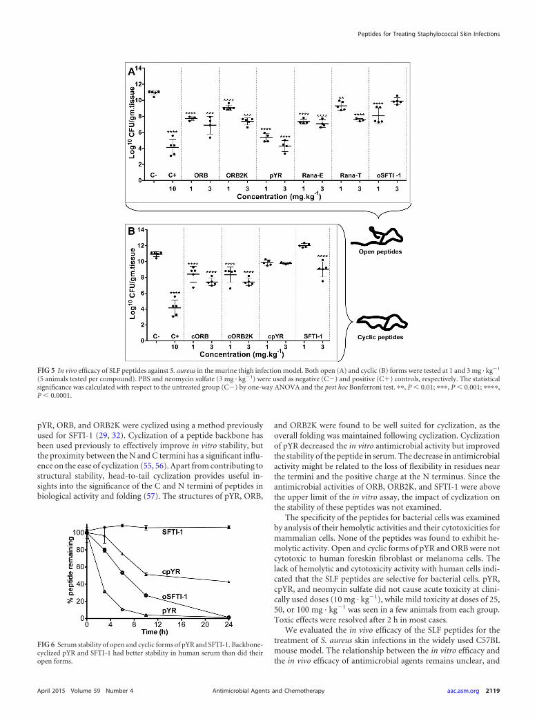
In vivo antimicrobial activity. The therapeutic efficacy of theanuran defense peptides against S. aureus was studied in a murinewound infection model (Fig. 5). The wound was infected with 2 109 CFU of S. aureus ATCC 29213 at the start of therapy, andchanges in bacterial loads were evaluated after 7 days of daily an-timicrobial therapy with antimicrobial peptides. The negative-control group of mice (untreated group) was given PBS instead ofantimicrobial agent, whereas the positive-control group wastreated with neomycin sulfate antibiotic at 10 mg · kg1 (antibi-otic-treated group). Neomycin sulfate is an antibiotic from theaminogylcoside family that is used in topical ointments, and thishas been used to study S. aureus infections (47, 48). The bacterialrecoveries for the antibiotic-treated group and the untreatedgroup were found to be 4.1 1.0 and 10.9 0.3 log10CFU/g oftissue, respectively.
The antimicrobial peptides were administered daily at the siteof infection at doses of 1 or 3 mg · kg1. The bacterial recovery for
TABLE 2 Structural statistics for NMR solution structures of pYR and cpYR
Parameter pYR cpYR
Experimental restraintsa
No. of interproton distance restraints 170 202No. of intraresidue restraints 39 51No. of sequential restraints 72 80No. of medium-range (i j of �5) restraints 27 27No. of long-range (i j of �5) restraints 32 44No. of hydrogen bond restraintsb 4 4No. of disulfide bond restraints 2 2No. of dihedral angle restraints (� and �) 20 22
Residues in most favored Ramachandran region (%) 96.47 4.83 87.50 2.99Ramachandran outliers (%) 0.29 1.31 0.31 1.40Unfavorable side chain rotamers (%) 8.82 7.27 11.00 8.72Clash score, all atoms (no. of overlaps/1,000 atoms)e 13.55 2.30 8.97 3.16Overall MolProbity score 2.41 0.50 2.74 0.34
a Only structurally relevant restraints, as defined by CYANA, are included.b Two restraints were used per hydrogen bond.c RMS, root mean square; SD, standard deviation.d Stereochemical properties were obtained by MolProbity (http://molprobity.biochem.duke.edu).e The clash score is defined as the number of steric overlaps of �0.4 Å per 1,000 atoms.
FIG 3 Relative trypsin inhibitory activities of SLF peptides, compared withSFTI-1. SFTI-1 has an equilibrium dissociation constant Ki of 17 pM (29). Thestatistical significance was calculated with respect to SFTI-1 by one-way anal-ysis of variance (ANOVA) and the post hoc Bonferroni test. ����, P � 0.0001.
TABLE 3 In vitro efficacy of SLF peptides against Staphylococcus aureusstrain ATCC 29213
pYR at 3 mg · kg1 was found to be 4.3 0.7 log10CFU/g of tissue,which was equivalent to the antibiotic control (20 mg · kg1).Lowering the dose of pYR by one-third increased the bacterialrecovery 1.5-fold. The other open-form anuran peptides signif-icantly reduced the numbers of log10CFU/g of tissue, comparedto the untreated group of mice; however, they were less efficientthan pYR.
Backbone cyclization of the peptides resulted in a decrease orloss of antimicrobial activity. The cyclic analogue (cpYR) of themost active open peptide (pYR) completely lost therapeutic effi-cacy even at 3 mg · kg1. cORB and cORB2K showed reducedbacterial clearance in wounds by 1 to 1.5 log10 CFU/g of tissue,compared to their respective open forms.
Serum stability. The open and cyclic forms of pYR and SFTI-1were incubated in human serum (Fig. 6), and the cyclic peptideswere found to have improved stability over the open forms over 24h. SFTI-1 did not degrade at all, whereas 57.2 0.9% of cpYRdegraded after 24 h. oSFTI-1 was more stable than pYR.
DISCUSSION
Antimicrobial peptides are found in both animal and plant speciesand are used in host defense mechanisms. There is growing inter-est in exploring the potential of antimicrobial peptides as antibi-otics to combat the decreased efficacy of conventional small-mol-ecule antibiotics (49, 50). Some antimicrobial peptides killmicrobes directly, while others are effective by modulating theinnate immune system (51). The ability of antimicrobial peptidesto neutralize endotoxemia/sepsis and to stimulate host innate re-sponses while dampening potentially deleterious inflammatoryresponses offers an added advantage over small-molecule antibi-otics (17). In this study, five antimicrobial peptides of anuranorigin, containing a reactive site loop of BBIs, were evaluated fortheir therapeutic efficacy to treat S. aureus-infected wounds in
mice. Three active peptides were backbone cyclized in an attemptto improve their biological properties.
Several SLF peptides have been reported to have in vitro anti-microbial activity against a range of microbes, but only ORB andORB2K have been tested against S. aureus ATCC 25923 (21–23).In light of these observations, we evaluated the in vitro antimicro-bial activities of five SLF peptides against S. aureus strain ATCC29213, a strain isolated from wounds (Table 3). ORB and ORB2Kwere found to have much greater inhibitory activities than re-ported (21). Variations in the antimicrobial susceptibility testingmethods have been found to have significant effects on the MICsof compounds, as demonstrated using the cysteine-rich antimi-crobial peptide protegrin-1, for example (52). Differences are alsodependent on the characteristics of the antimicrobial agents, in-cluding hydrophobic moments, exposed charges, amphipathicity,and peptide flexibility (53, 54). pYR was found to be the mostactive SLF peptide, while others had moderate-to-poor antimicro-bial activity. Loop 2 of these SLF peptides has significant sequencediversity, compared to loop 1, and this diversity in the length andnumber of hydrophobic and cationic residues might be responsi-ble for the observed variations in antimicrobial activity.
The high sequence similarity of the anuran peptides to SFTI-1,a stable and naturally cyclic peptide, guided this study toward thedevelopment of cyclic antimicrobial peptides that could poten-tially have improved stability and biological activities. Therefore,
TABLE 4 Evaluation of acute toxicity in mice treated with pYR, cpYR,or neomycin sulfate (positive control)
Dose (mg · kg1) andeffecta
No. of mice (n � 5/group)
Neomycin sulfate pYR cpYR
0No effect 5 5 5Toxicity level I 0 0 0Toxicity level II 0 0 0
5No effect 5 5 5Toxicity level I 0 0 0Toxicity level II 0 0 0
10No effect 5 5 5Toxicity level I 0 0 0Toxicity level II 0 0 0
25No effect 3 4 5Toxicity level I 2 1 0Toxicity level II 0 0 0
50No effect 2 2 4Toxicity level I 0 2 1Toxicity level II 3 1 0
100No effect 2 1 3Toxicity level I 0 2 2Toxicity level II 3 2 0
a Toxicity grading was as follows: level I, narrowing of eyes; level II, crouching andcuddling.
FIG 4 Cytotoxic activity of selected ORB and pYR peptides against noncan-cerous (HFF-1) (A) and cancerous (MM96L) (B) cells. Both open and cyclicforms of the peptides were tested, and melittin was used as a positive control(0% viability corresponds to 100% cell death).
Malik et al.
2118 aac.asm.org April 2015 Volume 59 Number 4Antimicrobial Agents and Chemotherapy
pYR, ORB, and ORB2K were cyclized using a method previouslyused for SFTI-1 (29, 32). Cyclization of a peptide backbone hasbeen used previously to effectively improve in vitro stability, butthe proximity between the N and C termini has a significant influ-ence on the ease of cyclization (55, 56). Apart from contributing tostructural stability, head-to-tail cyclization provides useful in-sights into the significance of the C and N termini of peptides inbiological activity and folding (57). The structures of pYR, ORB,
and ORB2K were found to be well suited for cyclization, as theoverall folding was maintained following cyclization. Cyclizationof pYR decreased the in vitro antimicrobial activity but improvedthe stability of the peptide in serum. The decrease in antimicrobialactivity might be related to the loss of flexibility in residues nearthe termini and the positive charge at the N terminus. Since theantimicrobial activities of ORB, ORB2K, and SFTI-1 were abovethe upper limit of the in vitro assay, the impact of cyclization onthe stability of these peptides was not examined.
The specificity of the peptides for bacterial cells was examinedby analysis of their hemolytic activities and their cytotoxicities formammalian cells. None of the peptides was found to exhibit he-molytic activity. Open and cyclic forms of pYR and ORB were notcytotoxic to human foreskin fibroblast or melanoma cells. Thelack of hemolytic and cytotoxicity activity with human cells indi-cated that the SLF peptides are selective for bacterial cells. pYR,cpYR, and neomycin sulfate did not cause acute toxicity at clini-cally used doses (10 mg · kg1), while mild toxicity at doses of 25,50, or 100 mg · kg1 was seen in a few animals from each group.Toxic effects were resolved after 2 h in most cases.
We evaluated the in vivo efficacy of the SLF peptides for thetreatment of S. aureus skin infections in the widely used C57BLmouse model. The relationship between the in vitro efficacy andthe in vivo efficacy of antimicrobial agents remains unclear, and
FIG 5 In vivo efficacy of SLF peptides against S. aureus in the murine thigh infection model. Both open (A) and cyclic (B) forms were tested at 1 and 3 mg · kg1
(5 animals tested per compound). PBS and neomycin sulfate (3 mg · kg1) were used as negative (C) and positive (C�) controls, respectively. The statisticalsignificance was calculated with respect to the untreated group (C) by one-way ANOVA and the post hoc Bonferroni test. ��, P � 0.01; ���, P � 0.001; ����,P � 0.0001.
FIG 6 Serum stability of open and cyclic forms of pYR and SFTI-1. Backbone-cyclized pYR and SFTI-1 had better stability in human serum than did theiropen forms.
Peptides for Treating Staphylococcal Skin Infections
April 2015 Volume 59 Number 4 aac.asm.org 2119Antimicrobial Agents and Chemotherapy
prediction of in vivo activity is very challenging, due to variationsin potency that can occur due to many uncontrolled parameters inthe host systems (58, 59). Despite the challenges of using in vitropotency to predict in vivo potency, we found that the most potentpeptide in the in vitro study, pYR, was also the most potent peptidein treating S. aureus skin infections in mice. pYR (at 3 mg · kg1)reduced bacterial loads in the wounds similarly to the positive-control compound neomycin sulfate (at 10 mg · kg1), a broad-spectrum antibiotic that is used in topical ointments and is mosteffective against S. aureus (48).
In conclusion, this study has investigated a class of �-sheetmultifunctional antimicrobial peptides that are effective in treat-ing S. aureus skin infections in a murine model. Although cycliza-tion was shown previously to enhance stability and bioactivity (28,60), in this case an improvement in stability was observed butbioactivity was reduced. Structural or charge distribution changesupon cyclization might be responsible for the decreased antimi-crobial activity. The choice of peptide for further development(open versus cyclic) reflects a balance between stability andbioactivity. Further studies are needed to investigate the mech-anisms of action of these peptides, to fully explore their poten-tial for clinical use.
ACKNOWLEDGMENTS
This work was supported by a grant from the National Health and MedicalResearch Council (grant APP1028509) and a University of Queenslandresearcher exchange travel grant. R.J.C. and N.L.D. are Australian Re-search Council Future Fellows (grants FT100100476 and FT1100100226,respectively). D.J.C. is a National Health and Medical Research CouncilProfessorial Fellow (grant APP1026501). Work at the UniversidadeCatólica de Brasília was supported by grants from the Conselho Nacionalde Desenvolvimento Científico e Tecnológico, Coordenação de Aper-feiçoamento de Pessoal de Nível Superior, the Fundação de Amparo aPesquisa do Distrito Federal, and the Universidade Católica de Brasília.
REFERENCES1. Krut O, Sommer H, Krönke M. 2004. Antibiotic-induced persistence of
cytotoxic Staphylococcus aureus in non-phagocytic cells. J AntimicrobChemother 53:167–173. http://dx.doi.org/10.1093/jac/dkh076.
2. Chambers HF, Deleo FR. 2009. Waves of resistance: Staphylococcus au-reus in the antibiotic era. Nat Rev Microbiol 7:629 – 641. http://dx.doi.org/10.1038/nrmicro2200.
3. Inoshima N, Wang Y, Wardenburg JB. 2012. Genetic requirement forADAM10 in severe Staphylococcus aureus skin infection. J Invest Dermatol132:1513–1516. http://dx.doi.org/10.1038/jid.2011.462.
4. McLoughlin RM, Solinga RM, Rich J, Zaleski KJ, Cocchiaro JL, RisleyA, Tzianabos AO, Lee JC. 2006. CD4� T cells and CXC chemokinesmodulate the pathogenesis of Staphylococcus aureus wound infections.Proc Natl Acad Sci U S A 103:10408 –10413. http://dx.doi.org/10.1073/pnas.0508961103.
5. Foster TJ, Geoghegan JA, Ganesh VK, Höök M. 2014. Adhesion, inva-sion and evasion: the many functions of the surface proteins of Staphylo-coccus aureus. Nat Rev Microbiol 12:49 – 62. http://dx.doi.org/10.1038/nrmicro3161.
7. Kazakova SV, Hageman JC, Matava M, Srinivasan A, Phelan L, GarfinkelB, Boo T, McAllister S, Anderson J, Jensen B. 2005. A clone of methicillin-resistant Staphylococcus aureus among professional football players. N Engl JMed 352:468–475. http://dx.doi.org/10.1056/NEJMoa042859.
8. Klevens RM, Morrison MA, Nadle J, Petit S, Gershman K, Ray S,Harrison LH, Lynfield R, Dumyati G, Townes JM. 2007. Invasive me-thicillin-resistant Staphylococcus aureus infections in the United States.JAMA 298:1763–1771. http://dx.doi.org/10.1001/jama.298.15.1763.
9. Gastmeier P, Schröder C, Behnke M, Meyer E, Geffers C. 2014. Dra-
matic increase in vancomycin-resistant enterococci in Germany. J Anti-microb Chemother 69:1660 –1664. http://dx.doi.org/10.1093/jac/dku035.
10. Hancock RE, Sahl H-G. 2006. Antimicrobial and host-defense peptides asnew anti-infective therapeutic strategies. Nat Biotechnol 24:1551–1557.http://dx.doi.org/10.1038/nbt1267.
11. Zasloff M. 2002. Antimicrobial peptides of multicellular organisms. Na-ture 415:389 –395. http://dx.doi.org/10.1038/415389a.
12. Nguyen LT, Haney EF, Vogel HJ. 2011. The expanding scope of antimi-crobial peptide structures and their modes of action. Trends Biotechnol29:464 – 472. http://dx.doi.org/10.1016/j.tibtech.2011.05.001.
13. Brogden KA. 2005. Antimicrobial peptides: pore formers or metabolicinhibitors in bacteria? Nat Rev Microbiol 3:238 –250. http://dx.doi.org/10.1038/nrmicro1098.
14. Lohner K. 2009. New strategies for novel antibiotics: peptides targetingbacterial cell membranes. Gen Physiol Biophys 28:105–116. http://dx.doi.org/10.4149/gpb_2009_02_105.
15. Strauss J, Kadilak A, Cronin C, Mello CM, Camesano TA. 2010.Binding, inactivation, and adhesion forces between antimicrobial peptidececropin P1 and pathogenic E. coli. Colloids Surf B Biointerfaces 75:156 –164. http://dx.doi.org/10.1016/j.colsurfb.2009.08.026.
16. Wimley WC, Hristova K. 2011. Antimicrobial peptides: successes, chal-lenges and unanswered questions. J Membr Biol 239:27–34. http://dx.doi.org/10.1007/s00232-011-9343-0.
18. Conlon JM. 2011. The contribution of skin antimicrobial peptides to thesystem of innate immunity in anurans. Cell Tissue Res 343:201–212. http://dx.doi.org/10.1007/s00441-010-1014-4.
19. Simmaco M, Mignogna G, Barra D. 1998. Antimicrobial peptides fromamphibian skin: what do they tell us? Biopolymers 47:435– 450.
20. Conlon JM, Mechkarska M. 2014. Host-defense peptides with therapeu-tic potential from skin secretions of frogs from the family Pipidae. Phar-maceuticals 7:58 –77. http://dx.doi.org/10.3390/ph7010058.
21. Li J, Zhang C, Xu X, Wang J, Yu H, Lai R, Gong W. 2007. Trypsininhibitory loop is an excellent lead structure to design serine proteaseinhibitors and antimicrobial peptides. FASEB J 21:2466 –2473. http://dx.doi.org/10.1096/fj.06-7966com.
22. Mangoni ML, Papo N, Mignogna G, Andreu D, Shai Y, Barra D,Simmaco M. 2003. Ranacyclins, a new family of short cyclic antimi-crobial peptides: biological function, mode of action, and parametersinvolved in target specificity. Biochemistry 42:14023–14035. http://dx.doi.org/10.1021/bi034521l.
23. Graham C, Irvine AE, McClean S, Richter SC, Flatt PR, Shaw C. 2005.Peptide tyrosine arginine, a potent immunomodulatory peptide isolatedand structurally characterized from the skin secretions of the dusky go-pher frog, Rana sevosa. Peptides 26:737–743. http://dx.doi.org/10.1016/j.peptides.2004.12.006.
24. Brauer AB, Domingo GJ, Cooke RM, Matthews SJ, Leatherbarrow RJ.2002. A conserved cis peptide bond is necessary for the activity of Bow-man-Birk inhibitor protein. Biochemistry 41:10608 –10615. http://dx.doi.org/10.1021/bi026050t.
25. Luckett S, Garcia RS, Barker JJ, Konarev AV, Shewry PR, Clarke AR,Brady RL. 1999. High-resolution structure of a potent, cyclic proteinaseinhibitor from sunflower seeds. J Mol Biol 290:525–533. http://dx.doi.org/10.1006/jmbi.1999.2891.
26. Korsinczky ML, Schirra HJ, Rosengren KJ, West J, Condie BA, Otvos L,Anderson MA, Craik DJ. 2001. Solution structures by 1H NMR of thenovel cyclic trypsin inhibitor SFTI-1 from sunflower seeds and an acyclicpermutant. J Mol Biol 311:579 –591. http://dx.doi.org/10.1006/jmbi.2001.4887.
27. Otvos L, Bokonyi K, Varga I, Otvos BI, Hoffmann R, Ertl HC, WadeJD, McManus AM, Craik DJ, Bulet P. 2000. Insect peptides with im-proved protease-resistance protect mice against bacterial infection. Pro-tein Sci 9:742–749. http://dx.doi.org/10.1110/ps.9.4.742.
28. Chan LY, Zhang VM, Huang YH, Waters NC, Bansal PS, Craik DJ,Daly NL. 2013. Cyclization of the antimicrobial peptide gomesin withnative chemical ligation: influences on stability and bioactivity. Chem-BioChem 14:617– 624. http://dx.doi.org/10.1002/cbic.201300034.
29. Quimbar P, Malik U, Sommerhoff CP, Kaas Q, Chan LY, Huang YH,Grundhuber M, Dunse K, Craik DJ, Anderson MA, Daly NL. 2013.High-affinity cyclic peptide matriptase inhibitors. J Biol Chem 288:13885–13896. http://dx.doi.org/10.1074/jbc.M113.460030.
Malik et al.
2120 aac.asm.org April 2015 Volume 59 Number 4Antimicrobial Agents and Chemotherapy
30. Chan LY, Gunasekera S, Henriques ST, Worth NF, Le S-J, Clark RJ,Campbell JH, Craik DJ, Daly NL. 2011. Engineering pro-angiogenicpeptides using stable disulfide-rich cyclic scaffolds. Blood 118:6709 –6717. http://dx.doi.org/10.1182/blood-2011-06-359141.
31. Dawson PE, Muir TW, Clark-Lewis I, Kent SB. 1994. Synthesis ofproteins by native chemical ligation. Science 266:776 –779. http://dx.doi.org/10.1126/science.7973629.
32. Daly NL, Chen Y-K, Foley FM, Bansal PS, Bharathi R, Clark RJ,Sommerhoff CP, Craik DJ. 2006. The absolute structural requirement fora proline in the P3=-position of Bowman-Birk protease inhibitors is sur-mounted in the minimized SFTI-1 scaffold. J Biol Chem 281:23668 –23675. http://dx.doi.org/10.1074/jbc.M601426200.
33. Marion D, Wuthrich K. 1983. Application of phase sensitive two-dimensional correlated spectroscopy (COSY) for measurements of 1H-1Hspin-spin coupling-constants in proteins. Biochem Biophys Res Commun113:967–974. http://dx.doi.org/10.1016/0006-291X(83)91093-8.
34. Bax A, Davis DG. 1985. MLEV-17-based two-dimensional homonuclearmagnetization transfer spectroscopy. J Magn Reson 65:355–360.
35. Jeener J, Meier B, Bachmann P, Ernst R. 1979. Investigation of exchangeprocesses by two-dimensional NMR spectroscopy. J Chem Phys 71:4546.http://dx.doi.org/10.1063/1.438208.
36. Shen Y, Delaglio F, Cornilescu G, Bax A. 2009. TALOS�: a hybridmethod for predicting protein backbone torsion angles from NMR chem-ical shifts. J Biomol NMR 44:213–223. http://dx.doi.org/10.1007/s10858-009-9333-z.
37. Chen VB, Arendall WB III, Headd JJ, Keedy DA, Immormino RM,Kapral GJ, Murray LW, Richardson JS, Richardson DC. 2010. Mol-Probity: all-atom structure validation for macromolecular crystallogra-phy. Acta Crystallogr D Biol Crystallogr 66:12–21. http://dx.doi.org/10.1107/S0907444909042073.
38. Erlanger BF, Kokowsky N, Cohen W. 1961. The preparation and prop-erties of two new chromogenic substrates of trypsin. Arch Biochem Bio-phys 95:271–278. http://dx.doi.org/10.1016/0003-9861(61)90145-X.
39. Wiegand I, Hilpert K, Hancock RE. 2008. Agar and broth dilutionmethods to determine the minimal inhibitory concentration (MIC) ofantimicrobial substances. Nat Protoc 3:163–175. http://dx.doi.org/10.1038/nprot.2007.521.
40. Petersen PJ, Bradford PA, Weiss WJ, Murphy TM, Sum P, Projan SJ.2002. In vitro and in vivo activities of tigecycline (GAR-936), daptomycin,and comparative antimicrobial agents against glycopeptide-intermediateStaphylococcus aureus and other resistant Gram-positive pathogens. Anti-microb Agents Chemother 46:2595–2601. http://dx.doi.org/10.1128/AAC.46.8.2595-2601.2002.
41. Chan LY, Wang CK, Major JM, Greenwood KP, Lewis RJ, Craik DJ,Daly NL. 2009. Isolation and characterization of peptides from Mo-mordica cochinchinensis seeds. J Nat Prod 72:1453–1458. http://dx.doi.org/10.1021/np900174n.
42. Navon-Venezia S, Feder R, Gaidukov L, Carmeli Y, Mor A. 2002.Antibacterial properties of dermaseptin S4 derivatives with in vivoactivity. Antimicrob Agents Chemother 46:689 – 694. http://dx.doi.org/10.1128/AAC.46.3.689-694.2002.
43. Wishart DS, Bigam CG, Holm A, Hodges RS, Sykes BD. 1995. 1H, 13Cand 15N random coil NMR chemical shifts of the common amino acids. I.Investigations of nearest-neighbor effects. J Biomol NMR 5:67– 81.
44. Sharman GJ, Griffiths-Jones SR, Jourdan M, Searle MS. 2001. Effects ofamino acid �, � propensities and secondary structure interactions inmodulating H� chemical shifts in peptide and protein �-sheet. J AmChem Soc 123:12318 –12324. http://dx.doi.org/10.1021/ja0116369.
45. Davis IW, Leaver-Fay A, Chen VB, Block JN, Kapral GJ, Wang X,Murray LW, Arendall WB, Snoeyink J, Richardson JS. 2007. MolPro-bity: all-atom contacts and structure validation for proteins and nucleicacids. Nucleic Acids Res 35:W375–W383. http://dx.doi.org/10.1093/nar/gkm216.
46. Park D, Song YS, Hong S, Womack JE, Kwon HW, Jung JW, Lee MO,Lee SY, Kim B, Jin HJ, Kim J, Ahn Y-J, Lee KW. 2014. Functionalcharacterization of naturally occurring melittin peptide isoforms in twohoney bee species, Apis mellifera and Apis cerana. Peptides 53:185–193.http://dx.doi.org/10.1016/j.peptides.2014.01.026.
47. Waksman SA, Lechevalier HA, Harris DA. 1949. Neomycin: productionand antibiotic properties. J Clin Invest 28:934 –939. http://dx.doi.org/10.1172/JCI102182.
49. Hancock RE, Diamond G. 2000. The role of cationic antimicrobial pep-tides in innate host defences. Trends Microbiol 8:402– 410. http://dx.doi.org/10.1016/S0966-842X(00)01823-0.
50. Boman H. 2003. Antibacterial peptides: basic facts and emerging con-cepts. J Intern Med 254:197–215. http://dx.doi.org/10.1046/j.1365-2796.2003.01228.x.
51. Fox JL. 2013. Antimicrobial peptides stage a comeback. Nat Biotechnol31:379 –382. http://dx.doi.org/10.1038/nbt.2572.
52. Steinberg DA, Hurst MA, Fujii CA, Kung A, Ho J, Cheng F, Loury DJ,Fiddes JC. 1997. Protegrin-1: a broad-spectrum, rapidly microbicidalpeptide with in vivo activity. Antimicrob Agents Chemother 41:1738 –1742.
53. Luangtongkum T, Morishita TY, El-Tayeb AB, Ison AJ, Zhang Q. 2007.Comparison of antimicrobial susceptibility testing of Campylobacter spp.by the agar dilution and the agar disk diffusion methods. J Clin Microbiol45:590 –594. http://dx.doi.org/10.1128/JCM.00986-06.
54. Porto WF, Pires ÁS, Franco OL. 2012. CS-AMPPred: an updated SVMmodel for antimicrobial activity prediction in cysteine-stabilized peptides.PLoS One 7:e51444. http://dx.doi.org/10.1371/journal.pone.0051444.
55. Clark RJ, Jensen J, Nevin ST, Callaghan BP, Adams DJ, Craik DJ. 2010.The engineering of an orally active conotoxin for the treatment of neuro-pathic pain. Angew Chem Int Ed Engl 49:6545– 6548. http://dx.doi.org/10.1002/anie.201000620.
56. Clark RJ, Akcan M, Kaas Q, Daly NL, Craik DJ. 2012. Cyclization ofconotoxins to improve their biopharmaceutical properties. Toxicon 59:446 – 455. http://dx.doi.org/10.1016/j.toxicon.2010.12.003.
57. Camarero JA, Muir TW. 1999. Biosynthesis of a head-to-tail cyclizedprotein with improved biological activity. J Am Chem Soc 121:5597–5598.http://dx.doi.org/10.1021/ja990929n.
58. Fantin B, Leggett J, Ebert S, Craig W. 1991. Correlation between in vitroand in vivo activity of antimicrobial agents against Gram-negative bacilliin a murine infection model. Antimicrob Agents Chemother 35:1413–1422. http://dx.doi.org/10.1128/AAC.35.7.1413.
59. Wider G, Dreier L. 2006. Measuring protein concentrations by NMRspectroscopy. J Am Chem Soc 128:2571–2576. http://dx.doi.org/10.1021/ja055336t.
60. Barry DG, Daly NL, Clark RJ, Sando L, Craik DJ. 2003. Linearizationof a naturally occurring circular protein maintains structure but elim-inates hemolytic activity. Biochemistry 42:6688 – 6695. http://dx.doi.org/10.1021/bi027323n.
Peptides for Treating Staphylococcal Skin Infections
April 2015 Volume 59 Number 4 aac.asm.org 2121Antimicrobial Agents and Chemotherapy