Research Article In vivo generation of a mature and functional artificial skeletal muscle Claudia Fuoco 1 , Roberto Rizzi 2,3 , Antonella Biondo 1 , Emanuela Longa 4 , Anna Mascaro 4 , Keren Shapira- Schweitzer 6 , Olga Kossovar 6 , Sara Benedetti 5,‡ , Maria L Salvatori 1 , Sabrina Santoleri 1,† , Stefano Testa 1 , Sergio Bernardini 1 , Roberto Bottinelli 4,7 , Claudia Bearzi 2,3 , Stefano M Cannata 1,*** , Dror Seliktar 6 , Giulio Cossu 5,8,** & Cesare Gargioli 1,2,* Abstract Extensive loss of skeletal muscle tissue results in mutilations and severe loss of function. In vitro-generated artificial muscles undergo necrosis when transplanted in vivo before host angiogenesis may provide oxygen for fibre survival. Here, we report a novel strategy based upon the use of mouse or human mesoangioblasts encapsu- lated inside PEG-fibrinogen hydrogel. Once engineered to express placental-derived growth factor, mesoangioblasts attract host vessels and nerves, contributing to in vivo survival and maturation of newly formed myofibres. When the graft was implanted underneath the skin on the surface of the tibialis anterior, mature and aligned myofibres formed within several weeks as a complete and functional extra muscle. Moreover, replacing the ablated tibialis anterior with PEG-fibrinogen-embedded mesoangioblasts also resulted in an artifi- cial muscle very similar to a normal tibialis anterior. This strategy opens the possibility for patient-specific muscle creation for a large number of pathological conditions involving muscle tissue wasting. Keywords artificial skeletal muscle; mesoangioblasts; PEG-fibrinogen Subject Categories Musculoskeletal System; Regenerative Medicine DOI 10.15252/emmm.201404062 | Received 13 March 2014 | Revised 16 January 2015 | Accepted 22 January 2015 | Published online 25 February 2015 EMBO Mol Med (2015) 7: 411–422 Introduction Tissue engineering aims to create a microenvironment similar to the one where organogenesis took place. This requires the combined use of progenitor cells and biomaterials that allow optimal cell- matrix interactions, thus promoting better engraftment, differentia- tion and, consequently, better tissue generation. Skin, bone and cartilage, and recently trachea and urinary bladder, are successful examples of this strategy (Horch et al, 2000; Koop et al, 2004; Vangsness et al, 2004; Bader & Macchiarini, 2010; Watanabe et al, 2011). In contrast, tissue engineering for skeletal muscle still repre- sents a difficult task despite its potential for the treatment of a vari- ety of pathological conditions including post-traumatic muscle damages, post-surgery tissue ablation and incontinent sphincters (Guettier-Sigrist et al, 1998; Rizzi et al, 2012). Three-dimensional skeletal muscle tissue has been developed exploiting the influence of mechanical stretch (Vandenburgh & Kaufman, 1979), revealing the pivotal role played by kinetic forces on myofibre organization and muscle maturation. For example, Powell and colleagues (Powell et al, 2002) improved engineered three-dimensional (3D) human skeletal muscle on a collagen and matrigel scaffold by applying mechanical stimulation, thus increasing elasticity, and cross- sectional area (CSA). Moreover, several other recent developments, using decellularized natural scaffold to repair massive muscle injury, exploit host stem cells regenerative capabilities. The latter approach has shown only partial integration with damaged muscle and limited vascularization and innervation at the interface between the artificial and the host muscle (Corona et al, 2012; Sicari et al, 2012). Hence, this method still needs optimization, especially in terms of supporting blood vessel and nerves for artificial tissue survival and function. To overcome this hurdle, we looked for an innovative strategy exploiting a biomaterial able to promote myogenic cell differentiation in vivo so that angiogenesis and inner- vation may occur during muscle fibre formation and maturation. 1 Department of Biology, Tor Vergata Rome University, Rome, Italy 2 IRCCS MultiMedica, Milan, Italy 3 Cell Biology and Neurobiology Institute, National Research Council of Italy, Rome, Italy 4 Department of Molecular Medicine and Interdepartmental Centre for Research in Sport Biology and Medicine, University of Pavia, Pavia, Italy 5 Department of Cell and Developmental Biology, University College London, London, UK 6 Faculty of Biomedical Engineering, Technion—Israel Institute of Technology, Haifa, Israel 7 Fondazione Salvatore Maugeri (IRCCS), Scientific Institute of Pavia, Pavia, Italy 8 Institute of Inflammation and Repair, University of Manchester, Manchester, UK *Corresponding author. Tel: +39 6 72594815; E-mail: cesare.gargioli@uniroma2.it **Corresponding author. Tel: +44 161 3062526; E-mail: [email protected]***Corresponding author. Tel: +39 6 72594815; E-mail: cannata@uniroma2.it † Present address: Biocenter Oulu, Institute of Biomedicine, University of Oulu Finland ‡ Correction added on 10 March 2015, after first online publication: author affiliations have been corrected. ª 2015 The Authors. Published under the terms of the CC BY 4.0 license EMBO Molecular Medicine Vol 7 | No 4 | 2015 411 Published online: February 25, 2015
Transcript
Research Article
In vivo generation of a mature and functionalartificial skeletal muscleClaudia Fuoco1, Roberto Rizzi2,3, Antonella Biondo1, Emanuela Longa4, Anna Mascaro4, Keren Shapira-
Schweitzer6, Olga Kossovar6, Sara Benedetti5,‡, Maria L Salvatori1, Sabrina Santoleri1,†,
Extensive loss of skeletal muscle tissue results in mutilations andsevere loss of function. In vitro-generated artificial muscles undergonecrosis when transplanted in vivo before host angiogenesis mayprovide oxygen for fibre survival. Here, we report a novel strategybased upon the use of mouse or human mesoangioblasts encapsu-lated inside PEG-fibrinogen hydrogel. Once engineered to expressplacental-derived growth factor, mesoangioblasts attract hostvessels and nerves, contributing to in vivo survival and maturation ofnewly formed myofibres. When the graft was implanted underneaththe skin on the surface of the tibialis anterior, mature and alignedmyofibres formed within several weeks as a complete and functionalextra muscle. Moreover, replacing the ablated tibialis anterior withPEG-fibrinogen-embedded mesoangioblasts also resulted in an artifi-cial muscle very similar to a normal tibialis anterior. This strategyopens the possibility for patient-specific muscle creation for a largenumber of pathological conditions involving muscle tissue wasting.
tion and, consequently, better tissue generation. Skin, bone and
cartilage, and recently trachea and urinary bladder, are successful
examples of this strategy (Horch et al, 2000; Koop et al, 2004;
Vangsness et al, 2004; Bader & Macchiarini, 2010; Watanabe et al,
2011). In contrast, tissue engineering for skeletal muscle still repre-
sents a difficult task despite its potential for the treatment of a vari-
ety of pathological conditions including post-traumatic muscle
damages, post-surgery tissue ablation and incontinent sphincters
(Guettier-Sigrist et al, 1998; Rizzi et al, 2012). Three-dimensional
skeletal muscle tissue has been developed exploiting the influence
of mechanical stretch (Vandenburgh & Kaufman, 1979), revealing
the pivotal role played by kinetic forces on myofibre organization
and muscle maturation. For example, Powell and colleagues (Powell
et al, 2002) improved engineered three-dimensional (3D) human
skeletal muscle on a collagen and matrigel scaffold by applying
mechanical stimulation, thus increasing elasticity, and cross-
sectional area (CSA). Moreover, several other recent developments,
using decellularized natural scaffold to repair massive muscle
injury, exploit host stem cells regenerative capabilities. The latter
approach has shown only partial integration with damaged muscle
and limited vascularization and innervation at the interface between
the artificial and the host muscle (Corona et al, 2012; Sicari et al,
2012). Hence, this method still needs optimization, especially in
terms of supporting blood vessel and nerves for artificial tissue
survival and function. To overcome this hurdle, we looked for an
innovative strategy exploiting a biomaterial able to promote
myogenic cell differentiation in vivo so that angiogenesis and inner-
vation may occur during muscle fibre formation and maturation.
1 Department of Biology, Tor Vergata Rome University, Rome, Italy2 IRCCS MultiMedica, Milan, Italy3 Cell Biology and Neurobiology Institute, National Research Council of Italy, Rome, Italy4 Department of Molecular Medicine and Interdepartmental Centre for Research in Sport Biology and Medicine, University of Pavia, Pavia, Italy5 Department of Cell and Developmental Biology, University College London, London, UK6 Faculty of Biomedical Engineering, Technion—Israel Institute of Technology, Haifa, Israel7 Fondazione Salvatore Maugeri (IRCCS), Scientific Institute of Pavia, Pavia, Italy8 Institute of Inflammation and Repair, University of Manchester, Manchester, UK
*Corresponding author. Tel: +39 6 72594815; E-mail: [email protected]**Corresponding author. Tel: +44 161 3062526; E-mail: [email protected]***Corresponding author. Tel: +39 6 72594815; E-mail: [email protected]†Present address: Biocenter Oulu, Institute of Biomedicine, University of Oulu Finland‡Correction added on 10 March 2015, after first online publication: author affiliations have been corrected.
ª 2015 The Authors. Published under the terms of the CC BY 4.0 license EMBO Molecular Medicine Vol 7 | No 4 | 2015 411
which are able to undergo robust myogenesis in vivo and in vitro.
PF, which is clinically approved, creates a favourable microenviron-
ment by coupling natural and synthetic features, adjustable to the
needs of each specific cell type; Mabs are also already clinically
approved, and a clinical trial (Eudract Number: 2011-000176-33) for
Duchenne Muscular Dystrophy has been recently completed
(G. Cossu et al, in preparation). The adult mouse C57 Mabs
(Dı́az-Manera et al, 2010) embedded into PF showed a remarkable
muscle differentiation (Fig 1A and B) and were therefore selected
for subsequent experiments and indicated as mouse Mabs (mMabs).
A cylindrical silicon mould 0.5 cm high × 0.2 cm diameter was
loaded with 100 ll solution of 8 mg/ml PF with 5 × 10⁵ mMabs
transduced with a lentiviral vector encoding the reporter gene
nuclear b-galactosidase (mMabs-nLacZ); the constructs were formed
by exposing the moulds to non-toxic long-wave UV light, as
described in the Materials and Methods. The constructs were
cultured for 24 h in serum-supplemented growth medium and then
transferred into serum-depleted differentiation medium for 5 days in
order to promote muscle fibre formation (Fig 1A and B). The Mabs
rapidly underwent cell fusion in the PF within 24 h as documented
by the time course performed in parallel with mMabs cultured into
2D standard plastic culture (TCP) versus mMabs encapsulated into
PF (Supplementary Fig S1). Mabs moved well inside the gel (Supple-
mentary Movie S1) and showed a much faster differentiation in the
3D PF cultures (Supplementary Fig S1D–F) than TCP one (Supple-
mentary Fig S1A–C), leading to the formation of mature well-
differentiated contractile myotubes. Of notice, even newly formed
myotubes contracted spontaneously (Supplementary Movie S2)
something usually not observed on TCP. We did not quantify prolif-
eration and differentiation inside PF, as compared to control cells
grown on plastic, due to the difficulty counting cells in the multiple
focal plans within the gel. However, after 5 days in vitro, immuno-
fluorescence analysis for myosin heavy chain (MyHC) and Western
blot analysis revealed robust expression of skeletal muscle-specific
proteins such as tropomyosin, troponin, creatine kinase and MyHC
(Fig 1C and D). Furthermore, after 15 days of 3D culture into 8 mg/ml
PF, mMabs generated a very thick three-dimensional network of
fibres able to contract the plug as whole (Supplementary Movie S3).
PlGF promotes in vivo vessel recruitment
Undifferentiated mMabs embedded in PF were subsequently
implanted under the back skin of 2-month-old RAG2/cchain null
mice (Cao et al, 1995). Immunodeficient mice were used to prevent
immune reaction against the bacterial LacZ gene used as reporter.
Angiogenesis in the implanted tissue was enhanced by transduction
of the mMabs-nLacZ with a lentiviral vector expressing placental-
derived growth factor (PlGF) that we had previously shown to
enhance angiogenesis of transplanted mMabs (Gargioli et al, 2008).
To rule out the possibility that PlGF expression may interfere with
mMabs differentiation, we performed immunofluorescence analysis
for MyHC which showed similar myogenic differentiation of both
the wild-type mMabs and mMabs-nLacZ expressing PlGF (mMabs-
nLacZ/PlGF) (Supplementary Fig S2A and B). In vivo analysis of
dorsal subcutaneous PF implants loaded with 1.5 × 10⁶ mMabs-
nLacZ/PlGF revealed increased blood vessel density in comparison
with control mMabs-nLacZ (Supplementary Fig S2C–G), quantified
by counting the number of blood vessels, stained for VE-cadherin
per muscle fibre (Supplementary Fig S2H). Robust muscle differenti-
ation of cells embedded into PF was observed, with enhanced
differentiation in mMabs-nLacZ/PlGF comparing with unmodified
mMabs (Supplementary Fig S2I–L). The mMabs-nLacZ/PlGF
showed an increased number of MyHC-positive myofibres in the
centre of the implant due to an enhanced vascularization and thus
to improved oxygenation and nutrition, while unmodified mMabs
generated fewer and smaller muscle fibres in the centre of the
implant (Supplementary Fig S2I and J). Because of these results,
only mMabs expressing PlGF were utilized for subsequent in vivo
studies. Hydrogel resorption was analysed in subcutaneous implants
of 1.5 × 10⁶ mMabs-nLacZ/PlGF encapsulated into 50 ll of 8 mg/ml
PF: after 3 days, the PF regular structure still surrounded the cells,
whereas after 7 days, the PF was almost completely resorbed
(Supplementary Fig S2M and N). In both conditions (with or without
PlGF), newly formed myofibre was randomly oriented and this did
not result in any structure anatomically recognizable as a skeletal
muscle (Supplementary Fig S2K and L).
Contracting muscle surface as ‘anatomical bioreactor’ promotingfibres alignment
We reasoned that a functional, contractile muscle could influence
the implant evoking myofibres alignment in response to its contrac-
tile activity mimicking a stretching stimulus. Thus, we implanted
mMabs-nLacZ/PlGF (1.5 × 10⁶) in PF constructs (cylindrically
shaped, 0.5 cm high × 0.2 cm diameter) under the skin covering
the surface of the tibialis anterior (TA) to exploit its contractile
activity and then promoting artificial muscle fibres alignment reca-
pitulating the normal skeletal muscle tissue architecture (Supple-
mentary Fig S3 and Fig 2A). The resulting structures were collected
at early (4 weeks) and late stages (8 weeks) after implantation. The
implants were completely incorporated by the host TA epimysium
(Fig 2B) and were revealed only when tendons were cut (Fig 2C).
The resulting structure showed size and morphology very similar to
the underlying TA muscle (Fig 2C and D). Macroscopic observation
of the whole muscle showed a network of blood vessels on the
muscle-like tissue (Fig 2E), while systemic ink injection via reci-
pient mouse femoral artery confirmed the expected connection with
the host vascular tree (Fig 2F). Western blot analysis of muscle and
vessel protein expression was performed on crude extracts from
8-week implanted tissue and revealed an expression pattern compa-
rable to control host TA (Fig 2G and H). The development of a
mature and vascularized artificial muscle tissue was further
confirmed by real-time quantitative PCR, which revealed compara-
ble expression levels of all the muscle-specific genes examined
(Supplementary Fig S4). b-Galactosidase staining on histological
sections of early (4 weeks) and mature (8 weeks) artificial muscle
revealed LacZ-positive donor nuclei centrally located at early stages
but peripherally located at 8 weeks, whereas cells derived from the
EMBO Molecular Medicine Vol 7 | No 4 | 2015 ª 2015 The Authors
EMBO Molecular Medicine Artificial skeletal muscle Claudia Fuoco et al
412
Published online: February 25, 2015
host were located mainly in the interstitial structures where the
LacZ-positive cells were mostly absent (Fig 2I–L). Immunofluores-
cence analysis with anti-laminin antibodies revealed an incomplete
basal lamina and residual interstitial tissue at 4 weeks; in contrast,
at 8 weeks, the artificial muscle showed an almost normal muscle
tissue organization with well-patterned basal lamina (Fig 2M–P).
Immunostaining for neurofilaments and bungarotoxin staining
(which binds the acetylcholine receptor) (Supplementary Fig S5A)
were performed at early and late stages and showed a progressive
maturation of the developing synapses; only axons were detected at
early stages (Supplementary Fig S5B), whereas bungarotoxin-
positive, neuromuscular plaques were detected at late stages
(Supplementary Fig S5C). Neuromuscular synapses were also
labelled by immunofluorescence and visualized by confocal micros-
copy; the isosurfaces showed the formation of mature neuromuscular
plaques in the generated artificial new muscle (Fig 2Q). Moreover,
the artificial muscle contained many small vessels identified by
immunostaining with antibodies against smooth muscle actin
(SMA) and vascular endothelial cadherin (VE-Cad) (Fig 2R). Despite
an apparently normal muscular morphology and gene expression
pattern (Supplementary Fig S4), careful examination of the artificial
muscle and histological longitudinal sections did not reveal tendons
at the extremities (Fig 2S). Interestingly, several nLacZ donor nuclei
were detected in the underlying TA—which was not injured during
the implantation—confirming that myogenic cells may colonize
adjacent muscles in small rodents (Hughes & Blau, 1990). This find-
ing may also explain why only a part of the artificial muscle had
LacZ-positive nuclei, due to incomplete transduction (about 80%) of
donor mesoangioblasts and possible cell migration from the under-
lying TA (Supplementary Fig S6A–F). Moreover, the new muscle
tissue showed re-constituted Pax7-positive satellite cell pool, in part
expressing b-galactosidase (Supplementary Fig S6G–I). Therefore, in
order to verify host contribution to artificial tissue generation, we
performed experiments into ubiquitous GFP mice background using
PF-embedded mMabs-PlGF. Because of an immune response against
implanted PF and mMabs-derived structure, we could analyse only
short-time engraftment (15 days), when we observed myogenic
differentiation of implanted mMabs in the absence of GFP-positive
cells that could be detected in the adjacent tissue (Supplementary
Fig S7). Furthermore, the artificial muscle ability to regenerate
was investigated by cardiotoxin injection that caused myofibre
degeneration, followed by muscle fibre regeneration with kinetics
comparable to that of a normal TA (Supplementary Fig S8). After
damage, the artificial muscle and the underlying TA contain satellite
cells with similar myogenic potency: magnetically sorted satellite
cells showed LacZ expression when isolated from the artificial
A B
C D
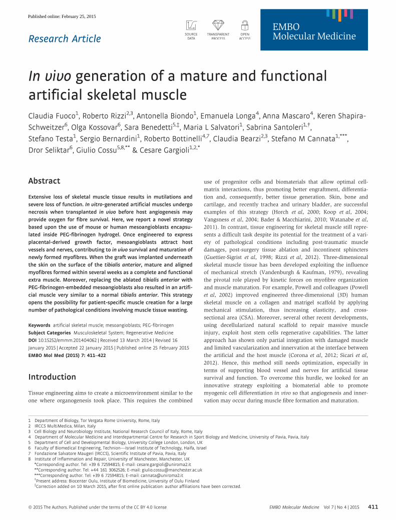
Figure 1. In vitro characterization of Mabs grown inside PF hydrogels.
A Phase contrast images of Mabs cultured into PF hydrogels (5 days): cells formed a thick three-dimensional myotube network with numerous thick myofibres(arrows). Scale bar: 50 lm.
B Confocal immunofluorescence analysis with an antibody against myosin heavy chain (MyHC) (red) and SYTOX green which labels nuclei (green), showing multi-nucleated, mature muscle fibres (arrows) developed from Mabs in PF (5 days). Scale bar: 50 lm.
C, D Western blot and densitometry analysis of different protein extracts of Mabs differentiated inside PF constructs (PF) showing expression levels comparable to adultTA (Ctrl). Group of n = 6 of Mabs differentiated inside PF and host TA has been tested, densitometric analysis from n = 3 different Western blots gauging the levelof expression of muscle markers is presented as means � standard error.
Source data are available online for this figure.
ª 2015 The Authors EMBO Molecular Medicine Vol 7 | No 4 | 2015
Claudia Fuoco et al Artificial skeletal muscle EMBO Molecular Medicine
413
Published online: February 25, 2015
A B C D
HG
I J M N Q
O P R SK L
E F
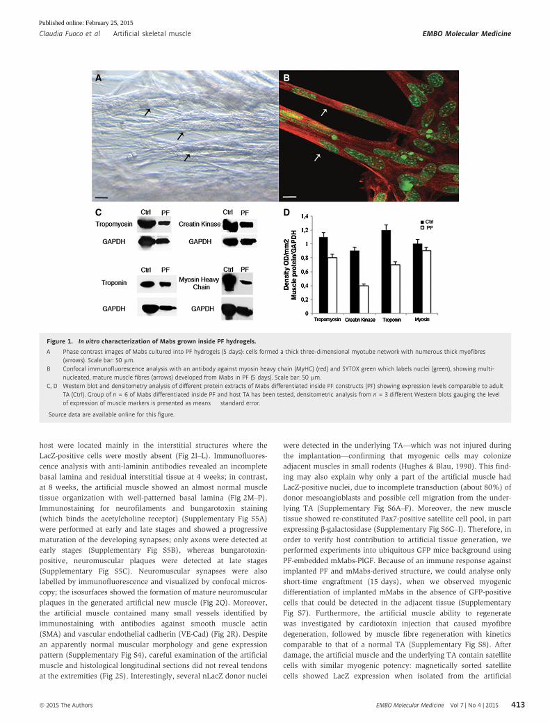
Figure 2. Implanted constructs of Mabs in PF on the surface of TA.
A Gross morphology of the PF implant containing Mabs-nLacZ/PlGF immediately after implantation on the surface of underlying TA (arrow).B, C Artificial muscles (arrows) developed within 8 weeks over the host TA surface (arrowheads), encapsulated by the host perimysium (B) and released after tendon
resection of the TA (C).D The isolated artificial muscle from the TA surface (arrow) is comparable in size to the underlying host TA (arrowhead).E Stereomicroscopy images revealing blood vessel formation in the artificial muscle. Scale bar: 100 lm.F X-Gal and H&E staining on artificial muscle section revealing vessel connection with host vasculature by Indian ink, injected into the femoral artery immediately
before sacrifice. Scale bar: 20 lm.G Western blot analysis of protein extracts from artificial muscles (PF) and host TA (Ctrl) reveals remarkable expression of muscle- and vessel-specific proteins (n = 5).H Densitometric analysis from n = 3 different Western blots gauging the level of expression of muscle- and vessel-specific proteins; presented values are expressed
as means � standard error.I–L X-Gal and H&E staining of artificial muscle sections at 4 (I, K) and 8 weeks (J, L) after implantation, showing donor origin of muscle cells (nLacZ+ nuclei) and
correlation between implant duration and myofibre maturation (I, J images are the results of the collage of several photos). Scale bar: (I, J) 200 lm, (K, L) 100 lm.M–P Immunofluorescence analysis on adjacent sections revealing laminin organization (green) and myosin heavy chain (MyHC) expression (red) at 4 weeks (M) and
8 weeks (N) after grafting (M, N displayed items are obtained from a multi-photo collage); DAPI staining (blue) shows centrally located nuclei in developing fibres(O) and peripheral nuclei in mature myofibres (P). Scale bar: (M, N) 200 lm, (O, P) 50 lm.
Q Confocal isosurface image of neurofilament (green) and bungarotoxin (red) immunofluorescence in the artificial muscle section shows a mature neuromuscularplaque. Scale bar: 10 lm.
R Immunofluorescence against smooth muscle actin (blue), laminin (green) and VE-Cad (red) superimposed on phase contrast image shows blood vessels (arrows)adjacent to the myofibres in the artificial muscle section. Scale bar: 20 lm.
S X-Gal staining on mature artificial muscle (arrowhead) and the underneath host TA (arrow) revealing no tendon development at the extremity of the artificialtissue; implanted LacZ-positive Mabs are present within the host TA. Scale bar: 200 lm.
Source data are available online for this figure.
EMBO Molecular Medicine Vol 7 | No 4 | 2015 ª 2015 The Authors
EMBO Molecular Medicine Artificial skeletal muscle Claudia Fuoco et al
414
Published online: February 25, 2015
muscle but not from the host TA and underwent myogenesis with
comparable efficiency (Supplementary Fig S9 and Supplementary
Table S1). Finally, in order to understand whether artificial muscle
would respond to hypertrophic or atrophic stimuli like a normal
muscle, clenbuterol administration and denervation were performed
(Supplementary Fig S10). The analysis revealed a striking atrophic
response to sciatic nerve resection both in the artificial muscle and
in the underlying TA (Supplementary Fig S10G–J), while the clenbu-
terol treatment showed only a modest, which did not reach statisti-
cal significance in both artificial and underlying muscles, possibly
due to inadequate response time or concentration (Supplementary
Fig S10C–F); quantification obtained evaluating CSA average of the
treated artificial and TA muscles in ten non-adjacent sections
(Supplementary Fig S10A and B).
Artificial muscle closely resembles a natural skeletal muscle
Ultrastructural and functional analyses were conducted to evaluate
to what extent the artificial muscle resembles a normal adult
muscle. Electron microscopy analysis revealed a completed sarco-
merogenesis typical of mature skeletal muscle while staining with
blu-O-gal revealed the presence of donor-derived LacZ-positive
nuclei adjacent to transversely sectioned sarcomeres (Fig 3A and B).
Single artificial myofibres were isolated from bunches of artificial
muscle fibres (LacZ-positive) and showed normal cross-striation
(Fig 3C and D). A subset of the artificial muscle fibres (n = 20)
was stained by immunofluorescence with antibodies against
b-galactosidase to assess the origin of their nuclei: results showed
that the large majority of nuclei were of donor origin (based on
nuclear LacZ expression), but few unlabelled nuclei, whose origin
remain unclear, were also present (Fig 3E and F) (Hughes & Blau,
1990). A large number of nLacZ-positive artificial muscle fibres
(n = 153) and underlying TA muscle control fibres (n = 103) from
4-month-old C57 RAG2/cchain�/� mice were mounted in a set-up
which enabled viewing their striation pattern at 320-times magni-
fication, determining CSA, specific force (Po/CSA) and maximum
shortening velocity (Vo) (Bottinelli et al, 1996; Pellegrino et al,
2003). The striation pattern, CSA, Po/CSA and Vo of muscle fibres
from artificial muscle and from control muscle were indistinguish-
able (Fig 3G–I). Po/CSA values were similar in single fibres from
the artificial muscle and the underlying control muscle. Moreover,
the values of specific force reported in Fig 3G are fully consistent
with our previous findings on muscle fibres from normal TA
muscles of mice (Torrente et al, 2004). Likewise, the value of Vo,
which is mainly dependent on the MyHC isoform content, indi-
cated that the kinetic of actomyosin interaction was virtually iden-
tical to that of the normal adult fibres of the underlying TA
(Bottinelli et al, 1994; Pellegrino et al, 2003; Torrente et al, 2004).
At the end of the functional analysis, the MyHC isoform content
of each fibre was also determined by SDS–PAGE. All fibres analy-
sed were found to contain adult MyHC isoforms and almost exclu-
sively MyHC-2B, which is the most expressed MyHC isoform in
fast mouse muscles (Pellegrino et al, 2003) (Fig 3J and K).
Human Mabs-derived artificial muscle
Human Mabs (hMabs) were isolated as described in the material
and methods (Tonlorenzi et al, 2007) and combined with PF as
described for mouse Mabs. Short-term analysis showed that the
human cells behave like their murine counterparts. Specifically,
myogenic differentiation of the hMabs in PF was observed using
in vitro cultures in as few as 5 days (Supplementary Fig S11A–F).
Moreover, hMabs exhibited an excellent capacity to form a new
human-derived artificial muscle in vivo when implanted into host
However, even in an immunodeficient background, over time the
xenogeneic graft with time attracted murine macrophages and
other non-lymphoid cells that infiltrated and prevented myogenic
maturation of the new muscle. Despite this problem, specifically
related to xenotransplantation, these data show that this method
can be applied to human cells, setting the stage for future clinical
application, even though extension of this method to large human
muscles may be technically very demanding and may require
significant additional work. Overall, the data presented above
establish a new paradigm for skeletal muscle tissue engineering,
by showing that it is possible and relatively simple to create an
artificial muscle by exploiting a contracting muscle surface as an
‘anatomical bioreactor’. In approximately 2 months, the artificial
muscle matures to become morphologically and functionally very
similar to the underlying, supporting host muscle. However, lack
of tendons prevented any functional test on the artificial muscle
in vivo or ex vivo.
Artificial muscle replacing the endogenous TA promotesfunctional recovery
Hence, in order to test whether the combination of mesoangio-
blasts and PEG-fibrinogen may lead to a biomechanically function-
ing artificial muscle, we performed implantations into a severe
injury muscle model. The TA of 2-month-old SCID mice was
almost entirely ablated surgically [80–90% of the TA was removed,
dislodged tissue weighting an average of 85 mg (TA weight
~100 mg) measured in five ablated mice, Supplementary Fig S12],
leaving the tendons intact in place to anchor the implant. The TA
was replaced with an implant comprised of 3 × 10⁶ mMabs
expressing PlGF (mMabs-PlGF) encapsulated into 8 mg/ml PF
within a total volume of 50 ll (Supplementary Movie S4 and
Supplementary Fig S13). Acellular PF gels were used as controls in
order to analyse the ability of the PF scaffold to induce host cell
migration and artificial muscle formation. The treated mice were
monitored for 6 months for their motor activity, running distance
and limb strength. Histological analysis was also performed at
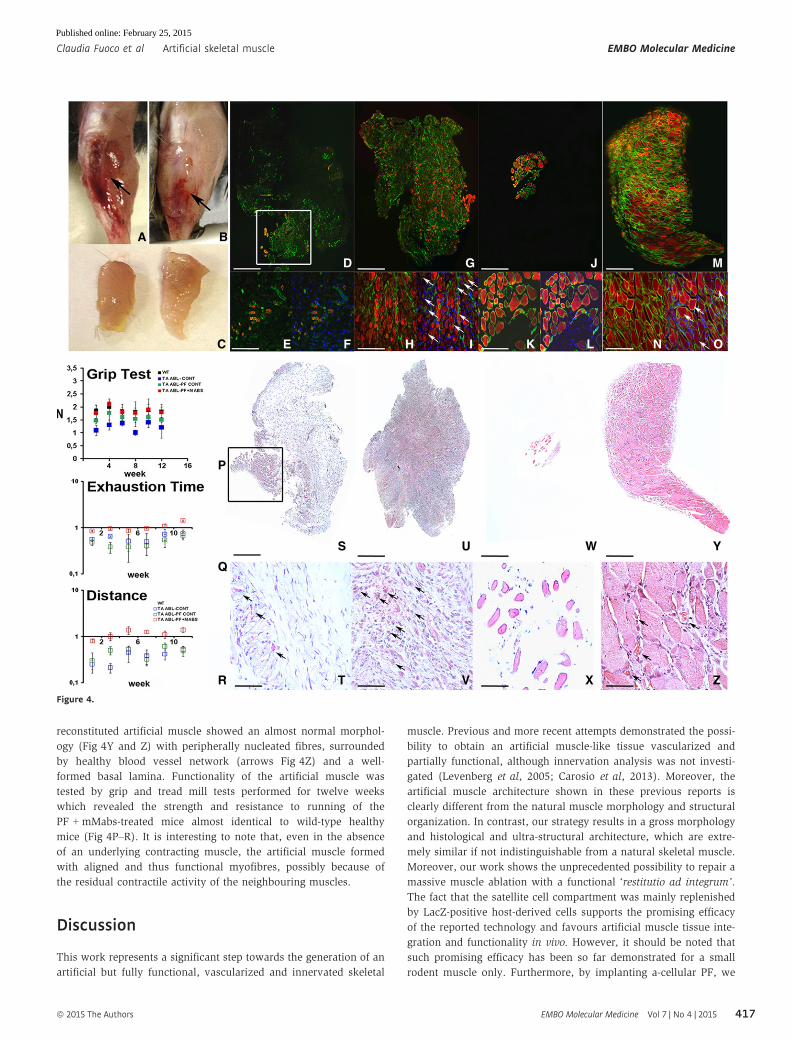
early and late time points after the TA replacement (Fig 4). Soon
after the intervention, all mice regain the ability of walking, but
they could not flex the foot as expected after the resection of the
TA preventing normal ambulatory activity (Supplementary Movie S5).
Six months after TA removal, mice treated with a-cellular PF
showed a complete lack of regeneration (Fig 4A), while the mice
treated with PF-embedded mMabs-PlGF showed the presence of a
new artificial muscle of approximately the same size as the ablated
TA (Fig 4B and C). Immunofluorescence and histological studies
were performed at early (10 days) and late (6 months) time points
after TA removal (Fig 4D–O and S–Z) in order to verify TA
ablation and its regeneration onset at very early time point, and
the artificial TA status at the end of reconstruction process. The
results obtained were comparable to those observed with the
ª 2015 The Authors EMBO Molecular Medicine Vol 7 | No 4 | 2015
Claudia Fuoco et al Artificial skeletal muscle EMBO Molecular Medicine
415
Published online: February 25, 2015
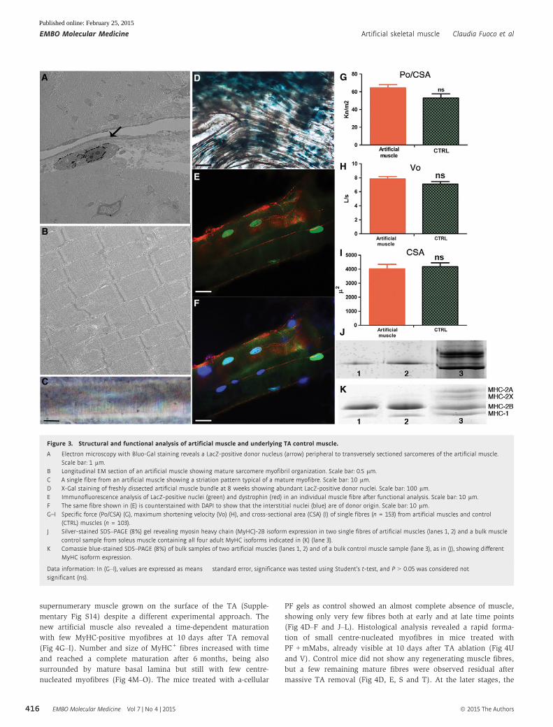
supernumerary muscle grown on the surface of the TA (Supple-
mentary Fig S14) despite a different experimental approach. The
new artificial muscle also revealed a time-dependent maturation
with few MyHC-positive myofibres at 10 days after TA removal
(Fig 4G–I). Number and size of MyHC+ fibres increased with time
and reached a complete maturation after 6 months, being also
surrounded by mature basal lamina but still with few centre-
nucleated myofibres (Fig 4M–O). The mice treated with a-cellular
PF gels as control showed an almost complete absence of muscle,
showing only very few fibres both at early and at late time points
(Fig 4D–F and J–L). Histological analysis revealed a rapid forma-
tion of small centre-nucleated myofibres in mice treated with
PF + mMabs, already visible at 10 days after TA ablation (Fig 4U
and V). Control mice did not show any regenerating muscle fibres,
but a few remaining mature fibres were observed residual after
massive TA removal (Fig 4D, E, S and T). At the later stages, the
E
F
A D G
H
I
J
K
B
C
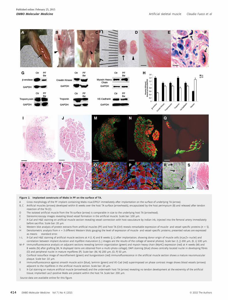
Figure 3. Structural and functional analysis of artificial muscle and underlying TA control muscle.
A Electron microscopy with Bluo-Gal staining reveals a LacZ-positive donor nucleus (arrow) peripheral to transversely sectioned sarcomeres of the artificial muscle.Scale bar: 1 lm.
B Longitudinal EM section of an artificial muscle showing mature sarcomere myofibril organization. Scale bar: 0.5 lm.C A single fibre from an artificial muscle showing a striation pattern typical of a mature myofibre. Scale bar: 10 lm.D X-Gal staining of freshly dissected artificial muscle bundle at 8 weeks showing abundant LacZ-positive donor nuclei. Scale bar: 100 lm.E Immunofluorescence analysis of LacZ-positive nuclei (green) and dystrophin (red) in an individual muscle fibre after functional analysis. Scale bar: 10 lm.F The same fibre shown in (E) is counterstained with DAPI to show that the interstitial nuclei (blue) are of donor origin. Scale bar: 10 lm.G–I Specific force (Po/CSA) (G), maximum shortening velocity (Vo) (H), and cross-sectional area (CSA) (I) of single fibres (n = 153) from artificial muscles and control
(CTRL) muscles (n = 103).J Silver-stained SDS–PAGE (8%) gel revealing myosin heavy chain (MyHC)-2B isoform expression in two single fibres of artificial muscles (lanes 1, 2) and a bulk muscle
control sample from soleus muscle containing all four adult MyHC isoforms indicated in (K) (lane 3).K Comassie blue-stained SDS–PAGE (8%) of bulk samples of two artificial muscles (lanes 1, 2) and of a bulk control muscle sample (lane 3), as in (J), showing different
MyHC isoform expression.
Data information: In (G–I), values are expressed as means � standard error, significance was tested using Student’s t-test, and P > 0.05 was considered notsignificant (ns).
EMBO Molecular Medicine Vol 7 | No 4 | 2015 ª 2015 The Authors
EMBO Molecular Medicine Artificial skeletal muscle Claudia Fuoco et al
416
Published online: February 25, 2015
reconstituted artificial muscle showed an almost normal morphol-
ogy (Fig 4Y and Z) with peripherally nucleated fibres, surrounded
by healthy blood vessel network (arrows Fig 4Z) and a well-
formed basal lamina. Functionality of the artificial muscle was
tested by grip and tread mill tests performed for twelve weeks
which revealed the strength and resistance to running of the
PF + mMabs-treated mice almost identical to wild-type healthy
mice (Fig 4P–R). It is interesting to note that, even in the absence
of an underlying contracting muscle, the artificial muscle formed
with aligned and thus functional myofibres, possibly because of
the residual contractile activity of the neighbouring muscles.
Discussion
This work represents a significant step towards the generation of an
artificial but fully functional, vascularized and innervated skeletal
muscle. Previous and more recent attempts demonstrated the possi-
bility to obtain an artificial muscle-like tissue vascularized and
partially functional, although innervation analysis was not investi-
gated (Levenberg et al, 2005; Carosio et al, 2013). Moreover, the
artificial muscle architecture shown in these previous reports is
clearly different from the natural muscle morphology and structural
organization. In contrast, our strategy results in a gross morphology
and histological and ultra-structural architecture, which are extre-
mely similar if not indistinguishable from a natural skeletal muscle.
Moreover, our work shows the unprecedented possibility to repair a
massive muscle ablation with a functional ‘restitutio ad integrum’.
The fact that the satellite cell compartment was mainly replenished
by LacZ-positive host-derived cells supports the promising efficacy
of the reported technology and favours artificial muscle tissue inte-
gration and functionality in vivo. However, it should be noted that
such promising efficacy has been so far demonstrated for a small
rodent muscle only. Furthermore, by implanting a-cellular PF, we
A B
Q
R
S U W Y
ZV XT
P
E F H I K L N OC
D G J M
Figure 4.
ª 2015 The Authors EMBO Molecular Medicine Vol 7 | No 4 | 2015
Claudia Fuoco et al Artificial skeletal muscle EMBO Molecular Medicine
417
Published online: February 25, 2015
were able to demonstrate that the scaffold matrix by itself is not able
to support formation of new muscle mediated by host stem cell
migration, thus showing that the combination of PF and Mabs only
can generate a complete and functional artificial muscle. Even if
in Mabs + PF-derived artificial muscle, there is a host cell co-
participation, this is mainly in the interstitial compartment and
probably advantageous for clinical reconstructive strategy. This by
no means implies that other biomaterials and other myogenic cell
types may not result in a similar functioning muscle, though this
remains to be tested. In the case of human cells, inflammatory host
cell infiltration prevented complete maturation of the human artifi-
cial muscle, but we believe this to be a problem of xenotransplanta-
tion and not relevant to an intraspecific, possibly autologous context.
Although differences were detected with bona fide mature
muscles such as the absence of MyHCs 2A and increased density of
nerves, both consistent with a still partially immature phenotype,
the contractile properties of newly formed fibres were indistinguish-
able from those of the underlying and wild-type TA. Clearly, a
number of issues still remain to be solved before this artificial
muscle may be used in a clinical setting. Importantly, tendons
should be developed in vivo by integrating the edge of the muscle
that already contains connective tissue to the existing tendons and
eventually adding tendon fibroblasts at the edge. Equally important
is the use of larger animal models that should be developed for
testing efficacy of this approach before clinical translation. Finally,
current Good Manufacturing Practices (cGMP) will have to be devel-
oped for this combination of cells and biomaterial, even though
both mesoangioblasts and PEG-fibrinogen are in clinical experimen-
tation. The discussed strategy offers the possibility of creating de
novo a functional artificial muscle in those anatomical districts
where it is needed. This opens up new scenarios for regenerative
medicine: ablated or irreversibly damaged muscles could be
replaced with patient’s own cells and biomaterial could be
implanted on top of a residual muscle, adjacent to the damaged area
where the new muscle will be later transferred with nerves and
vessels maintained. Clinical translation of this procedure should be
possible in a relatively near future, as already shown by Sicari and
colleagues (Sicari et al, 2014) experimenting in human patient mate-
rial scaffold to promote muscle reconstruction and function restora-
tion in a massive muscle ablation context.
Materials and Methods
Cells and culture conditions
We cultured mouse mesoangioblasts (Mabs) on Falcon dishes at
37°C with 5% CO2 in DMEM GlutaMAX (Gibco) supplemented with
heat-inactivated 10% foetal bovine serum (FBS), 100 international
units/ml penicillin and 100 mg/ml streptomycin. We transduced the
cells with third-generation lentiviral vectors encoding b-galactosidase(nLacZ) and/or PlGF as described (Gargioli et al, 2008). Human
Mabs (hMabs) were isolated and cultured as described before
(Tonlorenzi et al, 2007; Tedesco et al, 2012); briefly, hMabs were
isolated from cultured muscle biopsies provided by Dr. Roberto
Biagini head of orthopaedic oncology Rome IRE-ISG after obtaining
patient informed consensus. The human samples study protocol
was evaluated by the ethical committee of the Rome IRES-ISG and
approved by the scientific management. The hMabs were selected
for alkaline phosphatase (AP) expression and growth in Iscove’s
modified Dulbecco’s medium (IMDM; Sigma) containing 10% FBS,
2 mM glutamine, 0.1 mM beta-mercaptoethanol, 1% nonessential
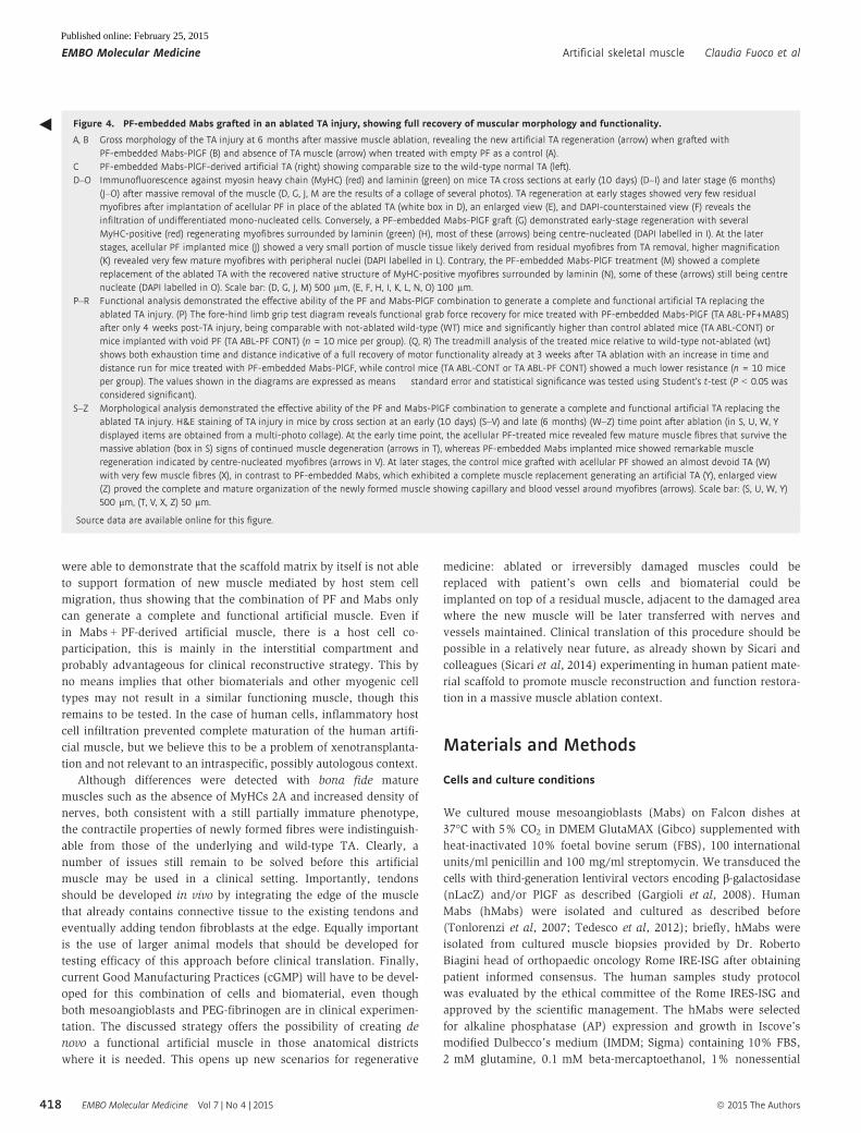
Figure 4. PF-embedded Mabs grafted in an ablated TA injury, showing full recovery of muscular morphology and functionality.
A, B Gross morphology of the TA injury at 6 months after massive muscle ablation, revealing the new artificial TA regeneration (arrow) when grafted withPF-embedded Mabs-PlGF (B) and absence of TA muscle (arrow) when treated with empty PF as a control (A).
C PF-embedded Mabs-PlGF-derived artificial TA (right) showing comparable size to the wild-type normal TA (left).D–O Immunofluorescence against myosin heavy chain (MyHC) (red) and laminin (green) on mice TA cross sections at early (10 days) (D–I) and later stage (6 months)
(J–O) after massive removal of the muscle (D, G, J, M are the results of a collage of several photos). TA regeneration at early stages showed very few residualmyofibres after implantation of acellular PF in place of the ablated TA (white box in D), an enlarged view (E), and DAPI-counterstained view (F) reveals theinfiltration of undifferentiated mono-nucleated cells. Conversely, a PF-embedded Mabs-PlGF graft (G) demonstrated early-stage regeneration with severalMyHC-positive (red) regenerating myofibres surrounded by laminin (green) (H), most of these (arrows) being centre-nucleated (DAPI labelled in I). At the laterstages, acellular PF implanted mice (J) showed a very small portion of muscle tissue likely derived from residual myofibres from TA removal, higher magnification(K) revealed very few mature myofibres with peripheral nuclei (DAPI labelled in L). Contrary, the PF-embedded Mabs-PlGF treatment (M) showed a completereplacement of the ablated TA with the recovered native structure of MyHC-positive myofibres surrounded by laminin (N), some of these (arrows) still being centrenucleate (DAPI labelled in O). Scale bar: (D, G, J, M) 500 lm, (E, F, H, I, K, L, N, O) 100 lm.
P–R Functional analysis demonstrated the effective ability of the PF and Mabs-PlGF combination to generate a complete and functional artificial TA replacing theablated TA injury. (P) The fore-hind limb grip test diagram reveals functional grab force recovery for mice treated with PF-embedded Mabs-PlGF (TA ABL-PF+MABS)after only 4 weeks post-TA injury, being comparable with not-ablated wild-type (WT) mice and significantly higher than control ablated mice (TA ABL-CONT) ormice implanted with void PF (TA ABL-PF CONT) (n = 10 mice per group). (Q, R) The treadmill analysis of the treated mice relative to wild-type not-ablated (wt)shows both exhaustion time and distance indicative of a full recovery of motor functionality already at 3 weeks after TA ablation with an increase in time anddistance run for mice treated with PF-embedded Mabs-PlGF, while control mice (TA ABL-CONT or TA ABL-PF CONT) showed a much lower resistance (n = 10 miceper group). The values shown in the diagrams are expressed as means � standard error and statistical significance was tested using Student’s t-test (P < 0.05 wasconsidered significant).
S–Z Morphological analysis demonstrated the effective ability of the PF and Mabs-PlGF combination to generate a complete and functional artificial TA replacing theablated TA injury. H&E staining of TA injury in mice by cross section at an early (10 days) (S–V) and late (6 months) (W–Z) time point after ablation (in S, U, W, Ydisplayed items are obtained from a multi-photo collage). At the early time point, the acellular PF-treated mice revealed few mature muscle fibres that survive themassive ablation (box in S) signs of continued muscle degeneration (arrows in T), whereas PF-embedded Mabs implanted mice showed remarkable muscleregeneration indicated by centre-nucleated myofibres (arrows in V). At later stages, the control mice grafted with acellular PF showed an almost devoid TA (W)with very few muscle fibres (X), in contrast to PF-embedded Mabs, which exhibited a complete muscle replacement generating an artificial TA (Y), enlarged view(Z) proved the complete and mature organization of the newly formed muscle showing capillary and blood vessel around myofibres (arrows). Scale bar: (S, U, W, Y)500 lm, (T, V, X, Z) 50 lm.
Source data are available online for this figure.
◀
EMBO Molecular Medicine Vol 7 | No 4 | 2015 ª 2015 The Authors
EMBO Molecular Medicine Artificial skeletal muscle Claudia Fuoco et al
418
Published online: February 25, 2015
amino acids, human basic fibroblast growth factor (5 ng/ml),
penicillin (100 IU/ml), streptomycin (100 mg/ml), 0.5 mM oleic and
linoleic acids (Sigma), 1.5 mM Fe2+ [iron(II) chloride tetrahydrate,
Sigma; or Fer-In-Sol, Mead Johnson], 0.12 mM Fe3+ [iron(III)
nitrate nonahydrate, Sigma; or Ferlixit, Aventis] and 1% insulin/
transferrin/selenium (Gibco).
PEG-fibrinogen
PEG-fibrinogen precursor solution was prepared and photopolymer-
ized as described elsewhere (Corona et al, 2012). We prepared PEG
hydrogels containing Mabs by mixing a PBS cell suspension and
PEGylated fibrinogen precursor solution containing 0.1% of Igracu-
retTM2959 photoinitiator (Ciba Specialty Chemicals) to have a final
concentration of 8 mg/ml with the desired cell concentration. We
added 30 ml aliquots of the suspension into cylindrical silicon
moulds and placed them under a long-wave UV lamp (365 nm,
4–5 mW/cm2) for 5 min in a laminar flow hood. DMEM culture
medium (containing 10% FBS) was added immediately to the poly-
merized hydrogels to ensure cell growth for the in vitro experi-
ments. The plugs were cultured for 24 h in serum-supplemented
growth medium and then transferred into serum-depleted differenti-
ation medium for 5 days in order to promote muscle fibre forma-
tion; for in vivo experiments, the moulds were directly implanted in
the animals subcutaneously on the back, underneath the skin on the
surface of the tibialis anterior (TA) or in its anatomical lodge, after
ablation, without in vitro culture.
Surgical procedure
Two-month-old male RAG2/cchain (mouse Mabs) or SCID (human
Mabs) transgenic mice were provided, respectively, by Taconic and
Charles Rivers, they were bred in ventilated cage at the Plaisant SPF
(Specific Pathogen Free) animal house of Castel Romano. Mice were
anesthetized with an intramuscular injection of physiologic saline
(10 ml/kg) containing ketamine (5 mg/ml) and xylazine (1 mg/ml)
and then implanted subcutaneously on the back, or underneath the
skin on the surface of the TA with PF constructs containing
1.5 × 10⁶ Mabs-nLacZ/PlGF or hMabs. In order to ensure a good
placement of the construct, we performed a limited incision on the
medial side of the back or the leg, separated the dorsal muscle or
the TA from the skin and placed the plug constructs as desired and
finally sutured. For the artificial muscle atrophy and hypertrophy,
RAG2/cchain null mice implanted with PF constructs containing
1.5 × 10⁶ Mabs-nLacZ/PlGF were subjected to denervation proce-
dure and clenbuterol (Sigma) administration, respectively, after
4 weeks of the artificial muscle development. The right sciatic nerve
was isolated in the mid-thigh region and cut, leading to denervation
of the lower limb muscles. Clenbuterol was administered at a dose of
2 mg/kg/day via intraperitoneal injection, continuously for 1 month.
To evaluate the host contribution in artificial muscle development,
PF constructs containing 1.5 × 10⁶ mMabs were implanted on the
surface of TA of ubiquitous GFP mice.
For TA removal, mice were anesthetized as previously described,
limited incision on the medial side of the leg has been performed in
order to reach the TA, and then utilizing a cautery to avoid bleeding,
the muscle fibres were completely removed, PF constructs contain-
ing 3 × 10⁶ mMabs-PlGF were polymerized in the TA venue as
already described and then the incision sutured. All the experi-
mental animal groups are summarized in Supplementary Table S2.
Analgesic treatment (Rimadyl, Pfizer, USA) was administered after
the surgery to reduce pain and discomfort. Mice were sacrificed at
different time points for molecular and morphological analysis.
Experiments on animals were conducted according to the rules of
good animal experimentation I.A.C.U.C. no 432 of 12 March 2006.
Satellite cells isolation
For artificial muscle satellite cell isolation, 8-week injured artificial
and TA muscles (n = 3) were minced and digested in HBSS (Gibco)
containing 2 lg/ml collagenase A (Roche), 2.4 U/ml dispase I
(Roche), 10 ng/ml DNase I (Roche), 0.4mM CaCl2 and 5mM
MgCl2 for 1 h at 37°C. Muscle satellite cells were magnetically
sorted as CD45�, CD31�, a7-integrin+ cells. First, the CD45+ cells
were magnetically labelled with anti-CD45 and CD31 MicroBeads
(Miltenyi). Then, the cell suspension was loaded onto a MACS
column (Miltenyi), which is placed in the magnetic field of a MACS
separator. The magnetically labelled CD45, CD31+ cells were
retained within the column. The unlabelled cells run through. Then,
these cells were labelled with anti-a7-integrin MicroBeads (Miltenyi)
and loaded onto a MACS column, which was placed in the magnetic
field of a MACS separator. After removing the column from the
magnetic field, the magnetically retained a7-integrin+ cells have
been eluted as the positively selected cell fraction.
Histology and immunocytochemistry
Cells and tissues were fixed in PFA 2% and processed for histology
and immunocytochemistry as previously described (Gargioli et al,
2008). The primary antibodies used were mouse anti-Pax7 (DSHB)
at 1:20, mouse MF20 (DSHB) at 1:2, rabbit anti-laminin (SIGMA
#9393) at 1:100, rabbit anti-LacZ (Cappel) at 1:100, rat anti-
VE-cadherin (clone BV13 homemade) at 1:100, mouse anti-SMA
(Sigma) at 1:100, mouse anti-dystrophin (Vector) at 1:100,
mouse anti-neuronal class III b-tubulin (COVANCE) and alpha-
bungarotoxin Alexa594 (Molecular probes) at 50 mg/ml. The
secondary antibodies used at 1:100 were anti-mouse Alexa555
(Molecular Probes), anti-rabbit Alexa488 (Molecular Probes) and
Mabs-PF implanted (8 weeks) RAG2/cchain null mice were injected
with 20 ml of 10 mM cardiotoxin (Latoxan) in PBS, into the artificial
muscle: limited incision in the medial side of the leg was performed
in order to visualize artificial muscle and inject cardiotoxin into it.
The treated artificial muscles were then collected 3, 7 and 14 days
following cardiotoxin injection. The muscles were harvested and
analysed to investigate artificial muscle fibres regeneration.
Treadmill
A 7° uphill treadmill protocol was performed using an Exer-3/6
open treadmill (Columbus Instruments, Columbus, OH, USA)
according to guidelines from the American Physiological Society.
The treadmill protocol consisted of five continuous days of incre-
mental training followed by experimental determination of maximal
running distance on day 6. Mice were first run at 10 m/min for
20 min, and treadmill speed was then increased by 1 m/min every
2 min until mice were exhausted. Exhaustion was defined as
spending > 10 s on the shocker without attempting to re-enter the
treadmill (McCullagh et al, 2008; Zeng et al, 2014).
Fore-hind leg grip test
A computerized grip-strength meter (Columbus Instruments) was
used to measure fore-hind limb grip strength in conscious mice.
Mice were acclimatized for 5 min before starting test. Gently the
mouse was lowered over the top of the grid so that both its front
paws and hind paws are allowed to grip the smooth metal pull bar
at the top of the apparatus. The mouse was then gently pulled back-
ward in the horizontal plane until it could no long grasp the bar.
The force at the time of release was recorded as the peak tension.
Each mouse was tested five times with a 20–40 s break between
tests. The average peak tension from three best attempts was
defined as fore-hind limb grip strength.
Cross-sectional area analysis
Morphometrical analyses to evaluate means of fibre per CSA were
carried out on 800 fibres per muscle on laminin-stained sections of
artificial muscle or TA using ImageJ (NIH) software. The CSA means
were scored in three different non-adjacent transverse sections from
the largest muscle portion for three mice per experimental group.
Statistical analysis
Data were analysed using GraphPad Prism 5, and values were
expressed as means � standard error (SEM). Statistical significance
was tested using Student’s t-test. A probability of less than 5%
(P < 0.05) was considered to be statistically significant. All the
experimental mice were analysed, and inclusion/exclusion criteria
were based on the presence or absence of artificial muscle genera-
tion after implantation procedure. Blind analyses were conducted
for quantification of PlGF effect on vessel density, clenbuterol
treatment and denervation procedure affecting muscle CSA in order
to minimize investigator bias.
Supplementary information for this article is available online:
http://embomolmed.embopress.org
AcknowledgementsWe thank M. Coletta for technical assistance, E. Dejana for the gift of BV13
antibody, the centre of Advanced Microscopy ‘P. Albertano’ for confocal imag-
ing, M.C. Panzeri and Alembic for electron microscopy imaging, R. Biagini and
C. Zoccali for providing human biopsies and surgical advices and L. Madaro for
CSA tips. This work was supported by EC-IP FP7 grants Biodesign (to DS and
GC), Duchenne Parent project Italia to GC and by the Cariplo Foundation, Italy
(grant no. 2010.0764) and by the European Commission, MYOAGE grant (no.
223576), funded under FP7 to RB.
Author contributionsGC and CG designed the research and wrote the paper. CF prepared vectors,
transduced cells and carried out most of the experimental work; RR and CB
performed artificial muscle functional analysis and helped with data interpre-
tation; AB tested different PF hydrogel concentration; EL, AM and RB designed
and performed physiological experiments; KS-S, OK and DS produced and
implemented PF for muscle experiments, tested in vitro PF compositions and
performed confocal time-lapse experiments; CL, MLS, SS and ST did the
histology and staining; SB did cell culture and immunostaining; CG and RR
performed surgical operation; SMC and DS helped with study design, data
analysis interpretation and paper writing.
The paper explained
ProblemDifferent causes such as major traumatisms or surgery for cancerresult in extensive loss of skeletal muscle tissue that cannot berepaired by the muscle itself. This unmet clinical need has stimulatedmany attempts to re-create a functional muscle either outside ordirectly inside the body. This has so far been a significant challengebecause it is difficult to reconstruct the complex architecture of theskeletal muscle tissue that has a high requirement of oxygen.
ResultsWe combined muscle progenitors (mesoangioblasts), engineered tostimulate blood vessel growth from the host, with a hydrogel (PEG-fibrinogen). When the graft was implanted underneath the skin on thesurface of a normal, contracting skeletal muscle (tibialis anterior),mature and aligned muscle fibres formed within several weeks as acomplete and functional extra muscle. Moreover, replacing the tibialisanterior ablated with the same combinations of cells and hydrogel, weobtained a complete and functional recovery of the ablated muscle.
ImpactThis strategy opens the possibility for patient-specific muscle genera-tion for a large number of pathological conditions involving muscletissue wasting. It should however be considered that a mouse muscleis very small and scaling up the process may require significant addi-tional work.
ª 2015 The Authors EMBO Molecular Medicine Vol 7 | No 4 | 2015
Claudia Fuoco et al Artificial skeletal muscle EMBO Molecular Medicine
421
Published online: February 25, 2015
Conflict of interestThe authors declare that they have no conflict of interest.
References
Almany L, Seliktar D (2015) Biosynthetic hydrogel scaffolds made from
fibrinogen and polyethylene glycol for 3D cell cultures. Biomaterials 7:
411 –422
Bader A, Macchiarini P (2010) Moving towards in situ tracheal regeneration:
the bionic tissue engineered transplantation approach. J Cell Mol Med 14: