Inactivation of p21WAF1/cip1 Enhances IntestinalTumor Formation in Muc2�/� Mice
WanCai Yang, Anna Velcich, Ioana Lozonschi,Jiao Liang, Courtney Nicholas, Min Zhuang,Laura Bancroft, and Leonard H. AugenlichtFrom the Department of Oncology, Montefiore Medical Center,
Albert Einstein Cancer Center, Bronx, New York
In the Apc1638�/� mouse model of intestinal tumor-igenesis, targeted inactivation of the cyclin-depen-dent kinase inhibitor p21WAF1/cip1 is highly effectivein enhancing Apc-initiated tumor formation in theintestine. Because p21WAF1/cip1 plays a critical role inregulating intestinal cell proliferation, maturation,and tumorigenesis, we examined whether its inacti-vation would enhance tumor formation in a differentmouse model of colon cancer. Therefore, we matedp21�/� mice with mice carrying a genetic deficiencyof the Muc2 gene, which encodes the major gastroin-testinal mucin. Muc2�/� mice develop tumors in thesmall and large intestine and the rectum, but incontrast to tumors in Apc1638�/� mice, this doesnot involve increased expression or nuclear localiza-tion of �-catenin. We found that inactivation ofp21WAF1/cip1 significantly increased the frequencyand size of intestinal tumors in Muc2 knockout miceand also led to development of more invasive adeno-carcinomas. This enhanced tumorigenesis signifi-cantly decreased mouse life span. Further, inactiva-tion of p21WAF1/cip1 increased cell proliferation,decreased apoptosis, and decreased intestinal trefoilfactor expression in the mucosa of both the small andlarge intestine. Surprisingly, reduced expression ofp27kip1 was also observed in the Muc2�/�, p21�/�,and p21�/� mice. In contrast, the expression of c-mycwas significantly elevated. Thus, p21 modulates theformation of tumors whose initiation does (Apc) ordoes not (Muc2) involve altered �-catenin-Tcf4 signal-ing, but which may converge on common elementsdownstream of this signaling pathway. (Am J Pathol2005, 166:1239–1246)
p21WAF1/cip1 is an inhibitor of cyclin-dependent kinase(cdk) activity and is therefore an important regulator of cellcycle progression, and overall cell maturation, including
differentiation and apoptosis.1–4 Despite this apparentlycentral role of p21, mice with a targeted, homozygous in-activation of the p21 gene are essentially normal, althoughembryonic fibroblasts from such mice are defective in cell-cycle arrest at the G1 checkpoint.5 Thus, either p21 is notcritical in cell maturation or its loss can be compensated forby other alterations that preserve pathways necessary fornormal development and tissue homeostasis.
In the intestinal tract, p21 appears to play a key role inthe maturation of cells as they migrate from the crypttoward the lumen.6 p21 is expressed as cells exit theproliferative compartment. This pattern of expression, aswell as the known functions of p21, suggested to us thatp21 levels would modulate tumor formation in the intes-tine, and indeed, the introduction of a targeted inactiva-tion of p21 into the Apc1638�/� mouse, in which intestinaltumors form when the wild-type Apc gene is lost or inac-tivated, substantially increased intestinal tumor forma-tion.3 More recently, the importance of p21 in homeosta-sis of the intestinal mucosa has been emphasized andclarified by the finding that expression of p21 is repressedby c-MYC protein,7–9 a target of APC-�-catenin-Tcf signal-ing.8,10–12 c-MYC sequesters the transcription factor MIZ ina MIZ/MYC complex,7,9,13,14 and data have been pre-sented that the down-regulation of c-myc gene expression,and consequent activation of p21 expression by MIZ, is acritical event in triggering intestinal cell differentiation.8
Interestingly, analysis of gene expression profiles incolonic tumor cells in which �-catenin-Tcf signaling isabrogated by expression of a dominant-negative Tcf -4demonstrated up-regulation of gene markers that arecharacteristic of either the mucosecretory or the absorp-tive cell lineages in the intestinal mucosa,15–18 consistentwith our report that down-regulation of �-catenin-Tcf sig-naling accompanied, and was mechanistically linked to,colonic cell differentiation in tissue culture.12 This there-fore suggests that loss of normal cell differentiation pat-terns in the intestinal mucosa, possibly attributed to the
Supported in part by the National Cancer Institute (grants CA96605,CA100926, CA87559, and P01 13330).
Accepted for publication January 11, 2005.
Address reprint requests to WanCai Yang, M.D., Department of Oncol-ogy, Montefiore Medical Center, Albert Einstein Cancer Center, 111 East210th St., Bronx, NY 10467. E-mail: [email protected].
American Journal of Pathology, Vol. 166, No. 4, April 2005
regulation of p21 expression through effects of �-catenin-Tcf signaling and c-myc expression, is a key event intumor formation, and indeed, introduction of the targetedinactivation of p21 in the Apc1638�/� mouse, which in-creased tumor formation, decreased the number of mu-cin-expressing goblet cells in the intestinal mucosa.3
We also showed that genetic inactivation of the Muc2gene, which encodes the principal colonic mucin,19–23
was sufficient to cause tumor formation.24 However, un-like the loss of Apc function, this loss of Muc2 expressionwas not accompanied by elevated �-catenin expression,or the relocalization of �-catenin to the nucleus.24 Thus,there was no evidence that �-catenin-Tcf signaling wasaberrant in this model, although c-myc expression waselevated in the tumors.24 Therefore, although the mech-anism of tumorigenesis in the Muc2 and the Apc modelsmay converge, the initial events seem to be distinct, anddiffer in their overt affects on �-catenin-Tcf signaling. Thequestion therefore arises whether the targeted inactiva-tion of p21 would also be effective in augmenting tumorformation in the Muc2�/� mouse model. Such data areessential for interpretation of the role that loss of p21plays in the central pathways involved in intestinal tumor-igenesis. We here report that introduction of a targetedinactivation of p21 increased tumor formation in the smallintestine, colon, and rectum, of mice with a targeted inacti-vation of Muc2, accompanied by increased cell prolifera-tion, decreased apoptosis, and decreased differentiationin the intestinal mucosa, which was associated withdown-regulation of p27kip and up-regulation of c-myc.
Materials and Methods
The p21 and Muc2 mouse models, and methods for geno-typing, have been reported.3,24 Muc2�/� mice (mixedC57BL/6J and 129/SvOla background) were mated withp21�/� mice (mixed 129S6/SvEvTac and NIH Black Swissbackground) to generate Muc2�/�, p21�/� offspring (F1).F1 mice were mated to produce desired genotypes:Muc2�/�, p21�/�, Muc2�/�, p21�/�, or Muc2�/�, p21�/�.At weaning (�3 to 4 weeks), littermates were fed AIN-76Adiet (Teklad, Madison, WI), ad libitum.
Mice were weighed weekly and maintained on diet for36 weeks, or until they exhibited significant weight loss orother signs of extensive tumor formation. Mice were killedby CO2 overdose and cervical dislocation, and then rap-idly dissected for evaluation of tumors and fixation oftissues, as described previously.3,4,24,25 Proliferation andapoptosis were evaluated by staining for proliferating cellnuclear antigen (Zymed, South San Francisco, CA) orterminal dUTP nick-end labeling (TUNEL) assay (Trevi-gen, Gaithersburg, MD), as described.3,26
Total RNA and protein were isolated from the frozentissues using TRIzol reagent (Invitrogen Life Technology,Carlsbad, CA), as described.25 The quantity of RNA andprotein were measured spectrophotometrically. As we de-scribed,25 cDNA was synthesized from DNase-treated totalRNA using TaqMan Multiscribe reverse transcriptase (Ap-plied Biosystems, Inc., Foster City, CA). Quantitative poly-merase chain reaction analysis was done using the ABIPrism 7900-HT sequence detection system (96-well, Ap-plied Biosystems, Inc., Foster City, CA). The primers forp21, p27, c-myc, and �-actin, the amplification conditionsfor the quantitative real-time polymerase chain reaction, anddata analysis, were reported previously.25
Western blot analyses of steady-state levels of specificproteins were done by standard methods, as described,25
using the following primary antibodies for detection: anti-p21, anti-p27, anti-c-myc (Santa Cruz Biotechnology, SantaCruz, CA); and anti-�-actin (Sigma, St. Louis, MO). Signalwas detected by the enhanced chemiluminescence tech-nique (Amersham Life Science, Piscataway, NJ). Immuno-histochemical staining for intestinal trefoil factor (ITF) waspreviously reported in detail.24 Briefly, 4-�m formalin-fixedand paraffin-embedded sections were deparaffinized andrehydrated, quenched with 1.5% H2O2, blocked with 10%normal goat serum, and probed with rabbit anti-ITF poly-clonal antibody (kindly provided by Catherine Tomasetto,Strasbourg, France). Detection was with biotinylated anti-rabbit IgG (Santa Cruz Biotechnology), followed by incuba-tion with avidin-biotin complex (Vector Labs, Burlingame,CA) and the substrate 3�,5�-diaminobenzidine, combinedwith hematoxylin counterstaining.
Figure 1. The incidence (a), frequency (b), and size (c) of intestinal tumors in the Muc2�/�, p21�/�, Muc2�/�, p21�/�, or Muc2�/�, p21�/� mice. a: *P � 0.025,in comparison to Muc2�/�, p21�/� mice by Fisher’s exact test. b: *P � 0.02. c: *P � 0.03 and **P � 0.02 in comparison to Muc2�/�, p21�/� mice by Mann-Whitneytest.
1240 Yang et alAJP April 2005, Vol. 166, No. 4
Results
Tumors developed throughout the intestinal tract in theMuc2�/�, p21 wild-type mice. Small intestinal tumors de-veloped in �60% of the Muc2�/�, p21 wild-type mice ata frequency of 1.0 tumor per mouse at an age of 36weeks (Figure 1, a and b). This is similar to the incidenceand frequency of tumors we previously reported for theMuc2�/� mice.24 However, littermates that were Muc2�/�
and either p21�/� or �/� had a significantly higher tumorincidence of 83% and 100% in small intestinal tumors,respectively (P � 0.025) (Figure 1a). In addition, thesmall intestinal tumor frequency per mouse was in-creased by 30% in the Muc2�/�, p21�/� mice and by�80% in the Muc2�/�, p21�/� mice (Figure 1b) (P �0.029). The effect on tumor size was also striking: smallintestinal tumors in Muc2�/�, p21�/� or Muc2�/�, p21�/�
mice were 35% larger than the tumors in Muc2�/�,p21�/� mice (P � 0.02 and P � 0.03, respectively, com-pared to the Muc2�/�, p21�/� mice) (Figure 1c). Mostimportant, 28% of the small intestinal tumors in theMuc2�/�, p21�/� and Muc2�/�, p21�/� mice (8 of 29 and8 of 28, respectively) were invasive adenocarcinomas(Figure 2a), more than the 20% in the Muc2�/�, p21wild-type mice (3 of 15) (Figure 2, a and c). BecauseMuc2�/�, p21�/� mice died earlier (Figure 3), theMuc2�/�, p21�/� mice that were sacrificed at 36 weeks,which provided the histopathological data, may overrep-resent those with less aggressive phenotype, accountingfor the similarity in the incidence, frequency, size, andpathology of these mice to the Muc2�/�, p21�/� mice.
The inactivation of p21 also increased tumor formation inthe colon and rectum of the mice. Although 53% ofMuc2�/�, p21 wild-type mice developed large intestinaltumors, in the Muc2�/�, p21�/� or Muc2�/�, p21�/� mice,this increased to 91% and 83%, respectively (Figure 1a),
and again the frequency was also significantly in-creased from 0.63 large intestinal tumors per mouse inthe Muc2�/�, p21�/� mice to 1.04 and 1.00 in theMuc2�/�, p21�/� or Muc2�/�, p21�/� mice (Figure 1b).Analysis of histopathology revealed that 50% of the largeintestinal tumors were early or advanced invasive adeno-carcinomas (Figure 2b) in the Muc2�/�, p21�/� orMuc2�/�, p21�/� mice (13 of 26 and 8 of 16, respectively)(Figure 2c), whereas 36% (4 of 11) of large intestinal tumors inthe Muc2�/�, p21�/� mice were invasive adenocarcinoma.More interesting, 33% (5/15) of Muc2�/�, p21�/� mice de-veloped rectal tumors. The incidence of this pathology wassomewhat higher in the p21 heterozygous mice (43%, 10 of23), and increased further, to 60% (9 of 15), in the Muc2�/�,p21�/� mice. All of the rectal tumors were either adenomas
Figure 2. Adenocarcinomas of the duodenum (a)and colon (b) in Muc2�/�, p21�/� mice. c: Inci-dence of adenocarcinoma in the small and largeintestine of Muc2�/�, p21�/�, Muc2�/�, p21�/�,or Muc2�/�, p21�/� mice.
Figure 3. Survival of Muc2�/�, p21�/�, Muc2�/�, p21�/�, or Muc2�/�,p21�/� mice.
p21WAF1 in Muc2 Knockout Mouse 1241AJP April 2005, Vol. 166, No. 4
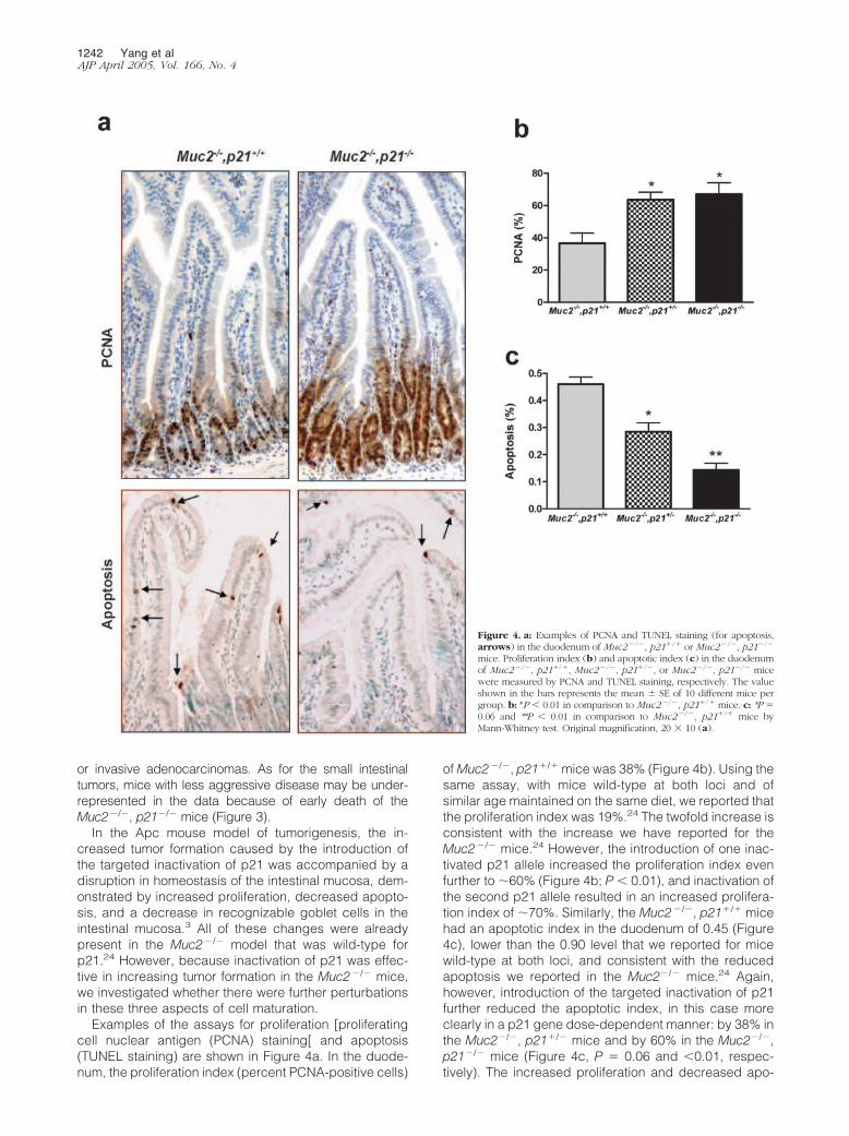
or invasive adenocarcinomas. As for the small intestinaltumors, mice with less aggressive disease may be under-represented in the data because of early death of theMuc2�/�, p21�/� mice (Figure 3).
In the Apc mouse model of tumorigenesis, the in-creased tumor formation caused by the introduction ofthe targeted inactivation of p21 was accompanied by adisruption in homeostasis of the intestinal mucosa, dem-onstrated by increased proliferation, decreased apopto-sis, and a decrease in recognizable goblet cells in theintestinal mucosa.3 All of these changes were alreadypresent in the Muc2�/� model that was wild-type forp21.24 However, because inactivation of p21 was effec-tive in increasing tumor formation in the Muc2�/� mice,we investigated whether there were further perturbationsin these three aspects of cell maturation.
Examples of the assays for proliferation [proliferatingcell nuclear antigen (PCNA) staining[ and apoptosis(TUNEL staining) are shown in Figure 4a. In the duode-num, the proliferation index (percent PCNA-positive cells)
of Muc2�/�, p21�/� mice was 38% (Figure 4b). Using thesame assay, with mice wild-type at both loci and ofsimilar age maintained on the same diet, we reported thatthe proliferation index was 19%.24 The twofold increase isconsistent with the increase we have reported for theMuc2�/� mice.24 However, the introduction of one inac-tivated p21 allele increased the proliferation index evenfurther to �60% (Figure 4b; P � 0.01), and inactivation ofthe second p21 allele resulted in an increased prolifera-tion index of �70%. Similarly, the Muc2�/�, p21�/� micehad an apoptotic index in the duodenum of 0.45 (Figure4c), lower than the 0.90 level that we reported for micewild-type at both loci, and consistent with the reducedapoptosis we reported in the Muc2�/� mice.24 Again,however, introduction of the targeted inactivation of p21further reduced the apoptotic index, in this case moreclearly in a p21 gene dose-dependent manner: by 38% inthe Muc2�/�, p21�/� mice and by 60% in the Muc2�/�,p21�/� mice (Figure 4c, P � 0.06 and �0.01, respec-tively). The increased proliferation and decreased apo-
Figure 4. a: Examples of PCNA and TUNEL staining (for apoptosis,arrows) in the duodenum of Muc2�/�, p21�/� or Muc2�/�, p21�/�
mice. Proliferation index (b) and apoptotic index (c) in the duodenumof Muc2�/�, p21�/�, Muc2�/�, p21�/�, or Muc2�/�, p21�/� micewere measured by PCNA and TUNEL staining, respectively. The valueshown in the bars represents the mean SE of 10 different mice pergroup. b: *P � 0.01 in comparison to Muc2�/�, p21�/� mice. c: *P �0.06 and **P � 0.01 in comparison to Muc2�/�, p21�/� mice byMann-Whitney test. Original magnification, 20 10 (a).
1242 Yang et alAJP April 2005, Vol. 166, No. 4
ptosis were also seen in the flat mucosa of the colonsfrom the Muc2�/�, p21�/� or Muc2�/�, p21�/� micecompared with Muc2�/� littermates wild-type for p21(data not shown). Therefore, changes in proliferation andapoptosis were consistently associated with the in-creased tumor formation because of p21 inactivation.
The most interesting finding regarded differentiation ofthe goblet cell lineage. The intestinal mucosa of theMuc2�/� mouse is characterized by the absence of rec-ognizable goblet cells.24 However, this cell lineage is notcompletely ablated, because cells in the mucosa stillstain immunohistochemically for ITF, another principalsecreted product of goblet cells and component of intes-tinal mucus.27,28 This can be clearly seen in the duode-num and colon of the Muc2�/�, p21�/� mice (Figure 5).However, on homozygous inactivation of the p21 gene,the goblet cell lineage is further perturbed as indicatedby the significant reduction in ITF staining (Figure 5).Thus, the loss of p21 expression in the intestinal mucosaof the Muc2�/� mice, which accelerates and enhancestumor formation, alters cell maturation—increasing cellproliferation, decreasing cell differentiation and apopto-sis—in the intestinal mucosa.
Gene expression was investigated in the mucosa ofthese mice. As expected, the levels of p21 mRNA de-creased in a gene dosage-dependent manner in theMuc2�/�, p21�/� and Muc2�/�, p21�/� mice comparedto the Muc2�/�, p21 wild-type mice (Figure 6a), and thiswas also reflected in the protein levels of p21 (Figure 6d).We have previously shown that the inactivation of p27kip1
in mice maintained on the AIN-76A diet was sufficient to
cause tumor formation (W. Yang, et al, manuscript inpreparation).25 It was therefore of interest that the inacti-vation of p21 in the Muc2�/� mice led to a concurrentreduction in p27 mRNA by 30% (Figure 6b) and proteinexpression by 50% (Figure 6, d and e).
As regards the reduction in p27 expression, it may bespecific for the compound knockout (ie, Muc2�/�,p21�/�) mice, because preliminary data indicated thatthere is no reduction in p27 expression in the Muc2�/�
mice compared to Muc2�/� mice when both are wild-type for p21 (N. Popova and A. Velcich, personal commu-nication), and it has been reported that p27 expression isnot reduced in p21�/� mice compared to wild-typemice.29–31
Finally, as outlined in the Introduction, p21 is under neg-ative control of myc expression in the intestinal mucosa. Wetherefore investigated whether there is a feedback, and thuswhether the decreased p21 expression alters c-myc ex-pression. Figure 6c illustrates that c-myc mRNA levels in-deed rose in the intestinal mucosa of the Muc2�/� mice inconjunction with the targeted inactivation of p21. This in-crease was �60% in the Muc2�/�, p21�/� mice comparedto the Muc2�/�, p21�/� mice (P � 0.05) (Figure 6c). Furtherquantifying Western blot signals demonstrated that c-mycprotein level in Muc2�/�, p21�/� mice was elevated by1.78-fold greater than the Muc2�/�, p21�/� mice (P � 0.05)(Figure 6, d and f).
Discussion
There is considerable evidence that p21WAF1/cip1 plays afundamental role in pathways that regulate intestinal cellmaturation and homeostasis of the intestinal muco-sa.1,3,4,6,32 In the intestinal tract, p21 is expressed ascells exit the proliferative compartment, and loss of bothexpression and topological regulation is detected early incolon tumor formation.1,6 Absence of p21 is linked toinability of colon tumor cells to arrest in the G1 phase ofthe cell cycle,13,33,34 when stimulated by the nonsteroidalanti-inflammatory drug sulindac,4,34,35 or by radiation.36
However, despite this evidence for a key role of p21, itstargeted inactivation does not grossly perturb the mu-cosa and does not cause tumor formation.5
Loss of p21 in mice that have an initiating mutation inApc does cause marked enhancement of tumor forma-tion.3 This report extends this observation to anotherinitiator of intestinal tumorigenesis: the inactivation ofMuc2, the gene that encodes the major intestinal mucin.Inactivation of Muc2 initiates tumor formation by a path-way that is distinct from that of loss of APC function, inthat loss of Muc2 expression does not involve elevation of�-catenin expression or accumulation in the nucleus,both of which are characteristic of APC-initiated tu-mors.24 The difference in mechanism is confirmed bymore recent evidence that combining the Apc and Muc2mutations in the mouse is synergistic in terms of tumorformation initiated by either mutation alone (A. Velcich,manuscript in preparation). Thus, the fact that inactivationof p21 enhances tumor formation in these two different
Figure 5. ITF expression in the duodenum and colon of Muc2�/�, p21�/�
or Muc2�/�, p21�/� mice. Original magnification, 20 10.
p21WAF1 in Muc2 Knockout Mouse 1243AJP April 2005, Vol. 166, No. 4
mouse models underlines the importance of p21 in intes-tinal homeostasis and tumorigenesis.
As in the Apc1638�/� model, the targeted inactivationof p21 causes a disruption in cell maturation pathwaysthat includes further elevation in proliferation, depressedapoptosis, and perturbation of differentiation lineages. Inthe Apc1638�/� model, the perturbation of differentiationby inactivation of p21 was manifest as a decrease in thenumber of mucin-containing goblet cells.3 However, inthe Muc2�/� mouse, this cell phenotype is already notdetectable in the intestinal mucosa, but the loss of p21appears to further perturb development of the lineagebecause it results in decreased expression of ITF, an-other marker of this cell type, which instead persists in theMuc2�/�, p21�/� mice (Figure 5).24 In this regard, it is ofinterest that in the Muc2�/�, p21�/� mice, there is down-regulation of another cdk inhibitor, p27kip1. Although theinactivation of p27 is able to initiate tumor formation inmice maintained on the AIN-76A diet,25 like Muc2�/�
mice, the p27�/� mice still express ITF, albeit assayed atthe mRNA level, while the mucin-expressing gobletcells are reduced.25 Therefore, it may be that in theMuc2�/�, p21�/� mice, the combined inactivation of p21,and down-regulation of p27, has a more extensive affecton the development of the goblet cell lineage than does
inactivation and/or down-regulation of only one of thesecdk inhibitors.
In the intestinal mucosa, tumor initiation by loss offunction of APC, either through mutation, deletion, orepigenetic events, is most often because of the effects ofAPC on �-catenin/Tcf signaling, and indeed, evidencethat altered �-catenin-Tcf signaling is sufficient to initiatetumor formation is very strong.15,18,37–39 One of the di-rect, key targets of �-catenin-Tcf signaling is c-myc. Briefinactivation of c-myc is sufficient to induce a sustainedloss of the transformed phenotype.40 The importance ofc-myc is also supported by the fact that Muc2�/� initiatedtumor formation, although not targeting �-catenin-Tcf sig-naling, still caused elevation of c-myc expression,24 andthat loss of p21, which increases tumor initiation andprogression, further elevates c-myc expression (Figure6). Thus, these data reinforce a key role for c-myc inintestinal tumor formation. This is consistent with our re-cent report that c-myc likely plays a key role in regulatingthe maturation pathway as cells migrate from the prolif-erative compartment toward the lumen in the intestinalmucosa.41
In summary, our data demonstrated that the loss ofp21 in the Muc2�/� mouse model of intestinal tumorformation is linked to further perturbation in cellular mech-
Figure 6. p21, p27 and c-myc expression in the colon of Muc2�/�, p21�/�, Muc2�/�, p21�/�, or Muc2�/�, p21�/� mice. a–c: Relative level of mRNA of p21(a), p27(b), and c-myc (c) in the colon, assayed by quantitative real-time reverse transcriptase-polymerase chain reaction. The value shown in the bars representsthe mean SE of five different mice per group. d: Protein expression of p21, p27, and c-myc, assayed by Western blot. The quantification of p27 (e) and c-myc(f) protein were normalized to �-actin. *P � 0.05 in comparison to Muc2�/�, p21�/� mice by Mann-Whitney test.
1244 Yang et alAJP April 2005, Vol. 166, No. 4
anisms of intestinal homeostasis. This is coincident withalterations of expression of at least two molecules criticalin the maturation of intestinal epithelial cells: down-regu-lation of p27 and up-regulation of c-myc. Each has mul-tiple targets and pathways that it can modulate. Thealterations of these targets and pathways by gene andproteomic profiling of cells as they migrate along thecrypt-villus axis of the intestinal tract in mouse geneticmodels, with and without modulation of tumorigenesis byother loci and environmental factors, will be fundamentalin defining the key events in establishing probability oftumor formation and progression.
Acknowledgments
We thank Dr. Philip Leder for providing the p21�/� miceand Dr. Maomi Li for reading the manuscript.
References
1. Polyak K, Hamilton SR, Vogelstein B, Kinzler KW: Early alteration ofcell-cycle-regulated gene expression in colorectal neoplasia. Am JPathol 1996, 149:381–387
2. Gartel AL, Tyner AL: Transcriptional regulation of the p21((WAF1/CIP1)) gene. Exp Cell Res 1999, 246:280–289
3. Yang WC, Mathew J, Velcich A, Edelmann W, Kucherlapati R, LipkinM, Yang K, Augenlicht LH: Targeted inactivation of the p21(WAF1/cip1) gene enhances Apc-initiated tumor formation and the tumor-promoting activity of a Western-style high-risk diet by altering cellmaturation in the intestinal mucosal. Cancer Res 2001, 61:565–569
4. Yang WC, Velcich A, Mariadason J, Nicholas C, Corner G, HoustonM, Edelmann W, Kucherlapati R, Holt PR, Augenlicht LH: p21(WAF1/cip1) is an important determinant of intestinal cell response to sulin-dac in vitro and in vivo. Cancer Res 2001, 61:6297–6302
5. Deng C, Zhang P, Harper JW, Elledge SJ, Leder P: Mice lackingp21CIP1/WAF1 undergo normal development, but are defective in G1checkpoint control. Cell 1995, 82:675–684
6. el-Deiry WS, Tokino T, Waldman T, Oliner JD, Velculescu VE, BurrellM, Hill DE, Healy E, Rees JL, Hamilton SR: Topological control ofp21WAF1/CIP1 expression in normal and neoplastic tissues. CancerRes 1995, 55:2910–2919
7. Wu S, Cetinkaya C, Munoz-Alonso MJ, von der Lehr N, Bahram F,Beuger V, Eilers M, Leon J, Larsson LG: Myc represses differentia-tion-induced p21CIP1 expression via Miz-1-dependent interactionwith the p21 core promoter. Oncogene 2003, 22:351–360
8. van de Wetering M, Sancho E, Verweij C, de Lau W, Oving I, HurlstoneA, van der Horn K, Batlle E, Coudreuse D, Haramis AP, Tjon-Pon-Fong M, Moerer P, van den Born M, Soete G, Pals S, Eilers M,Medema R, Clevers H: The beta-catenin/TCF-4 complex imposes acrypt progenitor phenotype on colorectal cancer cells. Cell 2002,111:241–250
9. Gartel AL, Shchors K: Mechanisms of c-myc-mediated transcriptionalrepression of growth arrest genes. Exp Cell Res 2003, 283:17–21
10. He TC, Sparks AB, Rago C, Hermeking H, Zawel L, da Costa LT,Morin PJ, Vogelstein B, Kinzler KW: Identification of c-MYC as a targetof the APC pathway. Science 1998, 281:1509–1512
11. Korinek V, Barker N, Morin PJ, van Wichen D, de Weger R, KinzlerKW, Vogelstein B, Clevers H: Constitutive transcriptional activation bya beta-catenin-Tcf complex in APC�/� colon carcinoma. Science1997, 275:1784–1787
12. Mariadason JM, Bordonaro M, Aslam F, Shi L, Kuraguchi M, VelcichA, Augenlicht LH: Down-regulation of beta-catenin TCF signaling islinked to colonic epithelial cell differentiation. Cancer Res 2001,61:3465–3471
13. Seoane J, Le HV, Massague J: Myc suppression of the p21(Cip1)Cdk inhibitor influences the outcome of the p53 response to DNAdamage. Nature 2002, 419:729–734
14. Schneider A, Peukert K, Eilers M, Hanel F: Association of Myc with the
zinc-finger protein Miz-1 defines a novel pathway for gene regulationby Myc. Curr Top Microbiol Immunol 1997, 224:137–146
15. Kolligs FT, Hu G, Dang CV, Fearon ER: Neoplastic transformation ofRK3E by mutant beta-catenin requires deregulation of Tcf/Lef tran-scription but not activation of c-myc expression. Mol Cell Biol 1999,19:5696–5706
16. Chen S, Guttridge DC, You Z, Zhang Z, Fribley A, Mayo MW, Kita-jewski J, Wang CY: Wnt-1 signaling inhibits apoptosis by activatingbeta-catenin/T cell factor-mediated transcription. J Cell Biol 2001,152:87–96
18. Morin PJ, Sparks AB, Korinek V, Barker N, Clevers H, Vogelstein B,Kinzler KW: Activation of beta-catenin-Tcf signaling in colon cancerby mutations in beta-catenin or APC. Science 1997, 275:1787–1790
19. Kim YS, Gum Jr JR: Diversity of mucin genes, structure, function, andexpression. Gastroenterology 1995, 109:999–1001
20. van Klinken BJ, Einerhand AW, Duits LA, Makkink MK, Tytgat KM,Renes IB, Verburg M, Buller HA, Dekker J: Gastrointestinal expres-sion and partial cDNA cloning of murine Muc2. Am J Physiol 1999,276:G115–G124
21. Aslam F, Palumbo L, Augenlicht LH, Velcich A: The Sp family oftranscription factors in the regulation of the human and mouse MUC2gene promoters. Cancer Res 2001, 61:570–576
22. Velcich A, Palumbo L, Selleri L, Evans G, Augenlicht L: Organizationand regulatory aspects of the human intestinal mucin gene (MUC2)locus. J Biol Chem 1997, 272:7968–7976
23. Velcich A, Palumbo L, Jarry A, Laboisse C, Racevskis J, Augenlicht L:Patterns of expression of lineage-specific markers during the in vitro-induced differentiation of HT29 colon carcinoma cells. Cell GrowthDiffer 1995, 6:749–757
24. Velcich A, Yang W, Heyer J, Fragale A, Nicholas C, Viani S, Kucher-lapati R, Lipkin M, Yang K, Augenlicht L: Colorectal cancer in micegenetically deficient in the mucin Muc2. Science 2002, 295:1726–1729
25. Yang WC, Bancroft L, Nicholas C, Lozonschi I, Augenlicht LH: Tar-geted inactivation of p27kip1 is sufficient for large and small intestinaltumorigenesis in the mouse, which can be augmented by a Western-style high-risk diet. Cancer Res 2003, 63:4990–4996
26. Augenlicht LH, Anthony GM, Church TL, Edelmann W, KucherlapatiR, Yang K, Lipkin M, Heerdt BG: Short-chain fatty acid metabolism,apoptosis, and Apc-initiated tumorigenesis in the mouse gastrointes-tinal mucosa. Cancer Res 1999, 59:6005–6009
27. Sands BE, Podolsky DK: The trefoil peptide family. Annu Rev Physiol1996, 58:253–273
29. Albrecht JH, Poon RY, Ahonen CL, Rieland BM, Deng C, Crary GS:Involvement of p21 and p27 in the regulation of CDK activity and cellcycle progression in the regenerating liver. Oncogene 1998,16:2141–2150
30. Kwon YH, Jovanovic A, Serfas MS, Kiyokawa H, Tyner AL: P21functions to maintain quiescence of p27-deficient hepatocytes. J BiolChem 2002, 277:41417–41422
31. Kwon YH, Jovanovic A, Serfas MS, Tyner AL: The Cdk inhibitor p21 isrequired for necrosis, but it inhibits apoptosis following toxin-inducedliver injury. J Biol Chem 2003, 278:30348–30355
32. Mahyar-Roemer M, Roemer K: p21 Waf1/Cip1 can protect humancolon carcinoma cells against p53-dependent and p53-independentapoptosis induced by natural chemopreventive and therapeuticagents. Oncogene 2001, 20:3387–3398
33. Archer S, Meng S, Wu J, Johnson J, Tang R, Hodin R: Butyrateinhibits colon carcinoma cell growth through two distinct pathways.Surgery 1998, 124:248–253
34. Goldberg Y, Nassif II, Pittas A, Tsai LL, Dynlacht BD, Rigas B, ShiffSJ: The anti-proliferative effect of sulindac and sulindac sulfide onHT-29 colon cancer cells: alterations in tumor suppressor and cellcycle-regulatory proteins. Oncogene 1996, 12:893–901
35. Augenlicht LH, Mariadason JM, Wilson A, Arango D, Yang WC,Heerdt BG, Velcich A: Short chain fatty acids and colon cancer. JNutr 2002, 132:3804S–3808S
p21WAF1 in Muc2 Knockout Mouse 1245AJP April 2005, Vol. 166, No. 4
36. McDonald III ER, Wu GS, Waldman T, El-Deiry WS: Repair defect inp21 WAF1/CIP1 �/� human cancer cells. Cancer Res 1996,56:2250–2255
37. Harada N, Tamai Y, Ishikawa T, Sauer B, Takaku K, Oshima M, TaketoMM: Intestinal polyposis in mice with a dominant stable mutation ofthe beta-catenin gene. EMBO J 1999, 18:5931–5942
38. Morin PJ: Beta-catenin signaling and cancer. Bioessays 1999,21:1021–1030
39. Munemitsu S, Albert I, Rubinfeld B, Polakis P: Deletion of an amino-terminal sequence beta-catenin in vivo and promotes hyperphos-
porylation of the adenomatous polyposis coli tumor suppressor pro-tein. Mol Cell Biol 1996, 16:4088–4094
40. Jain M, Arvanitis C, Chu K, Dewey W, Leonhardt E, Trinh M, SundbergCD, Bishop JM, Felsher DW: Sustained loss of a neoplastic pheno-type by brief inactivation of MYC. Science 2002, 297:102–104
41. Mariadason J, Nicholas C, L’Italien K, Zhuang M, Smartt H, HeerdtBG, Yang WC, Corner C, Wilson AJ, Klampfer L, Arango D, Augen-licht LH: The transcriptional reprogramming that defines intestinalepithelial cell maturation along the crypt-villus axis. Gastroenterology,2005 (in press)