Incomplete branchial arch syndromes, branchialcleft cyst and vascular hamartoma in apatient with multiple neurofibromatosis
HALLVARD VINDENES, KJELL SVEEN AND RUNE NILSEN
Department of Oral Surgery, Department of Plastic and Recollstructive Surgery, andDepartment of Oral Pathology and Forensic Odontology, University of Bergen,Bergen, Norway
ABSTIti\CT - A case report with simultaneous occurrence of neurofibromatosis, incomplete branchial arch syndromes, a branchial cleft cystand a pseudocyst in connection with a vascular hamartoma anterior tothe right ear of a 38-year-old woman is presented. A possible commonpathogenesis of the vascular hamartoma and the incomplete branchialarch syndromes as well as that of the neurofibromatosis is suggested.The pr.eudocyst is interpreted as a branchial cleft cyst showing inflammatory changes due to a pharyngitis shortly before the preauricular tumor appeared.
(Received for publication 11 May, accepted 6 June 1978)
Neurofibromatosis was first described byVON RECKLINGHAUSEN14 as a disease characterized by fibrous tumors of the skinand peripheral nerves together with cutaneous pigmentations (cafe au lait spots). Thedisease is complex and may involve any ofthe oral structures1,2,O,13,10,10-21, in combinations with classical systemic manifestations.Involvement of the facial bones is not uncommon. The disease is inherited as anautosomal dominant trait with low penetrance, without sex or racial preferences2.
The first and second branchial arch syndromes include defects which are usually,but not invariably, LlnilateraP2. Underdevelopment of the mandibular condyle andramus, the zygomatical arch and the malar
bone are often found. The masticatorymuscles and those of facial expression mayalso be affected, but in a large proportionof cases the manifestations of the syndrome are incompletel2.
The branchial cleft cyst (lateral cervicalcyst) is characterized as a painless circumscribed soft fluctuant and movable swelling.The size may, however, occasionally vary ifthe cyst is associated with a regional inflammatory process such as dental or upper repiratory tract infections3,5. The cysts areusually located in the lateral aspects of theneck or at the angle of the mandible5, inthe preauricular region3,17, in the floor ofthe mouth as well as within the parotidglancl I7,18,22. The branchial cleft cyst may
be present together with branchial archsyndromeS12•
Case reportA 38-year-old woman was referred to the Department of Oral Surgery for a tumor anteriorto the right ear. She bad been aware of theswelling for several months and sought medicalhelp because of its rapidly increasing size.Aspiration of the tumor performed by herlocal physician was negative. The lesion wasthought to be of dental origin though the patient had suffered from pharyngitis shortlybefore the tumor appeared.
At the age of 11 the patient had acute anterior poliomyelitis. Her medical records revealed changes of her facial expression on theright side interpreted as a sequela of a slightparalysis of the facial nerve. The right side ofher face was reported to be slightly enlarged.She exhibited a pronounced kyphoscoliosis.Her hearing had been slightly reduced on herright ear and she had used glasses since shewas 12. The family history disclosed that herfather had small nodular tumors in the skin
~"",,--:::::._-g,,,,"
Fig. 1. Photo of the patient showing swellingof the right cheek. Note the large ear lobe(arrow) and the enlarged right half of her face.
Fig. 2. Two fibromatous tumors and a large"cafe au lait" spot on the right arm of thepatient.
together with brown pigmented areas identicalto her own. The patient was a single child. Nofurther information about her family was available.
The clinical examination showed a soft, relatively mobile swelling with a diameter of approximately 5 em in her right cheek anteliorto the ear. The right half of her face was significantly larger than the left side and the rightear was slightly deformed, having a large lobe.Small fibromatous tumors were scattered overthe face and neck. On ber body and extremitiesextensive areas of pigmentation (cafe au laitspots) as well as scattered fibromas measuring0.5-1 em in diameter were seen (Fig. 2).
Intraorally there was a swelling in the rightcheek. The vestibulum in the right mandibularmolar region was obliterated. The teeth wereneglected and the oral hygiene poor. Themajor salivary glands all secreted abundantclear saliva on stimulation. There was no trismus but on opening the mouth a pronouncedmandibular deviation to the right was observed. The localization of the tumor did notindicate a dental pathogenesis.
The orthopantomogram showed reduced vertical height of the right mandibular body andthe condylar head was one third the size ofthat on the contralateral side. The condylarneck was slender and dorsally there was a sharpdemarcation. In the region of her right mandibular angle a notch-like deformity was seen,forming an obtuse angle. Occipito-mentalradiographs demonstrated a marked reductionof the malar bone. The zygomatic arch wasslender and showed an abrupt angie towardsthe temporal bone. The right masseter muscle
NEUROFIBROMATOSIS AND OTHER ANOMALIES 377
Fig. 3. Orthopantomogram showing hypoplastic right mandibular body, condylar head and muscular process, and a deep mandibular notch. Note the spherical picture of the: supraimposedradiopaque tumor (arrows) and notch-like deformity at the right mandibular angle.
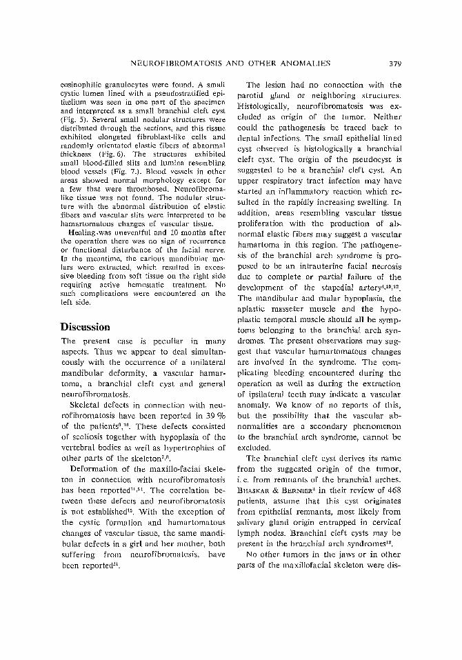
Fig. 4. Histological section showing a cystic lumen without epithelial delineation (up). Severalareas (arrow and arrowhead with inset) exhibiting abnormal elastic fibers in nodular-like areas.Inset is shown in Fig. 6. Elastin, van Gieson, X 23.
378 VINDENES, SVEEN AND NILSEN
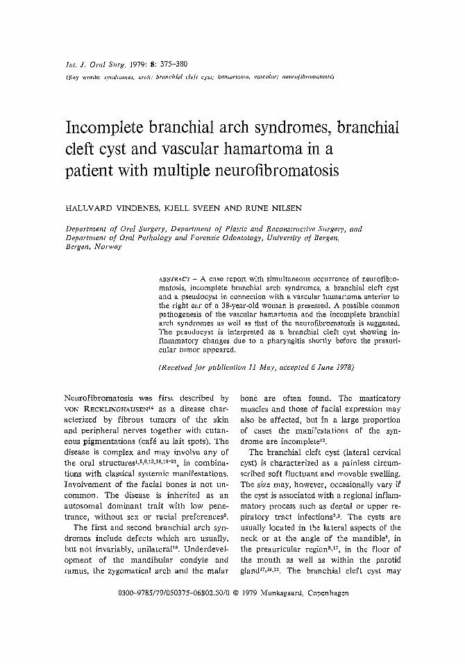
Fig. 5. Branchial cleft cyst with pseudostratifiedciliated epithelium. H & E, X 75.
appeared hypoplastic. Radiographic examination of the vertebral column revealed severekyphoscoliosis. Blood analyses were within
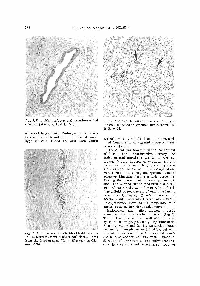
Fig. 6. Nodular areas with fibroblast-like cellsand randomly oriented abnormal elastic fibersfrom the inset area of Fig. 4. Elastin, van Gieson, X 96.
Fig. 7. Micrograph from similar area as Fig. 6showing blood-filled vascular slits (arrows). H.& E, X 96.
normal limits. A blood-stained fluid was aspirated from the tumor containing predominantly macrophages.
The patient was admitted to the Depatimentof Plastic and Reconstructive Surgery anduncler general anesthesia the tumor was extirpated in toto through an extraoral, slightlycurvecl incision 5 cm in length, starting abollt3 cm anterior to the ear lobe. Complicationswere encountered during the operation clue toexcessive bleeding from the soft tissue, indicating the presence of a capillary hemangioma. The excised tumor measured 5 X 3 X 2cm, and contained a cystic lumen with a bloodtinged fluid. A postoperative hematoma had tobe evacuated. However, Duke's test was withinnormal limits. Antibiotics were administered.Postoperatively there was a temporary mildpartial palsy of her right facial nerve.
Histological examination showed a cysticlumen without any epithelial lining (Fig. 4).The thick connective tissue wall was infiltratedby many macrophages and young fibroblasts.Bleeding was found in the connective tissue,and many macrophages contained hemosiderin.Lateral to this zone, dilated thin-walled vesselsand a loose connective tissue with a slight infiltration of lymphocytes and polymorphonuclear leukocytes as well as scattered groups of
NEUROFIBROMATOSIS AND OTHER ANOMALIES 379
eosinophilic granulocytes were found. A smallcystic lumen lined with a pseudostratified epithelium was seen in one part of the specimenand interpreted as a small branchial cleft cyst(Fig. 5). Several small nodular stnlctures weredistributed through the sections, and this tissueexhibited elongated fibroblast-like cells andrandomly orientated elastic fibers of abnormalthickness (Fig. 6). The structures exhibitedsmall blood-filled slits and lumina resemblingblood vessels (Fig. 7.). Blood vessels in otherareas showed normal morphology except fora few that were thrombosed. Neurofibromalike tissue was not found. The nodular structure with the abnormal distribution of elasticfibers and vascular slits were interpreted to behamartomatous changes of vascular tissue.
Healing.was uneventful and 10 months afterthe operation there was no sign of recurrenceor functional disturbance of the facial nerve.In the meantime, the carious mandibular molars were extracted, which resulted in excessive bleeding from soft tissue on the right siderequiring active hemostatic treatment. Nosuch complications were encountered on theleft side.
DiscussionThe present case is peculiar in manyaspects. Thus we appear to deal simultaneously with the occurrence of a unilateralmandibular deformity, a vascular hamartoma, a branchial cleft cyst and generalneurofibromatosis.
Skeletal defects in connection with neurofibromatosis have been reported in 39 %of the patients!l,!!). These defects consistedof scoliosis together with hypoplasia of thevertebral bodies as well as hypertrophies ofother parts of the skeleton7,~.
Deformation of the maxillo-facial skeleton in connection with neurofibromatosishas been reported!!,'!. The correlation between these defects and neurofibromatosisis not established's. With the exception ofthe cystic formation and hamartomatouschanges of vascular tissue, the same mandibular defects in a girl and her mother, bothsuffering from neurofibromatosis, havebeen reportec]2l.
The lesion had no connection with theparotid gland or neighboring structures.Histologically, neurofibromatosis was excluded as origin of the tumor. Neithercould the pathogenesis be traced back todental infections. The small epithelial lin edcyst observed is histologically a branchialcleft cyst. The origin of the pseudocyst issuggested to be a branchial cleft cyst. Anupper respiratory tract infection may havestarted an inflammatory reaction which resulted in the rapidly increasing swe.\ling. Inaddition, areas resembling vascular tissueproliferation with the production of abnormal elastic fibers may suggest a vascularhamartoma in this region. The pathogenesis of the branchial arch syndrome is proposed to be an intrauterine facial necrosisdue to complete or partial failure of thedevelopment of the stapedial artery4,lO,12.The mandibular and malar hypoplasia, theaplastic masseter muscle and the hypoplastic temporal muscle should all be symptoms belonging to the branchial arch syndromes. The present observations may suggest that vascular hamartomatous changesare involved in the syndrome. The complicating bleeding encountered during theoperation as well as during the extractionof ipsilateral teeth may indicate a vascularanomaly. We know of no reports of this,but the possibility that the vascular abnormalities are a secondary phenomenonto the branchial arch syndrome, cannot beexcluded.
The branchial cleft cyst derives its namefrom the suggested origin of the tumor,i. e. from remnants of the branchial arches.BHASKAR & BERNfER3 in their review of 468patients, assume that this cyst originatesfrom epithelial remnants, most likely fromsalivary gland origin entrapped in cervicallymph nodes. Branchial cleft cysts may bepresent in the branchial arch syndromesl2,
No other tumors in the jaws or in otherparts of the maxillofacial skeleton were dis-
380 VINDENES, SVEEN AND NILSEN
covered from the clinical and radiographicexamination.
We were not able to observe connectionsbetween the maxillofacial defects and theneurofibromatosis. The relatively low-gradepenetration of the neurofibromatosis together with the incomplete branchial archsyndrome may thus suggest a commonpathogenesis. Teratogenic, infectious ortoxic theories may be considered. However,there is no evidence in the literature ofgenetic predisposition of the arch syndrome.
Acknowledgments - The authors wish to thankthe head of Department of Plastic and Reconstructive Surgery, Dr. H. SCHlELDRUP forpermission to publish the case and for his helpand encouragement. We will also thank Professor O. GILHUUS-MoE and Professor G. BANGfor valuable advice and constructive criticism.
References1. Au.ALoUF, J.: Contribution a !'etude de let
neurofibromatose generalisee. Thesis, Faculty of Medicine. Samie fils Freres, Bordeaux 1920.
2. BomEN, E., PIERcE, H. E. & JACKSON, W.F.: Multiple neurofibromatosis with orallesions. Review of the literature and reportof a case. Oral Surg. 1955: 8: 263-280.
3. BHASKAR, S. N. & BERNlER, J. F.: Histogenesis of branchial cysts. Am. J. Pathol.1959: 35: 407-423.
4. BRAITIlWAITE, F. & WATSON, J.: A reporton three unusual cleft lips. Br. J. Plast.Surg. 1949: 2: 38-49.
5. GAISFORD, J. C. & ANDERSON, V. S.: Firstbranchial cleft cysts and sinuses. PIast.Recollstr. Surg. 1975: 55: 299-304.
6. GEMERT, R. J. VAN, YAMASHITA, D.-D. R.& GOODSELL, J. F.: Multiple neurofibromatosis (von Recklinghausen's disease) withconcurrent micrognathia. Review of theliterature and report of a case. Oral Surg.1977: 43: 165-173.
7. GOULD, E. P.: Bone changes occurring invon Recklinghausen's disease. O. J. Med.1917-18: 11: 21-32.
8. HOLT, J. F. & WlUGfIT, E. M.: The radio-
logic features of neurofibromatosis. Ra·diology 1948: 51: 647-663.
9. HUNT, 1. C. & PUGH, D. J.: Skeletal lesionsin neurofibromatosis. Radiology 1961: 76:1-20.
10. KEITH, A.: Three demonstrations of congenital malformations of palate, face andneck. Br. J. Dent. Sci. 1909: 7: 865, 913.
11. LoRSON, E., DE LONG, P. E., OSBON, D. B.& DOLAND, K. D.: Neurofibromatosis withcentral neurofibroma of the mandible: Review of the literature and report of case.J. Oral. Surg. 1977: 35: 733-738.
12. POSSWILl.:O, D.: The pathogenesis of thefirst and second branchial arch syndrome.Oral Surg. 1973: 35: 302-328.
13. RApPAPORT, H. M.: Neurofibromatosis ofthe oral cavity. Report of a case. Oral Surg.1953: 6: 599-604.
14. RECKLINOHAUSEN, F. VON: tiber die multiplen Fibroma del' Haut und ihre Beziehungzu den multiplen Neuromen, A. Hirschwald, Berlin 1882.
15. Rnn!RSMA, 1., TEN KATE, L. B. & WESTERINK, P.: Neurofibromatosis with mandibulardeformities. Oral Surg. 1972: 33: 718-727.
16. ROSEDALE, R. S.: Massive fibroma of themaxillary antrum as part of multiple neuro~
fibromatosis in sibling. Arch. Otolaryngol.1945: 42: 208-211.
17. SHAFER, W. G., HINE, M. K. & LEVY, B.M.: A textbook of oral pathology. W. B.Saunders, Philadelphia 1974, pp. 72-74.
18. STEWARTS, S., LEVY, R., KARPEL, J. &S'J100PACK, J.: Lymphoepithelial (branchial)cyst of the parotid gland. J. Oral Surg.1974: 32: 100-106.
19. THURSFIELD, J. H.: Macroglossia neurofibromatosa. Path, Soc. London. Lancet1902: ii, 1126-1128.