2
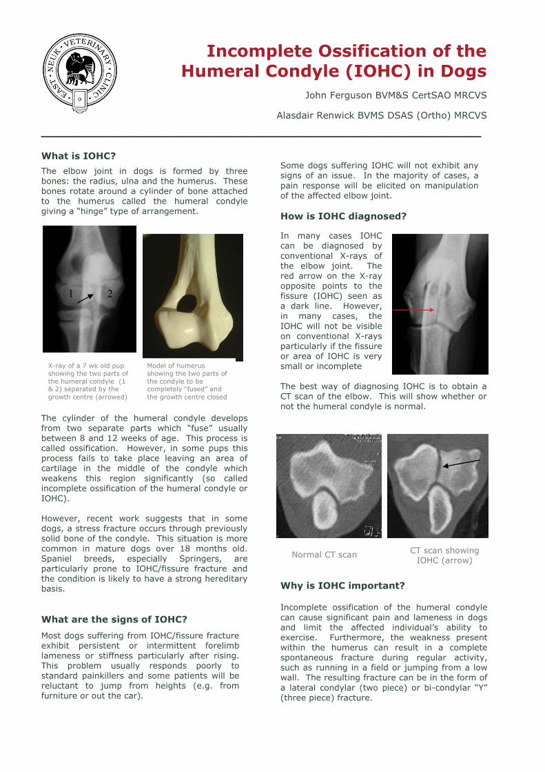
Incomplete Ossification of the Humeral Condyle (IOHC) in Dogs John Ferguson BVM&S CertSAO MRCVS Alasdair Renwick BVMS DSAS (Ortho) MRCVS ____________________________________________________________ What is IOHC? The elbow joint in dogs is formed by three bones: the radius, ulna and the humerus. These bones rotate around a cylinder of bone attached to the humerus called the humeral condyle giving a “hinge” type of arrangement. The cylinder of the humeral condyle develops from two separate parts which “fuse” usually between 8 and 12 weeks of age. This process is called ossification. However, in some pups this process fails to take place leaving an area of cartilage in the middle of the condyle which weakens this region significantly (so called incomplete ossification of the humeral condyle or IOHC). However, recent work suggests that in some dogs, a stress fracture occurs through previously solid bone of the condyle. This situation is more common in mature dogs over 18 months old. Spaniel breeds, especially Springers, are particularly prone to IOHC/fissure fracture and the condition is likely to have a strong hereditary basis. What are the signs of IOHC? Most dogs suffering from IOHC/fissure fracture exhibit persistent or intermittent forelimb lameness or stiffness particularly after rising. This problem usually responds poorly to standard painkillers and some patients will be reluctant to jump from heights (e.g. from furniture or out the car). Some dogs suffering IOHC will not exhibit any signs of an issue. In the majority of cases, a pain response will be elicited on manipulation of the affected elbow joint. How is IOHC diagnosed? In many cases IOHC can be diagnosed by conventional X-rays of the elbow joint. The red arrow on the X-ray opposite points to the fissure (IOHC) seen as a dark line. However, in many cases, the IOHC will not be visible on conventional X-rays particularly if the fissure or area of IOHC is very small or incomplete The best way of diagnosing IOHC is to obtain a CT scan of the elbow. This will show whether or not the humeral condyle is normal. Why is IOHC important? Incomplete ossification of the humeral condyle can cause significant pain and lameness in dogs and limit the affected individual’s ability to exercise. Furthermore, the weakness present within the humerus can result in a complete spontaneous fracture during regular activity, such as running in a field or jumping from a low wall. The resulting fracture can be in the form of a lateral condylar (two piece) or bi-condylar “Y” (three piece) fracture. CT scan showing IOHC (arrow) Normal CT scan X-ray of a 7 wk old pup showing the two parts of the humeral condyle (1 & 2) separated by the growth centre (arrowed) Model of humerus showing the two parts of the condyle to be completely “fused” and the growth centre closed 1 2


![Transcriptional Network Controlling Endochondral Ossification · branous ossification and endochondral ossification.[1] During intramembranous ossification, osteoblasts produce type](https://static.documents.pub/doc/80x56/5e8cf0c24763783dcf0d78ef/transcriptional-network-controlling-endochondral-ossification-branous-ossification.jpg)