Increase in serum 25-hydroxyvitamin-D 3 in humans after sunbed exposures compared to previtamin D 3 synthesis in vitro Tetiana Orlova a,⇑ , Johan Moan b,c , Zoya Lagunova b , Lage Aksnes d,e , Irina Terenetskaya a , Asta Juzeniene b a Department of Optical Quantum Electronics, Institute of Physics, National Academy of Sciences of Ukraine, 03680 Kiev, Ukraine b Department of Radiation Biology, Institute for Cancer Research, Oslo University Hospital, Montebello, N-0310 Oslo, Norway c Institute of Physics, University of Oslo, Blindern, 0316 Oslo, Norway d Department of Pediatrics, Haukeland University Hospital, 5021 Bergen, Norway e Department of Clinical Medicine, Section of Pediatrics, University of Bergen, N-5020 Bergen, Norway article info Article history: Received 24 December 2012 Received in revised form 12 March 2013 Accepted 18 March 2013 Available online 28 March 2013 Keywords: UV biodosimeter Vitamin D status 25(OH)D level Sunbed UV exposure Previtamin D photosynthesis abstract Ultraviolet (UV) radiation is liable to cause skin cancer but it is the main source of vitamin D. Vitamin D pho- tosynthesis takes place in skin at sub-erythemogenic UV doses, while larger exposures destroy vitamin D and increase DNA damage. Proper UV dosimetry is needed to obtain an optimal vitamin D status when skin cancer risk is minimal. A simple approach to such dosimetry using physically measured accumulated UV dose cannot provide a satisfactory quantification of vitamin D because of the complexity of the processes involved in vitamin D synthesis. A biological dosimeter of vitamin D synthetic UV radiation (‘D-dosimeter’) has been introduced earlier on the basis of an in vitro model of previtamin D photosynthesis. In the present study in vivo generation of 25-hydroxyvitamin D (25(OH)D) in serum of healthy volunteers exposed to UV radiation from the sunbed was accompanied by in vitro measurements of vitamin D formation using ‘D- dosimeter’. It was found that the increase in serum 25(OH)D concentration depended both on the initial 25(OH)D level and on the cumulative sunbed exposure time. The observed linear correlation between in vivo and in vitro data can be used to estimate changes in vitamin D status after UV exposure using only one pre-exposure blood sample combined with further in vitro measurements. Ó 2013 Elsevier B.V. All rights reserved. 1. Introduction Numerous studies of the vitamin D role for human health, undertaken during last two decades, indicate that vitamin D is more important for optimal health than previously assumed [1,2]. A sufficient vitamin D level is important for lowering the risk of different internal cancers, multiple sclerosis, diabetes types 1 and 2 together with the well-studied essential role for mineraliza- tion and maintenance of healthy skeleton and healthy bones [3–7]. Ultraviolet B (UVB) radiation (280–315 nm) converts 7-dehy- drocholesterol (7-DHC) into previtamin D which further isomerizes to vitamin D 1 by heat [8]. Afterwards vitamin D is metabolized to 25-hydroxyvitamin D (25(OH)D) in liver and in several other tissues [9]. Being the major circulating form of vitamin D in blood, 25(OH)D is metabolized to its active form 1,25-dihydroxyvitamin D (1,25(OH) 2 D) in kidneys and many nonrenal tissues, including bone, placenta, prostate, keratinocytes, etc. [9]. As steroid hormone, 1,25(OH) 2 D regulates calcium metabolism and bone health, a wide variety of genes in more than 30 different tissues, including brain, liver, kidney, prostate, and have important function in regulating cell growth, modulating immune system and cardiovascular health [2,7]. Selective sensitivity of vitamin D synthesis to the UVB part of solar spectrum makes almost impossible to give recommendations for effective solar exposure times because of daily, seasonal and latitudinal variability in the UVB intensity caused by changes in the ozone layer thickness, clouds, aerosols and air pollutions as well as personal sensitivity [10–12]. Insufficiency in solar UVB irradiation can lead to vitamin D deficiency in humans and thus create or increase already existing health problems. On the other hand, indoor tanning became popular procedure, especially in high-latitude countries. Most of tanning devices have fluorescent lamps with erythemal-effective radiant exposure H er (in the EU by law: H er = 0.3 W/m 2 ) close to that in natural sunlight, but the ratio between UVA (315–400 nm) and UVB irradiances of these lamps is very different from the ratio in the midday summer sun [13]. Nevertheless, sunbed use may lead not only to cosmetic effects (tanning), but can also increase the human vitamin D level [14–16]. Thus the dosimetry of UV radiation is needed not only to 1011-1344/$ - see front matter Ó 2013 Elsevier B.V. All rights reserved. http://dx.doi.org/10.1016/j.jphotobiol.2013.03.006 ⇑ Corresponding author. Tel.: +380 44 5250813; fax: +380 44 5251589. E-mail address: [email protected](T. Orlova). 1 Vitamin D is represented by cholecalciferol (vitamin D 3 ) and ergocalciferol (vitamin D 2 ) that are structurally similar secosteroids produced by the action of sunlight on 7-dehydrocholesterol (provitamin D 3 ) in mammalian skin or on ergos- terol (provitamin D 2 ) in plants, fungi and yeasts correspondingly. The structure differs only in the C-17 side-chain which in vitamin D 2 has a double-bond and an additional methyl group. Journal of Photochemistry and Photobiology B: Biology 122 (2013) 32–36 Contents lists available at SciVerse ScienceDirect Journal of Photochemistry and Photobiology B: Biology journal homepage: www.elsevier.com/locate/jphotobiol
Transcript
Journal of Photochemistry and Photobiology B: Biology 122 (2013) 32–36
Contents lists available at SciVerse ScienceDirect
Journal of Photochemistry and Photobiology B: Biology
Increase in serum 25-hydroxyvitamin-D3 in humans after sunbed exposurescompared to previtamin D3 synthesis in vitro
Tetiana Orlova a,⇑, Johan Moan b,c, Zoya Lagunova b, Lage Aksnes d,e, Irina Terenetskaya a, Asta Juzeniene b
a Department of Optical Quantum Electronics, Institute of Physics, National Academy of Sciences of Ukraine, 03680 Kiev, Ukraineb Department of Radiation Biology, Institute for Cancer Research, Oslo University Hospital, Montebello, N-0310 Oslo, Norwayc Institute of Physics, University of Oslo, Blindern, 0316 Oslo, Norwayd Department of Pediatrics, Haukeland University Hospital, 5021 Bergen, Norwaye Department of Clinical Medicine, Section of Pediatrics, University of Bergen, N-5020 Bergen, Norway
a r t i c l e i n f o
Article history:Received 24 December 2012Received in revised form 12 March 2013Accepted 18 March 2013Available online 28 March 2013
Keywords:UV biodosimeterVitamin D status25(OH)D levelSunbedUV exposurePrevitamin D photosynthesis
1011-1344/$ - see front matter � 2013 Elsevier B.V. Ahttp://dx.doi.org/10.1016/j.jphotobiol.2013.03.006
1 Vitamin D is represented by cholecalciferol (vi(vitamin D2) that are structurally similar secosteroidsunlight on 7-dehydrocholesterol (provitamin D3) in mterol (provitamin D2) in plants, fungi and yeasts corresponly in the C-17 side-chain which in vitamin D2 has a dmethyl group.
a b s t r a c t
Ultraviolet (UV) radiation is liable to cause skin cancer but it is the main source of vitamin D. Vitamin D pho-tosynthesis takes place in skin at sub-erythemogenic UV doses, while larger exposures destroy vitamin Dand increase DNA damage. Proper UV dosimetry is needed to obtain an optimal vitamin D status when skincancer risk is minimal. A simple approach to such dosimetry using physically measured accumulated UVdose cannot provide a satisfactory quantification of vitamin D because of the complexity of the processesinvolved in vitamin D synthesis. A biological dosimeter of vitamin D synthetic UV radiation (‘D-dosimeter’)has been introduced earlier on the basis of an in vitro model of previtamin D photosynthesis. In the presentstudy in vivo generation of 25-hydroxyvitamin D (25(OH)D) in serum of healthy volunteers exposed to UVradiation from the sunbed was accompanied by in vitro measurements of vitamin D formation using ‘D-dosimeter’. It was found that the increase in serum 25(OH)D concentration depended both on the initial25(OH)D level and on the cumulative sunbed exposure time. The observed linear correlation betweenin vivo and in vitro data can be used to estimate changes in vitamin D status after UV exposure using onlyone pre-exposure blood sample combined with further in vitro measurements.
� 2013 Elsevier B.V. All rights reserved.
1. Introduction
Numerous studies of the vitamin D role for human health,undertaken during last two decades, indicate that vitamin D ismore important for optimal health than previously assumed[1,2]. A sufficient vitamin D level is important for lowering the riskof different internal cancers, multiple sclerosis, diabetes types 1and 2 together with the well-studied essential role for mineraliza-tion and maintenance of healthy skeleton and healthy bones [3–7].
Ultraviolet B (UVB) radiation (280–315 nm) converts 7-dehy-drocholesterol (7-DHC) into previtamin D which further isomerizesto vitamin D1 by heat [8]. Afterwards vitamin D is metabolized to25-hydroxyvitamin D (25(OH)D) in liver and in several other tissues[9]. Being the major circulating form of vitamin D in blood, 25(OH)Dis metabolized to its active form 1,25-dihydroxyvitamin D
ll rights reserved.
: +380 44 5251589.
tamin D3) and ergocalciferols produced by the action of
ammalian skin or on ergos-ondingly. The structure differsouble-bond and an additional
(1,25(OH)2D) in kidneys and many nonrenal tissues, including bone,placenta, prostate, keratinocytes, etc. [9]. As steroid hormone,1,25(OH)2D regulates calcium metabolism and bone health, a widevariety of genes in more than 30 different tissues, including brain,liver, kidney, prostate, and have important function in regulating cellgrowth, modulating immune system and cardiovascular health [2,7].
Selective sensitivity of vitamin D synthesis to the UVB part ofsolar spectrum makes almost impossible to give recommendationsfor effective solar exposure times because of daily, seasonal andlatitudinal variability in the UVB intensity caused by changes inthe ozone layer thickness, clouds, aerosols and air pollutions aswell as personal sensitivity [10–12]. Insufficiency in solar UVBirradiation can lead to vitamin D deficiency in humans and thuscreate or increase already existing health problems.
On the other hand, indoor tanning became popular procedure,especially in high-latitude countries. Most of tanning devices havefluorescent lamps with erythemal-effective radiant exposure Her
(in the EU by law: Her = 0.3 W/m2) close to that in natural sunlight,but the ratio between UVA (315–400 nm) and UVB irradiances ofthese lamps is very different from the ratio in the midday summersun [13]. Nevertheless, sunbed use may lead not only to cosmeticeffects (tanning), but can also increase the human vitamin D level[14–16]. Thus the dosimetry of UV radiation is needed not only to
Fig. 1. The UV irradiance spectra from top (1) and bottom (2) sunbed parts andsolar spectrum in Oslo (3) combined to CIE action spectra of erythema (4) andprevitamin D synthesis in vivo (5) and with action spectrum of previtamin Dformation in vitro (6).
T. Orlova et al. / Journal of Photochemistry and Photobiology B: Biology 122 (2013) 32–36 33
avoid the harmful effects of UV radiation, such as sunburn, photo-aging, and skin cancer, but because of the positive effect of UVradiation to synthesize vitamin D.
The estimation of harmful UV levels is traditionally carried outby a variety of commonly used broadband UV detectors whichhave an output in terms of sunburn units. Because of the complex-ity of the processes involved in vitamin D synthesis, a dosimetryusing measured accumulated UV dose will not provide a satisfac-tory quantification of vitamin D synthesis, especially in view ofsignificant difference between the CIE erythema and ‘Vitamin Dsynthesis’ action spectra (Fig. 1) [12,17,18].2
With the aim to provide correct measurement of the vitamin Dsynthetic capacity of different UV sources, the original ‘D-dosime-ter’ has been developed [20–22]. This method is based on UVexposing of 7-DHC solution, recording UV absorption spectra andfurther spectrophotometric analysis of formed vitamin D photo-isomer mixture, i.e. on the first photochemical stage of vitamin Dsynthesis in vitro. As a result, the quantity of previtamin D (directvitamin D precursor) is obtained which is the biologic measure ofaccepted ‘antirachitic’ UV dose. Earlier laboratory and field testshave revealed that the ‘D-dosimeter’ is useful for in situ measure-ments of the vitamin D synthetic capacity of different UV sources[12,22–24]. Furthermore, thin film UV sensors (polymeric andliquid–crystalline) based on provitamin D photoconversions havebeen developed to avoid the inconvenience of liquid solvent useand to simplify the read-out [25,26].
The main goal of the present study is to perform direct mea-surements of the vitamin D level in blood of healthy volunteersexposed to artificial UV source (sunbed), in parallel with measure-ments of vitamin D generation in vitro using ‘D-dosimeter’. Then,such study can provide a missing link between in vivo andin vitro measurements and will be useful for obtainment of ade-quate vitamin D status by sunbed exposure.
2. Materials and methods
2.1. UV source
The UV source was a commercially available and approved sun-bed Wolff Suveren 53IG equipped with 2 types of fluorescentlamps, ‘Brun og blid’ 100W and ‘Suveren S’ 25W spaghetti tubes
2 Nevertheless, it was shown that the individual UV-erythemal sensitivity was agood marker of the individual efficiency of the resulting 25(OH)D3 in blood serumafter solar or solar-simulated UV exposure [19]. Besides, for several sunbedscorrelation was found between exposures determined on the basis of erythema doseand increase of 25(OH)D3 in serum [14–16].
3 The bottom of the sunbed contains only ‘Brun og blid’ 100W fluorescent lampswhereas the top part includes both ‘Brun og blid’ 100W and ‘Suveren S’ 25W tubes.
(Wolff System, Basel, Switzerland). The UV radiation spectra of thesunbed were measured using portable spectrometer Avantes Ava-Spec-2048x14 Fiber Optic Spectrometers. The erythema weightedirradiance was calculated and exposure time for 1 standard ery-thema dose (1 SED, or 100 J/m2) was determined.
2.2. In vitro estimation of previtamin D accumulation using ‘D-dosimeter’
As was mentioned, UV irradiation of 7-DHC leads to formationof previtamin D which further is thermally converted into vitaminD. In this manner the amount of previtamin D accumulated duringUV exposure is a measure of the biologically active ‘antirachitic’ UVdose.
However, previtamin D absorbs at the same spectral region asprovitamin D and thus undergoes a number of side photoconver-sions [27]. An important point is that the photoisomers mixturecomposition (the ratio between previtamin D and the side-prod-ucts concentrations) strongly depends on the UV irradiation wave-length [21,27].
With the aim to measure the concentration of formed previta-min D in vitro, solutions of 7DHC in ethanol (C = (2.5 ± 0.3) � 10�5 -mol/L) were placed in a rectangular quartz cuvettes (d = 1 cm), andfurther cuvettes were exposed inside the sunbed from two sides byupper and lower lamps. The UV absorption spectra were recordedbefore and after several exposures with a Perkin–Elmer Lambda40 UV/VIS spectrophotometer (Norwalk, CT, USA). At last, the pre-vitamin D concentration was determined from the recorded UVspectra using specially designed PC software [21,22].
2.3. In vivo study of 25(OH)D level in healthy volunteers
Thirty-two healthy volunteers living in Oslo (59�N) haveparticipated in the study which was approved by Regional EthicalCommittee, and each participant gave informed consent.
All the participants filled out the questionnaire with pointsabout age, weigh, height and skin type. Age of volunteers rangedfrom 21 to 61 years, and the average age was 31.3 years. All theparticipants had normal weight with body mass index (BMI) from20 to 25, and the average BMI in the group was 23.4. Most of theparticipants had Fitzpatrick skin Type II.
The study lasted 10 weeks. Each participant was exposed2 times per week during irradiation time corresponded to ery-thema dose 0.74 MED (or 185 J/m2, or 1.85 SED) in conditions ofwhole body exposure. To avoid any contribution from solar radia-tion, the study was conducted during the winter months (Januaryto March) when no vitamin D is synthesized in skin under sunlightin Oslo [12].
Their blood was sampled before the start of the investigation,then before 4, 8, 12 and 16 exposure, and 3 days after all 20 sunbedsessions. Serum was separated from the blood cells by centrifuga-tion and then frozen to �20 �C. All samples were analyzed atHaukeland University Hospital (Bergen, Norway) using a modifiedliquid chromatography-mass spectrometry method (LC/MSD SL;Agilent Technology, CA) [28].
3. Results
Measured sunbed irradiance spectra3 are shown in Fig. 1 incomparison with calculated solar spectra in Oslo (59�570�N,10�450�E) using FASTRT program (http://nadir.nilu.no/’olaeng/fast-rt/fastrt.html, June, 22, GMT 11:00, ozone layer thickness 300 DU,
,
240 260 280 300 3200,0
0,2
0,4
0,6
0,8
1,0 1 - initial2 - sunlight3 - sunbed
Abso
rban
ce, r
el. u
nits
Wavelength, nm
1
3
2
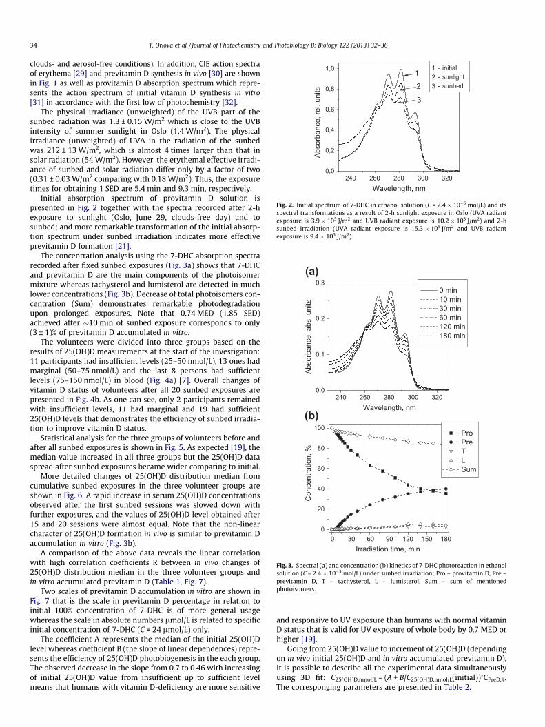
Fig. 2. Initial spectrum of 7-DHC in ethanol solution (C = 2.4 � 10�5 mol/L) and itsspectral transformations as a result of 2-h sunlight exposure in Oslo (UVA radiantexposure is 3.9 � 105 J/m2 and UVB radiant exposure is 10.2 � 103 J/m2) and 2-hsunbed irradiation (UVA radiant exposure is 15.3 � 105 J/m2 and UVB radiantexposure is 9.4 � 103 J/m2).
240 260 280 300 3200,0
0,1
0,2
0,3 0 min10 min30 min60 min120 min180 min
Abso
rban
ce, a
bs. u
nits
Wavelength, nm
0 30 60 90 120 150 1800
20
40
60
80
100
Con
cent
ratio
n, %
Irradiation time, min
Pro Pre T L Sum
(a)
(b)
Fig. 3. Spectral (a) and concentration (b) kinetics of 7-DHC photoreaction in ethanolsolution (C = 2.4 � 10�5 mol/L) under sunbed irradiation; Pro – provitamin D, Pre –previtamin D, T – tachysterol, L – lumisterol, Sum – sum of mentionedphotoisomers.
34 T. Orlova et al. / Journal of Photochemistry and Photobiology B: Biology 122 (2013) 32–36
clouds- and aerosol-free conditions). In addition, CIE action spectraof erythema [29] and previtamin D synthesis in vivo [30] are shownin Fig. 1 as well as provitamin D absorption spectrum which repre-sents the action spectrum of initial vitamin D synthesis in vitro[31] in accordance with the first low of photochemistry [32].
The physical irradiance (unweighted) of the UVB part of thesunbed radiation was 1.3 ± 0.15 W/m2 which is close to the UVBintensity of summer sunlight in Oslo (1.4 W/m2). The physicalirradiance (unweighted) of UVA in the radiation of the sunbedwas 212 ± 13 W/m2, which is almost 4 times larger than that insolar radiation (54 W/m2). However, the erythemal effective irradi-ance of sunbed and solar radiation differ only by a factor of two(0.31 ± 0.03 W/m2 comparing with 0.18 W/m2). Thus, the exposuretimes for obtaining 1 SED are 5.4 min and 9.3 min, respectively.
Initial absorption spectrum of provitamin D solution ispresented in Fig. 2 together with the spectra recorded after 2-hexposure to sunlight (Oslo, June 29, clouds-free day) and tosunbed; and more remarkable transformation of the initial absorp-tion spectrum under sunbed irradiation indicates more effectiveprevitamin D formation [21].
The concentration analysis using the 7-DHC absorption spectrarecorded after fixed sunbed exposures (Fig. 3a) shows that 7-DHCand previtamin D are the main components of the photoisomermixture whereas tachysterol and lumisterol are detected in muchlower concentrations (Fig. 3b). Decrease of total photoisomers con-centration (Sum) demonstrates remarkable photodegradationupon prolonged exposures. Note that 0.74 MED (1.85 SED)achieved after �10 min of sunbed exposure corresponds to only(3 ± 1)% of previtamin D accumulated in vitro.
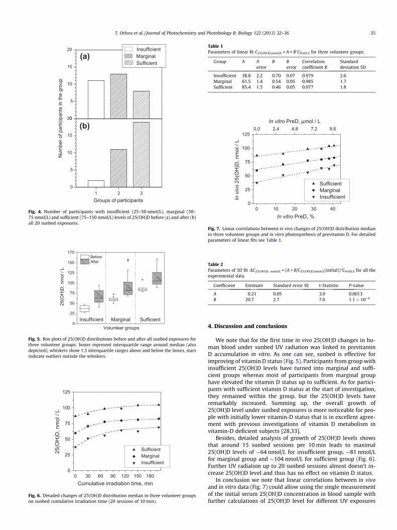
The volunteers were divided into three groups based on theresults of 25(OH)D measurements at the start of the investigation:11 participants had insufficient levels (25–50 nmol/L), 13 ones hadmarginal (50–75 nmol/L) and the last 8 persons had sufficientlevels (75–150 nmol/L) in blood (Fig. 4a) [7]. Overall changes ofvitamin D status of volunteers after all 20 sunbed exposures arepresented in Fig. 4b. As one can see, only 2 participants remainedwith insufficient levels, 11 had marginal and 19 had sufficient25(OH)D levels that demonstrates the efficiency of sunbed irradia-tion to improve vitamin D status.
Statistical analysis for the three groups of volunteers before andafter all sunbed exposures is shown in Fig. 5. As expected [19], themedian value increased in all three groups but the 25(OH)D dataspread after sunbed exposures became wider comparing to initial.
More detailed changes of 25(OH)D distribution median fromcumulative sunbed exposures in the three volunteer groups areshown in Fig. 6. A rapid increase in serum 25(OH)D concentrationsobserved after the first sunbed sessions was slowed down withfurther exposures, and the values of 25(OH)D level obtained after15 and 20 sessions were almost equal. Note that the non-linearcharacter of 25(OH)D formation in vivo is similar to previtamin Daccumulation in vitro (Fig. 3b).
A comparison of the above data reveals the linear correlationwith high correlation coefficients R between in vivo changes of25(OH)D distribution median in the three volunteer groups andin vitro accumulated previtamin D (Table 1, Fig. 7).
Two scales of previtamin D accumulation in vitro are shown inFig. 7 that is the scale in previtamin D percentage in relation toinitial 100% concentration of 7-DHC is of more general usagewhereas the scale in absolute numbers lmol/L is related to specificinitial concentration of 7-DHC (C = 24 lmol/L) only.
The coefficient A represents the median of the initial 25(OH)Dlevel whereas coefficient B (the slope of linear dependences) repre-sents the efficiency of 25(OH)D photobiogenesis in the each group.The observed decrease in the slope from 0.7 to 0.46 with increasingof initial 25(OH)D value from insufficient up to sufficient levelmeans that humans with vitamin D-deficiency are more sensitive
and responsive to UV exposure than humans with normal vitaminD status that is valid for UV exposure of whole body by 0.7 MED orhigher [19].
Going from 25(OH)D value to increment of 25(OH)D (dependingon in vivo initial 25(OH)D and in vitro accumulated previtamin D),it is possible to describe all the experimental data simultaneouslyusing 3D fit: C25(OH)D,nmol/L = (A + B/C25(OH)D,nmol/L(initial))�CPreD,%.The corresponging parameters are presented in Table 2.
(a)
(b)
Fig. 4. Number of participants with insufficient (25–50 nmol/L), marginal (50–75 nmol/L) and sufficient (75–150 nmol/L) levels of 25(OH)D before (a) and after (b)all 20 sunbed exposures.
Fig. 5. Box plots of 25(OH)D distributions before and after all sunbed exposures forthree volunteer groups: boxes represent interquartile range around median (alsodepicted), whiskers show 1.5 interquartile ranges above and below the boxes, starsindicate outliers outside the whiskers.
600 30 90 120 150 1800
25
50
75
100
125
Sufficient Marginal Insufficient
Cumulative irradiation time, min
25(O
H)D
, nm
ol /
L
Fig. 6. Detailed changes of 25(OH)D distribution median in three volunteer groupson sunbed cumulative irradiation time (20 sessions of 10 min).
Table 1Parameters of linear fit C25(OH)D,nmol/L = A + B�CPreD,% for three volunteer groups.
Fig. 7. Linear correlations between in vivo changes of 25(OH)D distribution medianin three volunteer groups and in vitro photosynthesis of previtamin D. For detailedparameters of linear fits see Table 1.
Table 2Parameters of 3D fit DC25(OH)D, nmol/L = (A + B/C25(OH)D,nmol/L(initial))�CPreD,% for all theexperimental data.
Coefficient Estimate Standard error SE t-Statistic P-value
A 0.21 0.05 3.9 0.0013B 20.7 2.7 7.6 1.1 � 10�6
T. Orlova et al. / Journal of Photochemistry and Photobiology B: Biology 122 (2013) 32–36 35
4. Discussion and conclusions
We note that for the first time in vivo 25(OH)D changes in hu-man blood under sunbed UV radiation was linked to previtaminD accumulation in vitro. As one can see, sunbed is effective forimproving of vitamin D status (Fig. 5). Participants from group withinsufficient 25(OH)D levels have turned into marginal and suffi-cient groups whereas most of participants from marginal grouphave elevated the vitamin D status up to sufficient. As for partici-pants with sufficient vitamin D status at the start of investigation,they remained within the group, but the 25(OH)D levels haveremarkably increased. Summing up, the overall growth of25(OH)D level under sunbed exposures is more noticeable for peo-ple with initially lower vitamin-D status that is in excellent agree-ment with previous investigations of vitamin D metabolism invitamin-D deficient subjects [28,33].
Besides, detailed analysis of growth of 25(OH)D levels showsthat around 15 sunbed sessions per 10 min leads to maximal25(OH)D levels of �64 nmol/L for insufficient group, �81 nmol/Lfor marginal group and �104 nmol/L for sufficient group (Fig. 6).Further UV radiation up to 20 sunbed sessions almost doesn’t in-crease 25(OH)D level and thus has no effect on vitamin D status.
In conclusion we note that linear correlations between in vivoand in vitro data (Fig. 7) could allow using the single measurementof the initial serum 25(OH)D concentration in blood sample withfurther calculations of 25(OH)D level for different UV exposures
36 T. Orlova et al. / Journal of Photochemistry and Photobiology B: Biology 122 (2013) 32–36
for predictions of vitamin D status based on the measurements ofprevitamin D accumulation in vitro. Certainly, obtained in thisstudy correlations should be re-measured for sunbed equippedwith other fluorescent lamps because previtamin D photosynthesisstrongly depends on the irradiation spectrum of an UV source [21].
Taking into account the non-linear dependence of previtamin Dphotoinduced accumulation correlating with the results of differ-ent UV exposures [19], the lower sunbed exposures would be pref-erable from the point of view of the safety requirements especiallyfor the lamps with elevated ratio UVB/UVA.
Furthermore these data need to be detailed depending on age,BMI, Fitzpatrick skin type, presence or absence of regular vitaminD intake, etc., that requires large epidemiological studies forcompleteness statistics.
Acknowledgments
The work of Dr. Tetiana Orlova at the Oslo University Hospitalwas supported by The Research Council of Norway, Yggdrasylprogramme, Personal mobility Grant 202615. The work of Asta Juz-eniene and Zoya Lagunova was supported by the South-EasternNorway Regional Health Authority.
References
[1] H. Reichel, H.P. Koeffler, A.W. Norman, The role of the vitamin D endocrynesystem in health and disease, N. Engl. J. Med. 320 (1989) 980–991.
[2] A. Mayar, A.W. Norman, in: R. Dulbecco (Ed.), Encyclopedia of Human Biology,Academic Press, San Diego, 1991, pp. 859–871.
[3] W.B. Grant, An estimation of excess cancer mortality in the United States dueto inadequate doses of solar ultraviolet-B radiation and/or vitamin D, Cancer94 (2002) 1867–1875.
[4] M.F. Holick, Vitamin D: importance in the prevention of cancers, type 1diabetes, heart disease and osteoporosis, Am. J. Clin. Nutr. 79 (2004) 362–371.
[5] M.F. Holick, Sunlight and vitamin D for bone health and prevention ofautoimmune diseases, cancers, and cardiovascular disease, Am. J. Clin. Nutr. 80(2004) 1678S–1688S.
[6] W.B. Grant, Epidemiology of disease risks in relation to vitamin D insufficiency,Prog. Biophys. Mol. Biol. 92 (2006) 65–79.
[7] A.W. Norman, R. Bouillon, Vitamin D nutritional policy needs a vision for thefuture, Exp. Biol. Med. 235 (2010) 1034–1045.
[8] A.R. Webb, M.F. Holick, The role of sunlight in the cutaneous production ofvitamin D3, Ann. Rev. Nutr. 8 (1988) 375–399.
[9] B. Lehmann, Meurer vitamin D metabolism, Dermatol. Ther. 23 (2010) 2–12.[10] A.R. Webb, L.W. Kline, M.F. Holick, Influence of season and latitude on the
cutaneous synthesis of vitamin D3: exposure to winter sunlight in Boston andEdmonton will not promote vitamin D3 in human skin, J. Clin. Endocrinol.Metabol. 67 (1988) 373–378.
[11] A.V. Parisi, D.J. Turnbull, N.J. Downs, Influence of high levels of cloud cover onvitamin D effective and erythemal solar UV irradiances, Photochem. Photobiol.Sci. 11 (2012) 1855–1859.
[12] I. Terenetskaya, T. Orlova, Variability of solar UV-B irradiance: in situmonitoring and model calculation of the vitamin D synthetic capacity ofsunlight, Int. J. Remote Sens. 32 (2011) 6205–6218.
[13] L.T. Nilsen, M. Hannevik, T.N. Aalerud, B. Johnsen, E.G. Friberg, M.B. Veierød,Trends in UV irradiance of tanning devices in Norway: 1983–2005,Photochem. Photobiol. 84 (2008) 1100–1108.
[14] A.C. Porojnicu, O.S. Bruland, L. Aksnes, W.B. Grant, J. Moan, Sun beds and codliver oil as vitamin D sources, J. Photochem. Photobiol. B 91 (2008) 125–131.
[15] P. Knushke, P. Bocionek, B. Lehnmann, B. Pinzer, M. Meurer, in: M.F. Holick,E.G. Jung (Eds.), Biologic Effects of Light 1998, Kluwer Academic Pub, Boston/London/Dordrecht, 1999, pp. 75–77.
[16] P. Datta, M.K. Bogh, P. Olsen, P. Eriksen, A.V. Schmedes, M.M.-L. Grage, P.A.Philipsen, H.C. Wulf, Increase in serum 25-hydroxyvitamin-D3 in humans aftersolar exposure under natural conditions compared to artificial UVB exposureof hands and face, Photochem. Photobiol. Sci. 11 (2012) 1817–1824.
[17] M. Norval, L.O. Bjorn, F.R. de Gruijl, Is the action spectrum for the UV-inducedproduction of previtamin D3 in human skin correct?, Photochem Photobiol.Sci. 9 (2010) 11–17.
[18] I. Terenetskaya, Duality of solar UV-B radiation and relevant dosimetry:vitamin D synthesis versus skin erytema, Proc. SPIE 4896 (2003) 144–150.
[19] P. Knuschke, B. Lehmann, A. Püschel, H. Rönsch, UV-abhängige vitamin Dsynthese – bilanzierung der expositionszeit durch UV zur produktion desoptimalen vitamin D3-bedarfs im menschlichen körper: vorhaben3607S04538, Bundesamt für Strahlenschutz, 2012 (in German).
[20] I. Terenetskaya, Provitamin D photoisomerization as possible UVB monitor:kinetic study using tunable dye-laser, Proc. SPIE 2134B (1994) 135–140.
[21] O.N. Galkin, I.P. Terenetskaya, Vitamin D biodosimeter: basic characteristicsand potential applications, J. Photochem. Photobiol. B 53 (1999) 12–19.
[22] I.P. Terenetskaya, Spectral monitoring of biologically active solar UVBradiation using an in vitro model of vitamin D synthesis, Talanta 53 (2000)195–203.
[23] I. Gvozdovskyy, T. Orlova, E. Salkova, I. Terenetskaya, G. Milinevsky, Ozone andsolar UVB radiation: monitoring of the vitamin D synthetic capacity of sunlightin Kiev and Antarctica, Int. J. Remote Sens. 26 (2005) 3555–3559.
[24] I. Terenetskaya, T. Orlova, I. Gvozdovskyy, G. Milinevsky, Solar UVB radiationand vitamin D synthesis: direct monitoring of the vitamin D synthetic capacityof sunlight in Kiev and in Antarctic, Annalen der Meteorologie 12 (2005) 676–678.
[25] T.N. Orlova, I.P. Terenetskaya, UV-biosensor for visual indication of vitamin Dsynthesis, Proc. SPIE 7003 (2008) (70031O(1)-70031O(8)).
[26] T.N. Orlova, I.P. Terenetskaya, A.M. Eremenko, N.I. Surovtseva, Provitamin Ddoped silica and polymeric films: new materials for UV biosensor, Mater. Sci.Appl. 1 (2010) 267–271.
[27] H.J.C. Jacobs, E. Havinga, in: J.N. Pitts, J.S. Hammond, K. Gollnick (Eds.),Advances in Photochemistry, 11, Wiley, New York, 1979, pp. 305–373.
[28] J. Moan, Z. Lagunova, E. Cicarma, L. Aksnes, A. Dahlback, W.B. Grant, A.C.Porojnicu, Sunbeds as vitamin D sources, Photochem. Photobiol. 85 (2009)1474–1479.
[29] A.F. McKinlay, B.L. Diffey, in: W.F. Passchier, B.F.M. Bosnjakovich (Eds.),Human Exposure to Ultraviolet Radiation: Risks and Regulations, Elsevier,Amsterdam, 1987, pp. 83–87.
[30] R. Bouillon, J. Eisman, M. Garabedian, M. Holick, J. Kleinschmidt, T. Suda, I.Terenetskaya, A. Webb, Action spectrum for the production of previtamin D3in human skin, CIE J. 174 (2006) 1–12.
[31] I. Terenetskaya, Inter-relation between the in vivo and in vitro action spectraof vitamin D synthesis, in: Proc 2nd CIE Expert Symposium ‘‘Lighting andHealth’’, Ottawa, Canada, September 7–8, 2006, pp. 182–185.
[32] N.J. Turro, Modern Molecular Photochemistry, Benjamin/Cummings, MenloPark, 1978.
[33] J.S. Adams, T.L. Clements, J.A. Parrish, M.F. Holick, Vitamin D synthesis andmetabolism after ultraviolet radiation of normal and vitamin D-deficientsubjects, N. Engl. J. Med. 306 (1982) 722–725.