Increased Presynaptic and Postsynaptic �2-Adrenoceptor Activityin the Spinal Dorsal Horn in Painful Diabetic Neuropathy
Shao-Rui Chen, Hong Chen, Wei-Xiu Yuan, and Hui-Lin PanDepartment of Anesthesiology and Perioperative Medicine (S.-R.C., H.C., W.-X.Y., H.-L.P.), University of Texas MD AndersonCancer Center, Houston, Texas; and Program in Neuroscience, University of Texas Graduate School of Biomedical Sciences,Houston, Texas (H.-L.P.)
Received October 26, 2010; accepted January 19, 2011
ABSTRACTDiabetic neuropathy is a common cause of chronic pain that is notadequately relieved by conventional analgesics. The �2-adrenoceptors are involved in the regulation of glutamatergic inputand nociceptive transmission in the spinal dorsal horn, but theirfunctional changes in diabetic neuropathy are not clear. The pur-pose of the present study was to determine the plasticity ofpresynaptic and postsynaptic �2-adrenoceptors in the control ofspinal glutamatergic synaptic transmission in painful diabetic neu-ropathy. Whole-cell voltage-clamp recordings of lamina II neuronswere performed in spinal cord slices from streptozotocin-induceddiabetic rats. The amplitude of glutamatergic excitatory postsyn-aptic currents (EPSCs) evoked from the dorsal root and the fre-quency of spontaneous EPSCs (sEPSCs) were significantly higherin diabetic than vehicle-control rats. The specific �2-adrenoceptoragonist 5-bromo-6-(2-imidazolin-2-ylamino)quinoxaline (UK-
14304) (0.1–2 �M) inhibited the frequency of sEPSCs more indiabetic than vehicle-treated rats. UK-14304 also inhibited theamplitude of evoked monosynaptic and polysynaptic EPSCsmore in diabetic than control rats. Furthermore, the amplitude ofpostsynaptic G protein-coupled inwardly rectifying K� channel(GIRK) currents elicited by UK-14304 was significantly larger in thediabetic group than in the control group. In addition, intrathecaladministration of UK-14304 increased the nociceptive thresholdmore in diabetic than vehicle-control rats. Our findings suggestthat diabetic neuropathy increases the activity of presynaptic andpostsynaptic �2-adrenoceptors to attenuate glutamatergic trans-mission in the spinal dorsal horn, which accounts for the potenti-ated antinociceptive effect of �2-adrenoceptor activation in dia-betic neuropathic pain.
IntroductionDiabetic neuropathy is one of the most serious complications
that afflicts people with diabetes and is frequently painful. Painassociated with diabetic neuropathy can occur either spontane-ously or as a result of exposure to mildly painful stimuli (hy-peralgesia) or stimuli not normally perceived as painful (allo-dynia) (Clark and Lee, 1995; Veves et al., 2008). Thedevelopment of diabetic neuropathic pain is associated withincreased nociceptive input, neuronal hyperactivity, and sus-tained stimulation of certain glutamate receptors in the spinalcord (Calcutt and Chaplan, 1997; Malcangio and Tomlinson,1998; Chen and Pan, 2002; Wang et al., 2007; Li et al., 2010).Because diabetic neuropathic pain often is not adequately re-
lieved by available analgesics, it represents an important un-met medical need.
Many experimental studies have demonstrated that intra-thecal or systemic administration of �2-adrenoceptor agonistscan produce a potent analgesic effect in neuropathic paincaused by traumatic nerve injury and diabetic neuropathy(Courteix et al., 1994; Yaksh et al., 1995; Calcutt and Chaplan,1997; Buerkle and Yaksh, 1998; Pan et al., 1999; Omiya et al.,2008). In addition, clinical studies have shown that spinallyadministered �2-adrenoceptor agonists reduce the intractableneuropathic pain caused by nerve injury and cancer (Rauck etal., 1993; Hassenbusch et al., 2002). At the spinal level, the�2-adrenoceptor is present presynaptically at the primary affer-ent terminals and postsynaptically on dorsal horn neurons.Although the �2A-adrenoceptor subtype is located predomi-nantly on the terminals of primary afferents (Sullivan et al.,1987; Roudet et al., 1994; Stone et al., 1998; Overland et al.,2009; Riedl et al., 2009), the �2C-adrenoceptor subtype is pres-ent mainly on spinal dorsal horn neurons (Stone et al., 1998).Stimulation of �2-adrenoceptors with clonidine dose-depend-
This study was supported by the National Institutes of Health NationalInstitute of General Medical Sciences [Grant GM64830], the National Insti-tutes of Health National Institute of Neurological Disorders and Stroke [GrantNS45602], and the N.G. and Helen Hawkins Endowment (to H.L.P.).
Article, publication date, and citation information can be found athttp://jpet.aspetjournals.org.
ently reduces excitation of dorsal horn neurons evoked by pri-mary afferent stimulation (Sullivan et al., 1987). The �2-adrenoceptor agonists can reduce glutamate release fromprimary afferents (Pan et al., 2002) and hyperpolarize the dor-sal horn neurons through a postsynaptic action (North andYoshimura, 1984). However, the plasticity and role of presyn-aptic and postsynaptic �2-adrenoceptors in the regulation ofglutamatergic input to spinal dorsal horn neurons in diabeticneuropathic pain remain unknown.
Therefore, in this study, we used a rat model of diabeticneuropathy and electrophysiological approaches to determinethe functional changes in presynaptic and postsynaptic �2-adrenoceptors in the control of spinal glutamatergic transmis-sion and how these changes affect the antinociceptive effect ofstimulation of �2-adrenoceptors at the spinal level in diabeticneuropathic pain.
Materials and MethodsAnimal Model of Diabetic Neuropathic Pain. Male Sprague-
Dawley rats (Harlan, Indianapolis, IN) initially weighing 200 to220 g were used in this study. Diabetes was induced by a singleintraperitoneal injection of streptozotocin (STZ; 60 mg/kg; Sigma-Aldrich, St. Louis, MO) freshly dissolved in 0.9% sterile saline (Chenand Pan, 2002; Wang et al., 2007; Li et al., 2010). Diabetes wasconfirmed in STZ-injected rats by measuring the blood glucose con-centration. Glucose levels in blood obtained from the tail vein wereassayed using ACCU-CHEK test strips (Roche Diagnostics, India-napolis, IN). The blood glucose level was measured 2 weeks after STZadministration, and only rats with high levels (� 300 mg/dl) wereused for the diabetic groups. Age-matched vehicle-injected rats wereused as controls. The low dose of STZ was used to minimize theeffects of STZ administration on the overall health of the animal(Chen and Pan, 2002; Chen et al., 2009). This model of neuropathicpain mimics the symptoms of neuropathy in diabetic patients, withalterations in pain sensitivity and poor responses to � opioid admin-istered systemically or intrathecally (Courteix et al., 1993; Malcan-gio and Tomlinson, 1998; Zurek et al., 2001; Chen and Pan, 2002). Ithas been shown that early insulin intervention can impede thedevelopment of painful diabetic neuropathy induced by STZ in rats(Sasaki et al., 1998; Hoybergs and Meert, 2007). Neuropathic pain indiabetic rats was confirmed by examining nociceptive thresholdsusing an analgesimeter (Ugo Basile, Comerio, Italy), and all diabeticrats developed mechanical hyperalgesia in our study. All of thediabetic rats remained relatively healthy, although their growth ratewas reduced (body weight gain: 5 g/week in the diabetic group versus20 g/week in the nondiabetic group). The final electrophysiologicaland behavioral experiments were performed on rats 3 weeks afterSTZ or vehicle treatment. The experiments were performed accord-ing to National Institutes of Health guidelines and approved by theAnimal Care and Use Committee of the University of Texas MDAnderson Cancer Center.
Intrathecal Cannulation and Behavioral Assessment of No-ciception. Intrathecal catheters (PE-10 polyethylene tubing) wereinserted in diabetic and control rats during isoflurane-induced an-esthesia. The catheter was advanced 8 cm caudally through anincision in the cisternal membrane and secured to the musculatureat the incision site (Chen and Pan, 2001, 2006). The rats wereallowed to recover for at least 5 days before drug testing began. Onlyrats with no evidence of neurological deficit after catheter insertionwere used in the study. Drugs for intrathecal injections were dis-solved in normal saline and administered in a volume of 5 �l followedby a 10-�l flush with normal saline.
The nociceptive mechanical threshold was measured using an UgoBasile analgesimeter to apply a noxious pressure to a hindpaw. Bypressing a pedal that activated a motor, the force increased at a
constant rate on the linear scale. When the rat responded by with-drawal of the paw or vocalization, the pedal was immediately re-leased and the nociceptive threshold was read on a scale. A cutoff of400 g was used to avoid tissue injury (Chen and Pan, 2002, 2006).Both hindpaws were tested in each rat, and the mean value was usedas the nociceptive withdrawal threshold.
Spinal Cord Slice Preparation. The rats were anesthetizedwith 2 to 3% isoflurane, and the lumbar segment of the spinal cordwas removed by means of laminectomy. The spinal cord segment atthe L5 and L6 levels was immediately placed in ice-cold sucrose-artificial cerebrospinal fluid (aCSF) presaturated with 95% O2 and5% CO2. The sucrose-aCSF contained 234 mM sucrose, 3.6 mM KCl,1.2 mM MgCl2, 2.5 mM CaCl2, 1.2 mM NaH2 PO4, 12 mM glucose,and 25 mM NaHCO3. The tissue was then placed in a shallow grooveformed in a gelatin block and glued to the stage of a vibratome.Transverse spinal cord slices (400 �m) were cut in the ice-coldsucrose-aCSF and then preincubated in Krebs’ solution oxygenatedwith 95% O2 and 5% CO2 at 34°C for at least 1 h before they weretransferred to the recording chamber. The Krebs’ solution contained117 mM NaCl, 3.6 mM KCl, 1.2 mM MgCl2, 2.5 mM CaCl2, 1.2 mMNaH2 PO4, 11 mM glucose, and 25 mM NaHCO3. The perfusionbuffer had been optimized to best preserve the spinal cord tissue forthe in vitro recording. On the basis of our experience, the highglucose level (11 mM) in the buffer is required to maintain theviability of spinal slices in vitro at 34 to 36°C. If the glucose level inthe buffer is lowered to 4 to 5 mM, we can obtain stable whole-cellrecordings only at room temperature (24–25°C).
Electrophysiological Recordings. Excitatory postsynaptic cur-rents (EPSCs) were recorded using the whole-cell voltage-clampmethod as described previously (Wang et al., 2007; Li et al., 2010).Each spinal cord slice was placed in a glass-bottomed chamber andfixed with parallel nylon threads supported by a U-shaped stainless-steel weight. The slice was continuously perfused with Krebs’ solu-tion at 5.0 ml/min at 34°C maintained by an inline solution heaterand a temperature controller. Neurons in the lamina II of the spinalcord slice were identified with differential interference contrast/infrared illumination on a fixed-stage microscope (BX50WI; Olym-pus, Tokyo, Japan).
Monosynaptic or polysynaptic EPSCs were evoked by electricalstimulation through a bipolar stimulation electrode placed on thedorsal root. We used fixed stimulation intensity (0.2 ms and 0.6 mA)to evoke EPSCs from primary afferents in both diabetic and controlrats. At the stimulation intensity used, both A- and C-afferent fibersthat were in close contact with the electrode tip were stimulated(Wang et al., 2007; Zhou et al., 2010). The evoked EPSCs wereconsidered to be monosynaptic if 1) the latency was constant withrepeated electrical stimulation at 0.1 Hz, and 2) there was no con-duction failure or changes in the latency when the stimulation fre-quency was increased to 20 Hz (stimulus train duration, 1 s) (Wanget al., 2007; Li et al., 2010). In contrast, evoked EPSCs were consid-ered to be polysynaptic if the latency was variable and conductionfailure occurred at a higher stimulation frequency (20 Hz). Theelectrode for the whole-cell recordings was pulled from borosilicateglass capillaries. The impedance of the pipette was 5 to 8 M� whenfilled with an internal solution containing 110 mM Cs2SO4, 5 mMtetraethylammonium, 2 mM MgCl2, 0.5 mM CaCl2, 5 mM HEPES, 5mM EGTA, 5 mM ATP-Mg, 0.5 mM Na-GTP, and 10 mM lidocaineN-ethyl bromide that had been adjusted to pH 7.2 to 7.3 with 1 MCsOH (290–300 mOsm). In the recordings of spontaneous EPSCs(sEPSCs) or evoked EPSCs, 1 mM guanosine 5�-O-(2-thiodiphosphate),a general G protein inhibitor (Zhou et al., 2008, 2010), was included inthe pipette internal solution to block the potential postsynaptic effectproduced by the �2-adrenoceptor agonist. Miniature EPSCs (mEPSCs)were recorded in the presence of 1 �M tetrodotoxin (TTX).
We recorded G protein-coupled inwardly rectifying K� channel(GIRK) currents elicited by 5-bromo-6-(2-imidazolin-2-ylamino)qui-noxaline (UK-14304; also known as brimonidine) as a measure ofpostsynaptic �2-adrenoceptor activity in the spinal cord. GIRK cur-
rents were recorded from lamina II neurons at a holding potential of�60 mV using a pipette internal solution containing 135.0 mMpotassium gluconate, 5.0 mM KCl, 2.0 mM MgCl2, 0.5 mM CaCl2, 5.0mM HEPES, 5.0 mM EGTA, 5.0 mM ATP-Mg, and 0.5 mM Na-GTP;the solution was adjusted to pH 7.2 to 7.4 with 1 M KOH (290–300m�sm) (Zhou et al., 2008, 2010). Recordings of EPSCs or GIRKcurrents began approximately 6 min after whole-cell access wasestablished and the current reached a steady state. The input resis-tance was monitored, and the recording was abandoned if it changedby more than 15%. All signals were recorded using an amplifier(MultiClamp 700B; Molecular Devices, Sunnyvale, CA) at a holdingpotential of �60 mV, filtered at 1 to 2 kHz, digitized at 10 kHz, andstored in a computer using pCLAMP 9.2 (Molecular Devices).
Yohimbine and guanosine 5�-O-(2-thiodiphosphate) were obtainedfrom Sigma-Aldrich. UK-14304 was purchased from Tocris Biosci-ence (Ellisville, MO). TTX was obtained from Ascent Scientific(Princeton, NJ).
Data Analysis and Statistics. Data are presented as means �S.E.M. In general, two to three neurons were recorded from each rat,and at least five rats were used for each group. The amplitude ofevoked EPSCs and GIRK currents were analyzed using Clampfit(Molecular Devices). The frequency and amplitude of sEPSCs andmEPSCs were analyzed off-line using a peak detection program(MiniAnalysis; Synaptosoft, Decatur, GA). Detection of events wasaccomplished by setting a threshold above the noise level. ThesEPSCs and mEPSCs were detected by the fast rise time of the signalover an amplitude threshold above the background noise. We man-ually excluded the event when the background noise was erroneouslyidentified as an sEPSC by the program. The cumulative probabilityof the amplitude and interevent interval of sEPSCs and mEPSCswas compared by using the Komogorov-Smirnov test, which esti-mates the probability that two cumulative distributions are similar.The effects of UK-14304 on GIRK currents, evoked EPSCs, sEPSCs,and mEPSCs were determined using repeated-measures analysis ofvariance (ANOVA), and the differences between the control anddiabetic groups were tested using two-way ANOVA with Bonferroni’spost hoc test. The effect of drug treatments on the paw withdrawalthreshold was determined by using repeated-measures ANOVA fol-lowed by Dunnett’s post hoc test. P 0.05 was considered to bestatistically significant.
ResultsIncreased Effect of UK-14304 on Glutamatergic In-
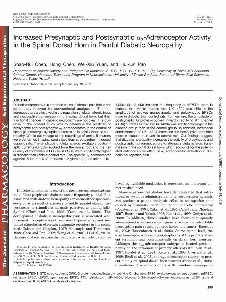
put to Spinal Dorsal Horn Neurons in Diabetic Rats.Three weeks after induction of diabetes, the diabetic ratsshowed significantly lower paw withdrawal thresholds(94.73 � 4.11 g) in response to the pressure stimulus appliedto the hindpaw than the age-matched control rats treatedwith vehicle (134.14 � 4.48 g; P 0.05, t test). The baselinefrequency, but not the amplitude, of glutamatergic sEPSCs oflamina II neurons was significantly higher in diabetic than invehicle control rats (Fig. 1).
To determine changes in the activity of �2-adrenoceptors inthe control of glutamatergic input to spinal dorsal horn neu-rons in diabetic neuropathy, we compared the inhibitoryeffects of UK-14304 on the frequency of sEPSCs of lamina IIneurons in diabetic and control rats. We selected UK-14304for our study because it is a highly potent and selective�2-adrenoceptor agonist (Buerkle and Yaksh, 1998). UnlikeFig. 1. Effects of UK-14304 on glutamatergic sEPSCs of spinal lamina II
neurons in control and diabetic rats. A, original traces show sEPSCs duringbaseline control, application of 0.1, 0.5, and 1 �M UK-14304, and washout in onelamina II neuron from a control rat and another from a diabetic rat. B, cumu-lative probability plots of sEPSCs of the same neuron in A showing the distri-bution of the interevent interval and amplitude of sEPSCs during control,application of UK-14304, and washout. C, summary data compare the effect of0.1 to 2 �M UK-14304 on the frequency of sEPSCs in 16 neurons from control
rats and 21 neurons from diabetic rats. Note that the baseline frequencyof sEPSCs of lamina II neurons was significantly higher in diabetic thanin control rats. Data are presented as means � S.E.M. �, P 0.05compared with the baseline control. #, P 0.05 compared with thecorresponding value in the control group.
Plasticity of Spinal �2-Adrenoceptors in Neuropathic Pain 287
clonidine and dexmedetomidine, the effect of UK-14304 isfast and can be easily washed out upon bath application.Bath application of 0.1 to 2 �M UK-14304 decreased thefrequency, but not the amplitude, of sEPSCs in all 21 laminaII neurons from diabetic rats in a concentration-dependentmanner (Fig. 1). The cumulative probability analysis ofsEPSCs revealed that the distribution pattern of the in-terevent interval of sEPSCs was shifted toward the right(i.e., reduced frequency of sEPSCs) in response to UK-14304 treatment. However, the distribution pattern of theamplitude of sEPSCs was not significantly changed (Fig.1B). UK-14304 also inhibited the frequency, but not theamplitude, of sEPSCs in 16 lamina II neurons from controlrats. However, the inhibitory effect of UK-14304 wasnearly maximal at 0.5 �M. Because the baseline frequencyof sEPSCs was significantly different between the diabeticand control rats, we normalized the effect of UK-14304 tothe baseline of sEPSCs in each group. UK-14304 caused asignificantly greater decrease in the frequency of sEPSCsin diabetic than in control rats (Fig. 1C). Similar to whatwe reported previously (Wang et al., 2007; Li et al., 2010),bath application of 10 �M 6-cyano-7-nitroquinoxaline-2,3-dione, a non-N-methyl-D-aspartate receptor antagonist,eliminated sEPSCs in all neurons examined in control anddiabetic rats (data not shown).
To further determine whether the effect of the �2-adreno-ceptor agonist on spinal glutamatergic transmission occursthrough a presynaptic site of action, we compared the effectsof UK-14304 on sEPSCs and mEPSCs in the same neuronsobtained from spinal cord slices of vehicle-control and dia-betic rats. Bath application of 1 �M UK-14304 had a similarinhibitory effect on the frequency, but not the amplitude, ofsEPSCs and mEPSCs in 14 lamina II neurons from controlrats and 11 lamina II neurons from diabetic rats (Fig. 2).
Increased Effect of UK-14304 on Glutamate Releasefrom Primary Afferent Terminals in Diabetic Rats. Todetermine the activity of �2-adrenoceptors on primary affer-ent terminals in the regulation of glutamatergic synapticinput to dorsal horn neurons in diabetic neuropathy, we nextexamined the effects of UK-14304 on monosynaptic and poly-synaptic EPSCs evoked from the dorsal root. The baselineamplitude of both monosynaptic and polysynaptic EPSCs ofspinal lamina II neurons was significantly higher in diabeticthan control rats (Fig. 3).
Bath application of 0.1 to 2 �M UK-14304 concentration-dependently reduced the peak amplitude of monosynapticEPSCs in most neurons (13/14, 92.3%) from diabetic rats(Fig. 3). Although UK-14304 (0.5–2 �M) also significantlyinhibited the amplitude of monosynaptic EPSCs in 13 of 15neurons (86.7%) tested in the control group, its effect wasnearly maximal at 0.5 �M. Because the baseline amplitude ofthe evoked EPSCs was significantly different between thetwo groups, we normalized the effect of UK-14304 to thebaseline control of evoked EPSCs in each group. At concen-trations of 1 and 2 �M, UK-14304 had a significantly greatereffect on the amplitude of monosynaptic EPSCs in diabeticthan control rats (Fig. 3). Furthermore, blocking �2-adreno-ceptors with yohimbine (2 �M) (Pan et al., 2002) abolishedthe inhibitory effect of 1 �M UK-14304 on monosynapticEPSCs in all neurons from diabetic rats (Fig. 3C).
In separate groups of lamina II neurons, bath applicationof 0.1 to 2 �M UK-14304 inhibited the peak amplitude of
evoked polysynaptic EPSCs in the control and diabeticgroups in a dose-dependent manner (Fig. 4). When the effectof UK-14304 on evoked EPSCs was normalized to the base-line control, it also produced a significantly greater effect onthe amplitude of polysynaptic EPSCs in diabetic rats than incontrol rats (Fig. 4B).
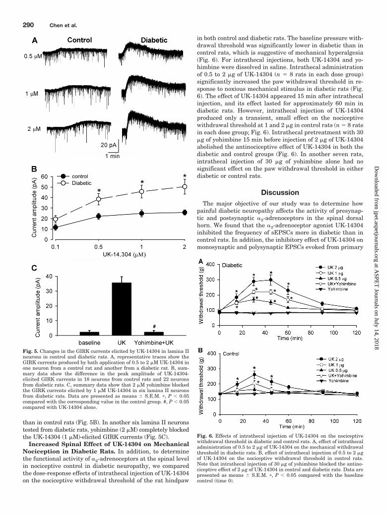
Increased GIRK Currents Elicited by UK-14304 inSpinal Dorsal Horn Neuron in Diabetic Rats. GIRKchannels are present postsynaptically in spinal lamina II neu-rons (Marker et al., 2006; Zhou et al., 2008). To determine thediabetes-induced changes in the activity of postsynaptic �2-adrenoceptors in the spinal dorsal horn, we measured GIRKcurrents elicited by UK-14304 in lamina II neurons of diabeticand control rats. Bath application of 0.1 to 2 �M UK-14304
Fig. 2. Comparison of the effects of UK-14304 on sEPSCs and mEPSCs ofspinal lamina II neurons in control and diabetic rats. A, original tracesshow sEPSCs during baseline control and application of 1 �M UK-14304,0.5 �M TTX, and 1 �M UK-14304 plus 0.5 �M TTX in one lamina IIneuron from a control rat and another from a diabetic rat. B, summarydata compare the effects of 1 �M UK-14304 on the frequency of sEPSCsand mEPSCs (in the presence of TTX) in 14 neurons from control rats and11 neurons from diabetic rats. Data are presented as means � S.E.M. �,P 0.05 compared with the baseline control. #, P 0.05 compared withTTX only.
readily produced GIRK currents in a concentration-dependentmanner in a small population of lamina II neurons (22/90,24.4%) from diabetic rats (Fig. 5, A and B). These concentra-tions of UK-14304 also elicited GIRK currents in 18 of 82(22.0%) lamina II neurons from control rats, but its effect wasnearly maximal at 0.5 �M. By comparison, UK-14304 at 1 and2 �M elicited significantly larger GIRK currents in diabetic
Fig. 3. Effects of UK-14304 on monosynaptic EPSCs of lamina IIneurons evoked from primary afferents in control and diabetic rats.A, original recordings show evoked monosynaptic EPSCs during base-line control, application of 0.1 to 2 �M UK-14304, and washout in oneneuron from a control rat and another from a diabetic rat. B, summarydata compare the effect of UK-14304 on the peak amplitude of evokedmonosynaptic EPSCs in 13 neurons from control rats and 13 neuronsfrom diabetic rats. C, summary data show that 2 �M yohimbineabolished the inhibitory effect of 1 �M UK-14304 on evoked monosyn-aptic EPSCs in six neurons from diabetic rats. Data are presentedas means � S.E.M. �, P 0.05 compared with the baseline control. #,P 0.05 compared with the corresponding value in the controlgroup.
Fig. 4. Effects of UK-14304 on polysynaptic EPSCs of lamina II neuronsevoked from primary afferents in control and diabetic rats. A, originalrecordings show evoked polysynaptic EPSCs during control, applicationof 0.1 to 2 �M UK-14304, and washout in one neuron from a control ratand another from a diabetic rat. B, summary data compare the effect of0.1 to 2 �M UK-14304 on the peak amplitude of evoked polysynapticEPSCs in 14 neurons from control rats and 15 neurons from diabetic rats.Data are presented as means � S.E.M. �, P 0.05 compared with thebaseline control. #, P 0.05 compared with the corresponding value inthe control group.
Plasticity of Spinal �2-Adrenoceptors in Neuropathic Pain 289
than in control rats (Fig. 5B). In another six lamina II neuronstested from diabetic rats, yohimbine (2 �M) completely blockedthe UK-14304 (1 �M)-elicited GIRK currents (Fig. 5C).
Increased Spinal Effect of UK-14304 on MechanicalNociception in Diabetic Rats. In addition, to determinethe functional activity of �2-adrenoceptors at the spinal levelin nociceptive control in diabetic neuropathy, we comparedthe dose-response effects of intrathecal injection of UK-14304on the nociceptive withdrawal threshold of the rat hindpaw
in both control and diabetic rats. The baseline pressure with-drawal threshold was significantly lower in diabetic than incontrol rats, which is suggestive of mechanical hyperalgesia(Fig. 6). For intrathecal injections, both UK-14304 and yo-himbine were dissolved in saline. Intrathecal administrationof 0.5 to 2 �g of UK-14304 (n 8 rats in each dose group)significantly increased the paw withdrawal threshold in re-sponse to noxious mechanical stimulus in diabetic rats (Fig.6). The effect of UK-14304 appeared 15 min after intrathecalinjection, and its effect lasted for approximately 60 min indiabetic rats. However, intrathecal injection of UK-14304produced only a transient, small effect on the nociceptivewithdrawal threshold at 1 and 2 �g in control rats (n 8 ratsin each dose group; Fig. 6). Intrathecal pretreatment with 30�g of yohimbine 15 min before injection of 2 �g of UK-14304abolished the antinociceptive effect of UK-14304 in both thediabetic and control groups (Fig. 6). In another seven rats,intrathecal injection of 30 �g of yohimbine alone had nosignificant effect on the paw withdrawal threshold in eitherdiabetic or control rats.
DiscussionThe major objective of our study was to determine how
painful diabetic neuropathy affects the activity of presynap-tic and postsynaptic �2-adrenoceptors in the spinal dorsalhorn. We found that the �2-adrenoceptor agonist UK-14304inhibited the frequency of sEPSCs more in diabetic than incontrol rats. In addition, the inhibitory effect of UK-14304 onmonosynaptic and polysynaptic EPSCs evoked from primary
Fig. 5. Changes in the GIRK currents elicited by UK-14304 in lamina IIneurons in control and diabetic rats. A, representative traces show theGIRK currents produced by bath application of 0.5 to 2 �M UK-14304 inone neuron from a control rat and another from a diabetic rat. B, sum-mary data show the difference in the peak amplitude of UK-14304-elicited GIRK currents in 18 neurons from control rats and 22 neuronsfrom diabetic rats. C, summary data show that 2 �M yohimbine blockedthe GIRK currents elicited by 1 �M UK-14304 in six lamina II neuronsfrom diabetic rats. Data are presented as means � S.E.M. �, P 0.05compared with the corresponding value in the control group. #, P 0.05compared with UK-14304 alone.
Fig. 6. Effects of intrathecal injection of UK-14304 on the nociceptivewithdrawal threshold in diabetic and control rats. A, effect of intrathecaladministration of 0.5 to 2 �g of UK-14304 on the mechanical withdrawalthreshold in diabetic rats. B, effect of intrathecal injection of 0.5 to 2 �gof UK-14304 on the nociceptive withdrawal threshold in control rats.Note that intrathecal injection of 30 �g of yohimbine blocked the antino-ciceptive effect of 2 �g of UK-14304 in control and diabetic rats. Data arepresented as means � S.E.M. �, P 0.05 compared with the baselinecontrol (time 0).
afferents was significantly greater in diabetic than in controlrats. Furthermore, the amplitude of GIRK currents elicitedby UK-14304 in dorsal horn neurons was significantly largerin the diabetic than in the control group. The presynaptic andpostsynaptic effects of UK-14304 were completely antago-nized by the �2-adrenoceptor antagonist yohimbine. In addi-tion, intrathecal administration of UK-14304 increased thenociceptive withdrawal threshold more in diabetic rats thanin control rats. Collectively, our electrophysiological datasuggest that diabetic neuropathy up-regulates �2-adrenocep-tors on primary afferent terminals and interneurons in thespinal dorsal horn. The profound antinociceptive effect of�2-adrenoceptor agonists on diabetic neuropathic pain prob-ably results from the inhibition of glutamatergic transmis-sion caused by stimulation of presynaptic and postsynaptic�2-adrenoceptors at the spinal level.
The spinal dorsal horn is a critical site for the transmissionand modulation of nociception. Glutamate released from thecentral terminals of primary afferents and glutamatergicinterneurons is an important neurotransmitter in the spinaldorsal horn. It has been reported that the basal glutamateconcentrations in the CSF, measured using a dialysis cathe-ter placed in the intrathecal space, is lower in diabetic ratsthan in control rats (Malmberg et al., 2006). However, thesources of glutamate in the CSF cannot be determined by thedialysis technique. In our study, the frequency of glutama-tergic sEPSCs and the amplitude of monosynaptic and poly-synaptic EPSCs evoked from primary afferents were signifi-cantly higher in diabetic than in control rats. Our data suggestthat glutamate release from the primary afferents to spinal dorsalhorn neurons is increased in diabetic neuropathic pain. The en-hanced glutamate input probably contributes to the hyperexcit-ability of the dorsal horn neurons and the maintenance of diabeticneuropathic pain (Chen and Pan, 2002; Wang et al., 2007; Chen etal., 2009). Thus, effective reduction in glutamatergic input to spi-nal dorsal horn neurons represents an important therapeuticstrategy for alleviating diabetic neuropathic pain.
At the spinal level, one of the important G protein-coupledreceptors that regulate nociceptive transmission is the �2-adrenoceptor (Pan et al., 2008). We found in the presentstudy that activation of �2-adrenoceptors with UK-14304significantly inhibited glutamatergic sEPSCs and monosyn-aptic and polysynaptic EPSCs evoked from primary afferentsin control rats. UK-14304 also significantly reduced the fre-quency of mEPSCs, suggesting that �2-adrenoceptors arepresent at presynaptic terminals in the spinal cord. Thesepresynaptic effects of UK-14304 are similar to the effectswe observed with another �2-adrenoceptor agonist, clonidine(Pan et al., 2002). Because the baseline sEPSC frequency andevoked EPSC amplitude were significantly different betweenthe diabetic and control rats, we normalized the effect ofUK-14304 to the baseline of sEPSCs and evoked EPSCs. It isnoteworthy that we found that UK-14304 inhibited sEPSCsand evoked EPSCs more in diabetic than in control rats. Ourfindings suggest that the �2-adrenoceptor at the primaryafferent terminals is up-regulated in diabetic neuropathy.The presynaptic effect of �2-adrenoceptor agonists probablyresults from inhibition of voltage-gated Ca2� channels pres-ent on primary afferent terminals. In this regard, �2-adreno-ceptor agonists can inhibit voltage-gated Ca2� channels inthe locus coeruleus neurons and sympathetic neurons (Lip-scombe et al., 1989; Ingram et al., 1997).
In addition to the presynaptic sites of actions, stimulationof �2-adrenoceptors could directly suppress the excitability ofspinal dorsal horn neurons through activation of GIRK chan-nels (North and Yoshimura, 1984; Mitrovic et al., 2003).Because the analgesic effect produced by the �2-adrenoceptoragonist is reduced in GIRK2-knockout mice (Mitrovic et al.,2003), the postsynaptic effect of �2-adrenoceptor agonistsprobably is involved in their antinociceptive effect on painfuldiabetic neuropathy. We found in this study that the GIRKcurrents elicited by UK-14304 were present in approximately20% of the lamina II neurons examined in control and dia-betic rats. Moreover, the amplitude of the UK-14304-pro-duced GIRK currents in lamina II neurons was significantlylarger in diabetic rats than in control rats. These data sug-gest that the postsynaptic �2-adrenoceptor activity is alsoincreased in interneurons in the spinal dorsal horn in dia-betic neuropathy. The mechanisms underlying increased �2-adrenoceptor activity in the spinal cord in diabetic neuropa-thy are not fully known. It has been reported that diabeticneuropathy reduces the norepinephrine release and causes acompensatory up-regulation of the �2-adrenoceptor in thespinal cord (Omiya et al., 2008). Nevertheless, because theendogenous ligand for �2-adrenoceptors may be reduced indiabetic rats, this could explain why the baseline glutamaterelease at the spinal level is still elevated in diabetic rats.The role of �2-adrenoceptors in the control of spinal glutama-tergic transmission in other types of neuropathic pain hasnot been determined. Results from the [35S]GTP�S bindingstudy suggest that nerve injury also increases �2-adrenocep-tor activity in the spinal cord (Bantel et al., 2005). However,it has been reported that nerve injury decreases presynaptic�2-adrenoceptors but increases postsynaptic �2-adrenocep-tors in the spinal cord (Stone et al., 1999).
Studies using �2-adrenoceptor subtype-knockout micehave shown that the �2A-adrenoceptor is involved primarilyin the analgesic effect produced by �2-adrenoceptor agonists(Stone et al., 1997; Fairbanks et al., 2002; Mansikka et al.,2004). Moreover, the �2C-adrenoceptors, but not the �2B-adrenoceptors, in the spinal cord contribute to the analgesiceffect produced by moxonidine, an imidazoline/�2-adrenocep-tor agonist (Fairbanks et al., 2002). The mRNA of all three�2-adrenoceptor subtypes is expressed in the human spinalcord and dorsal root ganglion (Smith et al., 1995; Ongioco etal., 2000). However, very little mRNA of the �2B-adrenocep-tor is detected in the rat spinal dorsal horn (Shi et al., 1999).Capsaicin treatment in neonatal rats or resiniferatoxin treat-ment in adult rats removes transient receptor potential cat-ion channel V1-expressing sensory neurons and induces alarge reduction in the �2A-adrenoceptor, but not the �2C-adrenoceptor, immunoreactivity in the spinal dorsal horn(Stone et al., 1998; Chen et al., 2007). Therefore, the �2A-adrenoceptor is located predominantly on the central termi-nals of primary afferents, whereas the �2C-adrenoceptor sub-type is located postsynaptically on spinal dorsal hornneurons. Because our electrophysiological data suggest thatthe activity of both presynaptic and postsynaptic �2-adreno-ceptors is increased in diabetic rats, it is possible that boththe �2A- and �2C-adrenoceptors in the spinal cord are up-regulated in painful diabetic neuropathy.
We found that intrathecal injection of UK-14304 pro-foundly increased the paw withdrawal threshold in responseto a noxious mechanical stimulus in diabetic rats. By com-
Plasticity of Spinal �2-Adrenoceptors in Neuropathic Pain 291
parison, intrathecal administration of UK-14304 producedonly a small and transient effect on mechanical nociceptionin control rats. These behavioral data provide further func-tional evidence for the up-regulation of �2-adrenoceptors inthe spinal cord in diabetic neuropathy. The combined presyn-aptic (�2A-adrenoceptors) and postsynaptic (�2C-adrenocep-tors) effects could account for the profound antinociceptiveeffect of �2-adrenoceptor agonists on diabetic neuropathicpain. By inhibiting increased glutamatergic input from pri-mary afferents and direct hyperpolarization of dorsal hornneurons, the �2-adrenoceptor agonist can reduce central sen-sitization and nociceptive transmission at the spinal level indiabetic neuropathic pain.
In summary, we provide electrophysiological data showingthat the �2-adrenoceptor is up-regulated on primary afferentterminals and dorsal horn interneurons in painful diabetic neu-ropathy. Our study provides new evidence for the importantrole of presynaptic and postsynaptic �2-adrenoceptors in thecontrol of glutamatergic input and nociceptive transmission atthe spinal level in diabetic neuropathic pain. The �2-adrenocep-tor agonists may represent a class of nonopioid analgesics foreffective treatments of painful diabetic neuropathy.
Authorship Contributions
Participated in research design: S.-R. Chen and Pan.Conducted experiments: S.-R. Chen, H. Chen, and Yuan.Performed data analysis: S.-R. Chen, H. Chen, Yuan, and Pan.Wrote or contributed to the writing of the manuscript: S.-R. Chen
and Pan.
ReferencesBantel C, Eisenach JC, Duflo F, Tobin JR, and Childers SR (2005) Spinal nerve
ligation increases �2-adrenergic receptor G-protein coupling in the spinal cord.Brain Res 1038:76–82.
Buerkle H and Yaksh TL (1998) Pharmacological evidence for different �2-adrenergic receptor sites mediating analgesia and sedation in the rat. Br J An-aesth 81:208–215.
Calcutt NA and Chaplan SR (1997) Spinal pharmacology of tactile allodynia indiabetic rats. Br J Pharmacol 122:1478–1482.
Chen SR and Pan HL (2001) Spinal endogenous acetylcholine contributes to theanalgesic effect of systemic morphine in rats. Anesthesiology 95:525–530.
Chen SR and Pan HL (2002) Hypersensitivity of spinothalamic tract neurons asso-ciated with diabetic neuropathic pain in rats. J Neurophysiol 87:2726–2733.
Chen SR and Pan HL (2006) Loss of TRPV1-expressing sensory neurons reducesspinal � opioid receptors but paradoxically potentiates opioid analgesia. J Neuro-physiol 95:3086–3096.
Chen SR, Pan HM, Richardson TE, and Pan HL (2007) Potentiation of spinal�(2)-adrenoceptor analgesia in rats deficient in TRPV1-expressing afferent neu-rons. Neuropharmacology 52:1624–1630.
Chen SR, Samoriski G, and Pan HL (2009) Antinociceptive effects of chronic admin-istration of uncompetitive NMDA receptor antagonists in a rat model of diabeticneuropathic pain. Neuropharmacology 57:121–126.
Clark CM Jr and Lee DA (1995) Prevention and treatment of the complications ofdiabetes mellitus. N Engl J Med 332:1210–1217.
Courteix C, Bardin M, Chantelauze C, Lavarenne J, and Eschalier A (1994) Study ofthe sensitivity of the diabetes-induced pain model in rats to a range of analgesics.Pain 57:153–160.
Courteix C, Eschalier A, and Lavarenne J (1993) Streptozocin-induced diabetic rats:behavioural evidence for a model of chronic pain. Pain 53:81–88.
Fairbanks CA, Stone LS, Kitto KF, Nguyen HO, Posthumus IJ, and Wilcox GL (2002)�(2C)-adrenergic receptors mediate spinal analgesia and adrenergic-opioid syn-ergy. J Pharmacol Exp Ther 300:282–290.
Hassenbusch SJ, Gunes S, Wachsman S, and Willis KD (2002) Intrathecal clonidinein the treatment of intractable pain: a phase I/II study. Pain Med 3:85–91.
Hoybergs YM and Meert TF (2007) The effect of low-dose insulin on mechanical sensi-tivity and allodynia in type I diabetes neuropathy. Neurosci Lett 417:149–154.
Ingram S, Wilding TJ, McCleskey EW, and Williams JT (1997) Efficacy and kineticsof opioid action on acutely dissociated neurons. Mol Pharmacol 52:136–143.
Li JQ, Chen SR, Chen H, Cai YQ, and Pan HL (2010) Regulation of increasedglutamatergic input to spinal dorsal horn neurons by mGluR5 in diabetic neuro-pathic pain. J Neurochem 112:162–172.
Lipscombe D, Kongsamut S, and Tsien RW (1989) �-Adrenergic inhibition of sym-pathetic neurotransmitter release mediated by modulation of N-type calcium-channel gating. Nature 340:639–642.
Malcangio M and Tomlinson DR (1998) A pharmacologic analysis of mechanicalhyperalgesia in streptozotocin/diabetic rats. Pain 76:151–157.
Malmberg AB, O’Connor WT, Glennon JC, Cesena R, and Calcutt NA (2006) Im-paired formalin-evoked changes of spinal amino acid levels in diabetic rats. BrainRes 1115:48–53.
Mansikka H, Lahdesmaki J, Scheinin M, and Pertovaara A (2004) �(2A) adrenocep-tors contribute to feedback inhibition of capsaicin-induced hyperalgesia. Anesthe-siology 101:185–190.
Marker CL, Lujan R, Colon J, and Wickman K (2006) Distinct populations of spinalcord lamina II interneurons expressing G-protein-gated potassium channels.J Neurosci 26:12251–12259.
Mitrovic I, Margeta-Mitrovic M, Bader S, Stoffel M, Jan LY, and Basbaum AI (2003)Contribution of GIRK2-mediated postsynaptic signaling to opiate and �2-adrenergic analgesia and analgesic sex differences. Proc Natl Acad Sci USA100:271–276.
North RA and Yoshimura M (1984) The actions of noradrenaline on neurones of therat substantia gelatinosa in vitro. J Physiol 349:43–55.
Omiya Y, Yuzurihara M, Suzuki Y, Kase Y, and Kono T (2008) Role of �2-adrenoceptors in enhancement of antinociceptive effect in diabetic mice. EurJ Pharmacol 592:62–66.
Ongioco RR, Richardson CD, Rudner XL, Stafford-Smith M, and Schwinn DA (2000)�2-Adrenergic receptors in human dorsal root ganglia: predominance of �2b and�2c subtype mRNAs. Anesthesiology 92:968–976.
Overland AC, Kitto KF, Chabot-Dore AJ, Rothwell PE, Fairbanks CA, Stone LS, andWilcox GL (2009) Protein kinase C mediates the synergistic interaction betweenagonists acting at �2-adrenergic and �-opioid receptors in spinal cord. J Neurosci29:13264–13273.
Pan HL, Chen SR, and Eisenach JC (1999) Intrathecal clonidine alleviates allodyniain neuropathic rats: interaction with spinal muscarinic and nicotinic receptors.Anesthesiology 90:509–514.
Pan HL, Wu ZZ, Zhou HY, Chen SR, Zhang HM, and Li DP (2008) Modulation of paintransmission by G-protein-coupled receptors. Pharmacol Ther 117:141–161.
Pan YZ, Li DP, and Pan HL (2002) Inhibition of glutamatergic synaptic input tospinal lamina II(o) neurons by presynaptic �(2)-adrenergic receptors. J Neuro-physiol 87:1938–1947.
Rauck RL, Eisenach JC, Jackson K, Young LD, and Southern J (1993) Epiduralclonidine treatment for refractory reflex sympathetic dystrophy. Anesthesiology79:1163–1169; discussion 27A.
Riedl MS, Schnell SA, Overland AC, Chabot-Dore AJ, Taylor AM, Ribeiro-da-Silva A,Elde RP, Wilcox GL, and Stone LS (2009) Coexpression of �2A-adrenergic and�-opioid receptors in substance P-containing terminals in rat dorsal horn. J CompNeurol 513:385–398.
Roudet C, Mouchet P, Feuerstein C, and Savasta M (1994) Normal distribution of�2-adrenoceptors in the rat spinal cord and its modification after noradrenergicdenervation: a quantitative autoradiographic study. J Neurosci Res 39:319–329.
Sasaki T, Yasuda H, Maeda K, and Kikkawa R (1998) Hyperalgesia and decreasedneuronal nitric oxide synthase in diabetic rats. Neuroreport 9:243–247.
Shi TJ, Winzer-Serhan U, Leslie F, and Hokfelt T (1999) Distribution of �2-adrenoceptor mRNAs in the rat lumbar spinal cord in normal and axotomized rats.Neuroreport 10:2835–2839.
Smith MS, Schambra UB, Wilson KH, Page SO, Hulette C, Light AR, and SchwinnDA (1995) �2-Adrenergic receptors in human spinal cord: specific localized expres-sion of mRNA encoding �2-adrenergic receptor subtypes at four distinct levels.Brain Res Mol Brain Res 34:109–117.
Stone LS, Broberger C, Vulchanova L, Wilcox GL, Hokfelt T, Riedl MS, and Elde R(1998) Differential distribution of �2A and �2C adrenergic receptor immunoreac-tivity in the rat spinal cord. J Neurosci 18:5928–5937.
Stone LS, MacMillan LB, Kitto KF, Limbird LE, and Wilcox GL (1997) The �2aadrenergic receptor subtype mediates spinal analgesia evoked by �2 agonists andis necessary for spinal adrenergic-opioid synergy. J Neurosci 17:7157–7165.
Stone LS, Vulchanova L, Riedl MS, Wang J, Williams FG, Wilcox GL, and Elde R(1999) Effects of peripheral nerve injury on �2A and �2C adrenergic receptorimmunoreactivity in the rat spinal cord. Neuroscience 93:1399–1407.
Sullivan AF, Dashwood MR, and Dickenson AH (1987) �2-Adrenoceptor modulationof nociception in rat spinal cord: location, effects and interactions with morphine.Eur J Pharmacol 138:169–177.
Veves A, Backonja M, and Malik RA (2008) Painful diabetic neuropathy: epidemiol-ogy, natural history, early diagnosis, and treatment options. Pain Med 9:660–674.
Wang XL, Zhang HM, Chen SR, and Pan HL (2007) Altered synaptic input andGABAB receptor function in spinal superficial dorsal horn neurons in rats withdiabetic neuropathy. J Physiol 579:849–861.
Yaksh TL, Pogrel JW, Lee YW, and Chaplan SR (1995) Reversal of nerve ligation-induced allodynia by spinal �2 adrenoceptor agonists. J Pharmacol Exp Ther272:207–214.
Zhou HY, Chen SR, Chen H, and Pan HL (2008) Sustained inhibition of neurotrans-mitter release from nontransient receptor potential vanilloid type 1-expressingprimary afferents by �-opioid receptor activation-enkephalin in the spinal cord.J Pharmacol Exp Ther 327:375–382.
Zhou HY, Chen SR, Chen H, and Pan HL (2010) Opioid-induced long-term potenti-ation in the spinal cord is a presynaptic event. J Neurosci 30:4460–4466.
Zurek JR, Nadeson R, and Goodchild CS (2001) Spinal and supraspinal componentsof opioid antinociception in streptozotocin induced diabetic neuropathy in rats.Pain 90:57–63.
Address correspondence to: Dr. Shao-Rui Chen, Department of Anesthesi-ology and Perioperative Medicine, Unit 110, University of Texas MD AndersonCancer Center, 1515 Holcombe Blvd., Houston, TX 77030-4009. E-mail:[email protected]