ORIGINAL RESEARCH published: 10 May 2016 doi: 10.3389/fmicb.2016.00672 Frontiers in Microbiology | www.frontiersin.org 1 May 2016 | Volume 7 | Article 672 Edited by: Akio Adachi, Tokushima University Graduate School, Japan Reviewed by: Mario Clerici, Università degli Studi di Milano, Italy Pasquale Ferrante, University of Milano, Italy *Correspondence: Valeria Pietropaolo [email protected]† These authors have contributed equally to this work. Specialty section: This article was submitted to Virology, a section of the journal Frontiers in Microbiology Received: 29 February 2016 Accepted: 22 April 2016 Published: 10 May 2016 Citation: Rodio DM, Anzivino E, Mischitelli M, Bellizzi A, Scrivo R, Scribano D, Conte G, Prezioso C, Trancassini M, Valesini G, Palamara AT and Pietropaolo V (2016) Increased Prevalence of Human Polyomavirus JC Viruria in Chronic Inflammatory Rheumatic Diseases Patients in Treatment with Anti-TNF α: A 18 Month Follow-Up Study. Front. Microbiol. 7:672. doi: 10.3389/fmicb.2016.00672 Increased Prevalence of Human Polyomavirus JC Viruria in Chronic Inflammatory Rheumatic Diseases Patients in Treatment with Anti-TNF α: A 18 Month Follow-Up Study Donatella Maria Rodio 1† , Elena Anzivino 1† , Monica Mischitelli 1† , Anna Bellizzi 2 , Rossana Scrivo 3 , Daniela Scribano 4 , Gianlorenzo Conte 1 , Carla Prezioso 1 , Maria Trancassini 1 , Guido Valesini 3 , Anna Teresa Palamara 2, 5 and Valeria Pietropaolo 1, 6 * 1 Department of Public Health and Infectious Diseases, “Sapienza” University of Rome, Rome, Italy, 2 Department of Public Health and Infectious Diseases, Institute Pasteur, Cenci-Bolognetti Foundation, “Sapienza” University of Rome, Rome, Italy, 3 Department of Internal Medicine and Medical Disciplines, Rheumatology, “Sapienza” University of Rome, Rome, Italy, 4 Department of Experimental and Clinical Sciences, “G. D’Annunzio” University of Chieti, Chieti, Italy, 5 San Raffaele Pisana Scientific Institute for Research, Hospitalization and Health Care, Rome, Italy, 6 Sbarro Institute for Cancer Research and Molecular Medicine, Center for Biotechnology, College of Science and Technology, Temple University, Philadelphia, PA, USA Chronic inflammatory rheumatic diseases (CIRDs) are immune-mediated pathologies involving joints. To date, TNFα-blocking agents administration is the most promising therapy, although these treatments are associated with an increased Polyomavirus JC (JCPyV) reactivation, the etiological agent of the Progressive Multifocal Leukoencephalopathy (PML). The aim of this study was the recruitment and the analysis of a CIRDs cohort in order to investigate a possible correlation between JCPyV presence and the influence of anti-TNF-α agents on viral loads. Blood and urine samples were collected from 34 CIRDs subjects prior the first anti-TNF-α infusion (T0) and after 3 (T3), 6 (T6), 12 (T12), and 18 (T18) months. Results showed persistent JC viruria significantly higher than JC viremia throughout the 18 month follow-up study (p = 0.002). In JCPyV positive samples, the non-coding control region (NCCR) was analyzed. Results evidenced archetypal structures (type II-S) in all isolates with the exception of a sequence isolated from a plasma sample, that corresponds to the type II-R found in PML subjects. Finally, the viral protein 1 (VP1) genotyping was performed and results showed the prevalence of the European genotypes 1A, 1B, and 4. Since only few studies have been carried out to understand whether there is a PML risk in CIRDs population infected by JCPyV, this study contributes to enrich literature insight on JCPyV biology in this cluster. Further investigations are necessary in order to recognize the real impact of biologics on JCPyV life cycle and to identify possible and specific viral variants related to increased virulence in CIRDs patients. Keywords: human polyomavirus JC, chronic inflammatory rheumatic diseases, anti-TNF-α, NCCR, VP1
Transcript
ORIGINAL RESEARCHpublished: 10 May 2016
doi: 10.3389/fmicb.2016.00672
Frontiers in Microbiology | www.frontiersin.org 1 May 2016 | Volume 7 | Article 672
Increased Prevalence of HumanPolyomavirus JC Viruria in ChronicInflammatory Rheumatic DiseasesPatients in Treatment with Anti-TNFα: A 18 Month Follow-Up StudyDonatella Maria Rodio 1 †, Elena Anzivino 1 †, Monica Mischitelli 1 †, Anna Bellizzi 2,
Maria Trancassini 1, Guido Valesini 3, Anna Teresa Palamara 2, 5 and Valeria Pietropaolo 1, 6*
1Department of Public Health and Infectious Diseases, “Sapienza” University of Rome, Rome, Italy, 2Department of Public
Health and Infectious Diseases, Institute Pasteur, Cenci-Bolognetti Foundation, “Sapienza” University of Rome, Rome, Italy,3Department of Internal Medicine and Medical Disciplines, Rheumatology, “Sapienza” University of Rome, Rome, Italy,4Department of Experimental and Clinical Sciences, “G. D’Annunzio” University of Chieti, Chieti, Italy, 5 San Raffaele Pisana
Scientific Institute for Research, Hospitalization and Health Care, Rome, Italy, 6 Sbarro Institute for Cancer Research and
Molecular Medicine, Center for Biotechnology, College of Science and Technology, Temple University, Philadelphia, PA, USA
Chronic inflammatory rheumatic diseases (CIRDs) are immune-mediated pathologies
involving joints. To date, TNFα-blocking agents administration is the most promising
therapy, although these treatments are associated with an increased Polyomavirus
JC (JCPyV) reactivation, the etiological agent of the Progressive Multifocal
Leukoencephalopathy (PML). The aim of this study was the recruitment and the
analysis of a CIRDs cohort in order to investigate a possible correlation between
JCPyV presence and the influence of anti-TNF-α agents on viral loads. Blood and urine
samples were collected from 34 CIRDs subjects prior the first anti-TNF-α infusion (T0)
and after 3 (T3), 6 (T6), 12 (T12), and 18 (T18) months. Results showed persistent JC
viruria significantly higher than JC viremia throughout the 18 month follow-up study
(p = 0.002). In JCPyV positive samples, the non-coding control region (NCCR) was
analyzed. Results evidenced archetypal structures (type II-S) in all isolates with the
exception of a sequence isolated from a plasma sample, that corresponds to the type
II-R found in PML subjects. Finally, the viral protein 1 (VP1) genotyping was performed
and results showed the prevalence of the European genotypes 1A, 1B, and 4. Since
only few studies have been carried out to understand whether there is a PML risk in
CIRDs population infected by JCPyV, this study contributes to enrich literature insight on
JCPyV biology in this cluster. Further investigations are necessary in order to recognize
the real impact of biologics on JCPyV life cycle and to identify possible and specific viral
variants related to increased virulence in CIRDs patients.
Chronic Inflammatory Rheumatic Diseases (CIRDs) are severeand chronic pathologies that cause deterioration of the quality oflife and place a major burden on health care systems worldwide(Mócsai et al., 2014).
Rheumatic Arthritis (RA) is a multiple joints inflammatorydisease associated with an increased mortality whenautoantibodies can be detected in the serum (Gerlag et al., 2015;Lahiri and Dixon, 2015). Its prevalence rate is approximately1%, peaking between the ages of 35 and 50 years (Kelly,2015).
Ankylosing spondylitis (AS) is a chronic inflammatory diseasethat affects 1% of the general population. Pathogenesis causesdestruction and fusion of the spinal vertebrae and sacroiliacjoints and the ligament calcification process, which results in pain(Ghasemi-Rad et al., 2015).
Psoriatic arthritis (PsA) is a systemic inflammatory arthritischaracterized by chronic and progressive disease leading to lostproductivity and reduced quality of life. PsA affects men andwomen equally (Dewing, 2015). PsA is a distinct disease respectto RA and AS, nevertheless, disease-modifying anti-rheumaticdrugs (DMARDs) were introduced into the therapy of allthese inflammatory rheumatic diseases to provide symptomaticimprovement and to progress or maintain quality of life (Molland Wright, 1973; Bijlsma, 2012). Methotrexate (MTX) is theDMARD most widely used and it remains the first line therapynevertheless its cytotoxic nature and/or partial efficacy limit itsuse (Mócsai et al., 2014).
Pro-inflammatory cytokines are also involved in thepathogenesis of many inflammatory and autoimmune diseasessince their overexpression. Therefore, to counteract thisphenomenon, synthetic glucocorticoids (GCs) are widely used,unfortunately, many patients show unresponsiveness called GCresistance (GCR) (Dejager et al., 2014).
Therapeutics molecules of biologic origin are named biologicagents, among them, drugs that block tumor necrosis factor alpha(TNF-α) were introduced into the therapy of autoimmune andinflammatory diseases in the late 1990s (Mócsai et al., 2014).Unfortunately, treatments with biologicals are increasinglyassociated with amplified susceptibility to viral infections, inparticular, new studies are performed to understand the roleof Polyomavirus John Cunningham (JCPyV) in developmentof a demyelinating disease, named Progressive MultifocalLeukoencephalopathy (PML), in patients treated with anti-TNFα (Comar et al., 2013). PML occurs as a consequence of lyticinfection of oligodendrocytes with a tumultuous disease courseand poor prognosis within a few months. It is still unclearwhether the virus seeds into the Central Nervous System (CNS)during primary viremia or following latency escape (secondaryviremia), in fact, whether JCPyV is carried into the brain byinfected lymphocyte or whether it directly crosses the brainbarrier, is a matter of debate (Hirsch et al., 2013). JCPyV is anon-enveloped icosahedron constituted by three viral proteins(VP1-3) with VP1 being the major constituent and determinesreceptor specificity and viral genotypes (Agostini et al., 2001;Ferenczy et al., 2012).
There are several distinct PML risk populations. The largestis the human immunodeficiency virus positive PML population.the second one is represented by patients with various formsof hematological malignancies and at present, the third-largestrisk population is relapsing–remitting multiple sclerosis patientstreated with natalizumab. Anti-JCV antibody positive patientsare the main at risk and present a survival rate of 77% basedon 517 PML patients with average follow up of almost 3 yearsafter PML diagnosis. However, 40% of survivors had severedisability, 47% had moderate disability and 13% had milddisability (Pavlovic et al., 2015).
Because of the low PML incidence reported in patients withRA, the potential positive predictive value of the anti-JCV assaywould be too low to serve as a valuable risk-mitigation option.If patients with RA are risk stratified assuming an anti-JCVantibody seropositivity of 60%, theoretically 23,400 anti-JCVantibody-positive patients would have to receive rituximab beforepotentially observing 1 PML case. Cases of rituximab-associatedPML have often been characterized by underlying diagnoses andprior immunosuppressant use (Borie and Kremer, 2015).
Poor data are present in literature regarding the possiblelink between administration of etanercept and JCV infectionor reactivation in treated patients. However, in 2012, Nardiset al., found JCV DNA in 2/15 PsA patients and in the sameyear, in a 23-year-old Native American woman with a history ofsystemic lupus erythematosus and erosive polyarthritis treatedwith prednisone and etanercept, PML was diagnosed (Graff-Radford et al., 2012; Nardis et al., 2012).
Infliximab is used for the treatment of Crohn’s disease (CD),severe forms of plaque psoriasis, RA and spondyloarthritis hence,its association with JCV infection/reactivation in treated patientsis largely studied. In an observational study published in 2013 byBellizzi et al., JCV DNA was found in biological samples of eitherCIRDs and CD patients treated with this medication. In 2 CDsubjects a rearranged form of the viral non-coding control region(NCCR) was also found in colon-rectal biopsies after 16 monthsof therapy (Bellizzi et al., 2013a).
NCCR is hyper-variable, contains a promoter/enhancerarrangement responsible for neurotropism and neuro-virulenceaffecting viral transcription and replication (Ferenczy et al.,2012). Four distinct structural forms of NCCR (I-S, I-R, II-S, andII-R) are defined along with tissue tropisms: type II-S is identifiedas archetype CY and it is composed of six boxes named A-F.Each box contains binding sites for transcriptional cell factorsinvolved in viral transcription. These binding sites undergo todeletion and enhancement process generating variants that couldup-modulate viral expression in a specific anatomical site andcould alter the cellular host range (Jensen and Major, 2001; Tanand Koralnik, 2010). Type I-S is 98 bp long and it is composedof boxes A, C and E. Type I-R has repeats of this 98 bp unit,with various deletions, as seen in the JCPyV prototypeMad-1 andMad-4 strains; both of these types have no boxes B and D (Jensenand Major, 2001; Tan and Koralnik, 2010). Mad-1 strain wasisolated from tissues of PML patients (Jensen and Major, 2001;Tan and Koralnik, 2010) and it was named on the hypothesis thatit results from a rearrangement of the archetype (Marshall andMajor, 2010).
Frontiers in Microbiology | www.frontiersin.org 2 May 2016 | Volume 7 | Article 672
Finally, type II-R is composed by rearranged NCCRscharacterized by repeats of the 98 bp unit with inserts andvarious mutations (Jensen and Major, 2001; Tan and Koralnik,2010). Rearranged NCCRs can correlate with PML poor clinicaloutcome (Marshall and Major, 2010).
Although viral infection and replication occurs at very highincidence, as demonstrated by Egli et al., on 400 consecutiveblood donors (Egli et al., 2009), PML is a rare disease,nevertheless, its onset as a side effect of immune-modulatorytherapy, is a growing concern with reports of fatal cases (Molloyand Calabrese, 2012; Bellizzi et al., 2013b). Therefore, continuedresearch and a more detailed understanding of JCPyV biology,epidemiology and pathology are of increasing importance.
On these bases, the objective of this study was to recruit andto analyze a cohort of subjects affected by CIRDs, including RA,AS and PsA, in order to evaluate JCPyV circulation in CIRDspatients and the possible influence of anti-TNF-α agents onviral loads. In addition, in JCPyV positive samples, NCCR wasanalyzed to perceive whether particular rearrangements couldhave arisen in a specific CIRD. Finally, in order to identifythe most prevalent JCPyV genotype in CIRDs cohort, VP1genotyping was performed.
MATERIALS AND METHODS
Patients and SamplesIn this study, 34 subjects affected by CIRDs (12males, 22 females)were dynamically enrolled. Patient’s mean age ± standarddeviation (std. dev.) was 52 ± 12 years (range: 25–74 years) andthe mean disease duration was 115.4 ± 116.1 months (range:9–480 months). Among these 34 outpatients, 8 were affectedby RA (8 females), 8 were affected by AS (4 males, 4 females)and 18 patients were affected by PsA (10 males, 8 females). Thiscohort, referred to the Department of Internal Medicine andMedical Disciplines, Rheumatology Unit (Sapienza University ofRome, Italy), was composed by subjects enrolled from January2013 to May 2015. Patients were treated first with DMARDsand/or GCs and successively with anti-TNF-α (golimumab,adalimumab, etanercept, certolizumab pegol), DMARDs and/orGCs when they resulted unresponsiveness exclusively toDMARDs therapy. After obtaining informed consent, basicdemographic data including concomitant medications anddisease activity measures, were collected and recorded on astandardized form, just before the beginning of biologic therapy.All patients were classified according to standard criteria (Vander Linden et al., 1984; Arnett et al., 1988; Taylor et al., 2006),designated to start biologic treatment and consecutively enrolled.Each patient was evaluated by the same rheumatologist. RA andPsA clinical evaluation included: swollen (SJC) and tender jointcount (TJC), patient and physician global assessment on a visualanalog scale (VAS, 0–100 mm), and HAQ (Fries et al., 1982). TheHAQ scores range from 0 to 3, with a higher score indicating ahigher level of disability (Lesuis et al., 2012). A blood drawing wasalso performed to evaluate ESR (mm/h) and CRP (mg/l). Diseaseactivity was assessed by the DAS28-CRP and/or the DAS28-ESR, with a high score indicating more active disease (Lesuiset al., 2012). Current disease activity in patients with AS was
measured by the Bath Ankylosing Spondylitis Disease ActivityIndex (BASDAI), ranging from 0 (no activity) to 10 (maximumactivity) (Garrett et al., 1994) (Tables 1, 2).
From each of these 34 outpatients, a sample of urine, plasmaand peripheral blood mononuclear cells (PBMCs) was collectedprior anti-TNF-α agents treatment (baseline designated as t0)and during the follow-up at 3 (T3), 6 (T6), 12 (T12), and 18months (T18) after starting biological therapy. In particular, 33urine and blood samples were collected from 33 patients at T3, 31urine and 32 blood samples were collected from 32 patients at T6,31 urine and 32 blood samples were collected from 32 patients atT12 and finally 32 urine and blood samples were collected from32 patients at T18.
Blood & Tissue Kit (QIAGEN, S.p.A, Milan, Italy). PBMCs wereisolated from whole blood using the standard Ficoll Hypaquedensity gradient centrifugation technique (Bøyum et al., 1991),and the number of viable leukocytes was determined by trypanblue exclusion. PBMCs DNA extraction was performed on 106
Standard laboratory procedures for sterile DNA extractionand PCR were practiced for all specimens.
Quantitative PCR (Q-Pcr) for JCPyV TAgDNA extracted from each sample was tested for JCV genomedetection and quantification by Q-PCR following a publishedprotocol (Delbue et al., 2008). Each sample was analyzed induplicate and the viral loads were given as the mean of atleast two positive reactions. Standard precautions designed toprevent contamination were followed and a negative control wasincluded in each run. Viral DNA was quantified using a standardcurve consisted of serial dilutions at known titer of a plasmidcontaining the entire JCPyV genome. For urine and plasma,viral DNA was expressed as genome equivalents (gEq)/ml and asgenome equivalents (gEq)/106 cells for PBMCs.
PCR for JCPyV NCCRIn order to amplify the JCPyV NCCR, a nested-PCR employedtwo pairs of primers that anneal to the invariant regions flankingJCPyV NCCR was performed (Pietropaolo et al., 2003). Thefirst pair primers amplified a 724 bp DNA fragment in JCPyV(Mad-1) whereas the second pair annealed to a portion ofthe first round PCR product, generating a fragment of 308bp (Flaegstad et al., 1991; Markowitz et al., 1993). Standard
Frontiers in Microbiology | www.frontiersin.org 3 May 2016 | Volume 7 | Article 672
SD, standard deviation; DAS28, 28-joint Disease Activity Score; VAS, visual analog scale; BASDAI, Bath Ankylosing Spondylitis Diseease Activity Index; DMARDs, disease modifying
anti-rheumatic drugs. *For patients with RA and PsA. **For patients with AS and PsA. ◦Age: expressed in years. §By Kruskal-Wallis test. p < 0.05 was considered statistically significant.
Bold values are statistically significant.
precautions designed to prevent contamination were followedand a negative control was included in each run (Kwok andHiguchi, 1989).
PCR products were detected by electrophoresis on a ethidiumbromide-stained 2% agarose gel and visualized under UVlight.
PCR for JCPyV VP1In order to define the JCPyV genotype of the isolated viral strains,a 215-bp fragment of the JCPyV VP1 gene was amplified using asingle set of primers (Agostini et al., 2001).
PCR products were detected by electrophoresis on a 2%agarose gel stained with ethidium bromide and visualized underUV light.
Sequencing of JCPyV NCCR and VP1RegionsPCR products corresponding to JCPyV NCCR and VP1 regionswere purified prior to sequencing (Pietropaolo et al., 2003).DNA sequencing was performed in service (BioFab research s.r.l.,Rome, Italy). All sequences obtained fromNCCR amplicons werecompared to JCPyV NCCR of the prototype Mad-1 (GenBank:J02227) and of the archetype CY (GenBank: AB038249.1).Sequences corresponding to VP1 amplicons, were classified into
the known genotypes/subtypes according to the single nucleotidepolymorphisms (SNPs) patterns and aligned with those reportedby Jobes et al. (2001). Sequence alignments were performed withClustalW2 at the EMBL-EBI website using default parameters(ClustalW2 - multiple sequence alignment1).
Cloning and SequencingPCR amplicons corresponding to the exact size of JCPyV NCCR,were cloned into a pGEM-T Easy Vector (Promega Corporation,Madison, WI). Plasmid DNA was purified from up to 25clones of amplicons, visualized in 1% agarose gel electrophoresisand sequenced (BioFab research s.r.l., Rome, Italy). Homologybetween obtained sequences and/or the prototype Mad-1 andthe archetype CY was searched by ClustalW2 at the EMBL-EBIwebsite (ClustalW2 - multiple sequence alignment).
Data AnalysisData were summarized as medians and ranges or as mean ±
standard deviation, as appropriate. If Z-test indicated a non-normal distribution, we used non-parametric tests such asMann–Whitney U-test and Kruskal–Wallis test. Categorical datawere analyzed by using χ
2-test and Student’s t-test. P < 0.05 wereconsidered statistically significant.
SD, standard deviation; DAS28, 28-joint Disease Activity Score; VAS, visual analog scale; BASDAI, Bath Ankylosing Spondylitis Diseease Activity Index; DMARDs, disease modifying
anti-rheumatic drugs. *For patients with RA and PsA. **For patients with AS and PsA. ◦Age: expressed in years. §By Kruskal-Wallis test or by Mann–Whitney U-test. p < 0.05 was
considered statistically significant.
RESULTS
Follow-Up of JC Viral Load in Urine,Plasma and PBMCs Samples Collectedfrom CIRDs PatientsIn this study, 34 subjects affected by CIRDs (12males, 22 females)were dynamically enrolled. These outpatients were divided inthree different classes on the basis of their pathology: 8 RA (8females), 8 AS (4 males, 4 females) and 18 PsA (8 males, 10females). Patients demographic and clinical characteristics areshown inTables 1, 2. None of the patients developed neurologicalsymptoms suggestive of PML at any time point of this 18 monthfollow-up study.
At baseline, 34 urine were screened for the presence ofJCPyV DNA. Q-PCR revealed that 16/34 (47.1%) urine wereJCPyV-positive with a median value of 6.73 log10 (gEq)/ml(range: 4.23–8.10) (Tables 3, 4). The median values of each classare reported in Tables 3, 4. Regarding the 34 plasma samplescollected at baseline, 4/34 (11.8%) patients (1 RA, 1 AS and 2PsA) were found positive to JCPyV DNA with a median valueof 4.62 log10 (gEq)/ml (range: 4.51–4.74) (Tables 3, 4). Themedian values of each class are shown in Table 4. Moreover,3 out of these 4 patients showed also JC viruria (Figure 1).Finally, PBMCs samples were all negative for JCV DNA(Tables 3, 4).
Three months after starting anti-TNF-α treatment (T3), 33urine samples, 33 plasma and 33 PBMCs were collected (8 AR,8 AS, and 17 PsA). Viral DNA was found in 13/33 (39.4%) urinewith a median value of 5.10 log10 (gEq)/ml (range: 4.83–5.28)(Tables 3, 4). The median values of RA, AS and PsA groupsare described in Table 4. About plasma, JCPyV was found in5/33 (15.2%) samples (1 RA and 4 PsA) with a median valueof 4.83 log10 (gEq)/ml (range: 4.41–5.28) (Tables 3, 4). Themedian values calculated for PsA and RA groups both are shownin Table 4. JCPyV was not revealed in AS patients (Table 4).Among the 5 patients with viremia, 3 were also JCV-positive inurine samples (Figure 1). Regarding PBMCs, 3 out of 33 (9.1%)patients (1 RA, 1 AS, and 1 PsA) resulted positive for JCPyVDNAwith a median value of 3.38 log10 (gEq)/106cells (Tables 3, 4).
At T6, corresponding to 6 months of anti-TNF-α treatment,31 urine samples, 32 plasma and 32 PBMCs were collected from32 patients (8 AR, 7 AS, and 17 PsA). At this time point, 14/31(45.2%) urine were found JCPyV positive with a median value of6.05 log10 (gEq)/ml (range: 3.20–7.61) (Tables 3, 4). The medianvalues of each class are reported in Table 4.
Regarding plasma, 3/32 (9.4%) samples (1 RA, 1 AS, and1PsA) were found positive to viral genome with a medianvalue of 3.87 log10 (gEq)/ml (range: 3.45–5.20) (Tables 3, 4).The median values of the three studied classes are labeled inTable 4. 2 out of the 3 patients showed also viruria (Figure 1).
Frontiers in Microbiology | www.frontiersin.org 5 May 2016 | Volume 7 | Article 672
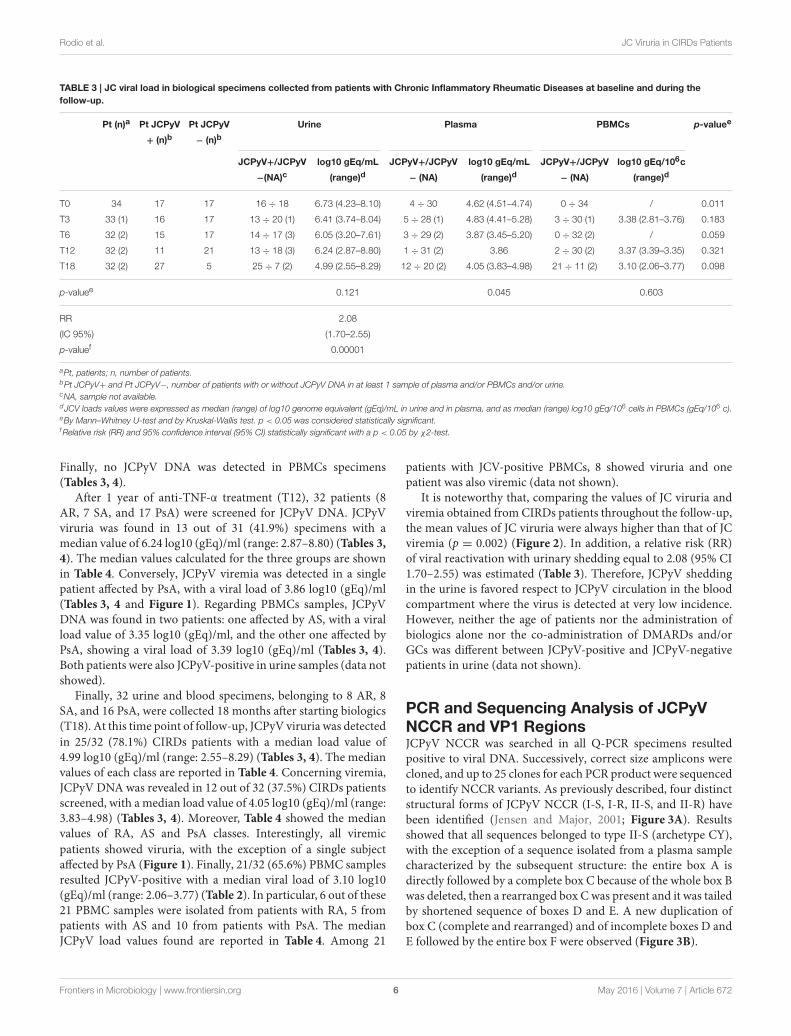
aPt, patients; n, number of patients.bPt JCPyV+ and Pt JCPyV−, number of patients with or without JCPyV DNA in at least 1 sample of plasma and/or PBMCs and/or urine.cNA, sample not available.dJCV loads values were expressed as median (range) of log10 genome equivalent (gEq)/mL in urine and in plasma, and as median (range) log10 gEq/106 cells in PBMCs (gEq/106 c).eBy Mann–Whitney U-test and by Kruskal-Wallis test. p < 0.05 was considered statistically significant.fRelative risk (RR) and 95% confidence interval (95% CI) statistically significant with a p < 0.05 by χ2-test.
Finally, no JCPyV DNA was detected in PBMCs specimens(Tables 3, 4).
After 1 year of anti-TNF-α treatment (T12), 32 patients (8AR, 7 SA, and 17 PsA) were screened for JCPyV DNA. JCPyVviruria was found in 13 out of 31 (41.9%) specimens with amedian value of 6.24 log10 (gEq)/ml (range: 2.87–8.80) (Tables 3,4). The median values calculated for the three groups are shownin Table 4. Conversely, JCPyV viremia was detected in a singlepatient affected by PsA, with a viral load of 3.86 log10 (gEq)/ml(Tables 3, 4 and Figure 1). Regarding PBMCs samples, JCPyVDNA was found in two patients: one affected by AS, with a viralload value of 3.35 log10 (gEq)/ml, and the other one affected byPsA, showing a viral load of 3.39 log10 (gEq)/ml (Tables 3, 4).Both patients were also JCPyV-positive in urine samples (data notshowed).
Finally, 32 urine and blood specimens, belonging to 8 AR, 8SA, and 16 PsA, were collected 18 months after starting biologics(T18). At this time point of follow-up, JCPyV viruria was detectedin 25/32 (78.1%) CIRDs patients with a median load value of4.99 log10 (gEq)/ml (range: 2.55–8.29) (Tables 3, 4). The medianvalues of each class are reported in Table 4. Concerning viremia,JCPyV DNA was revealed in 12 out of 32 (37.5%) CIRDs patientsscreened, with a median load value of 4.05 log10 (gEq)/ml (range:3.83–4.98) (Tables 3, 4). Moreover, Table 4 showed the medianvalues of RA, AS and PsA classes. Interestingly, all viremicpatients showed viruria, with the exception of a single subjectaffected by PsA (Figure 1). Finally, 21/32 (65.6%) PBMC samplesresulted JCPyV-positive with a median viral load of 3.10 log10(gEq)/ml (range: 2.06–3.77) (Table 2). In particular, 6 out of these21 PBMC samples were isolated from patients with RA, 5 frompatients with AS and 10 from patients with PsA. The medianJCPyV load values found are reported in Table 4. Among 21
patients with JCV-positive PBMCs, 8 showed viruria and onepatient was also viremic (data not shown).
It is noteworthy that, comparing the values of JC viruria andviremia obtained from CIRDs patients throughout the follow-up,the mean values of JC viruria were always higher than that of JCviremia (p = 0.002) (Figure 2). In addition, a relative risk (RR)of viral reactivation with urinary shedding equal to 2.08 (95% CI1.70–2.55) was estimated (Table 3). Therefore, JCPyV sheddingin the urine is favored respect to JCPyV circulation in the bloodcompartment where the virus is detected at very low incidence.However, neither the age of patients nor the administration ofbiologics alone nor the co-administration of DMARDs and/orGCs was different between JCPyV-positive and JCPyV-negativepatients in urine (data not shown).
PCR and Sequencing Analysis of JCPyVNCCR and VP1 RegionsJCPyV NCCR was searched in all Q-PCR specimens resultedpositive to viral DNA. Successively, correct size amplicons werecloned, and up to 25 clones for each PCR product were sequencedto identify NCCR variants. As previously described, four distinctstructural forms of JCPyV NCCR (I-S, I-R, II-S, and II-R) havebeen identified (Jensen and Major, 2001; Figure 3A). Resultsshowed that all sequences belonged to type II-S (archetype CY),with the exception of a sequence isolated from a plasma samplecharacterized by the subsequent structure: the entire box A isdirectly followed by a complete box C because of the whole box Bwas deleted, then a rearranged box Cwas present and it was tailedby shortened sequence of boxes D and E. A new duplication ofbox C (complete and rearranged) and of incomplete boxes D andE followed by the entire box F were observed (Figure 3B).
Frontiers in Microbiology | www.frontiersin.org 6 May 2016 | Volume 7 | Article 672
TABLE 4 | JC viral load in biological samples collected from patients with psoriatic arthritis, rheumatoid arthritis and ankylosing spondylitis at baseline
*Times of follow up: baseline (T0) and 3, 6, 12, and 18 months (T3, T6, T12, and T18).§JCPyV loads values were expressed as median (range) of log10 genome equivalent (gEq)/mL in urine and in plasma, and as median (range) log10 gEq/106 cells in PBMCs.
Frontiers in Microbiology | www.frontiersin.org 7 May 2016 | Volume 7 | Article 672
FIGURE 1 | JCPyV urine loads vs. the corresponding plasma viral loads in CIRDs patients during the 18 month follow-up study. The values of JCPyV
viruria for each patient at the different time-points were reported on the x-axis whereas the corresponding viremia values on the y-axis. All JCPyV loads values were
expressed as median of log10 genome equivalent (gEq)/mL in urine and in plasma. Viremia was mainly associated to viruria during the entire follow-up.
Regarding the binding sites present in this rearrangedsequence, the TATA box and the binding site for Tst-1, requiredfor JCPyV replication and transcription of early and lategenes, respectively, were both well conserved within the boxA (Figure 3B). About the multiple duplication of box C, itenhances the binding site for the cyclic AMP (cAMP) responseelement (CRE), a protein that up-modulates JCPyV expressionin cells, and for NF-1, responsible for the JCPyV neurotropism(Figure 3B). In addition, the binding sites for the cellular factorsNF-1, p53, and AP-1, involved in early viral transcription and inneurotropism of the virus, are present within the box F, althougha single point mutation occurred into the NF-1 binding site(Figure 3B).
Finally, an additional specific PCR was undertaken in orderto characterize the viral genotypes circulating within our CIRDscohort. Results showed a prevalence of the European genotypes1A, 1B, and 4, followed by the Eurasian genotype 2B and theAsian type 2E. In particular, genotypes 1A and 1B were identifiedin 7 patients (1A in 4 PsA and 3 RA; 1B in 4 PsA, 2 RA, and 1 AS),type 4 in 5 patients (3 PsA and 1 RA), whereas types 2B and 2E
were detected in a patient affected by PsA and in a patient withAS, respectively (data not shown).
DISCUSSION
Biologic therapies have successfully been introduced intothe treatment of several inflammatory rheumatic diseases inparticular, monoclonal antibodies or fusion proteins targetingTNF-α are widely used for the treatment of CIRDs patientsrefractory to conventional immune-suppressive medications.Nevertheless, treatments with biological drugs are associated withan increased susceptibility to viral infections including that byJCPyV, the etiological agent of the demyelinating disease namedPML (Comar et al., 2013; Iacobaeus et al., 2013). The incidenceof PML in immune-mediated diseases has recently increased as aconsequence of an improved use of biologics and other potentimmune-modulatory medications (Berger, 2010). Few studiesare present in literature that demonstrate a real risk of PMLdevelopment in CIRDs patients whereas several researches havefocused on a possible correlation between JCPyV viremia and the
Frontiers in Microbiology | www.frontiersin.org 8 May 2016 | Volume 7 | Article 672
FIGURE 2 | Comparison of JCPyV viruria and viremia detected in
CIRDs patients in a 18 month follow-up study. At T0, JCPyV DNA was
revealed in 47.1% urine with a median value of 6.73 log10 (gEq)/ml and in
11.8% plasma with a median value of 4.62 log10 (gEq)/ml. At T3, 39.4% of
urine and 15.2% of plasma resulted JCPyV positive with a median value of
6.41 log10 (gEq)/ml and of 4.83 log10 (gEq)/ml respectively. At T6, in 45.2%
urine (median value 6.05 log10 (gEq)/ml) and in 9.4% plasma (median value
3.87 log10 (gEq)/ml), JCPyV genome was found. At T12, JCPyV viruria was
found in 41.9% specimens with a median value of 6.24 log10 (gEq)/ml.
Conversely, JCPyV viremia was detected in a single PsA patient with a viral
load of 3.86 log10 (gEq)/ml. Finally, at T18, JCPyV viruria was detected in
78.1% of CIRDs patients with a median load value of 4.99 log10 (gEq)/ml,
whereas JCPyV viremia was revealed in 37.5% of samples (median value of
4.05 log10 (gEq)/ml). JCPyV viruria was significantly higher than JCPyV viremia
throughout the entire follow-up (p = 0.002). JCPyV load values are expressed
as log10 genome equivalent per milliliter (gEq/mL). T0: baseline; T3, T6, T12
and T18: 3, 6, 12, and 18 months of anti-TNF-α, therapy. *indicates the
highest viremia value detected in one patient.
biological therapy in patients with multiple sclerosis (MS) andCD (Lavagna et al., 2007; Verbeeck et al., 2008; Bellizzi et al., 2011,2013a,b; Bharat et al., 2012; Comar et al., 2013; Iacobaeus et al.,2013; Tur et al., 2013; Frohman et al., 2014). Therefore, it could beinteresting to understand whether there is a correlation betweenbiologics administered for CIRDs and the opportunity that thevirus escapes from latency, replicates actively and spreads to thebrain causing PML. In fact, despite the limited range of speciesand permissive cell types for viral replication, JCPyV is a verysuccessful pathogen because of it is able to tightly regulate its lifecycle in the infected host.
In this study, 34 patients affected by RA, AS and PsA werescreened to detect the presence of JCPyV in blood and in urinesamples in order to evaluate the risk of JCPyV dissemination tothe CNS under treatments with biologic agents and/or DMARDsand GCs. Patients were dynamically enrolled and studied frombaseline, before first anti-TNF-α infusion, up to 18 monthsafter.
A persistent JCPyV viruria significantly higher than JCPyVviremia was observed from baseline throughout the 18 monthfollow-up (p = 0.002). It could be explained taking in account
that the concomitant use of conventional therapies (DMARDsand GCs) and anti-TNF-α treatments, rather than a singlebiologic, endorsed JCPyV replication in urinary compartment.In fact, over the time, no difference was observed betweenJCPyV-positive and JCPyV-negative patients treated with singleadministration of biologics or co-administration of DMARDsand/or GC (Table 5). Conversely, a previous study demonstrateda positive correlation between the JCPyV DNA detection in theurine and the number of biologics consecutively used for RAtreatment (Verheyen et al., 2015). However, it is possible tohypothesize that the prolonged administration of biologic agentsover time had a causative role in the increasing number ofpatients with JC viruria after 18months of anti-TNF-α treatment.Considering that TNF-α cytokine plays an important role in hostdefense, it is feasible that the use of anti-TNF-α agents endorseJCV replication in the kidney. In fact, Q-PCR revealed thatviral replication occurred in the urinary tract at high copy levelsand that the virus could escape into the peripheral circulationas demonstrated by the fact that JCPyV DNA was detectedwith an increased frequency in plasma and PBMCs at T18. Inaddition, the effect of an ongoing immunosuppression on theJCPyV replication is confirmed by the striking number of urinepositive samples detected at T18. These data allow an intriguingspeculation: JCPyV might directly trigger joint inflammation.Indeed, it is well-known that viral infections can directly act onthe immune system through the secretion of pro-inflammatorycytokines or favoring the production of autoantibodies (FranssilaandHedman, 2006). Therefore, urine JCPyV loads not only couldbe supported by inflammatory state but also could be continuousdue to this insult.
The hypothesis of a role of biologics in promoting viruriais also sustained by the results of an our previous study, inwhich we observed a significantly increased JC viruria in youngpatients with Crohn’s disease treated with infliximab respectto those receiving a standard therapy (Bellizzi et al., 2011).Regarding viremia, it was mainly associated to viruria duringthe entire follow-up (Figure 1). Although JC viremia seems tobe essential for the development of PML and it was detectedin 37.5% (12/32) of our patients at T18, its short temporalityprecludes its usefulness in screening or diagnostic algorithms, asalready demonstrated by other Authors in comparable studies(Rinaldi et al., 2010; Bellizzi et al., 2015; Verheyen et al.,2015). Hence, monitoring urine JCPyV replication is a good,non-invasive method to check viral pathogenic potential. Inconclusion, results evidenced how viral replication and spreadingcould be cumulatively influenced by the use of various immune-suppressants, including biologics, rather than by a specificmedications, also considering their sequential administrationin the treatment refractory patients. However, due to the lownumber of patients analyzed in this study, how cumulative effectof various immunosuppressive agents really influence JCPyVpathogenesis, need further validation. Therefore, the cohort ofstudied subjects is being expanded.
In JCPyV DNA positive patients, NCCR sequencing alwaysrevealed the presence of archetype-like structures, according toother Authors (Giannecchini et al., 2012; Verheyen et al., 2015),except for a rearranged NCCR form detected in the plasma
Frontiers in Microbiology | www.frontiersin.org 9 May 2016 | Volume 7 | Article 672
FIGURE 3 | Comparison of NCCR structural forms and rearranged NCCR II-R found in a plasma sample at T3. (A) Type I-S is 98 base-pair (bp) long and it
is composed of box A (25 bp), box C (55 bp), box E (18 bp) and F (69 bp). Type I-R has repeats of this 98 bp unit, with various deletions, as seen in the JCPyV
prototype Mad-1 (GenBank no: J02227), that have no box B and box D (Jensen and Major, 2001). In particular, the prototype Mad-1 was isolated from tissues of
patients with PML (Tan and Koralnik, 2010) and it was named on the hypothesis that the prototype results from a rearrangement of the archetype sequence (Comar
et al., 2013). Type II-S is identified as archetype CY and it is composed of A (25 bp), B (23 bp), C (55 bp), D (66 bp), E (18 bp), and F (69 bp) boxes. It was isolated by
Yogo et al. (1990). Each box contains binding sites for transcriptional cell factors involved in viral early and late transcription. These binding sites undergo to deletion
and enhancement process that could generate variants that could up-modulate viral expression in a specific anatomical site (Jensen and Major, 2001). (B) In (B), the
rearranged sequence found in plasma at T3 is reported. This rearrangement presents the entire boxes A and C followed by a rearranged box C, shortened sequences
of boxes D and E, a new duplication of box C (complete and rearranged), incomplete boxes D and E and finally the entire box F. Asterisks represent single nucleotide
point mutations or deletions. Italicized capital letters indicate mutated nucleotides. The TATA box is presented by TATA. Boxes division from A to F is also shown. The
main binding sites for transcriptional cell factors are also indicated and the corresponding nucleotides sequences are underlined. Finally, the nucleotides sequences for
the transcriptional factor Spi-B are also shown in higher font.
sample of a patient affected by PsA (Figure 3B). Interestingly, thistype II-R rearrangement resembles the viral variants isolated insubjects who developed PML, and it is characterized by a markedneurotropism (Agostini et al., 2001; Marzocchetti et al., 2007).Indeed, it showed several repeats of the box C containing the CREelement, a specific enhancer of JCPyV replication in glial cells(Kumar et al., 1996). Moreover, it is noteworthy that this NCCRstructure presents a high-affinity binding site for the specifichematopoietic transcriptional factor Spi-B. In fact, it has beendemonstrated that Spi-B protein actively binds its site presenton Mad-1, but not in the non-pathogenic II-S (CY) (Marshallet al., 2010, 2012). Therefore, it is possible to hypothesize thatthis neurovirulent variant could enter PBMCs, using them as a
carrier to disseminate in the bloodstream and to reach the brain(Kumar et al., 1996). Furthermore, it is well-known that detectingviral genome in cerebrospinal fluid lied on PML diagnosis,however, its failure does not rule out the possibility that a patientmight have PML, particularly in the earlier stages (Mischitelliet al., 2013). Hence, identifying in the blood a virus withPML-associated NCCR rearrangements should alert clinicians,favoring an individual management of the patient.
Finally, regarding JCPyV genotyping, VP1 sequencingevidenced that genotype 1A, 1B, and 4 were the most prevalent,although genotypes 2B and 2E were also found. Type 1and type 4 are generally associated with Europeans andEuropean-Americans, whereas type 2B and 2E were typical
Frontiers in Microbiology | www.frontiersin.org 10 May 2016 | Volume 7 | Article 672
of Asians and Eurasians and of Western Pacific populations,respectively (Agostini et al., 2001). Interestingly, JCPyV subtype2B found in PsA patient with the PML-associated NCCRrearrangement in the blood, has been associated with increasedincidence of PML, while type 4 has been associated with lowerdisease risk (Agostini et al., 2001). Moreover, type 1 and type4 were found in urine of Italian patients affected by immune-mediated diseases, suggesting a possible JCPyV genotypeselection in response to pressure by immunomodulatory drugs(Zanotta et al., 2013).
Recently, observations of point mutations in the VP1 capsidgene have also been shown to be associated with PML (Delbueet al., 2009; Reid et al., 2011). Although VP1 gene is highlypolymorphic, mutations appear to be strongly patient-related andthey have been observed in virus characterized by rearrangedNCCR. Practically, their arising is only noted in a neuroaggressive viral variant evolved by the non-pathogenic form,according to cell alterations or global environment changes. Insummary, either VP1 mutations and NCCR rearrangements arethe most common viral alterations associated with PML (Delbueet al., 2009; Reid et al., 2011).
In conclusion, since biological therapies are promisingfor the treatment of immune-mediated disorders, little isknown about their contribution to the development of PML.However, it is clear that PML has been identified as aserious adverse event, hence it is interesting to clarifyhow anti TNF-α agents act on JCPyV immune-surveillanceendorsing viral reactivation and dissemination. To date,epidemiology of PML has been poorly characterized amongpatients with rheumatic diseases due to little population-based data existing. Therefore, this study contributes to enrichliterature insight on JCPyV biology in this cluster of patients,considering that the involvement of JC virus in developmentof adverse events in CIRDs is probably underestimated sincefew studies have been done about. Thus, it is necessaryto carry on investigations in order to understand the realimpact of biologic and/or other immunosuppressive therapieson JCPyV life cycle and to identify possible and specific viralvariants that, in CIRDs patients, could be related to increasedvirulence.
AUTHOR CONTRIBUTIONS
DMR, EA, MM, AB, RS, and VP designed research. DMR, EA,MM, AB, DS, GC, and CP performed experiments. All authorsanalyzed data. DMR, EA, MM, AB, and VP wrote the paper withcontribution of ATP and MT during revision.
ACKNOWLEDGMENTS
This work was supported by MIUR GRANT no C26A13CH,MIUR GRANT no C26A15PH4A and Minister of Health Grantno J82I14001080001. Anna Bellizzi was supported by post-doctoral fellowship “Teresa Ariaudo 2013” dispensed by InstitutePasteur Cenci-Bolognetti Foundation. The Authors gratefullyacknowledge Rocco Cetera for his precious suggestions and forhis friendly availability.
Frontiers in Microbiology | www.frontiersin.org 11 May 2016 | Volume 7 | Article 672
Agostini, H. T., Deckhut, A., Jobes, D. V., Girones, R., Schlunck, G.,Prost, M. G., et al. (2001). Genotypes of JC virus in East, Central andSouthwest Europe. J. Gen. Virol. 82, 1221–1331. doi: 10.1099/0022-1317-82-5-1221
Arnett, F. C., Edworthy, S. M., Bloch, D. A., McShane, D. J., Fries, J. F., Cooper, N.S., et al. (1988). The American Rheumatism Association 1987 revised criteriafor the classification of rheumatoid arthritis. Arthritis Rheum. 31, 315–324. doi:10.1002/art.1780310302
Bellizzi, A., Anzivino, E., Ferrari, F., Di Nardo, G., Colosimo, M. T., Fioriti,D., et al. (2011). Polyomavirus JC reactivation and noncoding control regionsequence analysis in pediatric Crohn’s disease patients treated with infliximab.J. Neurovirol. 17, 303–313. doi: 10.1007/s13365-011-0036-3
Bellizzi, A., Anzivino, E., Rodio, D. M., Cioccolo, S., Scrivo, R., Morreale, M.,et al. (2013a). Human Polyomavirus JC monitoring and noncoding controlregion analysis in dynamic cohorts of individuals affected by immune-mediateddiseases under treatment with biologics: an observational study. Virol. J. 30,298–316. doi: 10.1186/1743-422X-10-298
Bellizzi, A., Anzivino, E., Rodio, D. M., Palamara, A. T., Nencioni, L.,and Pietropaolo, V. (2013b). New insights on human polyomavirus JCand pathogenesis of progressive multifocal leukoencephalopathy. Clin. Dev.Immunol. 2013, 1–17. doi: 10.1155/2013/839719
Bellizzi, A., Mischitelli, M., Anzivino, E., Scrivo, R., Rodio, D. M., Scribano,D., et al. (2015). Human polyomavirus JC presence in chronic inflammatoryrheumatic diseases patients treated with anti-TNF-α: evaluation of JC viralloads in urine and plasma samples. Joint Bone Spine 82, 375–376. doi:10.1016/j.jbspin.2014.12.010
Berger, J. R. (2010). Progressive multifocal leukoencephalopathy and newerbiological agents. Drug Saf. 33, 969–983. doi: 10.2165/11537510-000000000-00000
Bharat, A., Xie, F., Baddley, J. W., Beukelman, T., Chen, L., Calabrese,L., et al. (2012). Incidence and risk factors for progressive multifocalleukoencephalopathy among patients with selected rheumatic diseases.Arthritis Care Res. 64, 612–615. doi: 10.1002/acr.21564
Bijlsma, J. W. (2012). Disease control with glucocorticoid therapy in rheumatoidarthritis. Rheumatology 51, iv9–iv13. doi: 10.1093/rheumatology/kes086
Borie, D., and Kremer, J. M. (2015). Considerations on the appropriatenessof the John Cunningham virus antibody assay use in patients withrheumatoid arthritis. Semin. Arthritis Rheum. 45, 163–166. doi:10.1016/j.semarthrit.2015.06.003
Bøyum, A., Løvhaug, D., Tresland, L., and Nordlie, E. M. (1991). Separationof leucocytes: improved cell purity by fine adjustments of gradient mediumdensity and osmolality. Scand. J. Immunol. 34, 697–712. doi: 10.1111/j.1365-3083.1991.tb01594.x
Comar, M., Delbue, S., Lepore, L., Martelossi, S., Radillo, O., Ronfani, L., et al.(2013). Latent viral infections in young patients with inflammatory diseasestreated with biological agents: prevalence of JC virus genotype 2. J. Med. Virol.
5, 716–722. doi: 10.1002/jmv.23525Dejager, L., Vandevyver, S., Petta, I., and Libert, C. (2014). Dominance of the
strongest: inflammatory cytokines versus glucocorticoids. Cytokine Growth
Factor Rev. 25, 21–33. doi: 10.1016/j.cytogfr.2013.12.006Delbue, S., Branchetti, E., Bertolacci, S., Tavazzi, E., Marchioni, E., Maserati,
R., et al. (2009). JC virus VP1 loop-specific polymorphisms are associatedwith favorable prognosis for progressive multifocal leukoencephalopathy. J.Neurovirol. 15, 51–56. doi: 10.1080/13550280802425467
Delbue, S., Branchetti, E., Boldorini, R., Vago, L., Zerbi, P., Veggiani, C., et al.(2008). Presence and expression of JCV early gene large T Antigen in the brainsof immunocompromised and immunocompetent individuals. J. Med. Virol. 80,2147–2152. doi: 10.1002/jmv.21313
Dewing, K. A. (2015). Management of patients with psoriatic arthritis.Nurse Pract.40, 40–46. doi: 10.1097/01.NPR.0000461950.23292.18
Egli, A., Infanti, L., Dumoulin, A., Buser, A., Samaridis, J., Stebler, C., et al. (2009).Prevalence of polyomavirus BK and JC infection and replication in 400 healthyblood donors. J. Infect. Dis. 199, 837–846. doi: 10.1086/597126
Ferenczy, M. W., Marshall, L. J., Nelson, C. D., Atwood, W. J., Nath, A., Khalili, K.,et al. (2012). Molecular biology, epidemiology, and pathogenesis of progressivemultifocal leukoencephalopathy, the JC virus-induced demyelinating disease
of the human brain. Clin. Microbiol. Rev. 25, 471–506. doi: 10.1128/CMR.05031-11
Flaegstad, T., Sundsfjord, A., Arthur, R. R., Pedersen, M., Traavik, T., andSubramani, S. (1991). Amplification and sequencing of the control regions ofBK and JC virus from human urine by polymerase chain reaction. Virology 180,553–560. doi: 10.1016/0042-6822(91)90069-N
Franssila, R., and Hedman, K. (2006). Infection and musculoskeletal conditions:viral causes of arthritis. Best Pract. Res. Clin. Rheumatol. 20, 1139–1157. doi:10.1016/j.berh.2006.08.007
Fries, J. F., Spitz, P. W., and Young, D. Y. (1982). The dimensions of healthoutcomes: the Health Assessment Questionnaire, disability and pain scales. J.Rheumatol. 9, 789–793.
Frohman, E. M., Monaco, M. C., Remington, G., Ryschkewitsch, C., Jensen, P. N.,Johnson, K., et al. (2014). JC Virus in CD34+ and CD19+ cells in patients withmultiple sclerosis treated with natalizumab. JAMA Neurol. 71, 596–602. doi:10.1001/jamaneurol.2014.63
Garrett, S., Jenkinson, T., Kennedy, L. G., Whitelock, H., Gaisford, P., and Calin,A. (1994). A new approach to defining disease status in ankylosing spondylitis:the Bath Ankylosing Spondylitis Disease Activity Index. J. Rheumatol. 21,2286–2291.
Gerlag, D. M., Norris, J. M., and Tak, P. P. (2015). RA: from risk factorsand pathogenesis to prevention. Towards prevention of autoantibody-positiverheumatoid arthritis: from lifestyle modification to preventive treatment.Rheumatology (Oxford) 53, 1–8. doi: 10.1093/rheumatology/kev347
Ghasemi-Rad, M., Attaya, H., Lesha, E., Vegh, A., Maleki-Miandoab, T., Nosair, E.,et al. (2015). Ankylosing spondylitis: a state of the art factual backbone. World
J. Radiol. 7, 236–252. doi: 10.4329/wjr.v7.i9.236Giannecchini, S., Clausi, V., Vultaggio, A., Macera, L., Maggi, F., Martelli, F.,
et al. (2012). Assessment of the risk of polyomavirus JC reactivation in patientswith immuno-mediated diseases during long-term treatment with infliximab.J. Neurovirol. 18, 55–61. doi: 10.1007/s13365-012-0078-1
Graff-Radford, J., Robinson, M. T., Warsame, R. M., Matteson, E. L., Eggers, S. D.,and Keegan, B. M. (2012). Progressive multifocal leukoencephalopathyin a patient treated with etanercept. Neurologist 18, 85–87. doi:10.1097/NRL.0b013e318247b868
Hirsch, H. H., Kardas, P., Kranz, D., and Leboeuf, C. (2013). The humanJC polyomavirus (JCPyV): virological background and clinical implications.APMIS 121, 685–727. doi: 10.1111/apm.12128
Iacobaeus, E., Hopia, L., Khademi, M., Lundén, M., Hammarin, A. L.,Svenungsson, E., et al. (2013). Analysis of JC virus DNA in NPSLE patientstreated with different immunomodulatory agents. Lupus 22, 307–311. doi:10.1177/0961203312470977
Jensen, P. N., and Major, E. O. (2001). A classification scheme for humanpolyomavirus JCV variants based on the nucleotide sequence of the noncodingregulatory region. J. Neurovirol. 7, 280–287. doi: 10.1080/13550280152537102
Jobes, D. V., Friedlaender, J. S., Mgone, C. S., Agostini, H. T., Koki, G.,Yanagihara, R., et al. (2001). New JC virus (JCV) genotypes from papua newguinea and micronesia (type 8 and type 2E) and evolutionary analysis of 32complete JCV genomes. Arch. Virol. 146, 2097–2113. doi: 10.1007/s007050170023
Kelly, J. C. (2015). Rheumatoid Arthritis: Updated Recommendations
Released. Medscape Medical News. Available online at:http://www.medscape.com/viewarticle/845495
Kumar, K. U., Tang, S. C., Pater, M. M., and Pater, A. (1996). Glial and muscleembryonal carcinoma cell specific independent regulation of expression ofhuman JC virus early promoter by cyclic AMP response elements and adjacentnuclear factor 1 binding sites. J. Med. Virol. 49, 199–204.
Kwok, S., and Higuchi, R. (1989). Avoiding false positive with PCR. Nature 339,232–238. doi: 10.1038/339237a0
Lahiri, M., and Dixon, W. G. (2015). Risk of infection with biologic antirheumatictherapies in patients with rheumatoid arthritis. Best Pract. Res. Clin. Rheumatol.
29, 290–305. doi: 10.1016/j.berh.2015.05.009Lavagna, A., Bergallo, M., Daperno, M., Sostegni, R., Costa, C., Leto, R.,
et al. (2007). Infliximab and the risk of latent viruses reactivation inactive Crohn’s disease. Inflamm. Bowel Dis. 13, 896–902. doi: 10.1002/ibd.20131
Lesuis, N., Befrits, R., Nyberg, F., and van Vollenhoven, R. F. (2012). Gender andthe treatment of immunemediated chronic inflammatory diseases: rheumatoid
Frontiers in Microbiology | www.frontiersin.org 12 May 2016 | Volume 7 | Article 672
arthritis, inflammatory bowel disease and psoriasis: an observational study.BMCMed. 10:82. doi: 10.1186/1741-7015-10-82
Markowitz, R. B., Thompson, H. C., Mueller, J. F., Cohen, J. A., and Dynan,W. S. (1993). Incidence of BK virus and JC virus viruria in humanimmunodeficiency virus infected and uninfected subjects. J. Infect. Dis. 167,13–20. doi: 10.1093/infdis/167.1.13
Marshall, L. J., Dunham, L., and Major, E. O. (2010). Transcription factor Spi-B binds unique sequences present in the tandem repeat promoter/enhancerof JC virus and supports viral activity. J. Gen. Virol. 91, 3042–3052. doi:10.1099/vir.0.023184-0
Marshall, L. J., and Major, E. O. (2010). Molecular regulation of JCvirus tropism: insights into potential therapeutic targets for progressivemultifocal leukoencephalopathy. J. Neuroimmune Pharmacol. 5, 404–417. doi:10.1007/s11481-010-9203-1
Marshall, L. J., Moore, L. D., Mirsky, M. M., and Major, E. O. (2012). JC viruspromoter/enhancers contain TATA box-associated Spi-B binding sites thatsupport early viral gene expression in primary astrocytes. J. Gen. Virol. 93,651–661. doi: 10.1099/vir.0.035832-0
Marzocchetti, A., Sanguinetti, M., Giambenedetto, S. D., Cingolani, A., Fadda, G.,Cauda, R., et al. (2007). Characterization of JC virus in cerebrospinal fluidfromHIV-1 infected patients with progressive multifocal leukoencephalopathy:insights into viral pathogenesis and disease prognosis. J. Neurovirol. 13,338–346. doi: 10.1080/13550280701381324
Mischitelli, M., Fioriti, D., Bellizzi, A., Anzivino, E., Chiarini, F., and Pietropaolo,V. (2013). “The Human Polyomavirus JC and progressive multifocalleukoencephalopathy,” in Neuroviral Infections: General Principles and DNA
Viruses, ed Taylor & Francis Group LLC (New York, NY: CRC Press), 347–364.Mócsai, A., Kovács, L., and Gergely, P. (2014). What is the future of targeted
therapy in rheumatology: biologics or small molecules? BMC Med. 12:43. doi:10.1186/1741-7015-12-43
Moll, J. M., and Wright, V. (1973). The pattern of chest and spinal mobilityin ankylosing spondylitis: un objective clinical study of 106 patients.Rheumatology 12, 115–134. doi: 10.1093/rheumatology/12.3.115
Molloy, E. S., and Calabrese, L. H. (2012). Progressive multifocalleukoencephalopathy associated with immunosuppressive therapy inrheumatic diseases: evolving role of biologic therapies. Arthritis Rheum.
64, 3043–3051. doi: 10.1002/art.34468Nardis, C., Anzivino, E., Bellizzi, A., Rodio, D. M., De Pità, O., and Pietropaolo, V.
(2012). Reactivation of human polyomavirus JC in patients affected by psoriasisvulgaris and psoriatic arthritis and treated with biological drugs: preliminaryresults. J. Cell Physiol. 227, 3796–3802. doi: 10.1002/jcp.24089
Pavlovic, D., Patera, A. C., Nyberg, F., Gerber, M., and Liu, M. (2015). ProgressiveMultifocal Leukeoncephalopathy Consortium. Progressive multifocalleukoencephalopathy: current treatment options and future perspectives.Ther. Adv. Neurol. Disord. 8, 255–273. doi: 10.1177/1756285615602832
Pietropaolo, V., Videtta, M., Fioriti, D., Mischitelli, M., Arancio, A., Orsi, N., et al.(2003). Rearrangement patterns of JC virus noncoding control region fromdifferent biological samples. J. Neurovirol. 9, 603–611. doi: 10.1080/714044482
Reid, C. E., Li, H., Sur, G., Carmillo, P., Bushnell, S., Tizard, R., et al. (2011).Sequencing and analysis of JC virus DNA from natalizumab-treated PMLpatients. J. Infect. Dis. 204, 237–244. doi: 10.1093/infdis/jir256
Rinaldi, L., Rinaldi, F., Perini, P., Calabrese, M., Seppi, D., Grossi, P., et al. (2010).No evidence of JC virus reactivation in natalizumab treated multiple sclerosispatients: an 18 month follow-up study. J. Neurol. Neurosurg. Psychiatr. 81,1345–1350. doi: 10.1136/jnnp.2009.201079
Tan, C. S., and Koralnik, I. J. (2010). Progressive multifocal leukoencephalopathyand other disorders caused by JC virus: clinical features and pathogenesis.Lancet Neurol. 9, 425–437. doi: 10.1016/S1474-4422(10)70040-5
Taylor, W., Gladman, D., Helliwell, P., Marchesoni, A., Mease, P., Mielants, H.,et al. (2006). Classification criteria for psoriatic arthritis. Development of newcriteria from a large international study. Arthritis Rheum. 54, 2665–2673. doi:10.1002/art.21972
Tur, C., Tintoré, M., Vidal-Jordana, Á., Bichuetti, D., Nieto González, P.,Arévalo, M. J., et al. (2013). Risk acceptance in multiple sclerosis patientson natalizumab treatment. PLoS ONE 8:e82796. doi: 10.1371/journal.pone.0082796
Van der Linden, S., Valkenburg, H. A., and Cats, A. (1984). Evaluation of diagnosticcriteria for ankylosing spondylitis. A proposal for modification of the NewYorkcriteria. Arthritis Rheum. 27, 361–368. doi: 10.1002/art.1780270401
Verbeeck, J., Van Assche, G., Ryding, J., Wollants, E., Rans, K., Vermeire,S., et al. (2008). JC viral loads in patients with Crohn’s disease treatedwith immunosuppression: can we screen for elevated risk of progressivemultifocal leukoencephalopathy? Gut 57, 1393–1397. doi: 10.1136/gut.2007.145698
Verheyen, J., Maizus, K., Feist, E., Tolman, Z., Knops, E., Saech, J., et al.(2015). Increased frequency of JC-polyomavirus detection in rheumatoidarthritis patients treated withmultiple biologics.Med.Microbiol. Immunol. 204,613–618. doi: 10.1007/s00430-015-0390-5
Yogo, Y., Kitamura, T., Sugimoto, C., Ueki, T., Aso, Y., Hara, K., et al.(1990). Isolation of a possible archetypal JC virus DNA sequence from non-immunocompromised individuals. J. Virol. 64, 3139–3143.
Zanotta, N., Delbue, S., Rossi, T., Pelos, G., D’Agaro, P., Monasta, L., et al. (2013).Molecular epidemiology of JCV genotypes in patients and healthy subjects fromNorthern Italy. J. Med. Virol. 85, 1286–1292. doi: 10.1002/jmv.23585
Conflict of Interest Statement: The authors declare that the research wasconducted in the absence of any commercial or financial relationships that couldbe construed as a potential conflict of interest.