Page 1
1
Incretin action in the pancreas: Potential promise, possible perils, and pathological pitfalls
Running title: Incretin biology and the pancreas
Daniel J. Drucker
From the Department of Medicine, Mt. Sinai Hospital, Samuel Lunenfeld Research Institute,
University of Toronto
Figures: 2 Supplementary Tables 1; Word count 4,645
Address correspondence to:
Dr. Daniel J Drucker
Samuel Lunenfeld Research Institute
Mt. Sinai Hospital 600 University Ave TCP5-1004
Toronto Ontario Canada M5G 1X5
416-361-2661 V 416-361-2669 F [email protected]
Page 1 of 31 Diabetes
Diabetes Publish Ahead of Print, published online July 1, 2013
Page 2
2
Glucagon-like peptide-1 (GLP-1) and glucose-dependent insulinotropic polypeptide (GIP) are incretin
hormones that control secretion of insulin, glucagon and somatostatin to facilitate glucose disposal. The
actions of incretin hormones are terminated via enzymatic cleavage by dipeptidyl peptidase-4 (DPP-4),
and through renal clearance. GLP-1 and GIP promote β-cell proliferation and β-cell survival in rodent β-
cells. DPP-4 inhibitors expand β-cell and reduce α-cell mass and inhibit glucagon secretion in preclinical
studies however whether incretin-based therapies sustain functional β-cell mass in human diabetic
subjects remains unclear. GLP-1 and GIP exert their actions predominantly through unique G protein
coupled receptors expressed on β-cells and other pancreatic cell types. Accurate localization of incretin
receptor expression in pancreatic ductal or acinar cells in normal or diabetic human pancreas is
challenging as antisera employed for detection of the GLP-1R are often neither sufficiently sensitive nor
specific to yield reliable data. We review recent advances and controversies in incretin hormone action in
the pancreas and contrast established mechanisms with areas of uncertainty. Furthermore, we highlight
methodological challenges and pitfalls and outline key areas requiring additional scientific investigation.
Page 2 of 31Diabetes
Page 3
3
Introduction
Incretins are gut-derived circulating peptide hormones that potentiate glucose-dependent insulin
secretion following meal ingestion. Glucose-dependent insulinotropic polypeptide (GIP), and
glucagon-like peptide-1 (GLP-1), are the major incretin hormones. The insulinotropic actions of
endogenously secreted GLP-1 and GIP are transient, as both peptides are rapidly cleared by the
kidney, and inactivated by cleavage at the N-terminus by a ubiquitous exopeptidase, dipeptidyl
peptidase-4 (DPP-4). Potentiation of incretin action underlines two therapeutic classes of
glucose-lowering agents, the GLP-1R agonists, and the DPP-4 inhibitors (1). Original concepts
of GIP and GLP-1 biology focused primarily on islet β-cells have been expanded to include
actions on other cell types within and outside the pancreas (2; 3). There is now considerable
interest in understanding how potentiation of incretin action controls multiple facets of
pancreatic biology, encompassing regulation of glucose sensing, hormone secretion, cell
proliferation, differentiation, and survival. Recent studies have suggested that incretin therapies
promote pancreatic inflammation, and aberrant cell proliferation within the endocrine and
exocrine pancreas (4; 5), however substantial technical and methodological issues limit the
generalizability of these findings. This Perspective evaluates the science supporting existing
dogma, and discusses new concepts, controversies, and uncertainties in the biology of incretin
action in the pancreas.
Localization of incretin receptor expression in the pancreas
Several dozen commercial antisera are available for detection of GLP-1 and GIP receptor
expression by immunohistochemical techniques and Western blotting, and Real Time PCR is
widely used to quantify incretin receptor gene expression in pancreatic exocrine and endocrine
Page 3 of 31 Diabetes
Page 4
4
compartments. Most antisera used to detect GLP-1R expression (by immunohistochemistry or
Western blot analysis) are neither sensitive nor specific (6; 7). Important control experiments
(absorption of the antibody with a peptide epitope, demonstration that the antibody recognizes
only a single protein, and fails to generate a signal in cells that do not express a full length
receptor mRNA transcript or in tissues from Glp1r-/- mice) are usually absent. Furthermore,
multiple studies describe GLP-1 receptor protein expression in cells or tissues that do not express
a full length Glp1r mRNA. The widespread use of tightly cropped bands in Western blot analysis
precludes accurate assessment of whether a putative band/protein detected by Western blotting is
the correct size, the only GLP-1R-immunoreactive protein visualized, or one of several unrelated
immunoreactive proteins detected by the same antisera.
Scientists interested in incretin hormone receptor expression face the challenging task of
assessing how much, if any, of the data published with these antisera is correct. For example,
immunoreactive GLP-1R protein expression or Glp1r mRNA transcripts have been detected
throughout the heart and ventricle, however we and others determined that cardiac Glp1r
expression was restricted to the atria, and absent from the ventricles in mice (8) and rats (9).
How do the limitations of available reagents impact our understanding of incretin action in the
pancreas? The putative localization of incretin receptor expression in the exocrine pancreas
provides an instructive example. Abundant immunohistochemical GLP-1 receptor expression in
ductal and acinar cells was reported in rodent and human pancreas, in papillary thyroid cancer
and pancreatic adenocarcinoma (10; 11). Characterization of multiple GLP-1R antisera,
including one of the reagents used in these studies, Abcam39072 (11), revealed major problems
with sensitivity and specificity. These antisera detected multiple spurious bands in Western blot
Page 4 of 31Diabetes
Page 5
5
analyses of fibroblasts that do not express the GLP-1R and in cellular extracts from Glp1r-/-
mice (6). We now extend these analyses to detection of the human GLP-1R. Western blot
analysis using fibroblasts transfected with the human GLP-1R cDNA shows that Abcam39072
does not detect the human GLP-1R (Figure 1). A second antiserum distributed by Novus
Biologicals (1940002), recognizes the human GLP-1R protein (Figure 1) however this antiserum
also detected multiple spurious bands/proteins in control cells that do not express the Glp1r
(Figure 1). Similar problems with sensitivity and specificity of GLP-1R antisera have been
described by others (7). Hence, the majority of published studies employing multiple GLP-1R
antisera must be discounted until the experimental data is independently verified with validated
highly sensitive and specific antisera.
Similar concerns relate to interpretation of some experiments using regular PCR (polymerase
chain reaction) or Real Time PCR to detect incretin receptor gene expression. Real Time PCR
detects Glp1r mRNA transcripts by generating an amplicon of less than 100 base pairs (b.p.) and
regular PCR frequently employs primer pairs that generate Glp1r PCR products several hundred
b.p. in length, both far smaller than the entire full length GLP-1R open reading frame. However,
cells may generate noncoding mRNA transcripts detectable by regular or Real Time PCR.
Analysis of Gipr expression revealed ~ 64 possible Gipr mRNA splice variants in RNA from
human adipose tissue, only two of which were predicted to contain an open reading frame
sufficient to give rise to a fully functional, membrane-spanning GIPR protein (12). Whether one
or more of these variant Gipr RNA transcripts encodes a truncated GIPR protein that might
exhibit dominant negative signaling activity, as described in mouse beta cells (13), requires
further investigation. Furthermore, using a polyclonal antiserum, an immunoreactive GIPR
Page 5 of 31 Diabetes
Page 6
6
protein was detected in human skeletal muscle (12), a tissue not previously reported to express
full length Gipr mRNA transcripts (14). Despite reports describing detection of a) partial Glp1r
mRNA transcripts by PCR, or b) immunoreactive GLP-1R proteins by Western blotting or
immunohistochemistry, in murine liver, macrophages, or ventricular cardiomyocytes (2), we
could not detect full length Glp1r mRNA transcripts in the same cells and tissues (6; 8).
Given the considerable limitations of commonly used reagents and techniques, how should we
interpret available data reporting localization of GLP-1R expression in the endocrine and
exocrine pancreas? The difficulty in isolating pure ductal, acinar, or islet cell RNA free from
contamination with other cell types renders use of such cell fractions suboptimal for analysis of
cell-specific gene expression. Some groups have localized GLP-1R expression in islet α-cells
(15), however, analysis of Glp1r mRNA transcripts in RNA from purified murine FACS-sorted
α-cells and β-cells failed to detect Glp1r mRNA transcripts in α-cells (Furuyama, K., and
Herrera, P., 2013, personal communication). Similarly, Glp1r and Gcgr mRNA transcripts were
not detected in rat and mouse α-cells, respectively, by in situ hybridization (16; 17). Although
Gipr mRNA transcripts were detected in rodent α cells (18), less information is available
regarding Glp1r or Gipr expression in human α-cells. GLP-1R activation stimulates islet
somatostatin secretion, however whether some, most or few somatostatin-producing δ-cells
express the GLP-1R has not been established. Cell surface DPP-4 expression has been identified
on murine α-cells, β-cells, and even more strongly on ductal cells (19), however whether DPP-4
activity locally regulates bioactive incretin activity within these pancreatic cell types has not
been determined.
Page 6 of 31Diabetes
Page 7
7
Glp1r mRNA transcripts have been detected in pancreatic ductal cell lines, and in human
pancreatic adenocarcinoma cell lines (20). However the GLP-1R agonist exendin-4 failed to
stimulate growth or enhance cell survival in 5 different human pancreatic cancer cell lines that
express an endogenous Glp1r mRNA transcript. Whether Glp1r mRNA transcripts are expressed
in non-immortalized pancreatic ductal or acinar cells remains uncertain. Tornehave and
colleagues were unable to demonstrate Glp1r mRNA transcripts in pancreatic duct cells from
mice and rats by in situ hybridization, despite detection of an immunoreactive protein in ducts
using a GLP-1R antibody subsequently shown to exhibit suboptimal specificity (6; 16).
Transcriptome analysis of human pancreatic endocrine and exocrine cells detected glucagon
receptor (Gcgr) expression in ductal cells, however Glp1r expression was not reported (21).
Despite immunohistochemical depiction of robust GLP-1R immunopositivity in human
pancreatic cancer cells (22), we have been unable to find evidence that Glp1r mRNA transcripts
are overexpressed in these tumors using transcriptome analysis of publicly available databases
(oncomine.com version 4.4.3, and Genome Expression Omnibus (GEO)
http://www.ncbi.nlm.nih.gov/geo/). Similarly, Korner and colleagues were unable to detect GLP-
1 binding sites in pancreatic adenocarcinomas using in situ ligand binding and autoradiography
(23). New studies employing individual endocrine or acinar cells purified by FACS analysis, or
isolation of single pancreatic cells using laser capture microdissection, followed by the use of
validated antisera and/or PCR analysis using primers that span the full length Glp1r open reading
frame should refine our understanding of the direct cellular targets of GLP-1 action in the
pancreas.
Incretin-mediated control of islet hormone secretion
Page 7 of 31 Diabetes
Page 8
8
The increasing realization that β-cells exhibit considerable functional heterogeneity begs the
question of whether there is a gradient of incretin receptor expression and action in different β-
cells and whether these putative gradients vary across species amongst islets of different size and
location. Although the insulin-stimulating properties of GLP-1R agonists are preserved in
experimental models of diabetes and human subjects with T2DM, the actions of GIP on the
diabetic β-cell are attenuated, likely due to downregulation of Gipr expression and/or attenuation
of signaling pathways coupling GIPR activation to insulin secretion (2). The loss of GIP action
in the diabetic pancreas is reversible in animal and human studies. Reduction of glycemia with
phlorizin restores islet GIPR expression and insulin secretion in response to GIP in diabetic rats
(24; 25), whereas treatment of human subjects with type 2 diabetes (T2D) with insulin for 4
weeks to reduce levels of glycated hemoglobin to ~7% significantly improves the insulin
secretory response to exogenous GIP (26).
GLP-1 and GIP exhibit different actions on islet α-cells. GLP-1R agonists (and DPP-4
inhibitors) inhibit glucagon secretion in normoglycemic and diabetic animals and humans (27),
most likely via GLP-1R-dependent stimulation of islet somatostatin secretion. Somatostatin in
turn inhibits glucagon secretion through SSTR2 expression on α-cells (28). Conversely, GIP
stimulates glucagon secretion in humans under conditions of hyperglycemia (29; 30), however
whether these actions reflect direct activation of α-cell GIP receptors (29) remains unclear.
Intriguingly, rodent and human α-cells express immunoreactive and bioactive GIP, hence an
intraislet paracrine or autocrine GIP axis, with locally-produced GIP acting through α-cell GIP
receptors cannot be excluded (31).
Pancreatic incretin receptor signaling, cell proliferation and apoptosis
Page 8 of 31Diabetes
Page 9
9
Expansion of β-cell mass
Multiple preclinical studies demonstrate proliferative and anti-apoptotic actions of GLP-1,
leading to expansion of β-cell mass (32). Early experiments promoted the concept that GLP-1R
agonists stimulated neogenesis of β-cells via activation of a ductal cell GLP-1R (2; 32). However
the contribution of β-cell neogenesis from ductal precursors to generation of new β-cells in adult
mice has been elegantly disputed (33). Anti-apoptotic actions of GLP-1R agonists have been
demonstrated in rodent and human islets (2; 32) and in preclinical studies of transplanted human
islet cells. More disappointing are results of clinical studies assessing whether GLP-1R agonists
preserve β-cell function in subjects with type 1 (T1D) or type 2 diabetes (T2D). There is little
evidence that prolonged therapy with GLP-1R agonists modifies the progressive decline in β-cell
function, an indirect surrogate of β-cell mass, independent of changes in weight loss, in subjects
with T2D (34). Similarly, treatment of C-peptide-positive subjects with long standing T1D with
exenatide, with or without immunosuppression (daclizumab), for 6-9 months, did not enhance β-
cell function or suppress meal-stimulated glucagon levels (35). The available evidence from
randomized controlled trials does not support the contention that therapy with exenatide or
liraglutide produces a sustained or progressive improvement in β-cell function in subjects with
T1D following islet transplantation.
Why have we not seen clinical evidence for expansion of functional β-cell mass in diabetic
subjects treated with GLP-1R agonists or DPP-4 inhibitors? The majority of positive preclinical
experiments were carried out in younger animals (2), whereas older rodent β-cells exhibit a
substantially diminished or absent proliferative response to multiple regenerative stimuli,
including GLP-1R agonists (36; 37). The diminution in β-cell replicative capacity in response to
Page 9 of 31 Diabetes
Page 10
10
GLP-1R agonists has been attributed to loss of cell cycle regulating proteins such as Skp2 (that
controls p27) and sustained expression of p16Ink4a in older rodent and human β-cells (38).
Human β-cells appear much less responsive to proliferative agents such as GLP-1compared to
rodent β-cells (39) and β-cell replication is substantially diminished in older human subjects
(40). Hence more work is required to understand whether an older diabetic human β-cell retains
a meaningful capacity to proliferate, resist cell death, or retain a functional differentiated state, in
response to GLP-1R agonists.
Control of α-cell mass
Multiple studies demonstrate that GLP-1R agonists and DPP-4 inhibitors inhibit glucagon
secretion (2; 27). Surprisingly, hyperplasia of glucagon-producing α-cells was described in
pancreata from diabetic human subjects who received sitagliptin (n=7) or exenatide (n=1) for at
least one year, leading to speculation that exposure to DPP-4 inhibitors and/or GLP-1R agonists
promotes α-cell hyperplasia via a reduction in glucagon secretion (5). Ki67+ proliferating α-
cells were not detected in these pancreata, hence putative mechanisms linking incretin action to
expansion of α-cell mass remain unknown. Remarkably, the diabetic controls and incretin-
treated subjects were substantially mismatched in regards to age, duration of diabetes, sex, age of
diabetes onset, medication profile, and history of ketoacidosis, precluding any meaningful
interpretation of the data. Furthermore, these observations are contradicted by extensive
preclinical studies in rodents and non-human primates that failed to detect α-cell hyperplasia,
despite systemic multiples of exposures to GLP-1R agonists or DPP-4 inhibitors much greater
than that achieved in human subjects (41-44). As the majority (7/8) of human pancreata studied
were from subjects taking sitagliptin (5) we reviewed preclinical studies reporting changes in α-
Page 10 of 31Diabetes
Page 11
11
cell numbers in preclinical studies with DPP-4 inhibitors (Supplementary Table 1). One of
twenty studies described an increase in α-cells, 6 studies reported no change in α-cells, and 13
papers described a reduction in α-cell number and/or decreased α-cell proliferation. Hence a
substantial body of independent scientific experimentation (Supplementary Table 1) taken
together with extensive preclinical data spanning thousands of mice, rats and monkeys (41-44),
consistently reports α-cell findings diametrically opposite to those reported in a small human
autopsy pancreas study (5).
Scientists reporting α-cell hyperplasia in pancreata from subjects treated with sitagliptin or
exenatide envisioned a pathway linking GLP-1-mediated reduction of glucagon secretion to
expansion of α-cell mass, independent of changes in α-cell proliferation (5). Complete genetic
attenuation of Gcgr expression in all tissues, or extinction of glucagon receptor signaling in the
liver leads to compensatory expansion of α-cell mass in an attempt to restore glucagon action,
achieved via mechanisms linked to increased α-cell proliferation (45; 46) (Figure 2). However,
the robust expansion of α-cell mass secondary to elimination of Gcgr signaling is independent of
GLP-1R signaling (47; 48). Furthermore, heterozygous Gcgr+/- mice do not exhibit α-cell
hyperplasia (14) and less than complete blockade of the Gcgr using a Gcgr antagonist
administered to high fat fed mice for 82 days did not result in α-cell hyperplasia (49). Complete
elimination of glucagon production also leads to α-cell hyperplasia (50), however DPP-4
inhibitors or GLP-1R agonists generally produce a 20-50% reduction in plasma glucagon levels
(27; 51), a scenario that has never been shown to trigger α-cell hyperplasia. Hence, a large
amount of independent experimentation refutes the existence of a speculative pathway (5)
Page 11 of 31 Diabetes
Page 12
12
linking partial reduction of glucagon secretion to expansion of α-cell mass and neuroendocrine
tumor formation independent of changes in α-cell proliferation.
Acinar and ductal cells
Notwithstanding the uncertainty about whether rodent or human acinar and ductal cells express a
functional GLP-1R, older rodent pancreatic ductal cells retain the capacity to proliferate
following GLP-1R activation. Indeed, a 3-fold increase in ductal proliferation was observed after
a 7 day course of exendin-4 in three 7 month old mice (38). Nevertheless, the hypothesis that
sustained GLP-1R signaling and/or inhibition of Gcgr signaling (which also increases levels of
GLP-1) will promote exocrine cell proliferation leading to expansion of exocrine mass (5) in
non-sensitized preclinical models has not been independently validated. Treatment of transgenic
mice expressing an activated K-ras oncogene, with exendin-4 for 12 weeks increased the
expression of low grade pancreatic intraepithelial neoplasia and enhanced ductal cell
proliferation, however acinar cell proliferation was not reported (10). The assertion (5) that
Gcgr-/- mice or humans with a Gcgr null mutation exhibit enhanced exocrine proliferation is not
supported by the published data (14; 52; 53) cited by the same authors (5). Although Gcgr-/-
mice exhibit pancreatic enlargement, increased acinar or ductal cell proliferation has not been
detected by multiple independent groups that have studied these animals (45; 46; 48; 52) .
Histological analyses of the pancreas have been carried out after extensive chronic treatment
of thousands of mice and rats and dozens of monkeys using high doses of GLP-1R agonists for
up to two years. None of the studies, involving multiple doses of structurally distinct GLP-1R
agonists has reported expansion of the ductal or exocrine compartments in rodents or non-human
primates (41; 42). Similarly, the DPP-4 inhibitors vildagliptin or sitagliptin administered to
Page 12 of 31Diabetes
Page 13
13
hundreds of mice and rats continuously for two years (43; 44) at doses producing high multiples
of systemic drug exposure did not result in acinar, ductal or endocrine cell neoplasia. Although
data from toxicology studies in diabetic animals is limited, a 3 month treatment regimen of
exenatide twice daily at doses of 6, 40 and 250 µg/kg/day produced no changes in pancreatic
exocrine structure or ductal proliferation (54). Similarly no proliferative effects of exenatide or
liraglutide were detected in the exocrine pancreas of diabetic ZDF rats after 13 weeks of drug
administration (55). Sitagliptin was administered for 3 months in monkeys, 12 months in dogs,
and 24 months in mice and rats, at doses producing levels of exposure considerably higher than
that achieved clinically, with no evidence of pancreatic abnormalities detected on gross or
histological analysis of the pancreas; however precise details on the actual analyses carried out in
these toxicology studies have not yet been published (44). As each pharmaceutical sponsor of a
DPP-4 inhibitor or GLP-1R agonist is required to carry out carcinogenicity studies of 2 years
duration in 2 species, there have now been thousands of animals exposed to DPP-4 inhibitors and
GLP-1R agonists, in addition to the studies reported above. However, reports of ductal or acinar
proliferation or pancreatic adenocarcinoma in preclinical studies have not yet been forthcoming,
either in the form of toxicology reports submitted as part of New Drug Applications to regulatory
authorities, or as published manuscripts.
Nevertheless, GLP-1R agonists increase the weight of the pancreas in some preclinical studies,
most notably in young rodents (10; 56), through incompletely understood mechanisms. Selective
restoration of hGLP-1R expression under the control of the Pdx1 promoter in β-cells and ducts
normalized glucose homeostasis in Glp1r-/- mice, but was not sufficient to mediate an increase
in pancreatic weight in response to exogenous exendin-4 (57). Hence, although insulin secretion
is not sufficient for the increase in pancreatic mass observed secondary to GLP-1R activation,
Page 13 of 31 Diabetes
Page 14
14
further studies are required to elucidate the precise cell types and mechanisms linking GLP-1R
activation to changes in pancreatic weight.
GLP-1R signaling, DPP-4 inhibition, and pancreatic inflammation
The glucose reduction achieved with DPP-4 inhibitors requires intact GLP-1R and GIPR
signaling (58; 59), however non-glucoregulatory actions may be mediated by other substrates,
including SDF-1α (3; 60). There is little data linking non-enzymatic signaling of DPP-4 to
specific actions in the endocrine or exocrine pancreas. The widespread expression of GLP-1
receptors on multiple immune cell populations (61), together with the expression and activity of
DPP-4( CD26) in the immune system, provides a logical basis for exploring whether GLP-1R
agonists and/or DPP-4 inhibitors modulate immune function. The majority of actions ascribed to
DPP-4 in immune cells are attributable to non-enzymatic actions of the enzyme; hence DPP-4
signaling in immune cells proceeds independent of its catalytic enzyme activity (62).
Accordingly, partial inhibition of the catalytic activity of DPP-4 using highly selective DPP-4
inhibitors would not be predicted to perturb immune function (60). Indeed T-cell dependent
immune responses are preserved in Dpp4-/- mice and in mice treated with a highly selective
DPP-4 inhibitor (63). Preclinical studies linking incretin action to enhanced pancreatic
inflammation include the observation that one of 8 human islet amyloid polypeptide (hIAPP)
transgenic rats treated with sitagliptin for 12 weeks developed focal pancreatic inflammation
(64).
In an attempt to reproduce abnormalities reported in the exocrine pancreas of hIAPP transgenic
rats treated with sitagliptin for 12 weeks (64), Aston-Mourney and colleagues treated high fat fed
non-hyperglycemic hIAPP transgenic mice for 12 months with sitagliptin, or metformin alone, or
Page 14 of 31Diabetes
Page 15
15
in combination (65). In contrast to findings observed in transgenic hIAPP rats (64), islet amyloid
deposition, ductal cell proliferation, and pancreatic mass were not increased by sitgaliptin in
hIAPP transgenic mice, however β-cell mass was increased, consistent with the known actions of
sitagliptin in mice (2). Furthermore, sitagliptin treatment was not associated with pancreatic
inflammation, necrosis, metaplasia, neoplasia, or periductal fibrosis; pancreatic mass was
increased in mice treated with metformin but not in mice treated with sitagliptin (65).
Two reports described non-diabetic rats treated with exenatide that developed pancreatic
damage and inflammation (66; 67). Notably, in both experiments, exenatide-treated rats
experienced profound weight loss of 25-30%, however no pair-fed controls were included in
these analyses, and mechanisms linking GLP-1R activation to increased pancreatic inflammation
were not identified (66; 67). Rapid profound weight loss is frequently associated with a catabolic
state, whereas more modest and gradual weight loss, particularly in the setting of pre-existing
obesity, is generally associated with reduced tissue and systemic markers of inflammation.
Increased pancreatic inflammation has not been detected in multiple preclinical studies
examining chronic effects (up to 2 years) of high dose administration of GLP-1R agonists or
DPP-4 inhibitors in non-diabetic rodents or non-human primates (41-44). For example, treatment
of diabetic rats with supratherapeutic doses of exenatide or liraglutide for 13 weeks was not
associated with histological or biochemical evidence of pancreatic inflammation (54; 55).
Moreover, administration of GLP-1R agonists prior to or following the induction of experimental
pancreatitis did not enhance pancreatic inflammation in normal or diabetic rats and mice (68;
69); unexpectedly GLP-1R agonists induced an anti-inflammatory gene expression profile in the
high fat fed insulin resistant murine pancreas (68).
Page 15 of 31 Diabetes
Page 16
16
Incretin-based therapies and inflammatory markers in humans
Small increases in plasma levels of amylase and lipase have been reported in diabetic subjects
treated with the DPP-4 inhibitors alogliptin and sitagliptin (70), and a separate observational
study of diabetic subjects treated with sitagliptin, saxagliptin, or exenatide reported that 35.6% of
subjects exhibited increases in plasma levels of amylase and/or lipase, with levels of lipase
increasing to a relatively greater extent (71); notably, elevated amylase and lipase levels were
also observed, albeit less frequently, in diabetic control subjects not receiving a DPP-4 inhibitor
or GLP-1R agonist . Whether the increase in amylase and lipase reflects subclinical pancreatic
inflammation, or dysregulated synthesis, secretion, or clearance of these enzymes, requires
further study. Administration of GLP-1R agonists or DPP-4 inhibitors is associated with
suppression of inflammation (72), however many of these experiments do not control for
concomitant reduction in glucose or body weight, which may also indirectly dampen
inflammation. Exenatide administered twice daily for 12 weeks in subjects with T2D reduced
circulating markers of inflammation in circulating mononuclear cells independent of changes in
body weight (73); a single acute 5 ug exenatide injection significantly and rapidly reduced levels
of reactive oxygen species, nuclear factor-κB binding activity, and expression of tumor necrosis
factor α , interleukin-1β , JNK-1, TLR-4 and SOCS-3 mRNA transcripts in RNA isolated from
circulating mononuclear cells (73). Similarly, administration of sitagliptin 100 mg once daily for
12 weeks to 12 subjects with T2D reduced expression of proinflammatory markers in circulating
mononuclear cells, whereas acute administration of 100 mg sitagliptin to fasting diabetic subjects
significantly reduced mononuclear cell expression of TLR-2, IKKb, CCR-2, CD-26 mRNA
transcripts and decreased nuclear factor-κB binding activity (74). Hence the available data
indicates that GLP-1R agonists and DPP-4 inhibitors independently exert anti-inflammatory
Page 16 of 31Diabetes
Page 17
17
actions in tissues such as the exocrine and endocrine pancreas, as well as in circulating blood
cells from diabetic subjects, although the mechanisms mediating these actions remain poorly
understood.
Summary and perspective
The potential promise of incretin-based therapies has been partially realized, in that we can now
implement anti-diabetic regimens associated with lower rates of hypoglycemia and weight gain.
Although the first actions of GLP-1 on pancreatic islet cells were described more than 25 years
ago, we still have much to learn about how GLP-1R signaling regulates β-cell function. For
example, the molecular mechanisms underlying glucose-sensitive GLP-1R signaling have
remained elusive. The precise cellular localization of the GLP-1R in islet and exocrine cell types
requires more careful study, not only in animals, but also in pancreata from human subjects, over
a broad range of ages, with and without pre-existing diabetes or diseases of the pancreas.
Possible perils of incretin therapies include the development of complications, including
pancreatitis and cancer. Although some studies combine groups of experimental subjects
exposed to DPP-4 inhibitors and GLP-1R agonists for pooled analyses of adverse events (5; 75),
these two distinct drug classes exhibit multiple fundamental differences in mechanisms of action
(2; 60). Hence it is not scientifically justifiable to pool subjects exposed to DPP-4 inhibitors and
GLP-1R agonists. The hypothesis that activation of GLP-1R signaling might promote increased
cell proliferation and increase the incidence or detection of specific neoplasms is reasonable.
Indeed rats, and to a lesser extent, mice, exhibit C cell hyperplasia and medullary thyroid cancer
after prolonged sustained exposure to GLP-1R agonists (76). Nevertheless, monkeys and humans
exhibit major differences in GLP-1R expression in their thyroid C cells, and calcitonin levels do
Page 17 of 31 Diabetes
Page 18
18
not rise into the abnormal range in the vast majority of subjects following prolonged exposure to
GLP-1R agonists (77). Studies assessing the pancreata of thousands and mice and rats have not
shown dysplasia or tumor formation following treatment with GLP-1R agonists or DPP-4
inhibitors for periods up to 2 years. Furthermore, GLP-1 levels remain substantially elevated for
years following many forms of bariatric surgery, yet rates of pancreatitis, medullary thyroid
cancer, glucagonomas, or cancer of the pancreas are not increased in this patient population
despite more than 10 years of follow-up (78). Hence, the hypothesis that GLP-1R agonists or
DPP-4 inhibitors will promote tumor formation (4) is not supported by the available preclinical
or clinical data.
Experimental evidence raising the possibility that incretin-based therapy may be associated
with a predisposition to develop pancreatitis or pancreatic cancer generates important hypotheses
that require testing in mechanistic preclinical studies, and independent validation in large
randomized controlled clinical trials. Pathological pitfalls of incretin-based science include the
use of non-specific antisera, mismatched cases and controls, the generation of non-validated
hypothesis and irreproducible data. As millions of patients with diabetes are being treated with
incretin-based therapies, our collective responsibility for carrying out higher quality science has
never been greater. Underpowered studies employing poorly validated reagents, or analysis of
mismatched cases and controls (5)will have a much greater certainty of not being reproducible,
and do not advance our understanding of incretin action in the pancreas. Emerging
pharmacovigilance studies, such as the Safety Evaluation of Adverse Reactions
in Diabetes (SAFEGUARD) study should shed additional clarity on the risk:benefit ratio of
medications used to treat diabetes.
Page 18 of 31Diabetes
Page 19
19
A great deal has been written about incretin action in the pancreas, including statements not
substantiated or contradicted by available data. For example, the claim that in Heloderma
suspectum, “production of exendin-4 causes rapid proliferation of intestinal tissue and a 50%
increase in the size of the pancreas” (79) is simply incorrect, and is clearly refuted by the actual
experimental data cited (80). The ongoing debate surrounding the mechanisms of action and
potential safety of incretin based therapies reminds one of a quotation variably attributed to
Daniel Patrick Moynihan, James Schlesinger, or Bernard Baruch. “Everyone is entitled to their
own opinions, but they are not entitled to their own facts”. The beauty of science is that it is self-
correcting, and provocative experiments and observations that are not highly reproducible are
ultimately discarded. Over the next several years, we will learn much more about the potential
risks and benefits of incretin-based therapies from large, randomized, ongoing cardiovascular
outcome studies, with rigorous independent adjudication of adverse events. Thoughtful scientists
await the results of these studies, and ongoing pharmacovigilance studies, with great interest.
The results of these trials will be extremely useful for increasing our understanding of incretin
action not only in the cardiovascular system but also in the diabetic pancreas.
Page 19 of 31 Diabetes
Page 20
20
References
1. Drucker DJ, Nauck MA: The incretin system: glucagon-like peptide-1 receptor agonists and dipeptidyl
peptidase-4 inhibitors in type 2 diabetes. Lancet 2006;368:1696-1705
2. Campbell JE, J. DD: Pharmacology physiology and mechanisms of incretin hormone action. Cell
metabolism 2013;17:819-837
3. Ussher JR, Drucker DJ: Cardiovascular Biology of the Incretin System. Endocrine reviews 2012;33:187-
215
4. Butler PC, Elashoff M, Elashoff R, Gale EA: A Critical Analysis of the Clinical Use of Incretin-Based
Therapies: Are the GLP-1 therapies safe? Diabetes care 2013; doi: 10.2337/dc12-2713
5. Butler AE, Campbell-Thompson M, Gurlo T, Dawson DW, Atkinson M, Butler PC: Marked Expansion of
Exocrine and Endocrine Pancreas with Incretin Therapy in Humans with increased Exocrine Pancreas
Dysplasia and the potential for Glucagon-producing Neuroendocrine Tumors. Diabetes 2013;March 22,
2013, doi: 10.2337/db12-1686
6. Panjwani N, Mulvihill EE, Longuet C, Yusta B, Campbell JE, Brown TJ, Streutker C, Holland D, Cao X,
Baggio LL, Drucker DJ: GLP-1 Receptor Activation Indirectly Reduces Hepatic Lipid Accumulation But
Does Not Attenuate Development of Atherosclerosis in Diabetic Male ApoE-/- Mice. Endocrinology
2013;154:127-139
7. Pyke C, Knudsen LB: The Glucagon-Like Peptide-1 Receptor--or Not? Endocrinology 2013;154:4-8
8. Kim M, Platt M, Shibasaki T, Quaggin S, Backx PH, Seino S, Simpson J, Drucker DJ: GLP-1 receptor
activation and Epac2 link atrial natriuretic peptide secretion to control of blood pressure. Nature
medicine 2013;19:567-575
9. Wohlfart P, Linz W, Hubschle T, Linz D, Huber J, Hess S, Crowther D, Werner U, Ruetten H:
Cardioprotective effects of lixisenatide in rat myocardial ischemia-reperfusion injury studies. J Transl
Med 2013;11:84
10. Gier B, Matveyenko AV, Kirakossian D, Dawson D, Dry SM, Butler PC: Chronic GLP-1 receptor
activation by exendin-4 induces expansion of pancreatic duct glands in rats and accelerates formation of
dysplastic lesions and chronic pancreatitis in the Kras(G12D) mouse model. Diabetes 2012;61:1250-1262
11. Gier B, Butler PC, Lai CK, Kirakossian D, DeNicola MM, Yeh MW: Glucagon like peptide-1 receptor
expression in the human thyroid gland. The Journal of clinical endocrinology and metabolism
2012;97:121-131
12. Ahlqvist E, Osmark P, Kuulasmaa T, Pilgaard K, Omar B, Brons C, Kotova O, Zetterqvist AV,
Stancakova A, Jonsson A, Hansson O, Kuusisto J, Kieffer TJ, Tuomi T, Isomaa B, Madsbad S, Gomez MF,
Poulsen P, Laakso M, Degerman E, Pihlajamaki J, Wierup N, Vaag A, Groop L, Lyssenko V: A link between
GIP and osteopontin in adipose tissue and insulin resistance. Diabetes 2013;62:2088-2094
13. Harada N, Yamada Y, Tsukiyama K, Yamada C, Nakamura Y, Mukai E, Hamasaki A, Liu X, Toyoda K,
Seino Y, Inagaki N: A novel GIP receptor splice variant influences GIP sensitivity of pancreatic -cells in
obese mice. American journal of physiology Endocrinology and metabolism 2008;294:E61-68
14. Yu R, Dhall D, Nissen NN, Zhou C, Ren SG: Pancreatic neuroendocrine tumors in glucagon receptor-
deficient mice. PloS one 2011;6:e23397
15. Heller RS, Kieffer TJ, Habener JF: Insulinotropic glucagon-like peptide I receptor expression in
glucagon-producing alpha-cells of the rat endocrine pancreas. Diabetes 1997;46:785-791
16. Tornehave D, Kristensen P, Romer J, Knudsen LB, Heller RS: Expression of the GLP-1 receptor in
mouse, rat, and human pancreas. J Histochem Cytochem 2008;56:841-851
17. Kedees MH, Grigoryan M, Guz Y, Teitelman G: Differential expression of glucagon and glucagon-like
peptide 1 receptors in mouse pancreatic alpha and beta cells in two models of alpha cell hyperplasia.
Mol Cell Endocrinol 2009;311:69-76
Page 20 of 31Diabetes
Page 21
21
18. Moens K, Heimberg H, Flamez D, Huypens P, Quartier E, Ling Z, Pipeleers D, Gremlich S, Thorens B,
Schuit F: Expression and functional activity of glucagon, glucagon-like peptide 1 and glucose-dependent
insulinotropic peptide receptors in rat pancreatic islet cells. Diabetes 1996;45:257-261
19. Dorrell C, Grompe MT, Pan FC, Zhong Y, Canaday PS, Shultz LD, Greiner DL, Wright CV, Streeter PR,
Grompe M: Isolation of mouse pancreatic alpha, beta, duct and acinar populations with cell surface
markers. Mol Cell Endocrinol 2011;339:144-150
20. Koehler JA, Drucker DJ: Activation of GLP-1 receptor signaling does not modify the growth or
apoptosis of human pancreatic cancer cells. Diabetes 2006;55:1369-1379
21. Dorrell C, Schug J, Lin CF, Canaday PS, Fox AJ, Smirnova O, Bonnah R, Streeter PR, Stoeckert CJ, Jr.,
Kaestner KH, Grompe M: Transcriptomes of the major human pancreatic cell types. Diabetologia
2011;54:2832-2844
22. Gier B, Butler PC: Glucagonlike Peptide 1-based drugs and pancreatitis: clarity at last, but what about
pancreatic cancer? JAMA internal medicine 2013;173:539-541
23. Korner M, Stockli M, Waser B, Reubi JC: GLP-1 receptor expression in human tumors and human
normal tissues: potential for in vivo targeting. J Nucl Med 2007;48:736-743
24. Xu G, Kaneto H, Laybutt DR, Duvivier-Kali VF, Trivedi N, Suzuma K, King GL, Weir GC, Bonner-Weir S:
Downregulation of GLP-1 and GIP Receptor Expression by Hyperglycemia: Possible Contribution to
Impaired Incretin Effects in Diabetes. Diabetes 2007;56:1551-1558
25. Piteau S, Olver A, Kim SJ, Winter K, Pospisilik JA, Lynn F, Manhart S, Demuth HU, Speck M, Pederson
RA, McIntosh CH: Reversal of islet GIP receptor down-regulation and resistance to GIP by reducing
hyperglycemia in the Zucker rat. Biochemical and biophysical research communications 2007;362:1007-
1012
26. Hojberg PV, Vilsboll T, Rabol R, Knop FK, Bache M, Krarup T, Holst JJ, Madsbad S: Four weeks of near-
normalisation of blood glucose improves the insulin response to glucagon-like peptide-1 and glucose-
dependent insulinotropic polypeptide in patients with type 2 diabetes. Diabetologia 2009;52:199-207
27. Dunning BE, Gerich JE: The role of alpha-cell dysregulation in fasting and postprandial hyperglycemia
in type 2 diabetes and therapeutic implications. Endocrine reviews 2007;28:253-283
28. de Heer J, Rasmussen C, Coy DH, Holst JJ: Glucagon-like peptide-1, but not glucose-dependent
insulinotropic peptide, inhibits glucagon secretion via somatostatin (receptor subtype 2) in the perfused
rat pancreas. Diabetologia 2008;51:2263-2270
29. Chia CW, Carlson OD, Kim W, Shin YK, Charles CP, Kim HS, Melvin DL, Egan JM: Exogenous glucose-
dependent insulinotropic polypeptide worsens post prandial hyperglycemia in type 2 diabetes. Diabetes
2009;58:1342-1349
30. Lund A, Vilsboll T, Bagger JI, Holst JJ, Knop FK: The separate and combined impact of the intestinal
hormones, GIP, GLP-1, and GLP-2, on glucagon secretion in type 2 diabetes. American journal of
physiology Endocrinology and metabolism 2011;300:E1038-1046
31. Fujita Y, Wideman RD, Asadi A, Yang GK, Baker R, Webber T, Zhang T, Wang R, Ao Z, Warnock GL,
Kwok YN, Kieffer TJ: Glucose-dependent insulinotropic polypeptide is expressed in pancreatic islet alpha-
cells and promotes insulin secretion. Gastroenterology 2010;138:1966-1975
32. Brubaker PL, Drucker DJ: Glucagon-like peptides regulate cell proliferation and apoptosis in the
pancreas, gut and central nervous system. Endocrinology 2004;145:2653-2659
33. Xiao X, Chen Z, Shiota C, Prasadan K, Guo P, El-Gohary Y, Paredes J, Welsh C, Wiersch J, Gittes GK: No
evidence for beta cell neogenesis in murine adult pancreas. J Clin Invest 2013;123:2207-2217
34. Bunck MC, Corner A, Eliasson B, Heine RJ, Shaginian RM, Taskinen MR, Smith U, Yki-Jarvinen H,
Diamant M: Effects of exenatide on measures of beta-cell function after 3 years in metformin-treated
patients with type 2 diabetes. Diabetes care 2011;34:2041-2047
Page 21 of 31 Diabetes
Page 22
22
35. Rother KI, Spain LM, Wesley RA, Digon BJ, 3rd, Baron A, Chen K, Nelson P, Dosch HM, Palmer JP,
Brooks-Worrell B, Ring M, Harlan DM: Effects of exenatide alone and in combination with daclizumab on
beta-cell function in long-standing type 1 diabetes. Diabetes care 2009;32:2251-2257
36. Rankin MM, Kushner JA: Adaptive Beta Cell Proliferation Is Severely Restricted with Advanced Age.
Diabetes 2009;58:1365-1372
37. Tschen SI, Dhawan S, Gurlo T, Bhushan A: Age-dependent Decline in Beta Cell Proliferation Restricts
the Capacity of Beta Cell Regeneration in Mice. Diabetes 2009;58:1312-1320
38. Tschen SI, Georgia S, Dhawan S, Bhushan A: Skp2 is required for incretin hormone-mediated beta-
cell proliferation. Molecular endocrinology 2011;25:2134-2143
39. Parnaud G, Bosco D, Berney T, Pattou F, Kerr-Conte J, Donath MY, Bruun C, Mandrup-Poulsen T,
Billestrup N, Halban PA: Proliferation of sorted human and rat beta cells. Diabetologia 2008;51:91-100
40. Perl S, Kushner JA, Buchholz BA, Meeker AK, Stein GM, Hsieh M, Kirby M, Pechhold S, Liu EH, Harlan
DM, Tisdale JF: Significant human beta-cell turnover is limited to the first three decades of life as
determined by in vivo thymidine analog incorporation and radiocarbon dating. The Journal of clinical
endocrinology and metabolism 2010;95:E234-239
41. Parkes DG, Mace KF, Trautmann ME: Discovery and development of exenatide: the first antidiabetic
agent to leverage the multiple benefits of the incretin hormone, GLP-1. Expert Opin Drug Discov
2013;8:219-244
42. Nyborg NC, Molck AM, Madsen LW, Bjerre Knudsen L: The Human GLP-1 Analog Liraglutide and the
Pancreas: Evidence for the Absence of Structural Pancreatic Changes in Three Species. Diabetes
2012;61:1243-1249
43. Busch SJ, Hoffmann P, Sahota P, Johnson R, Kothny W, Meyer F, Foley JE: Studies in rodents with the
dipeptidyl peptidase-4 inhibitor vildagliptin to evaluate possible drug-induced pancreatic histological
changes that are predictive of pancreatitis and cancer development in man. Diabetes, obesity &
metabolism 2013;15:72-76
44. Engel SS, Williams-Herman DE, Golm GT, Clay RJ, Machotka SV, Kaufman KD, Goldstein BJ: Sitagliptin:
review of preclinical and clinical data regarding incidence of pancreatitis. Int J Clin Pract 2010;64:984-
990
45. Longuet C, Robledo AM, Dean ED, Dai C, Ali S, McGuinness I, de Chavez V, Vuguin PM, Charron MJ,
Powers AC, Drucker DJ: Liver-Specific Disruption of the Murine Glucagon Receptor Produces alpha-Cell
Hyperplasia: Evidence for a Circulating alpha-Cell Growth Factor. Diabetes 2013;62:1196-1205
46. Vuguin PM, Kedees MH, Cui L, Guz Y, Gelling RW, Nejathaim M, Charron MJ, Teitelman G: Ablation of
the glucagon receptor gene increases fetal lethality and produces alterations in islet development and
maturation. Endocrinology 2006;147:3995-4006
47. Chen M, Mema E, Kelleher J, Nemechek N, Berger A, Wang J, Xie T, Gavrilova O, Drucker DJ,
Weinstein LS: Absence of the Glucagon-Like Peptide-1 Receptor Does Not Affect the Metabolic
Phenotype of Mice with Liver-Specific Gs{alpha} Deficiency. Endocrinology 2011;152:3343-3350
48. Ali S, Lamont BJ, Charron MJ, Drucker DJ: Dual elimination of the glucagon and GLP-1 receptors in
mice reveals plasticity in the incretin axis. J Clin Invest 2011;121:1917-1929
49. Mu J, Jiang G, Brady E, Dallas-Yang Q, Liu F, Woods J, Zycband E, Wright M, Li Z, Lu K, Zhu L, Shen X,
Sinharoy R, Candelore ML, Qureshi SA, Shen DM, Zhang F, Parmee ER, Zhang BB: Chronic treatment with
a glucagon receptor antagonist lowers glucose and moderately raises circulating glucagon and glucagon-
like peptide 1 without severe alpha cell hypertrophy in diet-induced obese mice. Diabetologia
2011;54:2381-2391
50. Hayashi Y, Yamamoto M, Mizoguchi H, Watanabe C, Ito R, Yamamoto S, Sun XY, Murata Y: Mice
deficient for glucagon gene-derived peptides display normoglycemia and hyperplasia of islet {alpha}-
cells but not of intestinal L-cells. Mol Endocrinol 2009;23:1990-1999
Page 22 of 31Diabetes
Page 23
23
51. Dunning BE, Foley JE, Ahren B: Alpha cell function in health and disease: influence of glucagon-like
peptide-1. Diabetologia 2005;48:1700-1713
52. Gelling RW, Du XQ, Dichmann DS, Romer J, Huang H, Cui L, Obici S, Tang B, Holst JJ, Fledelius C,
Johansen PB, Rossetti L, Jelicks LA, Serup P, Nishimura E, Charron MJ: Lower blood glucose,
hyperglucagonemia, and pancreatic {alpha} cell hyperplasia in glucagon receptor knockout mice.
Proceedings of the National Academy of Sciences of the United States of America 2003;100:1438-1443
53. Zhou C, Dhall D, Nissen NN, Chen CR, Yu R: A homozygous P86S mutation of the human glucagon
receptor is associated with hyperglucagonemia, alpha cell hyperplasia, and islet cell tumor. Pancreas
2009;38:941-946
54. Tatarkiewicz K, Belanger P, Gu G, Parkes D, Roy D: No evidence of drug-induced pancreatitis in rats
treated with exenatide for 13 weeks. Diabetes, obesity & metabolism 2013;15:417-426
55. Vrang N, Jelsing J, Simonsen L, Jensen AE, Thorup I, Soeborg H, Knudsen LB: The effects of 13 wk of
liraglutide treatment on endocrine and exocrine pancreas in male and female ZDF rats: a quantitative
and qualitative analysis revealing no evidence of drug-induced pancreatitis. American journal of
physiology Endocrinology and metabolism 2012;303:E253-264
56. Baggio LL, Huang Q, Cao X, Drucker DJ: The long-acting albumin-exendin-4 GLP-1R agonist CJC-1134
engages central and peripheral mechanisms regulating glucose homeostasis. Gastroenterology
2008;134:1137-1147
57. Lamont BJ, Li Y, Kwan E, Brown TJ, Gaisano H, Drucker DJ: Pancreatic GLP-1 receptor activation is
sufficient for incretin control of glucose metabolism in mice. J Clin Invest 2012;122:388-402
58. Flock G, Baggio LL, Longuet C, Drucker DJ: Incretin receptors for glucagon-like peptide 1 and glucose-
dependent insulinotropic polypeptide are essential for the sustained metabolic actions of vildagliptin in
mice. Diabetes 2007;56:3006-3013
59. Hansotia T, Baggio LL, Delmeire D, Hinke SA, Yamada Y, Tsukiyama K, Seino Y, Holst JJ, Schuit F,
Drucker DJ: Double incretin receptor knockout (DIRKO) mice reveal an essential role for the
enteroinsular axis in transducing the glucoregulatory actions of DPP-IV inhibitors. Diabetes
2004;53:1326-1335
60. Drucker DJ: Dipeptidyl peptidase-4 inhibition and the treatment of type 2 diabetes: preclinical
biology and mechanisms of action. Diabetes care 2007;30:1335-1343
61. Hadjiyanni I, Siminovitch KA, Danska JS, Drucker DJ: Glucagon-like peptide-1 receptor signalling
selectively regulates murine lymphocyte proliferation and maintenance of peripheral regulatory T cells.
Diabetologia 2010;53:730-740
62. Kirby M, Yu DM, O'Connor S, Gorrell MD: Inhibitor selectivity in the clinical application of dipeptidyl
peptidase-4 inhibition. Clin Sci (Lond) 2010;118:31-41
63. Vora KA, Porter G, Peng R, Cui Y, Pryor K, Eiermann G, Zaller DM: Genetic ablation or
pharmacological blockade of dipeptidyl peptidase IV does not impact T cell-dependent immune
responses. BMC Immunol 2009;10:19
64. Matveyenko AV, Dry S, Cox HI, Moshtaghian A, Gurlo T, Galasso R, Butler AE, Butler PC: Beneficial
endocrine but adverse exocrine effects of sitagliptin in the human islet amyloid polypeptide transgenic
rat model of type 2 diabetes: interactions with metformin. Diabetes 2009;58:1604-1615
65. Aston-Mourney K, Subramanian SL, Zraika S, Samarasekera T, Meier DT, Goldstein LC, Hull RL: One
year of sitagliptin treatment protects against islet amyloid-associated beta-cell loss and does not induce
pancreatitis or pancreatic neoplasia in mice. American journal of physiology Endocrinology and
metabolism 2013; doi: 10.1152/ajpendo.00025.2013
66. Yu X, Tang H, Huang L, Yang Y, Tian B, Yu C: Exenatide-induced chronic damage of pancreatic tissue in
rats. Pancreas 2012;41:1235-1240
Page 23 of 31 Diabetes
Page 24
24
67. Nachnani JS, Bulchandani DG, Nookala A, Herndon B, Molteni A, Pandya P, Taylor R, Quinn T, Weide
L, Alba LM: Biochemical and histological effects of exendin-4 (exenatide) on the rat pancreas.
Diabetologia 2009;53:153-159
68. Koehler JA, Baggio LL, Lamont BJ, Ali S, Drucker DJ: GLP-1 receptor activation modulates pancreatitis-
associated gene expression but does not modify the susceptibility to experimental pancreatitis in mice.
Diabetes 2009;58:2148-2161
69. Tatarkiewicz K, Smith PA, Sablan EJ, Polizzi CJ, Aumann DE, Villescaz C, Hargrove DM, Gedulin BR, Lu
MG, Adams L, Whisenant T, Roy D, Parkes DG: Exenatide does not evoke pancreatitis and attenuates
chemically induced pancreatitis in normal and diabetic rodents. American journal of physiology
Endocrinology and metabolism 2010;299:E1076-1086
70. Tokuyama H, Kawamura H, Fujimoto M, Kobayashi K, Nieda M, Okazawa T, Takemoto M, Shimada F:
A low-grade increase of serum pancreatic exocrine enzyme levels by dipeptidyl peptidase-4 inhibitor in
patients with type 2 diabetes. Diabetes Res Clin Pract 2013;Apr 22. doi:pii: S0168-8227(13)00133-2
71. Lando HM, Alattar M, Dua AP: Elevated amylase and lipase levels in patients using glucagonlike
peptide-1 receptor agonists or dipeptidyl-peptidase-4 inhibitors in the outpatient setting. Endocrine
practice : official journal of the American College of Endocrinology and the American Association of
Clinical Endocrinologists 2012;18:472-477
72. Drucker DJ, Rosen CF: Glucagon-like peptide-1 (GLP-1) receptor agonists, obesity and psoriasis:
diabetes meets dermatology. Diabetologia 2011;54:2741-2744
73. Chaudhuri A, Ghanim H, Vora M, Sia CL, Korzeniewski K, Dhindsa S, Makdissi A, Dandona P:
Exenatide exerts a potent antiinflammatory effect. The Journal of clinical endocrinology and metabolism
2012;97:198-207
74. Makdissi A, Ghanim H, Vora M, Green K, Abuaysheh S, Chaudhuri A, Dhindsa S, Dandona P:
Sitagliptin exerts an antinflammatory action. The Journal of clinical endocrinology and metabolism
2012;97:3333-3341
75. Singh S, Chang HY, Richards TM, Weiner JP, Clark JM, Segal JB: Glucagonlike Peptide 1-based
therapies and risk of hospitalization for acute pancreatitis in type 2 diabetes mellitus: a population-
based matched case-control study. JAMA internal medicine 2013;173:534-539
76. Bjerre Knudsen L, Madsen LW, Andersen S, Almholt K, de Boer AS, Drucker DJ, Gotfredsen C, Egerod
FL, Hegelund AC, Jacobsen H, Jacobsen SD, Moses AC, Molck AM, Nielsen HS, Nowak J, Solberg H, Thi TD,
Zdravkovic M: Glucagon-like Peptide-1 receptor agonists activate rodent thyroid C-cells causing
calcitonin release and C-cell proliferation. Endocrinology 2010;151:1473-1486
77. Hegedus L, Moses AC, Zdravkovic M, Thi TL, Daniels GH: GLP-1 and Calcitonin Concentration in
Humans: Lack of Evidence of Calcitonin Release from Sequential Screening in over 5000 Subjects with
Type 2 Diabetes or Nondiabetic Obese Subjects Treated with the Human GLP-1 Analog, Liraglutide. The
Journal of clinical endocrinology and metabolism 2011;96:853-860
78. Sjostrom L, Gummesson A, Sjostrom CD, Narbro K, Peltonen M, Wedel H, Bengtsson C, Bouchard C,
Carlsson B, Dahlgren S, Jacobson P, Karason K, Karlsson J, Larsson B, Lindroos AK, Lonroth H, Naslund I,
Olbers T, Stenlof K, Torgerson J, Carlsson LM: Effects of bariatric surgery on cancer incidence in obese
patients in Sweden (Swedish Obese Subjects Study): a prospective, controlled intervention trial. The
lancet oncology 2009;10:653-662
79. Gale E: Incretin therapy: should adverse consequences have been anticipated? BMJ 2013;346:f3617
80. Christel CM, DeNardo DF, Secor SM: Metabolic and digestive response to food ingestion in a binge-
feeding lizard, the Gila monster (Heloderma suspectum). The Journal of experimental biology
2007;210:3430-3439
Acknowledgements
Page 24 of 31Diabetes
Page 25
25
DJD wrote the manuscript, is the guarantor of the content, and takes full responsibility for all
aspects of the manuscript. We thank Laurie Baggio, Jackie Koehler, and Bernardo Yusta (all
from the Samuel Lunenfeld Research Institute, Mt. Sinai Hospital in Toronto, Ontario, Canada)
for constructive comments. JK assessed Glp1r expression in public databases and BY carried out
Western blotting with the transfected GLP-1R cDNA and two GLP-1R antisera. Pancreatic
incretin research in the Drucker lab is supported by CIHR grants 123391 and 82700. DJD is
supported by a Canada Research Chair in Regulatory Peptides and the Banting and Best Diabetes
Centre-Novo Nordisk Chair in Incretin biology. Dr. Drucker has served as an advisor or
consultant within the past 12 months to Arisaph Pharmaceuticals Inc., Eli Lilly Inc, Glaxo Smith
Kline, Intarcia, Merck Research Laboratories, Novo Nordisk Inc., NPS Pharmaceuticals Inc.,
Sanofi, Takeda, and Transition Pharmaceuticals Inc. Neither Dr. Drucker nor his family
members hold stock directly or indirectly in any of these companies. Dr. Drucker receives
operating grant support for incretin research, via grants to Mt. Sinai Hospital, from GSK, Merck,
and Novo Nordisk. DJD is a party to a DPP-4 inhibitor license agreement, together with the
University of Toronto, the University Health Network, Tufts University, and Arispah
Pharmaceuticals Inc.
Page 25 of 31 Diabetes
Page 26
26
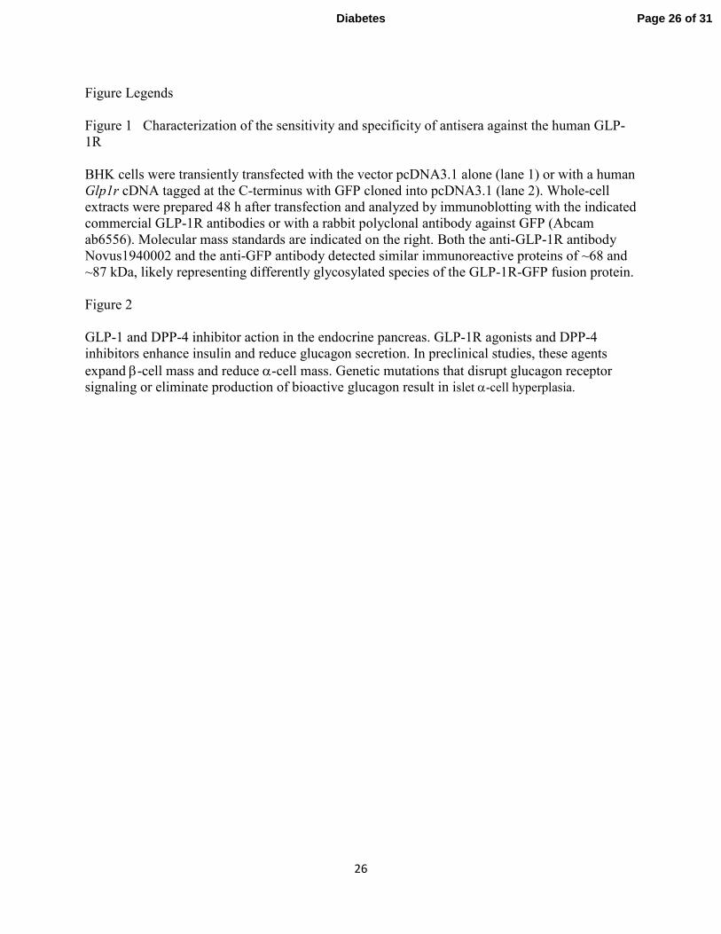
Figure Legends
Figure 1 Characterization of the sensitivity and specificity of antisera against the human GLP-
1R
BHK cells were transiently transfected with the vector pcDNA3.1 alone (lane 1) or with a human
Glp1r cDNA tagged at the C-terminus with GFP cloned into pcDNA3.1 (lane 2). Whole-cell
extracts were prepared 48 h after transfection and analyzed by immunoblotting with the indicated
commercial GLP-1R antibodies or with a rabbit polyclonal antibody against GFP (Abcam
ab6556). Molecular mass standards are indicated on the right. Both the anti-GLP-1R antibody
Novus1940002 and the anti-GFP antibody detected similar immunoreactive proteins of ~68 and
~87 kDa, likely representing differently glycosylated species of the GLP-1R-GFP fusion protein.
Figure 2
GLP-1 and DPP-4 inhibitor action in the endocrine pancreas. GLP-1R agonists and DPP-4
inhibitors enhance insulin and reduce glucagon secretion. In preclinical studies, these agents
expand β-cell mass and reduce α-cell mass. Genetic mutations that disrupt glucagon receptor
signaling or eliminate production of bioactive glucagon result in islet α-cell hyperplasia.
Page 26 of 31Diabetes
Page 27
For Peer Review Only
ab39072
1 2
Novus1940002
1 2
72
Anti-GFP
1 2
55
43
34
170 kDa
95
130
Anti-GLP-1R
Figure 1
Page 27 of 31 Diabetes
Page 28
For Peer Review OnlyInsulin SecretionGlucagon Secretion
Somatostatin Secretion
Diabetes
Gcgr-/- miceGcgrHep-/- miceLGsKO miceGcg-/- micePcsk2-/- miceGcgr human mutation
-cell hyperplasia
-cells
-cells
-cells
Normal islet
GLP-1R activationDPP-4 inhibition
-cells
-cells
Page 28 of 31Diabetes
Page 29
Supplementary Table 1 Publication Experimental Model Drug(s) Results & Exposure
Ahren (1) Regular and HFD mice 8 wks NVP DPP728 No change in islet cells Akarte (2) Neonatal STZ Rats 8 wks PKF-275-055 vs. Saline Reduced number of glucagon+ islet cells
Busch (3) Rats & mice 2 year Vildagliptin No changes in islet cell number Exposure > 200x
Duttaroy (4) Neonatal rats 19 days Vildagliptin No change in islet cells
Furuta (5) HFD-STZ mice 11 wks DSP-7238 Normalization of cell topography
Han (6) ZDF rats 5 wks Sitagliptin + metformin Reduced numbers of islet cells
Hou (7) STZ rats 12 wks Sitagliptin + mangiferin Reduced numbers of islet cells
Hu (8) STZ HFD rats 10 wks Sitagliptin + stem cells Reduced numbers of islet cells
Inaba (9) GK rats 18 weeks Vildagliptin Increased cell proliferation
Liang (10) STX mice 2-14 days Sitagliptin + losartan Reduced cell proliferation
Mu (11) ICR Mice STZ/HFD 10 wks Sitagliptin Reduced numbers of islet cells
Mu (12) ICR mice STZ HFD 11 wks Des-fluoro sitagliptin vs. SU Reduced numbers of cells
Omar (13) HFD mice 11 months Vildagliptin Reduced numbers of cells
Sato (14) HFD 20 wks IRS2-/- mice Vildagliptin No change in islet cells
Shimizu (15) C/EBPB TG mice 24wks Vildagliptin No change in islet cells
Souza-Mello (16) HFD mice 6 wks Sitagliptin + telmisartan Reduced numbers of cells
Tajima (17) HFD STZ mice 5 wks TS-021 Reduced cell; cell ratio
Takeda (18) STZ mice 5 wks Des-fluoro sitagliptin Reduced numbers of cells Yeom (19) Akita/db/db mice 4-6 wks Sitagliptin Decreased islet glucagon immunoreactivity
Zhang (20) HFD/STZ mice 10 wks Alogliptin Reduced numbers of cells
Page 29 of 31 Diabetes
Page 30
Supplementary Table 1 Legend
Preclinical studies describing effects of DPP-4 inhibitors on numbers of islet cells in normal or diabetic animals, exclusive of experiments in NOD mice. Two year toxicology data in rats and mice carried out as part of regulatory requirements for multiple DPP-4 inhibitors was not included in this Table.
References
1. Reimer MK, Holst JJ, Ahren B. Long-term inhibition of dipeptidyl peptidase IV improves glucose tolerance and preserves islet function in mice. Eur J Endocrinol. 2002;146(5):717-27. Epub 2002/05/01. 2. Akarte AS, Srinivasan BP, Gandhi S, Sole S. Chronic DPP-IV inhibition with PKF-275-055 attenuates inflammation and improves gene expressions responsible for insulin secretion in streptozotocin induced diabetic rats. Eur J Pharm Sci. 2012;47(2):456-63. Epub 2012/07/18. 3. Busch SJ, Hoffmann P, Sahota P, Johnson R, Kothny W, Meyer F, et al. Studies in rodents with the dipeptidyl peptidase-4 inhibitor vildagliptin to evaluate possible drug-induced pancreatic histological changes that are predictive of pancreatitis and cancer development in man. Diabetes, obesity & metabolism. 2013;15(1):72-6. Epub 2012/08/14. 4. Duttaroy A, Voelker F, Merriam K, Zhang X, Ren X, Subramanian K, et al. The DPP-4 inhibitor vildagliptin increases pancreatic beta cell mass in neonatal rats. European journal of pharmacology. 2011;650(2-3):703-7. Epub 2010/11/13. 5. Furuta Y, Horiguchi M, Sugaru E, Ono-Kishino M, Otani M, Sakai M, et al. Chronic administration of DSP-7238, a novel, potent, specific and substrate-selective DPP IV inhibitor, improves glycaemic control and beta-cell damage in diabetic mice. Diabetes, obesity & metabolism. 2010;12(5):421-30. Epub 2010/04/27. 6. Han SJ, Choi SE, Kang Y, Jung JG, Yi SA, Kim HJ, et al. Effect of sitagliptin plus metformin on beta-cell function, islet integrity and islet gene expression in Zucker diabetic fatty rats. Diabetes Res Clin Pract. 2011;92(2):213-22. Epub 2011/02/25. 7. Hou J, Zheng D, Fan K, Yu B, Xiao W, Ma J, et al. Combination of mangiferin and dipeptidyl peptidase-4 inhibitor sitagliptin improves impaired glucose tolerance in streptozotocin-diabetic rats. Pharmacology. 2012;90(3-4):177-82. Epub 2012/09/06. 8. Hu J, Wang F, Sun R, Wang Z, Yu X, Wang L, et al. Effect of combined therapy of human Wharton's jelly-derived mesenchymal stem cells from umbilical cord with sitagliptin in type 2 diabetic rats. Endocrine. 2013. Epub 2013/05/21. 9. Inaba W, Mizukami H, Kamata K, Takahashi K, Tsuboi K, Yagihashi S. Effects of long-term treatment with the dipeptidyl peptidase-4 inhibitor vildagliptin on islet endocrine cells in non-obese type 2 diabetic Goto-Kakizaki rats. European journal of pharmacology. 2012;691(1-3):297-306. Epub 2012/07/24. 10. Liang J, Leung KK, Lam SY, Leung PS. Combined treatment with a dipeptidyl peptidase-IV inhibitor (sitagliptin) and an angiotensin II type 1 receptor blocker (losartan) promotes islet regeneration via enhanced differentiation of pancreatic progenitor cells. Diabetes, obesity & metabolism. 2012;14(9):842-51. Epub 2012/04/24. 11. Mu J, Petrov A, Eiermann GJ, Woods J, Zhou YP, Li Z, et al. Inhibition of DPP-4 with sitagliptin improves glycemic control and restores islet cell mass and function in a rodent model of type 2 diabetes. European journal of pharmacology. 2009;623(1-3):148-54. Epub 2009/09/22. 12. Mu J, Woods J, Zhou YP, Roy RS, Li Z, Zycband E, et al. Chronic Inhibition of Dipeptidyl Peptidase-4 With a Sitagliptin Analog Preserves Pancreatic {beta}-Cell Mass and Function in a Rodent Model of Type 2 Diabetes. Diabetes. 2006;55(6):1695-704. 13. Omar BA, Vikman J, Winzell MS, Voss U, Ekblad E, Foley JE, et al. Enhanced beta cell function and anti-inflammatory effect after chronic treatment with the dipeptidyl peptidase-4 inhibitor vildagliptin in an advanced-aged diet-induced obesity mouse model. Diabetologia. 2013. Epub 2013/05/03. 14. Sato K, Nakamura A, Shirakawa J, Muraoka T, Togashi Y, Shinoda K, et al. Impact of the dipeptidyl peptidase-4 inhibitor vildagliptin on glucose tolerance and beta-cell function and mass in insulin receptor substrate-2-knockout mice fed a high-fat diet. Endocrinology. 2012;153(3):1093-102. Epub 2012/02/09. 15. Shimizu S, Hosooka T, Matsuda T, Asahara S, Koyanagi-Kimura M, Kanno A, et al. DPP4 inhibitor vildagliptin preserves beta-cell mass through amelioration of endoplasmic reticulum stress in C/EBPB transgenic mice. Journal of molecular endocrinology. 2012;49(2):125-35. Epub 2012/07/24.
Page 30 of 31Diabetes
Page 31
16. Souza-Mello V, Gregorio BM, Relvas-Lucas B, da Silva Faria T, Aguila MB, Mandarim-de-Lacerda CA. Pancreatic ultrastructural enhancement due to telmisartan plus sitagliptin treatment in diet-induced obese C57BL/6 mice. Pancreas. 2011;40(5):715-22. Epub 2011/05/24. 17. Tajima A, Hirata T, Taniguchi K, Kondo Y, Kato S, Saito-Hori M, et al. Combination of TS-021 with metformin improves hyperglycemia and synergistically increases pancreatic beta-cell mass in a mouse model of type 2 diabetes. Life sciences. 2011;89(17-18):662-70. Epub 2011/08/30. 18. Takeda Y, Fujita Y, Honjo J, Yanagimachi T, Sakagami H, Takiyama Y, et al. Reduction of both beta cell death and alpha cell proliferation by dipeptidyl peptidase-4 inhibition in a streptozotocin-induced model of diabetes in mice. Diabetologia. 2012;55(2):404-12. Epub 2011/11/11. 19. Yeom JA, Kim ES, Park HS, Ham DS, Sun C, Kim JW, et al. Both sitagliptin analogue & pioglitazone preserve the beta-cell proportion in the islets with different mechanism in non-obese and obese diabetic mice. BMB reports. 2011;44(11):713-8. Epub 2011/11/29. 20. Zhang X, Wang Z, Huang Y, Wang J. Effects of chronic administration of alogliptin on the development of diabetes and beta-cell function in high fat diet/streptozotocin diabetic mice. Diabetes, obesity & metabolism. 2011;13(4):337-47. Epub 2011/01/06.
Page 31 of 31 Diabetes
Page 32
Supplementary Table 1 Publication Experimental Model Drug(s) Results & Exposure
Ahren (1) Regular and HFD mice 8 wks NVP DPP728 No change in islet cells Akarte (2) Neonatal STZ Rats 8 wks PKF-275-055 vs. Saline Reduced number of glucagon+ islet cells
Busch (3) Rats & mice 2 year Vildagliptin No changes in islet cell number Exposure > 200x
Duttaroy (4) Neonatal rats 19 days Vildagliptin No change in islet cells
Furuta (5) HFD-STZ mice 11 wks DSP-7238 Normalization of cell topography
Han (6) ZDF rats 5 wks Sitagliptin + metformin Reduced numbers of islet cells
Hou (7) STZ rats 12 wks Sitagliptin + mangiferin Reduced numbers of islet cells
Hu (8) STZ HFD rats 10 wks Sitagliptin + stem cells Reduced numbers of islet cells
Inaba (9) GK rats 18 weeks Vildagliptin Increased cell proliferation
Liang (10) STX mice 2-14 days Sitagliptin + losartan Reduced cell proliferation
Mu (11) ICR Mice STZ/HFD 10 wks Sitagliptin Reduced numbers of islet cells
Mu (12) ICR mice STZ HFD 11 wks Des-fluoro sitagliptin vs. SU Reduced numbers of cells
Omar (13) HFD mice 11 months Vildagliptin Reduced numbers of cells
Sato (14) HFD 20 wks IRS2-/- mice Vildagliptin No change in islet cells
Shimizu (15) C/EBPB TG mice 24wks Vildagliptin No change in islet cells
Souza-Mello (16) HFD mice 6 wks Sitagliptin + telmisartan Reduced numbers of cells
Tajima (17) HFD STZ mice 5 wks TS-021 Reduced cell; cell ratio
Takeda (18) STZ mice 5 wks Des-fluoro sitagliptin Reduced numbers of cells Yeom (19) Akita/db/db mice 4-6 wks Sitagliptin Decreased islet glucagon immunoreactivity
Zhang (20) HFD/STZ mice 10 wks Alogliptin Reduced numbers of cells
Page 33
Supplementary Table 1 Legend
Preclinical studies describing effects of DPP-4 inhibitors on numbers of islet cells in normal or diabetic animals, exclusive of experiments in NOD mice. Two year toxicology data in rats and mice carried out as part of regulatory requirements for multiple DPP-4 inhibitors was not included in this Table.
References
1. Reimer MK, Holst JJ, Ahren B. Long-term inhibition of dipeptidyl peptidase IV improves glucose tolerance and preserves islet function in mice. Eur J Endocrinol. 2002;146(5):717-27. Epub 2002/05/01. 2. Akarte AS, Srinivasan BP, Gandhi S, Sole S. Chronic DPP-IV inhibition with PKF-275-055 attenuates inflammation and improves gene expressions responsible for insulin secretion in streptozotocin induced diabetic rats. Eur J Pharm Sci. 2012;47(2):456-63. Epub 2012/07/18. 3. Busch SJ, Hoffmann P, Sahota P, Johnson R, Kothny W, Meyer F, et al. Studies in rodents with the dipeptidyl peptidase-4 inhibitor vildagliptin to evaluate possible drug-induced pancreatic histological changes that are predictive of pancreatitis and cancer development in man. Diabetes, obesity & metabolism. 2013;15(1):72-6. Epub 2012/08/14. 4. Duttaroy A, Voelker F, Merriam K, Zhang X, Ren X, Subramanian K, et al. The DPP-4 inhibitor vildagliptin increases pancreatic beta cell mass in neonatal rats. European journal of pharmacology. 2011;650(2-3):703-7. Epub 2010/11/13. 5. Furuta Y, Horiguchi M, Sugaru E, Ono-Kishino M, Otani M, Sakai M, et al. Chronic administration of DSP-7238, a novel, potent, specific and substrate-selective DPP IV inhibitor, improves glycaemic control and beta-cell damage in diabetic mice. Diabetes, obesity & metabolism. 2010;12(5):421-30. Epub 2010/04/27. 6. Han SJ, Choi SE, Kang Y, Jung JG, Yi SA, Kim HJ, et al. Effect of sitagliptin plus metformin on beta-cell function, islet integrity and islet gene expression in Zucker diabetic fatty rats. Diabetes Res Clin Pract. 2011;92(2):213-22. Epub 2011/02/25. 7. Hou J, Zheng D, Fan K, Yu B, Xiao W, Ma J, et al. Combination of mangiferin and dipeptidyl peptidase-4 inhibitor sitagliptin improves impaired glucose tolerance in streptozotocin-diabetic rats. Pharmacology. 2012;90(3-4):177-82. Epub 2012/09/06. 8. Hu J, Wang F, Sun R, Wang Z, Yu X, Wang L, et al. Effect of combined therapy of human Wharton's jelly-derived mesenchymal stem cells from umbilical cord with sitagliptin in type 2 diabetic rats. Endocrine. 2013. Epub 2013/05/21. 9. Inaba W, Mizukami H, Kamata K, Takahashi K, Tsuboi K, Yagihashi S. Effects of long-term treatment with the dipeptidyl peptidase-4 inhibitor vildagliptin on islet endocrine cells in non-obese type 2 diabetic Goto-Kakizaki rats. European journal of pharmacology. 2012;691(1-3):297-306. Epub 2012/07/24. 10. Liang J, Leung KK, Lam SY, Leung PS. Combined treatment with a dipeptidyl peptidase-IV inhibitor (sitagliptin) and an angiotensin II type 1 receptor blocker (losartan) promotes islet regeneration via enhanced differentiation of pancreatic progenitor cells. Diabetes, obesity & metabolism. 2012;14(9):842-51. Epub 2012/04/24. 11. Mu J, Petrov A, Eiermann GJ, Woods J, Zhou YP, Li Z, et al. Inhibition of DPP-4 with sitagliptin improves glycemic control and restores islet cell mass and function in a rodent model of type 2 diabetes. European journal of pharmacology. 2009;623(1-3):148-54. Epub 2009/09/22. 12. Mu J, Woods J, Zhou YP, Roy RS, Li Z, Zycband E, et al. Chronic Inhibition of Dipeptidyl Peptidase-4 With a Sitagliptin Analog Preserves Pancreatic {beta}-Cell Mass and Function in a Rodent Model of Type 2 Diabetes. Diabetes. 2006;55(6):1695-704. 13. Omar BA, Vikman J, Winzell MS, Voss U, Ekblad E, Foley JE, et al. Enhanced beta cell function and anti-inflammatory effect after chronic treatment with the dipeptidyl peptidase-4 inhibitor vildagliptin in an advanced-aged diet-induced obesity mouse model. Diabetologia. 2013. Epub 2013/05/03. 14. Sato K, Nakamura A, Shirakawa J, Muraoka T, Togashi Y, Shinoda K, et al. Impact of the dipeptidyl peptidase-4 inhibitor vildagliptin on glucose tolerance and beta-cell function and mass in insulin receptor substrate-2-knockout mice fed a high-fat diet. Endocrinology. 2012;153(3):1093-102. Epub 2012/02/09. 15. Shimizu S, Hosooka T, Matsuda T, Asahara S, Koyanagi-Kimura M, Kanno A, et al. DPP4 inhibitor vildagliptin preserves beta-cell mass through amelioration of endoplasmic reticulum stress in C/EBPB transgenic mice. Journal of molecular endocrinology. 2012;49(2):125-35. Epub 2012/07/24.
Page 34
16. Souza-Mello V, Gregorio BM, Relvas-Lucas B, da Silva Faria T, Aguila MB, Mandarim-de-Lacerda CA. Pancreatic ultrastructural enhancement due to telmisartan plus sitagliptin treatment in diet-induced obese C57BL/6 mice. Pancreas. 2011;40(5):715-22. Epub 2011/05/24. 17. Tajima A, Hirata T, Taniguchi K, Kondo Y, Kato S, Saito-Hori M, et al. Combination of TS-021 with metformin improves hyperglycemia and synergistically increases pancreatic beta-cell mass in a mouse model of type 2 diabetes. Life sciences. 2011;89(17-18):662-70. Epub 2011/08/30. 18. Takeda Y, Fujita Y, Honjo J, Yanagimachi T, Sakagami H, Takiyama Y, et al. Reduction of both beta cell death and alpha cell proliferation by dipeptidyl peptidase-4 inhibition in a streptozotocin-induced model of diabetes in mice. Diabetologia. 2012;55(2):404-12. Epub 2011/11/11. 19. Yeom JA, Kim ES, Park HS, Ham DS, Sun C, Kim JW, et al. Both sitagliptin analogue & pioglitazone preserve the beta-cell proportion in the islets with different mechanism in non-obese and obese diabetic mice. BMB reports. 2011;44(11):713-8. Epub 2011/11/29. 20. Zhang X, Wang Z, Huang Y, Wang J. Effects of chronic administration of alogliptin on the development of diabetes and beta-cell function in high fat diet/streptozotocin diabetic mice. Diabetes, obesity & metabolism. 2011;13(4):337-47. Epub 2011/01/06.