Abstract Objective: The objective of our study was to successfully isolate progenitor cells from dental pulp and umbilical tissue and perform a comparative investigation of their potential transdifferentiation into osteo and neuronal-like cells. Methods: Progenitor cells were harvested from dental pulp tissue as well as cord tissue and cultured through explant culture method over the span of 4 weeks. Image-based cytometric analysis was done to determine the cell viability along with phenotypic analysis to validate the occurrence of stem cell surface markers such as CD13, CD29, CD31, CD34, CD45, CD73, CD90, CD105, HLA-DR, and HLA-ABC. After culturing mesenchymal progenitor cells, osteogenic and neurogenic differentiation potential of both tissue sources was studied. The cells were seeded in two different surfaces tissue culture treated dishes and titanium sheets and cultured along with osteogenic differentiation medium (for 28 days) and neurogenic differentiation medium (for 5 days). The osteogenic potential of the progenitor cells were checked with the detection of calcium deposits by Vonn kossa staining and PCR studies were done to confirm the presence of osteogenic genes like BMP2, HDAC1, HNF1A. The neurogenic potential of the progenitors were phenotypically determined by the observation of neuronal cells in the culture medium. Post differentiation PCR studies were done to confirm the presence of neuronal genes like NESTIN, AGRIN, MAG, DAPDH, NF-M. Findings: Progenitor cells extracted from cord tissue and dental pulp were positive for markers such as CD13, CD29, CD73, CD90, and CD105 and were found negative for markers such CD31, CD34, CD45, and HLA-DR. Progenitors obtained from dental pulp tissue showed a higher expression of cell surface markers indicating a stronger mesenchymal lineage. After culturing progenitor cells in osteogenic differentiation specific medium, these cells were successfully differentiated into cells of osteogenic lineage. Within 28 days of culture calcium deposits were detected by Von kossa staining. The differentiated cells were also found positive for osteogenic markers such as BMP2, HDAC1, HNF1A.In neural differentiation, post day 5 of culture neurospheres of varying sizes were observed floating in the culture medium. The fraction of the cells differentiated into osteogenic and neurogenic lineages were higher in progenitor cells derived from dental pulp in comparison with umbilical cord tissue. The higher potentiality of progenitor cells derived from dental pulp for neurogenic trans-differentiation could be explained by the fact that human adult dental pulp stem cells residing within the perivascular niche are thought to originate from the migrating cranial neural crest cells. There was no difference observed in the osteogenic and neurogenic differentiation capabilities of mesenchymal cells when plated in a plastic dish or titanium surface. Improvement: In-depth studies needs to be carried out on progenitor cells from dental pulp tissues in order to enhance the clinical efficacy of stem cells based therapies. Indian Journal of Science and Technology, Vol 10(32), DOI: 10.17485/ijst/2017/v10i32/116427, August 2017 ISSN (Print) : 0974-6846 ISSN (Online) : 0974-5645 Comparative Analysis of Mesenchymal Progenitor Cells from Dental Pulp and Cord Tissue and their Potentiality Towards Trans-Differentiation Ranjith Kumar Indarapu 1 , Leonie Grace Fernandes 2 and Cherukuri Pavana Jyothi 1 * 1 Department of Microbiology and Food Science and Technology GITAM Institute of Science, GITAM University, Visakhapatnam −530045, Andhra Pradesh, India; [email protected], [email protected]2 Transcell Biologics Pvt. Ltd, Plot No. 64, Road No. 5, ALEAP Industrial Estate, Pragathi Nagar Road, Gajularamaram, Hyderabad − 500092, Telangana, India; [email protected]*Author for correspondence Keywords: Dental Pulp, Mesenchymal Progenitor Cells, Neurons, Osteocyte, Umbilical Cord Tissue Stem Cells
Transcript
AbstractObjective: The objective of our study was to successfully isolate progenitor cells from dental pulp and umbilical tissue and perform a comparative investigation of their potential transdifferentiation into osteo and neuronal-like cells. Methods: Progenitor cells were harvested from dental pulp tissue as well as cord tissue and cultured through explant culture method over the span of 4 weeks. Image-based cytometric analysis was done to determine the cell viability along with phenotypic analysis to validate the occurrence of stem cell surface markers such as CD13, CD29, CD31, CD34, CD45, CD73, CD90, CD105, HLA-DR, and HLA-ABC. After culturing mesenchymal progenitor cells, osteogenic and neurogenic differentiation potential of both tissue sources was studied. The cells were seeded in two different surfaces tissue culture treated dishes and titanium sheets and cultured along with osteogenic differentiation medium (for 28 days) and neurogenic differentiation medium (for 5 days). The osteogenic potential of the progenitor cells were checked with the detection of calcium deposits by Vonn kossa staining and PCR studies were done to confirm the presence of osteogenic genes like BMP2, HDAC1, HNF1A. The neurogenic potential of the progenitors were phenotypically determined by the observation of neuronal cells in the culture medium. Post differentiation PCR studies were done to confirm the presence of neuronal genes like NESTIN, AGRIN, MAG, DAPDH, NF-M. Findings: Progenitor cells extracted from cord tissue and dental pulp were positive for markers such as CD13, CD29, CD73, CD90, and CD105 and were found negative for markers such CD31, CD34, CD45, and HLA-DR. Progenitors obtained from dental pulp tissue showed a higher expression of cell surface markers indicating a stronger mesenchymal lineage. After culturing progenitor cells in osteogenic differentiation specific medium, these cells were successfully differentiated into cells of osteogenic lineage. Within 28 days of culture calcium deposits were detected by Von kossa staining. The differentiated cells were also found positive for osteogenic markers such as BMP2, HDAC1, HNF1A.In neural differentiation, post day 5 of culture neurospheres of varying sizes were observed floating in the culture medium. The fraction of the cells differentiated into osteogenic and neurogenic lineages were higher in progenitor cells derived from dental pulp in comparison with umbilical cord tissue. The higher potentiality of progenitor cells derived from dental pulp for neurogenic trans-differentiation could be explained by the fact that human adult dental pulp stem cells residing within the perivascular niche are thought to originate from the migrating cranial neural crest cells. There was no difference observed in the osteogenic and neurogenic differentiation capabilities of mesenchymal cells when plated in a plastic dish or titanium surface. Improvement: In-depth studies needs to be carried out on progenitor cells from dental pulp tissues in order to enhance the clinical efficacy of stem cells based therapies.
Indian Journal of Science and Technology, Vol 10(32), DOI: 10.17485/ijst/2017/v10i32/116427, August 2017 ISSN (Print) : 0974-6846
ISSN (Online) : 0974-5645
Comparative Analysis of Mesenchymal Progenitor Cells from Dental Pulp and Cord Tissue and their
Potentiality Towards Trans-DifferentiationRanjith Kumar Indarapu1, Leonie Grace Fernandes2 and Cherukuri Pavana Jyothi1*
1Department of Microbiology and Food Science and Technology GITAM Institute of Science, GITAM University, Visakhapatnam −530045, Andhra Pradesh, India; [email protected], [email protected]
Comparative Analysis of Mesenchymal Progenitor Cells from Dental Pulp and Cord Tissue and their Potentiality Towards Trans-Differentiation
Indian Journal of Science and TechnologyVol 10 (32) | August 2017 | www.indjst.org 2
1. IntroductionMesenchymal progenitor cells are an unique population of stem cells, mostly found in major organ systems of the body. Some of the characteristic features of these cells are: they are easy to isolate, they have the ability to self-renew, amplify in numbers, their multi-lineage differentiation capabilities and they possess immunomodulatory abili-ties1. Hence, it seems to be a fascinating source of stem cells for research and therapeutic purposes. Mesenchymal progenitor cells have been harvested from numerous sources, such as cord tissue, peripheral blood2-4, bone marrow, adipose tissue5, placenta6,7, dermis, heart skeletal muscle8, synovium, periosteum, amniotic fluid9, skeletal muscle10 and dental pulp11. These cells are characterized for positive markers such as CD90+, CD73+, CD105+ with the additional expression of stage specific embry-onic antigen (SSEA-4) and low affinity nerve growth factor receptor (LNGFR) and are negative markers for CD34, CD45 and HLA DR12. Mesenchymal stem cells are known to be multipotent in nature; they predomi-nantly differentiate into adipo, osteo and chondrogenic lineages13. Certain studies on mesenchymal progenitor cells have shown their ability to differentiate into neuro-glial, hepatocyte like cells14 and also possess an affinity for endothelial differentiation15. Another outstanding feature of MSC's is immune-modulation and immunoprivilege. In immunomodulation MSC’s are able to suppress several functions of the immune system such as proliferation of immune cells, production of cytokines and prevent cellu-lar toxicity of immunological cells like T, B and NK cells.
Several studies have been done on these progenitor cells from the different sources, which showed that cord tissue was an ideal source for isolation of mesenchymal progeni-tor cells, it is rich in number, can be obtained non-invasively with minimum risk to the donors and can be cultured eas-ily without ethical controversy16. Till today UC tissue is inevitably discarded even though it is an excellent source for progenitor cells. In in-vitro cultures, comparison with BM MSC and Adipose-derived MSC, UC tissue derived -MSCs displayed higher frequency in forming of colony forming units12, high proliferative capacity with highest doubling number in all passages7. UC- MSCs are posi-tive for cell surface markers such as CD90, CD44, CD29, CD13, and CD10 and negative for CD45, CD34, CD31, CD56, CD33, CD14, and HLA-DR confirming their non-hematopoietic lineage. UC-MSCs show a higher affinity for osteogenic17 odontoblastic, chondrogenic, adipogenic
and neural lineages18. The progenitor cells harvested from the cord tissue have shown to trans-differentiate into neu-ron specific protein NEuN and neurofilament NF-positive neurons19,20 and hepatocytes21. Another advantage of cord tissue derived progenitor cells is that these cells express embryonic stem cell (ESC) markers, and therefore are more primitive in nature as compared to MSC found from other sources22. Several studies have been undertaken over the past couple of years asserting the fact that these cells can be used for therapeutic treatment of liver cirrhosis16.
The dental pulp stem cells (DPSCs) were 1st isolated in the year 2000; these multipotent cells were highly pro-liferative, capable of regenerating a tissue and showed the affinity for osteogenic differentiation. Gronthos identified human adult progenitor cells from dental pulp tissue and found that they could regenerate a dentin and pulp-like complex, which is composed of a mineralized matrix with tubules lined with odontoblasts, and fibrous tissue containing blood vessels in an arrangement similar to the dentin-pulp complex found in normal human teeth. Adult dental stem cells can differentiate into many den-tal components, such as dentin, periodontal ligament, cementum and dental pulp tissue, but not into the enamel. DPSCs area also able to adhere and proliferate in scaffolds and they can also differentiate into odontoblastic lineage cells23. A remove DPSCs are positive for markers such as β2 integrin, CD13, CD24, CD29, CD44, CD73, CD90, CD105, CD106, CD146, NANOG, OCT4, and STRO-1 and were negative for CD14, CD45, CD34 and HLA-DR, thus affirming that these cells are not derived from a hematopoietic source, but instead have a mesenchymal origin24.
DPSCs were also found to be capable of reprogram-ming into multiple cell types such as, odontoblast, osteoblast25, endotheliocyte, neurons26, chondrocyte, corneal epithelial cell, myocyte, melanoma cell, neu-rocyte, adipocyte, iPS cells27-29, neural crest-derived melanocytes30 and neural-like cells. DPSCs were capa-ble of forming ectopic dentin and associated pulp tissue in vivo28. DPSC have shown active potential for cellu-lar migration, organization and mineralization, which could produce 3D mineralized structures31. The potential of DPSC in dental tissue engineering is due to the high clonogenic capacity of these cells28. Like umbilical cord tissue progenitor cells dental pulp progenitor cells have the following benefits: extraction of stem cells from pulp tissue is difficult but and has a high-efficiency rate. They seem to possess immune privileges, they have an exten-
Ranjith Kumar Indarapu, Leonie Grace Fernandes and Cherukuri Pavana Jyothi
Indian Journal of Science and Technology 3Vol 10 (32) | August 2017 | www.indjst.org
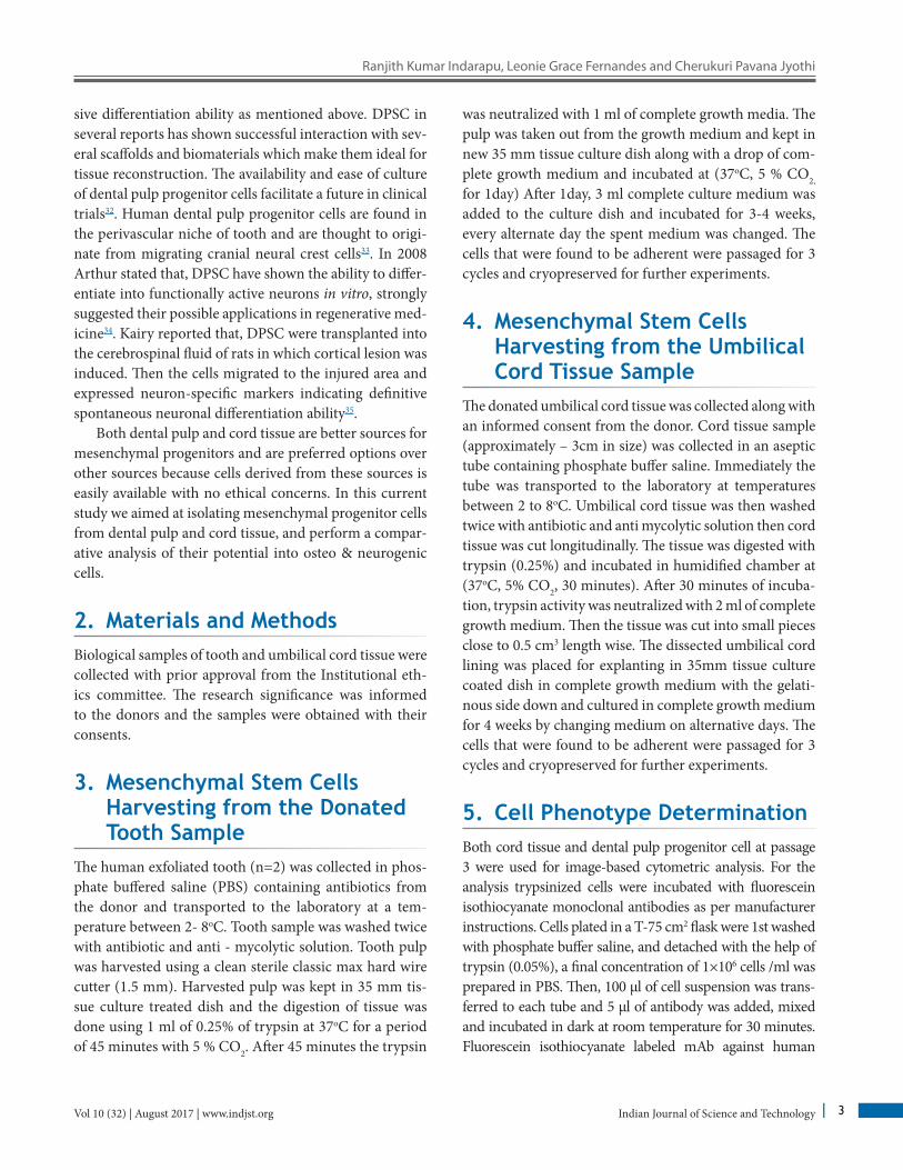
sive differentiation ability as mentioned above. DPSC in several reports has shown successful interaction with sev-eral scaffolds and biomaterials which make them ideal for tissue reconstruction. The availability and ease of culture of dental pulp progenitor cells facilitate a future in clinical trials32. Human dental pulp progenitor cells are found in the perivascular niche of tooth and are thought to origi-nate from migrating cranial neural crest cells33. In 2008 Arthur stated that, DPSC have shown the ability to differ-entiate into functionally active neurons in vitro, strongly suggested their possible applications in regenerative med-icine34. Kairy reported that, DPSC were transplanted into the cerebrospinal fluid of rats in which cortical lesion was induced. Then the cells migrated to the injured area and expressed neuron-specific markers indicating definitive spontaneous neuronal differentiation ability35.
Both dental pulp and cord tissue are better sources for mesenchymal progenitors and are preferred options over other sources because cells derived from these sources is easily available with no ethical concerns. In this current study we aimed at isolating mesenchymal progenitor cells from dental pulp and cord tissue, and perform a compar-ative analysis of their potential into osteo & neurogenic cells.
2. Materials and Methods Biological samples of tooth and umbilical cord tissue were collected with prior approval from the Institutional eth-ics committee. The research significance was informed to the donors and the samples were obtained with their consents.
3. Mesenchymal Stem Cells Harvesting from the Donated Tooth Sample
The human exfoliated tooth (n=2) was collected in phos-phate buffered saline (PBS) containing antibiotics from the donor and transported to the laboratory at a tem-perature between 2- 8oC. Tooth sample was washed twice with antibiotic and anti - mycolytic solution. Tooth pulp was harvested using a clean sterile classic max hard wire cutter (1.5 mm). Harvested pulp was kept in 35 mm tis-sue culture treated dish and the digestion of tissue was done using 1 ml of 0.25% of trypsin at 37oC for a period of 45 minutes with 5 % CO2. After 45 minutes the trypsin
was neutralized with 1 ml of complete growth media. The pulp was taken out from the growth medium and kept in new 35 mm tissue culture dish along with a drop of com-plete growth medium and incubated at (37oC, 5 % CO2,
for 1day) After 1day, 3 ml complete culture medium was added to the culture dish and incubated for 3-4 weeks, every alternate day the spent medium was changed. The cells that were found to be adherent were passaged for 3 cycles and cryopreserved for further experiments.
4. Mesenchymal Stem Cells Harvesting from the Umbilical Cord Tissue Sample
The donated umbilical cord tissue was collected along with an informed consent from the donor. Cord tissue sample (approximately – 3cm in size) was collected in an aseptic tube containing phosphate buffer saline. Immediately the tube was transported to the laboratory at temperatures between 2 to 8oC. Umbilical cord tissue was then washed twice with antibiotic and anti mycolytic solution then cord tissue was cut longitudinally. The tissue was digested with trypsin (0.25%) and incubated in humidified chamber at (37oC, 5% CO2, 30 minutes). After 30 minutes of incuba-tion, trypsin activity was neutralized with 2 ml of complete growth medium. Then the tissue was cut into small pieces close to 0.5 cm3 length wise. The dissected umbilical cord lining was placed for explanting in 35mm tissue culture coated dish in complete growth medium with the gelati-nous side down and cultured in complete growth medium for 4 weeks by changing medium on alternative days. The cells that were found to be adherent were passaged for 3 cycles and cryopreserved for further experiments.
5. Cell Phenotype DeterminationBoth cord tissue and dental pulp progenitor cell at passage 3 were used for image-based cytometric analysis. For the analysis trypsinized cells were incubated with fluorescein isothiocyanate monoclonal antibodies as per manufacturer instructions. Cells plated in a T-75 cm2 flask were 1st washed with phosphate buffer saline, and detached with the help of trypsin (0.05%), a final concentration of 1×10⁶ cells /ml was prepared in PBS. Then, 100 μl of cell suspension was trans-ferred to each tube and 5 μl of antibody was added, mixed and incubated in dark at room temperature for 30 minutes. Fluorescein isothiocyanate labeled mAb against human
Comparative Analysis of Mesenchymal Progenitor Cells from Dental Pulp and Cord Tissue and their Potentiality Towards Trans-Differentiation
Indian Journal of Science and TechnologyVol 10 (32) | August 2017 | www.indjst.org 4
HLA-DR, HLA-ABC, CD 13, CD 29, CD 31, CD 45, CD 34, CD 90, CD 105, CD 73 and CD 146 (AbD Serotec). After incubation, the cells were washed with 2ml of washing buf-fer twice, centrifuged at 400g for 5 minutes. Discard the supernatant. The cellular pellet was suspended in 200 µl of phosphate buffered saline and glycerol at 1:1 ratio and 25 µl of the cell suspension were loaded on the tali cellular analysis slide by pipetting the sample at sample loading area by capil-lary action. The slide was inserted into the slide port of the tali image-based cytometer and cells were observed. Data were acquired for 10,000 cells and the desired marker was determined from the given cell populations.
6. Cell ViabilityCell viability and cell number are determined using the tali viability dead cell red kit (Life Technologies, Invitrogen). The tali dead cell red reagent contains a solution of propidium iodide. This is a cell-impermeant fluorescent DNA binding dye which is used to detect the necrotic cells. For live cells this propidium iodide is impermeant, where as for dead cells this will penetrate into nucleic acids and become fluo-rescent. After trypsinizing, centrifuge the cells and remove the supernatant. To 100 μl of suspended cells add 1 μl of tali propidium iodide (PI, component B) mixed well and kept in the dark at room temperature for 5 minutes. From this 25 μl of sample was loaded on to tali cellular analysis slide by capil-lary action. The slide is inserted in to the slide port of the tali image-based cytometer and observed for live cells.
7. Osteogenic Trans-Differentiation in Tissue Culture Coated Plastic Surface
Osteogenic differentiation was done in monolayer cul-tures of both cord tissue and dental pulp progenitors. After the cells attained 80% confluency cells were then cultured in osteogenic differentiation medium (Invitrogen) for a period of 28 days in a humidified chamber at 37oC with 5% CO2 to determine osteogenesis. Microscopic images were captured and confirmed with von kossa staining by detecting calcium deposits.
8. Von Kossa StainingFor the detection of the calcium deposits, the cultured cells are washed twice with phosphate buffered saline and
later fixed with 70% ethanol for 1 hour at room tempera-ture without any disturbance. After fixation, the fixative is washed with double distilled water carefully. After aspi-rating double distilled water from the fixed cells. Then silver nitrate solution (5%) was added to cells and the cell container was placed under a UV light till the cal-cium deposits change color to dark brown/black. The cell culture plate was further washed three times with dou-ble distilled water by gentle shaking. Then 5% sodium thiosulphate was added at room temperature to remove unreacted silver, followed by another three washes with double distilled water (DDW).
9. Seeding Cells onto the Titanium Sheet
The pre-coated titanium sheets were seeded with both umbilical cord tissue and dental pulp progenitor cells at 3x10³ cells per sheet. The sheets kept in 50 ml sterile tube containing complete growth medium and incubated at 37oC with 5% CO2. Every alternative day the spent medium is replaced with fresh medium for a week. The titanium sheets were utilized for differentiation assay.
10. Osteogenic Trans-Differentiation on Titanium Surface
Both dental pulp and cord tissue progenitor cells were seeded on titanium sheets and then cultured in osteo-genic differentiation medium (Invitrogen) for 28 days at 37oC, 5% CO2. The spent medium was changed twice in a week to determine osteogenesis. After 28 days, RNA extracted from the cells were checked for specific osteo genes like BMP2, HDAC1, HNF1A. Microscopic images were captured to confirm the osteogenesis.
11. Neurogenic Trans-Differentiation on Tissue Culture Coated Plastic Surface
Neurogenesis is a cellular level event where in a cascade of steps occurs in forming a neuron from progenitor cells. Generally neurogenesis occurs during embryogen-esis. Neurogenic differentiation was done in a monolayer culture after the cells attained 80% confluency. The cells
Ranjith Kumar Indarapu, Leonie Grace Fernandes and Cherukuri Pavana Jyothi
Indian Journal of Science and Technology 5Vol 10 (32) | August 2017 | www.indjst.org
were then cultured in neurogenic differentiation medium (Invitrogen) for a period of 5 days by changing medium after 48 hours. Both cord tissue and dental pulp derived mesenchymal cells have been proven to have good neuro-genic potential but dental pulp progenitor cells showed a greater potential than cord tissue progenitor cells.
11.1 Neurogenic Trans-Differentiation on Titanium SheetsBoth dental pulp and umbilical cord derived progenitor cells seeded on titanium sheets were then cultured in neu-rogenic differentiation medium (Invitrogen) for a period of 5 days respectively at 37oC with 5% CO2. Medium changed after 48 hours. Microscopic images were cap-tured to determine the neurogenesis.
12. RNA Isolation from Stem Cells Ware Carried with Standard Kit (Takara RNA Isolation Kit)
The cells were trypsinized and collected in 1.5 ml eppen-dorf tube. Now the cells were centrifuged and a pellet was obtained. β mercapto ethanol along with RA1 buffer was added to lyse the cells. Now the lysate was added to a nucleospin filter along with a collection tube and spun for 1 min at 11000 X g. To the homogenized lysate, ETOH was added in order to obtain a visible precipitate. The precipitate obtained was disintegrated again and spun for 30 seconds at 11,000 X g along with a new nucleospin ribonucleic acid column and collection tube. MDM was added in to the column and spun at 11,000 Xg for 1 min-ute to dry the column. The column was further treated with DNase and Rxn for 15 min at room temperature. To the column RAW 2 buffer was added and spun for 30 seconds at 11,000 X g along with a new 2ml collection tube. The column was further treated with RA3 buffer and spun for 30 seconds at 11,000 g. The flow through was discarded and the column was placed back in a new collection tube. Additional buffer RA 3 was added to the spin column, spun again for 2 minutes at 11,000 x g. The membrane was then allowed to dry for 2 min and the RNA was eluted out and added to RNase-free water.
13. Polymerase Chain Reaction Post RNA extraction c-DNA was prepared for each sample using reverse transcriptase, c-DNA Synthesis kit (TAKARA) as per manufacturer’s manual. PCR stud-ies were carried out using standard kit components. A master mix was prepared which consists of nuclease free water, 10 X PCR buffer, dNTPs, and Taq DNA polymerase enzyme. Equal quantities of master mix were added to tubes containing c-DNA template along with forward and reverse primer specific for each gene in separate tubes all the reagents were added with the exception of tem-plate DNA which served as a negative control. All the components of the reaction mix were mixed and care was taken to avoid bubbles. The PCR tubes were placed on the thermal cycler for a 45 min long reaction, once the program had finished the products were detected by loading small quantities of each reaction on a agarose gel containing ethidium bromide. If the PCR product is present, the ethidium bromide intercalates with the DNA strands will be visualized with a UV Illuminator. The gel electrophoresis was done in order to visualize the DNA fragments and check for the presence of neural genes such as NESTIN, AGRIN, MAG, DADPH, and osteo genes such as NF-M, BMP2, HDAC1, HNF1A.
14. ResultsIn the current study, mesenchymal progenitor cells were harvested from two different sources dental pulp and cord tissue. The self-renewal capacities of these cells were observed to be remarkable and the cell culture reached confluency within 20 days. The harvested mesenchymal progenitors were fibroblastic in nature with compact cell body, prominent nucleus, spindle shaped.
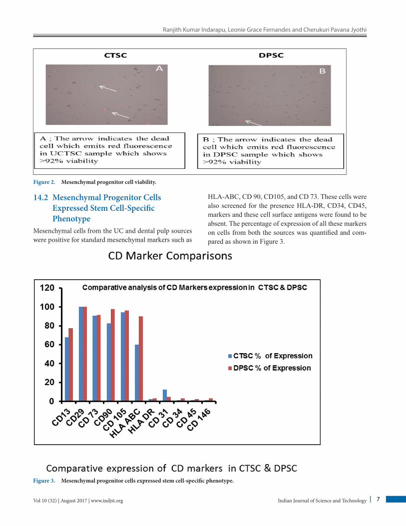
14.1 Mesenchymal Progenitor Cell ViabilityDental pulp and umbilical cord tissue derived mesen-chymal progenitor cells were cultured up to P5. These progenitor cells showed fibroblastic morphology and were plastic adherent (Figure 1a-b). The average size of cord and dental pulp derivedcells were 9-15µm and 9-13µm respectively. For the osteo derived cells and neuro differ-entiation assay passage 3 cells were used after performing the viability assay (Figure 2).
Comparative Analysis of Mesenchymal Progenitor Cells from Dental Pulp and Cord Tissue and their Potentiality Towards Trans-Differentiation
Indian Journal of Science and TechnologyVol 10 (32) | August 2017 | www.indjst.org 6
Figure 1A. Cord tissue mesenchymal stem cells
Figure 1B. Dental pulp mesenchymal stem cells.
Ranjith Kumar Indarapu, Leonie Grace Fernandes and Cherukuri Pavana Jyothi
Indian Journal of Science and Technology 7Vol 10 (32) | August 2017 | www.indjst.org
Mesenchymal cells from the UC and dental pulp sources were positive for standard mesenchymal markers such as
Figure 2. Mesenchymal progenitor cell viability.
HLA-ABC, CD 90, CD105, and CD 73. These cells were also screened for the presence HLA-DR, CD34, CD45, markers and these cell surface antigens were found to be absent. The percentage of expression of all these markers on cells from both the sources was quantified and com-pared as shown in Figure 3.
Comparative Analysis of Mesenchymal Progenitor Cells from Dental Pulp and Cord Tissue and their Potentiality Towards Trans-Differentiation
Indian Journal of Science and TechnologyVol 10 (32) | August 2017 | www.indjst.org 8
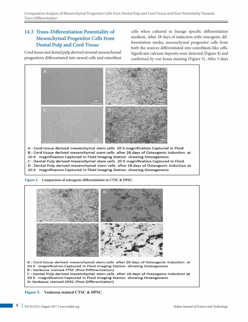
14.3 Trans-Differentiation Potentiality of Mesenchymal Progenitor Cells from Dental Pulp and Cord Tissue
Cord tissue and dental pulp derived stromal mesenchymal progenitors differentiated into neural cells and osteoblast
Figure 4. Comparison of osteogenic differentiation in CTSC & DPSC.
Figure 5. Vonkossa stained CTSC & DPSC.
cells when cultured in lineage specific differentiation medium. After 28 days of induction with osteogenic dif-ferentiation media, mesenchymal progenitor cells from both the sources differentiated into osteoblasts-like cells. Significant calcium deposits were detected (Figure 4) and confirmed by von kossa staining (Figure 5). After 5 days
Ranjith Kumar Indarapu, Leonie Grace Fernandes and Cherukuri Pavana Jyothi
Indian Journal of Science and Technology 9Vol 10 (32) | August 2017 | www.indjst.org
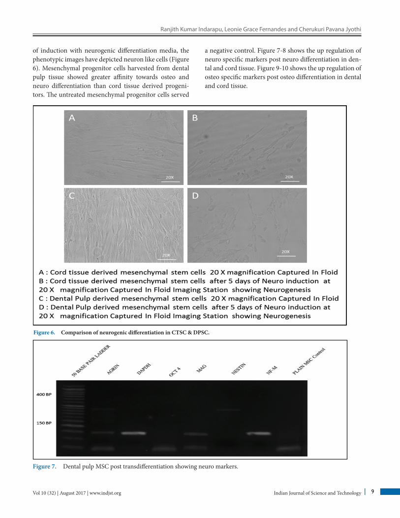
of induction with neurogenic differentiation media, the phenotypic images have depicted neuron like cells (Figure 6). Mesenchymal progenitor cells harvested from dental pulp tissue showed greater affinity towards osteo and neuro differentiation than cord tissue derived progeni-tors. The untreated mesenchymal progenitor cells served
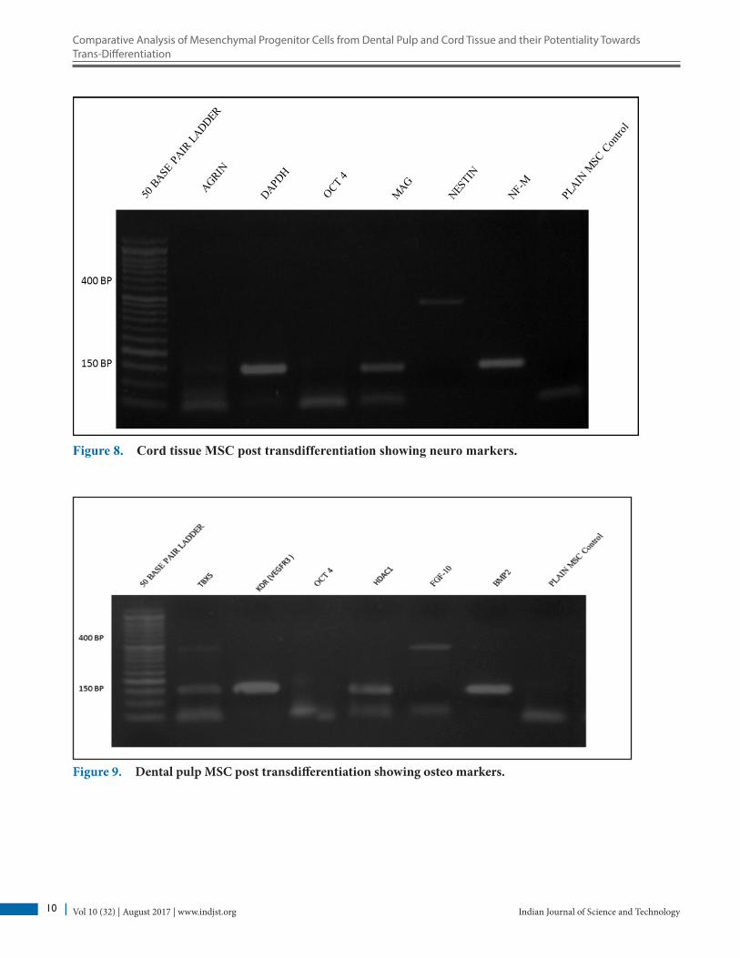
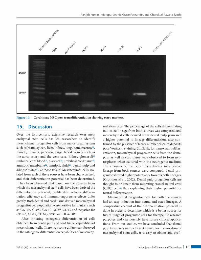
a negative control. Figure 7-8 shows the up regulation of neuro specific markers post neuro differentiation in den-tal and cord tissue. Figure 9-10 shows the up regulation of osteo specific markers post osteo differentiation in dental and cord tissue.
Figure 6. Comparison of neurogenic differentiation in CTSC & DPSC.
Figure 7. Dental pulp MSC post transdifferentiation showing neuro markers.
Comparative Analysis of Mesenchymal Progenitor Cells from Dental Pulp and Cord Tissue and their Potentiality Towards Trans-Differentiation
Indian Journal of Science and TechnologyVol 10 (32) | August 2017 | www.indjst.org 10
Figure 8. Cord tissue MSC post transdifferentiation showing neuro markers.
Figure 9. Dental pulp MSC post transdifferentiation showing osteo markers.
Ranjith Kumar Indarapu, Leonie Grace Fernandes and Cherukuri Pavana Jyothi
Indian Journal of Science and Technology 11Vol 10 (32) | August 2017 | www.indjst.org
15. DiscussionOver the last century, extensive research over mes-enchymal stem cells has led researchers to identify mesenchymal progenitor cells from major organ system such as brain, spleen, liver, kidney, lung, bone marrow36, muscle, thymus, pancreas, large blood vessels such as the aorta artery and the vena cava, kidney glomeruli37 umbilical cord blood38, placenta39, umbilical cord tissue40, amniotic membrane41, amniotic fluid42, dental pulp and adipose tissue43, adipose tissue. Mesenchymal cells iso-lated from each of these sources have been characterized, and their differentiation potential has been determined. It has been observed that based on the sources from which the mesenchymal stem cells have been derived the differentiation potential, proliferative activity, differen-tiation efficiency and immuno-suppressive effects differ greatly. Both dental and cord tissue derived mesenchymal progenitor cell population were positive for markers such as CD105, CD90, CD73, CD29, CD13 and negative for CD146, CD45, CD34, CD31 and HLA-DR.
After initiating osteogenic differentiation of cells obtained from dental pulp and cord tissue, capabilities of mesenchymal cells. There was some differences observed in the osteogenic differentiation capabilities of mesenchy-
mal stem cells. The percentage of the cells differentiating into osteo lineage from both sources was compared, and mesenchymal cells derived from dental pulp possessed a higher potential to lineage differentiation, also con-firmed by the presence of larger number calcium deposits post-Vonkossa staining. Similarly, for neuro trans-differ-entiation, mesenchymal progenitor cells from the dental pulp as well as cord tissue were observed to form neu-rospheres when cultured with the neurogenic medium. The amounts of the cells differentiating into neuron lineage from both sources were compared, dental pro-genitor showed higher potentiality towards both lineages. (Gronthos et al., 2002). Dental pulp progenitor cells are thought to originate from migrating cranial neural crest (CNC) cells44 thus explaining their higher potential for neural differentiation.
Mesenchymal progenitor cells for both the sources had an easy induction into neural and osteo lineages. A comparative account of their differentiation potential is done in order to determine which is a better source for future usage of progenitor cells for therapeutic research purposes and can possibly have future clinical applica-tions. From our studies, we have concluded that dental pulp tissue is a more efficient source for the isolation of mesenchymal stem cells, it is easy to obtain and avail-
Figure 10. Cord tissue MSC post transdifferentiation showing osteo markers.
Comparative Analysis of Mesenchymal Progenitor Cells from Dental Pulp and Cord Tissue and their Potentiality Towards Trans-Differentiation
Indian Journal of Science and TechnologyVol 10 (32) | August 2017 | www.indjst.org 12
able at high abundance, extraction of the tissue is easy, low morbidity rate and no ethical concerns. Cells derived from dental pulp showed higher mesenchymal stem cell characteristics, higher values of cell lineage markers and naturally had a higher potency for neural and osteo lin-eages.
Dental pulp stem cells have proven to be a more supe-rior source for isolating mesenchymal stem cells, along with observed osteogenic and neurogenic potential. And these cells also shown the ability to differentiate into other cell types such as odontoblast, cementobast, myo-blast, hepatocyte, melanocyte, chondrocyte, endothelial cells etc45. Over the last 10 years there has been extensive research carried out on dental pulp stem cells and the other stem cells from the dentine tissue, several positive reports have already been published on dental pulp stem cells with its potential clinical applications46. Isolated den-tal pulp stem cells and along with a collagen scaffold have used the cells for the regeneration oro-maxillo-facial bone tissue. In this setup dental pulp stem cells along with col-lagen sponge biocomplexes could completely restore the human mandible bone defects affirming this cell popula-tion repair ability. In another study dental pulp stem cells were capable of forming a living autologous fibrous bone (LAB) tissue invitro and post invivo transplantation the lab tissue also showed the ability to form bone contain-ing osteocytes47. Several studies have also followed the potential applications dental pulp stem cells for neuro regeneration. A comparative study between bone marrow stem cells, induced pluripotent stem cells and dental pulp stem cells showed that dental pulp stem cells were more effective in repair of spinal cord injury when transected into a rat spinal cord. The dental pulp stem cells facili-tated the regeneration of axons, replaced the lost cells and differentiated into adult oligodendrocyte, along with regeneration the dental pulp stem cells inhibited further apoptosis of neural cells, preserved myelin sheaths and neurofilaments and improved locomotor function48. In another similar study dental pulp stem cells were trans-planted into the spinal cord of a mice, which was subjected to laminectomy which caused compression of the spinal cord, post transplantation the number of oligodendro-cytes increased, the number of myelinated axons did not deplete instead remained constant, increased trophic fac-tor expression and overall improvement in locomotor functions was also observed. Thus, proving the fact that dental pulp stem cells can be a highly feasible candidate for therapeutic intervention for bone regeneration, cen-
tral nervous system repair as well as neuro degenerative disorders such as Parkinson’s, Alzheimer’s, etc.
R, Mosca JD, Moorman MA, Simonetti DW, Craig S. Multilineage potential of adult human mesenchymal stem cells, Science. 1999 Apr; 284(5411):143−47. Crossref. PMid:10102814.
2. Hu J, Zhou Z, Shi S, Zhu X, Wang X, Zhang W, Hu S, Qian H, Xu W. Mesenchymal stem-like cells isolated from human esophageal carcinoma and adjacent non-cancerous tissues, Oncology Letters. 2013 Jan; 5(1):179-84. Crossref. PMid:23255916.
3. Trivanović D, Kocić J, Mojsilović S, Krstić A, Ilić V, Okić-Đorđević I, Santibanez JF, Jovčić G, Terzić M, Bugarski D. Mesenchymal stem cells isolated from periph-eral blood and umbilical cord Wharton’s jelly, Srpski Arhiv Za Celokupno Lekarstvo. 2013; 141(3-4):178−86. Crossref. PMid:23745340.
4. Kern S, Bieback K, Klüter H, Eichler H. Comparative analysis of mesenchymal stem cells from bone marrow, cord blood or adipose tissue, The International Journal of Artificial Organs. 2005 Apr; 28(4):310.
5. Lee RH, Kim B, Choi I, Kim H, Choi HS, Suh K, Bae YC, Jung JS. Characterization and expression analysis of mesen-chymal stem cells from human bone marrow and adipose tissue, Cellular Physiology and Biochemistry. 2004; 14(4-6): 311−24. Crossref. PMid:15319535.
6. Talebian N, Parivar K, Kafami L, Marzban M, Shirmohammadi M, Joghataei MT. Comparative analysis of mesenchymal stem cells isolated from human bone mar-row and Wharton’s jelly, Anatomical Sciences Journal. 2013 May; 10(2):73−8.
7. Oswald J, Boxberger S, Jørgensen B, Feldmann S, Ehninger G, Bornhäuser M, Werner C. Mesenchymal stem cells can be differentiated into endothelial cells in vitro, Stem Cells. 2004 May; 22(3):377−84. Crossref. PMid:15153614.
8. Young HE, Steele TA, Bray RA, Hudson J, Floyd JA, Hawkins K, Thomas K, Austin T, Edwards C, Cuzzourt J, Duenzl M. Human reserve pluripotent mesenchymal stem cells are present in the connective tissues of skeletal muscle and dermis derived from fetal, adult, and geriatric donors, The Anatomical Record. 2001 Sep; 264(1):51−62. Crossref. PMid:11505371.
9. Majore I, Moretti P, Stahl F, Hass R, Kasper C. Growth and differentiation properties of mesenchymal stromal cell pop-ulations derived from whole human umbilical cord, Stem Cell Reviews and Reports. 2011 Mar; 7(1):17−31. Crossref. PMid:20596801.
Ranjith Kumar Indarapu, Leonie Grace Fernandes and Cherukuri Pavana Jyothi
Indian Journal of Science and Technology 13Vol 10 (32) | August 2017 | www.indjst.org
10. In’t Anker PS, Scherjon SA, Kleijburg-van der Keur C, de Groot-Swings GM, Claas FH, Fibbe WE, Kanhai HH. Isolation of mesenchymal stem cells of fetal or maternal origin from human placenta, Stem Cells (Dayton, Ohio). 2004; 22(7):1338−45. Crossref. PMid:15579651.
11. Mitchell KE, Weiss ML, Mitchell BM, Martin P, Davis D, Morales L, Helwig B, Beerenstrauch M, Abou‐Easa K, Hildreth T, Troyer D. Matrix cells from Wharton’s jelly form neurons and glia, Stem cells. 2003 Jan; 21(1):50−60. Crossref. PMid:12529551.
12. Lu LL, Liu YJ, Yang SG, Zhao QJ, Wang X, Gong W, Han ZB, Xu ZS, Lu YX, Liu DE, Chen ZZ. Isolation and charac-terization of human umbilical cord mesenchymal stem cells with hematopoiesis-supportive function and other poten-tials, haematologica. 2006 Jan; 91(8):1017−26.
13. Lee OK, Kuo TK, Chen WM, Lee KD, Hsieh SL, Chen TH. Isolation of multipotent mesenchymal stem cells from umbilical cord blood, Blood. 2004 Mar; 103(5):1669-75. Crossref. PMid:14576065.
14. Feng JX, La XL, Ma Y, Bi XJ, Wen H. Isolation of human pluripotent mesenchymal stem cells from second-tri-mester amniotic fluid using two kinds of culture protocol and their differentiation into neuron-like cells, Zhongguo Wei Zhong Bing Ji Jiu Yi Xue. 2009 Dec; 21(12):729−33.PMid:20042139.
15. Kim J, Ha C, Rhim J, Park Y, Han W, Choi S, Lee K, Park H. Different characteristics of mesenchymal stem cells isolated from different layers of full term placenta, Cytotherapy. 2017 May; 19(5):S164. Crossref.
16. Zhang Z, Lin H, Shi M, Xu R, Fu J, Lv J, Chen L, Lv S, Li Y, Yu S, Geng H. Human umbilical cord mesenchymal stem cells improve liver function and ascites in decompen-sated liver cirrhosis patients, Journal of Gastroenterology and Hepatology. 2012 Mar; 27 (s2):112−20. Crossref. PMid:22320928.
17. d’Aquino R, Graziano A, Sampaolesi M, Laino G, Pirozzi G, De Rosa A, Papaccio G. Human postnatal dental pulp cells co-differentiate into osteoblasts and endotheliocytes: a pivotal synergy leading to adult bone tissue formation, Cell Death and Differentiation. 2007 Jun; 14(6):1162−71. Crossref. PMid:17347663.
18. Zeddou M, Briquet A, Relic B, Josse C, Malaise MG, Gothot A, Lechanteur C, Beguin Y. The umbilical cord matrix is a better source of Mesenchymal Stem Cells (MSC) than the umbilical cord blood, Cell Biology International. 2010 Jul; 34(7):693−701. Crossref. PMid:20187873.
19. Sakaguchi Y, Sekiya I, Yagishita K, Muneta T. Comparison of human stem cells derived from various mesenchymal tissues: superiority of synovium as a cell source, Arthritis and Rheumatology. 2005 Aug; 52(8):2521−29. Crossref. PMid:16052568.
20. Yan X, Qin H, Qu C, Tuan RS, Shi S, Huang GT. iPS cells reprogrammed from human mesenchymal-like stem/progenitor cells of dental tissue origin, Stem Cells and Development. 2010 Apr; 19(4):469−80. Crossref. PMid:19795982 PMCid:PMC2851830.
21. Banas A, Teratani T, Yamamoto Y, Tokuhara M, Takeshita F, Quinn G, Okochi H, Ochiya T. Adipose tissue‐derived mesenchymal stem cells as a source of human hepa-tocytes, Hepatology. 2007 Jul; 46(1):219−28. Crossref. PMid:17596885.
22. Fu YS, Shih YT, Cheng YC, Min MY. Transformation of human umbilical mesenchymal cells into neurons in vitro, Journal of Biomedical Science. 2004; 11(5):652−60. Crossref. PMid:15316141.
23. Scherjon SA, Kleijburg-van der Keur C, Noort WA, Claas FH, Willemze R, Fibbe WE, Kanhai HH. Amniotic fluid as a novel source of mesenchymal stem cells for therapeutic transplantation, Blood. 2003 Aug 15; 102(4):1548−49.
24. Toupadakis CA, Wong A, Genetos DC, Cheung WK, Borjesson DL, Ferraro GL, Galuppo LD, Leach JK, Owens SD, Yellowley CE. Comparison of the osteogenic poten-tial of equine mesenchymal stem cells from bone marrow, adipose tissue, umbilical cord blood, and umbilical cord tis-sue, American Journal of Veterinary Research. 2010 Oct; 71(10):1237−45. Crossref. PMid:20919913.
25. Hendrijantini N, Kresnoadi U, Salim S, Agustono B, Retnowati E, Syahrial I, Mulawardhana P, Wardhana MP, Pramono C, Rantam FA. Study biocompatibility and osteo-genic differentiation potential of human umbilical cord mesenchymal stem cells (hUCMSCs) with gelatin solvent, Journal of Biomedical Science and Engineering. 2015 Jul; 8(7):420−28. Crossref.
26. Gronthos S, Mankani M, Brahim J, Robey PG, Shi S. Postnatal human Dental Pulp Stem Cells (DPSCs) in vitro and in vivo, Proceedings of the National Academy of Sciences. 2000 Dec; 97(25):13625−30. Crossref. PMid:11087820 PMCid:PMC17626.
27. da Silva Meirelles L, Chagastelles PC, Nardi NB. Mesenchymal stem cells reside in virtually all post-natal organs and tissues, Journal of Cell Science. 2006 Jun; 119(11):2204−13. Crossref. PMid:16684817.
28. Gronthos S, Brahim J, Li W, Fisher LW, Cherman N, Boyde A, DenBesten P, Robey PG, Shi S. Stem cell properties of human dental pulp stem cells, Journal of Dental Research. 2002 Aug; 81(8):531−5. Crossref. PMid:12147742.
29. Elluru V, Ramesh DN. Stem cell therapy: dental aspects, Journal of Indian Academy of Oral Medicine and Radiology. 2012 Jan; 24(1):45−50. Crossref.
30. Stevens A, Zuliani T, Olejnik C, LeRoy H, Obriot H, Kerr-Conte J, Formstecher P, Bailliez Y, Polakowska RR. Human dental pulp stem cells differentiate into neural crest-derived
Comparative Analysis of Mesenchymal Progenitor Cells from Dental Pulp and Cord Tissue and their Potentiality Towards Trans-Differentiation
Indian Journal of Science and TechnologyVol 10 (32) | August 2017 | www.indjst.org 14
melanocytes and have label-retaining and sphere-form-ing abilities, Stem Cells and Development. 2008 Dec; 17(6):1175-84. Crossref. PMid:18393638.
31. Zhang W, Walboomers XF, Shi S, Fan M, Jansen JA. Multilineage differentiation potential of stem cells derived from human dental pulp after cryopreservation, Tissue Engineering. 2006 Oct; 12(10):2813−23. Crossref. PMid:17518650.
32. Aquino RD, Papaccio G, Laino G, Graziano A. Dental pulp stem cells: A promising tool for bone regeneration, Stem Cell Reviews and Reports. 2008 Mar; 4(1):21−26. Crossref. PMid:18300003.
33. Bakopoulou A, Leyhausen G, Volk J, Tsiftsoglou A, Garefis P, Koidis P, Geurtsen W. Comparative analysis of in vitro osteo/odontogenic differentiation potential of human Dental Pulp Stem Cells (DPSCs) and stem cells from the apical papilla (SCAP), Archives of Oral Biology. 2011 Jul; 56(7):709−21. Crossref. PMid:21227403.
34. Király M, Kádár K, Horváthy DB, Nardai P, Rácz GZ, Lacza Z, Varga G, Gerber G. Integration of neuronally prediffer-entiated human dental pulp stem cells into rat brain in vivo, Neurochemistry International. 2011 Sep 30; 59(3):371−81. Crossref. PMid:21219952.
35. Estrela C, Alencar AH, Kitten GT, Vencio EF, Gava E. Mesenchymal stem cells in the dental tissues: perspectives for tissue regeneration, Brazilian Dental Journal. 2011; 22(2):91−98. Crossref. PMid:21537580.
36. Lee RH, Kim B, Choi I, Kim H, Choi HS, Suh K, Bae YC, Jung JS. Characterization and expression analysis of mesen-chymal stem cells from human bone marrow and adipose tissue, Cellular Physiology and Biochemistry. 2004; 14(4-6):311−24. Crossref. PMid:15319535.
37. Meirelles LSD, Chagastelles PC, Nardi NB. Mesenchymal stem cells reside in virtually all post-natal organs and tis-sues, Journal of Cell Science. 2006 Jun; 119(11):2204−13. Crossref. PMid:16684817.
38. Kern S, Eichler H, Stoeve J, Klüter H, Bieback K. Comparative analysis of mesenchymal stem cells from bone marrow, umbilical cord blood, or adipose tissue, Stem cells. 2006 May; 24(5):1294−301. Crossref. PMid:16410387.
39. Anker PSI, Scherjon SA, Kleijburg-van der Keur C, de Groot-Swings GM, Claas FH, Fibbe WE, Kanhai HH. Isolation of mesenchymal stem cells of fetal or maternal origin from human placenta, Stem Cells (Dayton, Ohio). 2004; 22(7):1338−45. Crossref. PMid:15579651.
40. Wang HS, Hung SC, Peng ST, Huang CC, Wei HM, Guo YJ, Fu YS, Lai MC, Chen CC. Mesenchymal stem cells in the Wharton’s jelly of the human umbilical cord, Stem Cells. 2004 Dec; 22(7):1330−37. Crossref. PMid:15579650.
41. Alviano F, Fossati V, Marchionni C, Arpinati M, Bonsi L, Franchina M, Lanzoni G, Cantoni S, Cavallini C, Bianchi
F, Tazzari PL. Term amniotic membrane is a high through-put source for multipotent mesenchymal stem cells with the ability to differentiate into endothelial cells in vitro, BMC Developmental Biology. 2007 Feb; 7:1−14. Crossref, Crossref. PMid:17313666, PMCid:PMC1810523.
42. Tsai MS, Lee JL, Chang YJ, Hwang SM. Isolation of human multipotent mesenchymal stem cells from second‐trimes-ter amniotic fluid using a novel two‐stage culture protocol, Human Reproduction. 2004 Jun; 19(6):1450−56. Crossref. PMid:15105397.
43. Musina RA, Bekchanova ES, Belyavskii AV, Sukhikh GT. Differentiation potential of mesenchymal stem cells of differ-ent origin, Bulletin of Experimental Biology and Medicine. 2006 Jan; 141(1):147−51. Crossref. PMid:16929987.
44. Arthur A, Rychkov G, Shi S, Koblar SA, Gronthos S. Adult human dental pulp stem cells differentiate toward functionally active neurons under appropriate environ-mental cues, Stem Cells. 2008 Jul; 26(7):1787−95. Crossref. PMid:18499892.
45. Liu J, Yu F, Sun Y, Jiang B, Zhang W, Yang J, Xu GT, Liang A, Liu S. Concise reviews: characteristics and potential applications of human dental tissue‐derived mesenchymal stem cells, Stem Cells. 2015 Mar; 33(3):627−38. Crossref. PMid:25447379.
46. Aquino RD, Rosa AD, Lanza V, Tirino V, Laino L, Graziano A, Desiderio V, Laino G, Papaccio G. Human mandible bone defect repair by the grafting of dental pulp stem/pro-genitor cells and collagen sponge biocomplexes, Eur Cell Mater. 2009 Nov; 18(7):75−83. PMid:19908196.
47. Laino G, d’Aquino R, Graziano A, Lanza V, Carinci F, Naro F, Pirozzi G, Papaccio G. A new population of human adult dental pulp stem cells: A useful source of Living Autologous fibrous Bone tissue (LAB), Journal of Bone and Mineral Research. 2005 Aug; 20(8):1394−402. Crossref.
48. Sakai K, Yamamoto A, Matsubara K, Nakamura S, Naruse M, Yamagata M, Sakamoto K, Tauchi R, Wakao N, Imagama S, Hibi H. Human dental pulp-derived stem cells promote locomotor recovery after complete transec-tion of the rat spinal cord by multiple neuro-regenerative mechanisms, The Journal of Clinical Investigation. 2012 Jan; 122(1):80−90. PMid:22133879.


![Comparative Statement on Indian GAAP and IFRS[1]](https://static.documents.pub/doc/80x56/577cd70f1a28ab9e789df14e/comparative-statement-on-indian-gaap-and-ifrs1.jpg)


