Individual Differences in the Expression of Tyrosine Hydroxylase mRNA in Neurosecretory Neurons of the Human Paraventricular and Supraoptic Nuclei: Positive Correlation with Vasopressin mRNA
Maria T. Panayotacopoulou
a,
b Yiannis Malidelis
a,
b,
c Joop van Heerikhuize
c Unga Unmehopa
c Dick Swaab
c
a Department of Psychiatry, University of Athens, and b
University Mental Health Institute, Athens , Greece; c
Netherlands’ Institute for Brain Research, Amsterdam , The Netherlands
the transcriptional level and (2) quantitative TH immuno-histochemistry and in situ hybridization for VP mRNA throughout the dorsolateral part of the SON (dl-SON) in order to elucidate whether indeed expression of TH in neurosecretory nuclei depends on activation of VP neu-rons. Postmortem formalin-fi xed, paraffi n-embedded hypothalamic sections of 16 control subjects were stud-ied for TH protein and TH and VP mRNAs. For 6 of the above cases, the number of TH-IR neurons and the total VP mRNA levels were estimated throughout the entire dl-SON using an image analysis system. Individual vari-ation was observed in TH mRNA expression which ap-pears to parallel the expression of TH-protein. Using Spearman’s bivariate test, a positive correlation was found between the number of TH-IR- and TH-mRNA-ex-pressing neurons in both PVN and SON (p ! 0.01) as well as between the number of TH-IR neurons and the total VP mRNA in the dl-SON (p ! 0.05). Our results show (1) that the individual variability in the number of TH-IR neurons within the neurosecretory nuclei might be due to differential expression and/or stability of TH mRNA and (2) that expression of TH-immunoreactivity in hu-man PVN and SON depends on the activation of VP neu-rons.
Abstract Previous studies indicated that in the human paraven-tricular nucleus (PVN) and in the supraoptic nucleus (SON) tyrosine hydroxylase (TH) – the fi rst and rate-lim-iting enzyme in catecholamine synthesis – is localized mainly in magnocellular neurosecretory neurons. Indi-vidual differences were observed among control sub-jects in number and distribution of TH-immunoreactive (IR) perikarya, indicating that antemortem factors may regulate TH expression. Since a large number of TH-IR perikarya were observed in subjects who suffered from somatic illnesses leading to prolonged osmotic or non-osmotic stimulation of vasopressin (VP) release, we sug-gested that TH expression is related to the activation of VP neurons. The purpose of our study was to apply (1) in situ hybridization for TH mRNA on human PVN and SON to investigate how the previously reported individ-ual differences in TH protein expression are depicted at
Received: January 27, 2005 Accepted after revision: August 5, 2005 Published online: October 4, 2005
Maria T. PanayotacopoulouDepartment of Psychiatry, University of Athens, Eginition Hospital74 Vassilissis Sophias AvenueAthens 115-28 (Greece)Tel. +30 210 6170884, Fax +30 210 7242020, E-Mail [email protected]
Tyrosine hydroxylase (TH) – the fi rst and rate-limiting enzyme in catecholamine synthesis converting L -tyrosine to L -DOPA [1] – has been extensively used for the im-munohistochemical localization and mapping of brain catecholamine systems. In the human hypothalamus, TH protein is expressed in a large number of magnocellular neurosecretory neurons of the paraventricular nucleus (PVN) and the supraoptic nucleus (SON) [2–4], where TH is found to colocalize with vasopressin (VP) [5, 6] or oxytocin (OXY) [2, 5] . TH immunoreactivity in the hu-man neurosecretory neurons is homogeneously distrib-uted over the entire cytoplasm and processes [3] , while large TH-immunoreactive (IR) axons can be followed from TH-IR perikarya to travel together with VP and OXY fi bers to the edge of the pituitary stalk [2] .
In a number of previous studies [3–7], we described striking individual differences concerning the number and distribution of TH-IR neurons in PVN and SON among control subjects, i.e., cases without known neuro-logical, psychiatric, or endocrinological illnesses. These differences were evident throughout the entire rostrocau-dal length of PVN and SON and appeared to be related neither to age or sex of the subjects nor to postmortem interval or staining procedures. We attributed these indi-vidual differences to antemortem factors that could infl u-ence the expression of the TH gene within the human neurosecretory neurons. Since a large number of TH-IR perikarya was observed specifi cally in subjects that had suffered from somatic illnesses, leading to prolonged os-motic or nonosmotic stimulation of VP release (i.e., de-hydration, right-sided heart failure due to pulmonary hy-pertension, or liver cirrhosis), we suggested that the ex-pression of TH within the human neurosecretory nuclei is related to the activation of VP neurons [7] .
In the rat PVN and SON, an increased expression of TH mRNA and TH protein was indeed demonstrated after experimental stimulation of the hypothalamoneuro-hypophyseal system (HNS) by dehydration [8–10] or dur-ing lactation [11] . Dehydration-triggered induction of TH gene disappeared after rehydration, suggesting that TH may have a role in the recovery of homeostasis in neurosecretory nuclei [12] . The promoter region of the TH gene of both rats and humans contains regulatory el-ements, such as CRE and AP-1, that are implicated in the control of the gene transcriptional activity by elevation of intracellular cAMP, by protein products of protoonco-genes (c-fos, c-jun), and by growth factors, hormones, and other proteins [for reviews see 13–15] .
The purposes of the present study were (1) to apply in situ hybridization (ISH) for TH mRNA on human PVN and SON in order to investigate how the observed differ-ences in TH protein expression are depicted at the tran-scriptional level and (2) to apply quantitative TH immu-nohistochemistry and ISH for VP mRNA throughout the entire rostrocaudal length of the dorsolateral part of the SON (dl-SON) in order to elucidate whether the expres-sion of TH indeed depends on neuronal activation of the human VP neurons. Our quantitative study was focused on the dl-SON, since this part of the SON contains more than 95% VP neurons in humans [for a review see 16] .
Materials and Methods
Our postmortem material consisted of the hypothalami of 16 control subjects – i.e., cases that had not suffered from neuro-logical, psychiatric, or endocrinological diseases – obtained from The Netherlands’ Brain Bank (coordinator Dr. R. Ravid). Permis-sion was obtained for brain autopsy and for the use of brain mate-rial and clinical data for research purposes. The clinicopathological data of the subjects (7 males and 9 females, age range 49–94 years) are presented in tables 1 and 2 . The hypothalami were dissected from the brain, fi xed in 10% neutral-buffered formalin, embedded in paraffi n, and serially cut in 6- � m sections. One section, every 100th, was routinely stained with thionin and immunohistochem-ically stained for VP (Truus antibody 1: 1,000, obtained from The Netherlands’ Institute for Brain Research) [for specifi city tests see 17 ] to delineate the neurosecretory nuclei.
The fi rst part of our study was performed on a sample of 10 cas-es ( table 1 ), the majority of whom were extensively described in our previous papers to show the individual differences in number and distribution of TH-IR perikarya within PVN and SON [4, 7] as well as TH colocalization with VP or OXY [5, 6] . For this part of the study, it was neccessary to use cases with available histological infor-mation concerning the number and distribution of TH-IR neurons within the neurosecretory neurons. This sample included previously studied [7] representative cases that had suffered from right-sided heart failure (NBB 90-031), liver cirrhosis (NBB 96-013), or dehy-dration (NBB 95-054), i.e., illnesses leading to increased VP synthe-sis and secretion due to a decrease in ‘effective’ blood volume or to osmotic stimulation. In this sample semiquantitative comparative estimation of the number of TH-IR and of TH-mRNA-synthesizing neurons was performed. Consecutive sections at two randomly se-lected levels of the hypothalamus caudal to the anterior commissure containing PVN and both parts of the SON were mounted on chrome-alum-coated slides using 0.5% albumin and processed as fol-lows: section 1 was stained for thionin, section 2 was stained for VP (Truus antibody 1: 1,000) to delineate the neurosecretory neurons [16] , and section 3 was stained for TH (Jacques-Boy Institute, France, 1: 1,000) to estimate the number and distribution of TH-IR perikarya within the human neurosecretory nuclei using the classic peroxidase-antiperoxidase-diaminobenzidine method with nickel intensifi ca-tion [4] . The specifi city of the TH immunohistochemical reaction/procedure was previously checked using serum absorbed with pure TH [4, 5] or by omitting the primary antibody [7] .
Tyrosine Hydroxylase mRNA in the Human Neurosecretory Nuclei
Neuroendocrinology 2005;81:329–338 331
Table 1. Clinicopathological data of the cases studied and results revealed by TH immunohistochemistry and TH mRNA in situ hybrid-ization (semiquantitative estimation)
NBBNo.
Ageyears/sex
Brainweightg
Postmortemdelay, h/fi xa-tion time, days
pH Clinical diagnosis – cause of death; medication TH-IR neurons in PVNand SON
95-106 74/m 1,317 0.08/60 6.75 angina pectoris – myocardial infarction +/– only in PVN +/– only in PVN
97-144 78/m 1,160 0.04/39 6.4 pulmonary carcinoma; prednisone(daily for 2 months before death)
+ only in PVN + only in PVN
90-031 82/m 1,250 0.11/34 5.85 urothelial carcinoma, multiple metastases in lung, liver and kidney, cachexia – respiratory and circulatory failure due to a combination of sepsis and massive thrombosis in the right cardiac ventricle; dopamine
NBB No. = Patient number in The Netherlands’ Brain Bank; n.d = not determined. Relative differences in numbers of TH-IR or TH mRNA positive neu-rons: ++++ = very large number, +++ = large number; ++ = many; + = some; +/– = a few.
Table 2. Clinicopathological data of the cases studied and results revealed by quantitative TH immunohistochemistry and in situ hy-bridization for VP mRNA throughout the dl-SON
NBBNo.
Ageyears/sex
Brain weightg
Postmortem delay, h/fi xationtime, days
pH Clinical diagnosis – cause of death; medication Number of TH-IR neuronsin dl-SON
Total VP mRNAin dl-SON, AU
94-118 49/m 1,254 22.3/33 6.6 adenocarcinoma of the colon with metastases, Pseudomonas pneumonia – septic shock due to peritonitis
0 1,415
96-010 63/m 1,250 0,10/32 6.4 chronic obstructive pulmonary disease, lung carcinoma, myocardial infarction, diabetes mellitus type 2, cachexia – acute enlargement of an old myocardial infarction; antibiotics, bronchodilators
0 714
98-035 65/f n.d. 0,20/55 n.d. thrombosis of the superior mesenteric artery, hypothyroidism, rheumatoid arthritis – complications of mesenteric ischemia; levothyroxine, diuretics, digoxin, anticoagulants, nonsteroidalanti-infl ammatory drugs, benzodiazepines
12 2,144
92-049 71/m 1,250 0, 6/32 7.4 sudden death (no cerebral abnormalities) 7 1,038
Sections consecutive to those stained for TH protein were mounted on 2% aminoalkylsilane (Sigma) coated microscope slides and proceeded for TH mRNA ISH according to a procedure previ-ously used for the demonstration of various mRNAs on human paraffi n-embedded postmortem brain tissue and described in detail elsewere [18–20] . In brief, the sections were deparaffi nized, brought to phosphate-buffered saline (pH 7.6) via descending series of eth-anols, treated with 0.2 N HCl, deproteinized with proteinase K (10 � g/ml), rinsed in glycine buffer, and delipidated with chloro-form or 0.1% Triton X-100 in phosphate-buffered saline. For hy-bridization, a synthetic 35 S-labeled oligonucleotide (30 bp) cDNA probe was used, complementary to bases 1228–1257 of the human TH mRNA (HTH-1), a sequence present in all three additional al-ternatively spliced human TH mRNAs [21, 22] . The probe was labeled using terminal deoxynucleotidyltransferase (Roche) and [ 35 S]dATP ( 1 1,250 Ci/mmol; Perkin Elmer – Life Analytical Sci-ences) and alcohol precipitated. The pellet was resuspended in for-mamide, and the labeled probe was diluted in hybridization buffer. The sections were covered with 70 � l of the diluted probe (about 10 6 cpm/section), coverslipped, and hybridized at 40 ° C overnight. After washing in standard saline sodium citrate (1 ! SSC and 0.1 ! SCC) at 45 ° C, the sections were dehydrated, dried, and apposed to autoradiography fi lm (Hyperfi lm B-max, Amersham, or Biomax MR-1, Sigma) for 1 week. The sections were then dipped in auto-radiographic emulsion (NTB-2, Kodak), developed after 4 weeks, counterstained with thionin, dehydrated in graded alcohols, washed in xylene, and coverslipped with Entellan. The specifi city of the hybridization reaction was checked by incubation of duplicated sections in 35 S-labeled sense probe, resulting in a negative ISH sig-nal, or by washing after TH mRNA hybridization in SSC at 55 or 65 ° C, causing complete detachment of the labeled probe.
The semiquantitative estimation of the number of TH (pro-tein)-IR and of TH mRNA synthesizing neurons of PVN and SON was made by two independent observers who did not know the source of the tissue. The cases were rated – as in our previous study [7] – using a fi ve-point scale as follows: +/– when ! 5% of the ob-served magnocellular neurosecretory neurons at each level were TH positive; +, ++, and +++ when the proportion increased to 5–15, 15–25 and 25–40%, respectively, and ++++ when this proportion was 1 40% ( table 1 ).
The second part of our study was performed on a sample of 6 cases ( table 2 ) and focused on dl-SON, since in humans this part of the nucleus contains 1 95% VP-synthesizing neurons and few OXY-synthesizing neurons which are usually located at the borders of the dl-SON with the lateral hypothalamus [for a review see 16] . In this sample, the number of TH-IR perikarya was counted at three central levels of the dl-SON, in order to have an adequate number of TH-IR/VP-synthesizing neurons for quantitative evaluation. The selection of these three central levels of the dl-SON was based on the microscopical observation of series of sections – every 100th section throughout the whole rostrocaudal lenght of the hypothala-mus – routinely stained for VP, as described above.
Three sections with a distance of at least 50 sections between them, at the level where the largest area of the dl-SON was detect-ed, were mounted on chrome-alum-coated slides and stained for TH using the classic peroxidase-antiperoxidase immunohisto-chemical method [4] . The counting of TH-IR/VP-synthesizing neu-rons was performed after exclusion of the few possibly TH-IR/OXY-synthesizing neurons – revealed in a consecutive section stained immunohistochemically for OXY using a monoclonal anti-
body (kindly offered by Dr. Anna Hou-You, Columbia University, New York, N.Y., USA) [23] – by means of an image analysis system (IBAS, Kontron). Only VP neurons with a nucleolus having a clear reaction for TH (gray value ! 110 in the IBAS) were counted.
The total VP mRNA was counted by applying ISH at eight lev-els throughout the dl-SON of each case in order to estimate the total volume of the dl-SON using Cavalieri’s principle [24] . For the selection of the above levels, the rostral and caudal limits of dl-SON were determined by observation of the routinely VP-stained sec-tions (every 100th throughout the whole hypothalamus). For each case eight sections with a constant distance between them through-out the whole rostrocaudal lenght of the dl-SON were mounted on 2% aminoalkylsilane (Sigma) coated microscope slides and pro-ceeded for quantitative ISH for VP mRNA.
For the quantitative evaluation of VP mRNA by ISH, a [ 35 S]dATP-labeled 48-bp oligonucleotide probe was used comple-mentary to bases 411–458 of the human prepro arginine VP pre-cursor (Eurogentec/Pharmacia Biotech), showing no homology with OXY mRNA [18, 25] . After posthybridization washings, the sections were exposed to autoradiographic fi lm for 72 h. In order to quantify the total amount of VP mRNA, radioactive standards were prepared using 20 repeated dilutions of our probes, with ra-dioactive values ranging from 30,000 to 25 cpm/ � l. One mocroliter of every sample was spotted on a fi lter paper attached to a glass slide. The fi lters were air-dried overnight and placed among the hybridized sections during autoradiography. A gray-value transfor-mation table was prepared, correlating the radioactive standards with the gray values of the spots on the fi lm. The radioactive area on the fi lms was outlined, and the total radioactivity was deter-mined using IBAS software [18] .
For the statistical analysis of our measurements, Spearman’s bivariate test was used in order to determine correlations between (1) the number of TH-IR and TH mRNA synthesizing neurons and (2) between the number of TH-IR neurons and the total VP mRNA levels. p ! 0.05 was considered to be statistically signifi cant.
Results
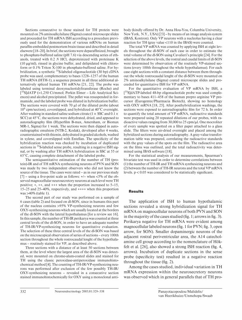
The application of ISH to human hypothalamicsections revealed a strong hybridization signal for TH mRNA on magnocellular neurons of both PVN and SON in the majority of the cases studied ( fi g. 1; arrows in fi g. 3 ). Perikarya negative for TH mRNA were evident among magnocellular labeled neurons ( fi g. 1 for PVN; fi g. 3 , open arrow, for SON). Smaller dopaminergic neurons of the adjacent rostral periventricular area, the A14 catechol-amine cell group according to the nomenclature of Hök-felt et al. [26], also showed a strong ISH reaction ( fi g. 4 , arrows). Incubation of duplicate sections in the sense probe (specifi city test) resulted in a negative reaction throughout the tissue ( fi g. 2 ).
Among the cases studied, individual variation in TH mRNA expression within the neurosecretory neurons was observed which in general parallels that of TH pro-
Tyrosine Hydroxylase mRNA in the Human Neurosecretory Nuclei
Neuroendocrinology 2005;81:329–338 333
tein revealed by immunohistochemistry ( table 1 ). Cases with the largest number of TH-IR neurons – such as NBB 95-054 and NBB 90-031 [also studied in our pre-vious papers as suffering from illnesses leading to os-motic or, respectively, nonosmotic stimulation and de-picted by TH immunohistochemistry in fi gures 12 and 13 of reference 7 ] – showed also the largest number of
intensely stained TH mRNA synthesizing neurons in both PVN ( fi g. 1 ) and SON ( fi g. 3 ). On the other hand, cases with a very limited number of TH-IR perikarya (such as NBB 95-106 or NBB 97-144) showed only few lightly labeled neurons in the PVN, while no TH-IR nor TH mRNA positive neurons were found in the SON (see fi g. 8 ). In the remaining cases, it appears that more
Fig. 1. PVN of case No. NBB 95-054 (suf-fered from dehydration) hybridized with the antisense probe for TH mRNA. Note that the majority of magnocellular neurons are strongly labeled for TH mRNA. Few mag-nocellular unlabelled perikarya are also vis-ible. Thionin counterstaining. Bar: 50 � m. Fig. 2. Adjacent section of that presented in fi gure1 hybridized with the sense probe for TH mRNA used to test the specifi city of the hybridization. Note the lack of signal on magnocellular neurons throughout the tis-sue. Thionin counterstaining. Bar: 50 � m. Fig. 3. SON of case No. NBB 90-031 (suf-fered from right-sided cardiac failure) stained for TH mRNA. Note that the ma-jority of neurosecretory neurons are posi-tive for TH mRNA (dark arrows). Negative magnocellular neurons were also evident (open arrow). Thionin counterstaining. Bar: 50 � m. Fig. 4. Rostral periventricular area (Pe) of case No. NBB 95-054. Note the presence of small dopaminergic neurons – A14 dopa-minergic cell group according to the nomen-clature of Hökfelt et al. [26] – with a strong signal for TH mRNA (arrows). Thionin counterstaining. Bar: 50 � m.
TH mRNA synthesizing neurons were detected by ISH than those considered as TH-IR by immunohistochem-istry. In those cases, many TH-IR neurons were re-vealed with impressive variation in the intensity of TH immunohistochemical reaction – from very intense to very light ( fi g. 5 , arrow), while the TH mRNA signal was intense in both PVN ( fi g. 6 ) and SON ( fi g. 7 ). The indi-vidual differences, although evident in both PVN and SON, were more impressive in the SON, where they were observable after autoradiography even by naked
eye. In some cases, almost all the SON neurons were strongly labeled for TH mRNA ( fi g. 7 ), while in others no positive neurons could be found (see fi g. 8 ). In such a case (NBB 97-144), no TH mRNA signal was detect-able in any magnocellular perikaryon in the center of the SON, while only one strongly labeled neuron was clearly evident at the border of SON with the lateral hypothalamus ( fi g. 8 , arrow). Spearman’s bivariate cor-relation test revealed a strong correlation between the number of TH-IR neurons and the number of TH
Fig. 5. PVN of case No. NBB 97-008 stained for TH protein by immunohisto-chemistry. Note the variability of TH-im-munohistochemical reaction. Two intense-ly TH-IR neurons are visible – a round one in the upper left corner and a long one with nucleus and nucleolus in the center – as well as three very lightly stained neurons. The arrow shows a very lightly stained TH-IR neuron with a nucleus. No counterstain-ing. Bar: 50 � m. Fig. 6. PVN of the same case as in fi gure 5 stained for TH mRNA by ISH. Note that almost all magnocellular neurons of this case are strongly labeled for TH mRNA. Thionin counterstaining. Bar: 50 � m. Fig. 7. SON of case No. NBB 97-008 stained for TH mRNA. Note that in this case al-most all neurosecretory neurons are posi-tive for TH mRNA. Thionin counterstain-ing. Bar: 50 � m. Fig. 8. SON of case No. NBB 97-144 stained for TH mRNA. Note that all magnocellular neurosecretory neurons of the SON are neg-ative for TH mRNA, while only one strong-ly labeled neuron appears at the border ofthe SON with the lateral hypothalamus (ar-row). Thionin counterstaining. Bar: 50 � m.
Tyrosine Hydroxylase mRNA in the Human Neurosecretory Nuclei
Neuroendocrinology 2005;81:329–338 335
mRNA expressing neurons in both PVN and SON (r = 0.93, p ! 0.01).
Using quantitative analysis performed at predeter-mined levels throughout the dl-SON, the variable expres-sion of TH immunoreactivity appears to parallel the ex-pression of VP mRNA ( table 2 ). The largest number of TH-IR/VP-synthesizing neurons was found in the dl-SON of case NBB 94-074 who suffered from cardiac de-compensation (i.e., an illness leading to nonosmotic stim-ulation of VP synthesis and release). In this case, also the highest values of VP mRNA were counted on the autora-diographic fi lms. On the other hand, no TH-IR/VP-syn-thesizing perikarya were observed in the dl-SON of case NBB 96-010 who died acutely due to myocardial infarc-tion. In this case the lowest values of VP mRNA were detected. Spearman’s bivariate test revealed a positive correlation between the number of TH-IR neurons and the total VP mRNA (r = 0.90, p ! 0.05) in the dl-SON. The TH expression was found to correlate neither with the age of the subjects nor with postmortem delay, fi xa-tion, or storage time of the tissues. The brain pH values known to affect the preservation of mRNA in the human postmortem brain [27–29] – although not determined for all our cases – did not appear to infl uence TH mRNA or TH protein expression in our sample and, therefore, can-not be considered a determining factor for the observed individual differences.
Discussion
Our ISH study indicated striking differences in TH mRNA expression within magnocellular neurosecretory neurons of PVN and SON among control subjects, i.e., cases without neurological, psychiatric, or endocrinologi-cal disorders. In general, the TH mRNA expression par-allels that of TH protein (revealed by immunohistochem-istry), indicating that the previously observed individual differences in TH immunoreactivity in the human neu-rosecretory neurons [7] might rather be due to differential expression and/or stability of TH mRNA than to differ-ential degradation of TH protein by cytosolic peptidases [30] . This differential expression of TH might be a dy-namic phenomenon related to the activation of VP neu-rons, since it is found to positively correlate with the total VP mRNA levels, as shown by quantitative analysis per-formed throughout the dl-SON. Such a dynamic expres-sion of TH has been shown in the rat after experimental stimulation of the HNS by dehydration [8–10, 12] , ap-pears to depend on the severity and the duration of the
inducing stimulus, and disappears after the recovery of homeostasis. This dynamic phenomenon could explain the impressive variation in the intensity of the TH-im-munohistochemical reaction – also reported by Panayo-tacopoulou et al. [7] – indicating a variable cytoplasmic TH protein content within the human neurosecretory neurons despite the intense signal for TH mRNA ob-served in some cases.
The observed interindividual variation did not de-pend on the age or sex of the subjects nor on laboratory postmortem conditions, indicating that antemortem fac-tors may regulate TH mRNA expression or stability. The TH activity in both the central and the peripheral nervous system is subject to short- and long-term regulation re-lated to changes in neuronal activity [13, 31–34] , stress [35–37] , lesions [38, 39] , drug effects [40–44], and other physiological or endocrinological manipulations [45–48] . As mentioned in the Introduction, several trans -acting factors (Fos, Jun, CREB, etc.) may bind to cis elements within the promoter region of the TH gene and affect its transcription [for reviews see 15, 49] . Conditions stimu-lating gene transcription may also increase the half-life of TH mRNA [50] . For example, hypoxia – a potent stimu-lus for TH expression in the human PVN and SON [4, 7] – was found to increase both TH transcriptional rate and TH mRNA stability, as indicated in PC12 pheochromo-cytoma cell lines [51–53] .
Although it is diffi cult to dissociate the effects of dif-ferent premortem conditions sustained by the patient, cases that suffered from somatic illnesses leading to pro-longed osmotic or nonosmotic activation of the HNS – such as dehydration (NBB 95-054), liver cirrhosis (NBB 96-013), or right-sided cardiac failure (NBB 90-031) – showed the largest numbers of neurosecretory neurons expressing TH mRNA and TH protein. Quantitative morphometry showed the highest number of TH-IR/VP-synthesizing neurons in the dl-SON of case NBB 94-074 – a patient that had suffered from cardiac decompensa-tion and in whom also the highest VP mRNA levels were detected. On the other hand, only a few TH-expressing neurosecretory neurons were found in the PVN of victims of sudden death – as, e.g., in subjects NBB 95-106 and NBB 96-010 who died acutely from myocardial infarc-tion. In these cases no TH expression was evident in the SON. Very limited TH mRNA neurons were also evident in the PVN (but not in the SON) of case NBB 97-144, a patient who suffered from pulmonary carcinoma and re-ceived corticosteroids before death, a treatment known to have a suppressive effect on VP [16, 54, 55] .
In summary, our study showed a differential expres-sion of TH in human neurosecretory neurons which cor-relates positively with VP mRNA, supporting our previ-ous hypothesis that the TH expression in PVN and SON depends on activation of VP neurons. A dynamic correla-tion of TH expression with VP turnover has been shown in magnocellular neurons of the supraoptico-posthypoph-yseal system of adult rats under long-term dehydration, suggesting similar mechanisms for the regulation of TH and VP gene expression and synthesis during prolonged stimulation [56] . An increased TH mRNA and protein synthesis was shown in many catecholaminergic brain areas after prolonged neuronal stimulation triggered by different types of stress conditions. Stress induces an ex-tensive catecholamine release, resulting in a reduction of intracellular catecholamine levels which is subsequently compensated for by increased biosynthesis [for review see 15] . However, in the neurosecretory nuclei, despite the apparent activity-related induction of TH after prolonged stimulation of the HNS, the biosynthesis of catechol-amine is still questionable [11, 57, 58] . TH in neurosecre-tory nuclei was considered to be inactive, since GTP cy-clohydrolase I – the fi rst and limiting enzyme for the bio-synthesis of tetrahydropterin, the natural obligatory cofactor of TH in catecholamine synthesis [59] – was not detected by immunohistochemistry in both human [60] and rat [11] neurosecretory nuclei (under osmotic stimu-lation). On the other hand, aromatic L -amino acid decar-boxylase (AADC) – the second enzyme in catecholamine synthesis – has still not been immunohistochemically de-tected in magnocelullar neurosecretory neurons of both humans [57, 58] and rats [61] . Since negative immuno-histochemical results should be interpreted with caution, further study is necessary to elucidate whether GTP cy-clohydrolase I and AADC are not expressed in these neu-rons or, alternatively, whether the applied procedures were not sensitive enough to demonstrate low levels of these enzymes. Dopamine, however, could be synthe-sized at the level of the neurohypophysis by AADC which is known to occur in blood vessels [62, 63] . A similar sug-gestion was proposed for the ventrolateral part of the ar-cuate nucleus of the rat, where TH-IR neurons also lack immunohistochemically detectable AADC [64] . In this nucleus, dopamine could be synthesized by neurons ex-pressing individual complementary enzymes of the dopa-mine synthetic pathway (TH or AADC) in cooperation [65, 66] .
In the rat, pharmacological studies provided strong evidence that dopamine acts as a neurotransmitter in the neurointermediate lobe of the hypophysis [for a review
see 67] . A single dose of the dopamine precursor L -DOPA to healthy volunteers suppresses the VP release, indicat-ing an inhibitory role of dopamine on the VP secretion [68] . If dopamine is indeed produced and secreted by hu-man neurosecretory neurons, it is important to note that neuroleptics, i.e., dopamine receptor blocking agents, un-der certain circumstances may facilitate VP secretion and induce the syndrome of inappropriate secretion of an-tidiuretic hormone [for a review see 69] which in combi-nation with polydipsia – an enduring problem of up to 20% of chronic psychiatric inpatients [70] – can induce life-threatening episodes of water intoxication due to im-paired excretory capacity [70–72] .
In conclusion, the present study showed (1) that the previously reported individual differences in number of TH-IR neurons within the human PVN and SON may be due to differential expression and/or stability of TH mRNA and (2) that TH expression within the human neurosecretory nuclei depends on activation of VP neu-rons. Increased TH immunoreactivity within magnocel-lular neurons could therefore be considered as indicative of long-term activation of VP neurons in human postmor-tem material.
Acknowledgments
Brain material was obtained from The Netherlands’ Brain Bank (coordinator Dr. R. Ravid). We wish to thank all the members of the above institution not only for collecting the brain material but also for providing us with detailed case reports on which the inter-pretation of our results is based. We are grateful to Dr. S.E.F. Gul-denaar for designing the probe for TH mRNA, D. Kontostavlaki for critically reading and preparing the manuscript, B. Fisser for his excellent technical assistance, and G. van der Meulen for pre-paring the photographs This study was supported by a grant from The Netherlands’ Brain Foundation (No. 7F 99.09 to Y.M.).
Tyrosine Hydroxylase mRNA in the Human Neurosecretory Nuclei
Neuroendocrinology 2005;81:329–338 337
References
1 Nagatsu T, Levitt M, Udenfriend S: Tyrosine hydroxylase, the initial step in NE synthesis. J Biol Chem 1964; 239: 2910–2917.
2 Li YW, Halliday GM, Joh TH, Geffen LB, Blessing WW: Tyrosine hydroxylase-contain-ing neurons in the supraoptic and paraventric-ular nuclei of the adult human. Brain Res 1988;
Issidorides MR, Constantinidis J: Tyrosine hy-droxylase-immunoreactive neurons in the paraventricular and supraoptic nuclei of the human brain demonstrated by a method adapted to prolonged formalin fi xation. J Neu-rosci Methods 1991; 39: 39–44.
4 Panayotacopoulou MT, Swaab DF: Develop-ment of tyrosine hydroxylase-immunoreactive neurons in the human paraventricular and su-praoptic nucleus. Brain Res Dev Brain Res 1993; 72: 145–150.
5 Panayotacopoulou MT, Raadsheer FC, Swaab DF: Colocalization of tyrosine hydroxylase with oxytocin and vasopressin in neurons of the human paraventricular and supraoptic nu-cleus. Brain Res Dev Brain Res 1994; 83: 59–66.
6 Panayotacopoulou MT, Goudsmit E, Van Heerikhuize JJ, Swaab DF: Simultaneous de-tection of tyrosine hydroxylase-immunoreac-tivity and vasopressin mRNA in neurons of the human paraventricular and supraoptic nucle-us. Brain Res 2000; 855: 181–185.
7 Panayotacopoulou MT, Malidelis YI, Fliers E, Bouras C, Ravid R, Swaab DF: Increased ex-pression of tyrosine hydroxylase immunoreac-tivity in paraventricular and supraoptic neu-rons in illnesses with prolonged osmotic or nonosmotic stimulation of vasopressin release. Neuroendocrinology 2002; 76: 254–266.
8 Kiss J, Mezey E: Tyrosine hydroxylase in mag-nocellular neurosecretory neurons. Neuroen-docrinology 1986; 43: 519–525.
9 Young WS, Warden M, Mezey E: Tyrosine hy-droxylase mRNA is increased by hyperosmot-ic stimuli in the paraventricular and supraoptic nuclei. Neuroendocrinology 1987; 46: 439–444.
10 Meister B, Cortes R, Villar MJ, Schalling M, Hökfelt T: Peptides and transmitter enzymes in hypothalamic magnocellular neurons after administration of hyperosmotic stimuli: com-parison between messenger RNA and peptide/protein levels. Cell Tissue Res 1990; 260: 279–297.
11 Marsais F, Calas A: Ectopic expression of non-catecholaminergic tyrosine hydroxylase in the rat hypothalamic magnocellular neurons. Neu-roscience 1999; 94: 151–161.
12 Yagita K, Okamura H, Ibata Y: Rehydration process from salt-loading: recovery of vaso-pressin and its coexisting galanin, dynorphin and tyrosine hydroxylase immunoreactivities in the supraoptic and paraventricular nuclei. Brain Res 1994; 667: 13–23.
13 Goldstein M: Long- and short-term regulation of tyrosine hydroxylase; in Bloom FE, Kupfer DJ (eds): Psychopharmacology: The Fourth Generation of Progress. New York, Raven Press, 1995, pp 189–195.
14 Joh TH: Progress in tyrosine hydroxylase gene research; in Naoi M, Parvez SH (eds): Tyrosine Hydroxylase: From Discovery to Cloning. Utrecht, VSP, 1993, pp 155–162.
15 Sabban E, Kvetnansky R: Stress-triggered acti-vation of gene expression in catecholaminergic systems: dynamics of transcriptional events. Trends Neurosci 2001; 24: 91–98.
16 Swaab DF: The human hypothalamus: basic and clinical aspects. I. Nuclei of the hypothala-mus; in Aminoff MJ, Boller F, Swaab DF (eds): Handbook of Clinical Neurology, 3rd Ser. Am-sterdam, Elsevier, 2003, vol 79.
17 Swaab DF, Hofman MA, Honnebier MB: De-velopment of vasopressin neurons in the hu-man suprachiasmatic nucleus in relation to birth. Brain Res Dev Brain Res 1990; 52: 289–293.
18 Lucassen PJ, Goudsmit E, Pool CW, Mengod G, Palacios JM, Raadsheer FC, Guldenaar SE, Swaab DF: In situ hybridization for vasopres-sin mRNA in the human supraoptic and para-ventricular nucleus: quantitative aspects of formalin-fi xed paraffi n-embedded tissue sec-tions as compared to cryostat sections. J Neu-rosci Methods 1995; 57: 221–230.
19 Guldenaar SE, Swaab DF: Estimation of oxy-tocin mRNA in the human paraventricular nucleus in AIDS by means of quantitative in situ hybridization. Brain Res 1995; 700: 107–114.
20 Guldenaar SE, Veldkamp B, Bakker O, Wiers-inga WM, Swaab DF, Fliers E: Thyrotropin-releasing hormone gene expression in the hu-man hypothalamus. Brain Res 1996; 743:
93–101. 21 Grima B, Lamouroux A, Boni C, Julien JF,
Javoy-Agid F, Mallet J. A single human gene encoding multiple tyrosine hydroxylases with different predicted functional characteristics. Nature 1987; 326: 707–711.
22 Lewis DA, Melchitzky DS, Haycock JW: Four isoforms of tyrosine hydroxylase are expressed in human brain. Neuroscience 1993; 54: 477–492.
23 Hou-Yu A, Lamme AT, Zimmerman EA, Sil-verman AJ: Comparative distribution of vaso-pressin and oxytocin neurons in the rat brain using a double-label procedure. Neuroendocri-nology 1986; 44: 235–246.
24 Verwer RW, Raber-Durlacher JE: Effi cient and unbiased estimation of volume and area of tissue components and cell number in gingival biopsies. J Periodontal Res 1993; 28: 313–323.
25 Mengod G, Goudsmit E, Probst A, Palacios JM: In situ hybridization histochemistry in the human hypothalamus. Prog Brain Res 1992;
93: 45–55.
26 Hökfelt T, Martensson R, Björklund A, Klein-au S, Goldstein M: Distributional maps of ty-rosine-hydroxylase-immunoreactive neurons in the rat brain; in Björklund A, Hökfelt T (eds): Handbook of Chemical Neuroanatomy. Vol 2: Classical Neurotransmitters in the CNS, pt 1. Amsterdam, Elsevier, 1984, pp 277–379.
27 Ravid R, Van Zwieten EJ, Swaab DF: Brain banking and the human hypothalamus – fac-tors to match for, pitfalls and potentials. Prog Brain Res 1992; 93: 83–95.
28 Kingsbury AE, Foster OJ, Nisbet AP, Cairns N, Bray L, Eve DJ, Lees AJ, Marsden CD: Tis-sue pH as an indicator of mRNA preservation in human post-morten brain. Brain Res Mol Brain Res 1995; 28: 311–318.
29 Harrison PJ, Heath PR, Eastwood SL, Burnet PW, McDonald B, Pearson RC: The relative importance of premortem acidosis and post-mortem interval for human brain gene expres-sion studies: selective mRNA vulnerability and comparison with their encoded proteins. Neurosci Lett 1995; 200: 151–154.
30 Fernandez E, Craviso GL: Protein synthesis blockade differentially affects the degradation of constitutive and nicotinic receptor-induced tyrosine hydroxylase protein level in isolated bovine chromaffi n cells. J Neurochem 1999;
lase regulation in the central nervous system. Mol Cell Biochem 1983; 53/54: 129–152.
32 Schalling M, Stieg PE, Lindquist C, Goldstein M, Hökfelt T: Rapid increase in enzyme and peptide mRNA in sympathetic ganglia after electrical stimulation in humans. Proc Natl Acad Sci U S A 1989; 86: 4302–4305.
33 Killbourne EJ, Nankova BB, Lewis EJ, McMa-hon A, Osaka H, Sabban DB, Sabban EL: Reg-ulated expression of the tyrosine hydroxylase gene by membrane depolarization. J Biol Chem 1992; 267: 7563–7569.
34 Cho JY, Min N, Franzen L, Baker H: Rapid down-regulation of tyrosine hydroxylase ex-pression in the olfactory bulb of naris-occluded adult rats. J Comp Neurol 1996; 369: 264–276.
35 Richard F, Faucon-Biguet N, Labatut R, Rollet D, Mallet J, Buda M: Modulation of tyrosine hydroxylase gene expression in rat brain and adrenals by exposure to cold. J Neurosci Res 1988; 20: 32–37.
36 Angulo A, Printz D, Ledoux M, McEwen BS: Isolation stress increases tyrosine hydroxylase mRNA in the locus coeruleus and midbrain and decreases proenkephalin mRNA in the striatum and nucleus accumbens. Brain Res Mol Brain Res 1991; 11: 301–308.
37 Watanabe Y, McKittrick CR, Blanchard DC, Blanchard RJ, McEwen BS, Sakai RR: Effects of chronic social stress on tyrosine hydroxylase mRNA and protein levels. Brain Res Mol Brain Res 1995; 32: 176–180.
38 Pasinetti GM, Osterburg HH, Kelly AB, Ko-hama S, Morgan DG, Reinhard JF, Stellwagen RH, Finch CE: Slow changes of tyrosine hy-droxylase gene expression in dopaminergic brain neurons after neurotoxin lesioning: a model of neuron aging. Brain Res Mol Brain Res 1992; 13: 63–73.
39 Shirao T, Evinger MJ, Iacovitti L, Reis DJ: Le-sions of nigrostriatal pathway reduce expres-sion of tyrosine hydroxylase gene in residual dopaminergic neurons of the substantia nigra. Neurosci Lett 1992; 141: 208–212.
40 Weiss-Wunder LT, Chesselet MF: Acute and repeated administration of fl uphenazine-N-mustard alters levels of tyrosine hydroxylase mRNA in subsets of mesencephalic dopaminer-gic neurons. Neuroscience 1992; 49: 297–305.
41 Lavergne A, Frain O, Guilbert B, Faucon-Big-uet N, Leviel V: Regulation of tyrosine hydrox-ylase gene expression in mesencephalic dopa-mine neurons: effect of imipramine treatment. Neurosci Lett 1994; 182: 167–171.
42 Stork O, Hashimoto T, Obata K: Haloperidol activates tyrosine hydroxylase gene expression in the rat substantia nigra, pars reticulata. Brain Res 1994; 633: 213–222.
43 Stork O, Hashimoto T, Obata K: Increase of tyrosine hydroxylase and its mRNA in the rat substantia nigra pars reticulata by diazepam and picrotoxin. Neurosci Res 1994; 19: 73–80.
44 Shishido T, Watanabe Y, Matsuoka I, Naka-nishi H, Niwa S: Acute methamphetamine ad-ministration increases tyrosine hydroxylase mRNA levels in the rat locus coeruleus. Brain Res Mol Brain Res 1997; 52: 146–150.
45 Blum M, McEwen B, Roberts J: Transcription-al analysis of tyrosine hydroxylase gene in tu-beroinfundibular dopaminergic neurons of the rat arcuate nucleus after estrogen treatment. J Biol Chem 1987; 262: 817–821.
46 Simerly RB: Hormonal control of the develop-ment and regulation of tyrosine hydroxylase expression within a sexually dimorphic popu-lation of dopaminergic cells in the hypothala-mus. Brain Res Mol Brain Res 1989; 6: 297–310.
47 Goldstein ME, Tank AW, Fossom LH, Hamill RW: Molecular aspects of the regulation of ty-rosine hydroxylase by testosterone. Brain Res Mol Brain Res 1992; 14: 79–86.
48 Beccavin JC, Malpaux B, Tillet Y: Effect of oestradiol and photoperiod on TH mRNA con-centrations in A15 and A12 dopamine cell groups in the ewe. J Neuroendocrinol 1998; 10:
59–66. 49 Kumer SC, Vrana KE: Intricate regulation of
50 Fossom LH, Sterling CR, Tank AW: Regula-tion of tyrosine hydroxylase gene transcription rate and tyrosine hydroxylase mRNA stability by cyclic AMP and glucocorticoid. Mol Phar-macol 1992; 42: 898–908.
51 Czyzyk-Krzeska MF, Bayliss DA, Lawson EE, Millhorn DE: Regulation of tyrosine hydroxy-lase gene expression in the rat carotid body by hypoxia. J Neurochem 1992; 58: 1538–1546.
52 Czyzyk-Krzeska MF, Furnari BA, Lawson EE, Millhorn DE: Hypoxia increases rate of tran-scription and stability of tyrosine hydroxylase mRNA in pheochromocytoma (PC12) cells. J Biol Chem 1994; 269: 760–764.
53 Czyzyk-Krzeska MF, Dominski Z, Kole R, Millhorn DE: Hypoxia stimulates binding of a cytoplasmic protein to a pyrimidine-rich se-quence in the 3 � -untranslated region of rat ty-rosine hydroxylase mRNA. J Biol Chem 1994; 269: 9940–9945.
54 Erkut ZA, Pool CW, Swaab DF: Glucocorti-coids suppress corticotropin-releasing hor-mone and vasopressin expression in human hypothalamic neurons. J Clin Endocrinol Metab 1998; 83: 2066–2073.
55 Erkut ZA, Gabreels BA, Eikelenboom J, Van Leeuwen J, Swaab DF: Glucocorticoid treat-ment is associated with decreased expression of processed AVP but not of proAVP, neuro-physin or oxytocin in the human hypothala-mus: are PC1 and PC2 involved? Neuro Endo-crinol Lett 2002; 23: 33–44.
56 Abramova M, Marsais F, Calas A, Thibault J, Ugrumov M: Dynamical study of tyrosine hy-droxylase expression and its correlation with vasopressin turnover in the magnocellular neu-rons of the supraoptico-posthypophysial sys-tem under long-term salt loading of adult rats. Brain Res 2002; 925: 67–75.
57 Kitahama K, Ikemoto K, Jouvet A, Nagatsu I, Geffard M, Okamura H, Pearson J: Dopamine synthesizing enzymes in paraventricular hypo-thalamic neurons of the human and monkey (Macaca fuscata) . Neurosci Lett 1998; 243: 1–4.
58 Kitahama K, Ikemoto K, Jouvet A, Nagatsu I, Sakamoto N, Pearson J: Aromatic L -amino acid decarboxylase- and tyrosine hydroxylase-immunohistochemistry in the adult human hy-pothalamus. J Chem Neuroanat 1998; 16: 43–55.
59 Kaufman S: Properties of the pterin-depen-dent aromatic amino acid hydroxylases. Ciba Found Symp 1974; 20: 85–115.
60 Nagatsu I, Ikemoto K, Kitahama K, Nishimu-ra A, Ichinose H, Nagatsu T: Specifi c localiza-tion of the guanosine triphosphate (GTP) cy-clohydrolase I-immunoreactivity in the human brain. J Neural Transm 1999; 106: 607–617.
61 Okamura H, Kitahama K, Raynaud B, Borri-Voltattorni C, Nagatsu I, Weber M: Immuno-cytochemistry of aromatic L -amino acid decar-boxylase in the rat preoptic area and anterior hypothalamus, with special reference to tyro-sine hydroxylase immunocytochemistry. Bio-genic Amines 1989; 7: 351–361.
62 Bertler A, Falck B, Rosengren E: The direct demonstration of a barrier mechanism in the brain capillaries. Acta Pharmacol Toxicol (Co-penh) 1963; 20: 317–321.
63 Constantinidis J, de la Torre JC, Tissot R, Geissbühler F: La barrière capillaire pour la dopa dans le cerveau et les différents organes. Psychopharmacologia 1969; 15: 75–87.
64 Meister B, Hökfelt T, Steinbusch HW, Skager-berg G, Lindvall O, Geffard M, Joh TH, Cuel-lo AC, Goldstein M: Do tyrosine hydroxylase-immunoreactive neurons in the ventrolateral arcuate nucleus produce dopamine or only L -dopa? J Chem Neuroanat 1988; 1: 59–64.
65 Ugrumov MV, Melnikova VI, Ershov P, Balan I, Calas A: Tyrosine hydroxylase- and/or aro-matic L -amino acid decarboxylase-expressing neurons in the rat arcuate nucleus: ontogenesis and functional signifi cance. Psychoneuroendo-crinology 2002; 27: 533–548.
66 Ugrumov MV, Melnikova VI, Lavrentyeva AV, Kudrin VS, Rayevsky KS: Dopamine syn-thesis by non-dopaminergic neurons express-ing individual complementary enzymes of the dopamine synthetic pathway in the arcuate nucleus of fetal rats. Neuroscience 2004; 124:
629–635. 67 Holzbauer M, Muscholl E, Racke K, Sharman
DF: Evidence that dopamine is a neurotrans-mitter in the neurointermediate lobe of the hy-pophysis. Prog Brain Res 1983; 60: 357–364.
68 Lightman SL, Forsling M: Evidence for dopa-mine as an inhibitor of vasopressin release in man. Clin Endocrinol (Oxf) 1980; 12: 39–46.
69 Spigset O, Hedenmalm K: Hyponatremia and the syndrome of inappropriate antidiuretic hormone secretion (SIADH) induced by psy-chotropic drugs. Drug Saf 1995; 12: 209–225.
70 de Leon J, Verghese C, Tracy JI, Josiassen RC, Simpson GM: Polydipsia and water intoxica-tion in psychiatric patients: a review of the ep-idemiological literature. Biol Psychiatry 1994;
35: 408–419. 71 Raskind MA, Orenstein H, Christopher TG:
Acute psychosis, increased water ingestion, and inappropriate antidiuretic hormone secre-tion. Am J Psychiatry 1975; 132: 907–910.
72 de Leon J: Polydipsia: a study in a long-term psychiatric unit. Eur Arch Psychiatry Clin Neurosci 2003; 253: 37–39.