Page 1
Pag
e45
74
Indo American Journal of Pharmaceutical Research, 2013 ISSN NO: 2231-6876
Journal home page:
http://www.iajpr.com/index.php/en/
INDO AMERICAN
JOURNAL OF
PHARMACEUTICAL
RESEARCH
COMPARATIVE STUDY OF ANTI DANDRUFF ACTIVITY OF SYZYGIUM AROMATICUM AND
ZINGIBER OFFICINALE
S. Mohanapriya, C. Mercy Bastine, R. Caroline Jeba* *Department of Industrial Biotechnology, Dr. MGR Educational and Research Institute, University, Maduravoyal, Chennai-600 095
Corresponding author
R. Caroline Jeba Department of Industrial Biotechnology
Dr M.G.R Educational & Research Institute University,Maduravoyal,
Chennai-95 , India
E-mail: [email protected]
Copy right © 2013 This is an Open Access article distributed under the terms of the Indo American journal of Pharmaceutical Research, which permits unrestricted
use, distribution, and reproduction in any medium, provided the original work is properly cited.
ARTICLE INFO ABSTRACT
Article history Received 01/06/2013
Available online
30/06/2-13
Keywords Dandruff,
malassezia
furfur,
Syzygium aromaticum
To study the inhibitory effect of Malassezia furfur by using the plants Syzygium
aromaticum and Zingiber officinale.The antidandruff activity of hexane, ethyl
acetate and methanol extracts of Zingiber officinalae and Syzygium aromaticum
was studied by agar well diffusion and broth dilution assay. The Minimum
inhibitory concentration (MIC) of the methanol extract of S.aromaticum was
studied as 100µg/ml and IC50 as 850µg/ml. Partial purification through TLC and
bio-autography were also studied. Out of the three extracts methanol extract,
showed good activity comparatively. MIC activity was good in S.aromaticum
when compared to Z.officinale. In S.aromaticum Rf value of 0.153 was the active
compound which showed good activity against dandruff.Comparing the inhibitory
activities of the two plants S.aromaticum showed Minimum inhibitory activity
against M.furfur.
Please cite this article in press as R. Caroline Jeba et.al. Comparative study of anti dandruff activity of syzygium aromaticum and
zingiber officinale. Indo American Journal of Pharm Research.2013:3(6).
Page 2
www.iajpr.com
Pag
e45
75
Vol 3, Issue 6, 2013. R. Caroline Jeba et al. ISSN NO: 2231-6876
INTRODUCTION
Medicinal plants have been used for centuries and have become part of complementary medicine worldwide
because of their potential health benefits. In India, earliest references are available in Rigveda which is said to
be written between 3500 – 1600 B.C. [1]. Plant metabolites are known to have direct positive effects in the
treatment and management of infectious diseases and cancer. In addition, the indirect effects of plant
metabolites through immunomodulation is well studied [2]. The medicinal plants are rich in secondary
metabolites and essential oils of therapeutic importance [3]. Plants as a therapeutic option were achieving
significance due to their safety profile besides being economical, effective and easily available. Plants play an
essential role in the health care needs for the treatment of diseases and to improve the immunological response
against much pathology [4].
Dandruff is a common scalp disorder affecting almost half of the pubertal population of any ethnicity and both
genders [5]. It is a chronic scalp condition characterized by visible flakes induced by rapid turnover of scalp
cells. In general, dandruff occurs after puberty and mainly affects males more than the females [6]. The
pathogenesis of dandruff involves hyper proliferation, resulting in deregulation of keratinization. The
corneocytes clump together, manifesting as large flakes of skin. The causative agents of dandruff belong to the
group of scalp commensal lipophilic yeasts of the genus, Malassezia. Malassezia species are normal flora of
skin and cause Pityriasis versicolor and foliculities under suitable conditions [7]. They are saprophytic
lipophilic yeasts characterized morphologically by small cells exhibiting unilateral, enteroblastic and repetitive
budding [8]. At present several species of Malassezia have been isolated which are M. furfur, M. globosa, M.
pachydermatis, M. restricta, M. obtusa, M. slooffiae , M. yamatoensis, M. dermatis, M. japonica, M. nana, M.
caprae, M. equina, M. cuniculi [9].
Several fungistatic compounds have been shown to improve dandruff condition. The main active ingredients
include imidazole derivatives such as ketoconazole and other compounds such as selenium sulphide, zinc
pyrithione (ZnPTO), piroctone olamine, cipropirox olamine, etc [5]. Herbal essential oils are promising sources
for new natural antifungal drugs, which have good effects against pathogenic fungi compared with commercial
synthetic antifungal drugs [8]. Syzygium aromaticum (cloves) contains a high percentage of eugenol, which has
been identified as a compound exhibiting antifungal properties [10]. Previous studies have reported antifungal
activity for clove oil and eugenol against yeasts and filamentous fungi, such as several food-borne fungal specie
and human pathogenic fungi [11]. Zingiber officinale (Ginger), one of the most important spices in the world, is
known for its medicinal and flavoring potentials. The medicinal properties are attributed to its spicy, pungent
constituents, mainly gingerols, which stimulate the thermoregulatory receptors. This stimulation influences
stomach and bile secretions by reflex action. [12].
In view of the above said facts the purpose of the current study is focused on evaluating the antidandruff
activity of Zingiber officinale (Ginger) and Syzygium aromaticum (Cloves) against Malassezia furfur.
MATERIALS AND METHODS
General laboratory techniques recommended by was followed for the preparation of media, inoculation and
maintenance of cultures.
Cleaning of glassware
The glassware were first soaked in chromic acid solution, (10% potassium dichromate in 25% sulphuric acid)
for a few hours to remove tough residues, washed twice in tap water, then they were rinsed in metal distilled
water and dried in an oven at 80ºC.
Sterilization
Sterilization of culture media and glassware were carried out in an autoclave at 121°C, 15 lbs for 20 minutes.
Thermo labile substances are sterilized through millipore filter. All the experiments were conducted under
laminar hood with strict aseptic conditions.
Page 3
www.iajpr.com
Pag
e45
76
Vol 3, Issue 6, 2013. R. Caroline Jeba et al. ISSN NO: 2231-6876
Chemicals Analytical grade chemicals supplied by Loba, Hi-media, S.D.Fine chemicals, E-Merck, Qualigens and Sigma
Chemicals (USA) were used.
MEDIA PREPARATION
Potato Dextrose Agar (g/L)
Potato 200.0
Dextrose 20.0
Agar 20.0
pH 6.5
Sabouraud’s Dextrose Agar (g/L)
Dextrose 40.000
Mycological peptone 10.000
Agar 15.000
Final pH (at 25°C) 5.6±0.2
Nitrate Reduction Medium (g/L)
Beef (meat) extract 3.0
Gelatin peptone 5.0
Potassium nitrate (KNO3) 1.0
pH 7.2±0.2
Nutrient Gelatin Medium (g/L)
Peptone 5.0
Beef extract 3.
Gelatin 120.0
pH 6.8±0.2
Urea Broth Base (g/L)
Peptone 1.0
Glucose 1.0
Sodium chloride 5.0
Disodium phosphate 1.2
KH2PO4 0.8
Phenol red 0.004
pH 6.8± 0.2
Plant material collection and preparation
The rhizome part of Zingiber officinale and the buds of Syzygium aromaticum were collected from Koyambedu
market, Chennai. The samples were authenticated, and specimens were deposited in Arvind Remedies
LTD.They were carefully washed with tap water, rinsed with distilled water, and air-dried for 1hr after which
they were shade dried for 7days, powdered coarsely and stored at room temperature.
Direct extraction Direct extraction with hexane, ethyl acetate and methanol was done following the method of [13]. In this
method, finely ground plant material was extracted with hexane, ethyl acetate and methanol in the ratio of 1:10
in conical flask in shaking condition for overnight. The extract was filtered through the Whatmann No.1 filter
paper in a separate container. The process was repeated 3times and the same plant material but using fresh
solvent. The combined filtrate was subjected to condensation. The solvent was removed by placing the extracts
Page 4
www.iajpr.com
Pag
e45
77
Vol 3, Issue 6, 2013. R. Caroline Jeba et al. ISSN NO: 2231-6876
in distillation unit in the respective temperature. The extracted residues were weighed and re-dissolved in
different solvents to yield 10mg/ml solutions ready for further analysis.
Isolation of dandruff causing agent
Samples were collected by scraping the lesions of patients and stored in sterile containers in refrigerator until
use. Different media formulations (potato dextrose agar, Sabouraud’s dextrose agar) were supplemented with
olive oil and inoculated with the sample. The plates were incubated at 37°C for 3- 5days [6].
Morphological characterization of the isolate
Microscopic examination of the samples was performed after the treatment with KOH (20%) and 5%
lactophenol cotton blue staining [14].
Biochemical tests The organism was biochemically analysed by the following assays.
Catalase test
Using a sterile inoculating loop a small amount of organism from a 24hr colony was placed onto the
microscopic slide. Using a dropper, 1drop of 3% H2O2 was placed on the microscopic slide and observed for
immediate bubble formation [15].
Nitrate reduction test
The nitrate reduction medium was dispensed in test tubes. They were autoclaved for 15mins at 121°C, 15 psi
and cooled before use [15].
REAGENTS
Sulfanilic acid solution (Reagent A) 8g of sulfanilic acid was dissolved in 1L of 5N acetic acid.
α-Naphthylamine solution (Reagent B) 6g of N, N-Dimethyl-1-naphthylamine was dissolved in 1L of 5N acetic acid. Procedure
The tubes were inoculated heavily with a fresh culture of the suspect organism. Another tube was kept
as negative control without organism. The tubes were incubated at 35 to 37°C for 24 to 48hrs in an incubator. 5
drops of Sulfanilic acid solution and 5 drops of α-Naphthylamine solution were added into the tube containing
culture to be tested and the negative control. The tubes were shaken well to mix reagents with medium and
observed for a distinct red or pink color, which should develop within a few minutes, indicates presence of
nitrate reduction.
Gelatin hydrolysis test About 2 to 3ml of medium was dispensed into test tubes. The medium was autoclaved at 121
oC (15 psi)
for 15 minutes. The tubed medium was cooled in an upright position before use. A heavy inoculum of 24hrs old
test culture was stab-inoculated into tubes containing nutrient gelatin. The inoculated tubes and an uninoculated
control tube are incubated at 25°C, for up to 1 week, and checked every day for gelatin liquefaction. The tubes
are immersed in an ice bath for 15 to 30 minutes. Afterwards, tubes were tilted to observe if gelatin has been
hydrolyzed [16].
Urea hydrolysis test
The urea broth base was sterilized by autoclaving at 115°C for 20 minutes and was cooled to 55°C. To
this 5ml of sterile 40% Urea Solution was added and mixed well. The tubes of Urea Broth were inoculated with
1ml of culture broth and incubated for 2 to 6hrs at 35°C and was observed for pink color [17].
Page 5
www.iajpr.com
Pag
e45
78
Vol 3, Issue 6, 2013. R. Caroline Jeba et al. ISSN NO: 2231-6876
Evaluation of antidandruff potential of the selected extracts
Various concentrations (250-1000µg/ml) of the extracts were prepared in DMSO from the resultant
extract to determine its antidandruff activity. Control experiments were performed by using DMSO with
identical concentration used to test the extract. Isolates from dandruff were inoculated by swabbing on the
surface of gelled PDA plates. Wells of 8mm in diameter were performed in the PDA media, and each well was
filled with 50µl of certain concentration of extract. The plates were kept in laminar air flow for 30 minutes for
proper diffusion of the extract and thereafter incubated at 37ºC for 3-5days. The radius for the zone of inhibition
was in millimeters and recorded against the corresponding concentration [6].
Broth dilution assay
Dilution assays are standard method used to compare the inhibition efficiency of the antimicrobial
agents. 5ml of the potato dextrose broth, 0.1ml of the 24hrs growing culture (M. furfur) and the different
concentration (100µg, 200µg….1000µg) of the crude extract dissolved in Dimethyl sulphoxide were taken in
test tubes. The tubes were incubated at 37ºC for 24hrs. The optical densities were measured spectrometically at
600 nm . The percentage of viable cells was calculated using the following formula.
% of inhibition = Control O.D - Test O.D X 100 Control O.D
O.D = Optical density
Qualitative phytochemical screening
The methanol extracts were subjected to the following qualitative phytochemical analysis following standard
techniques.
Detection of alkaloids
Solvent free extract (50mg) was stirred with few ml of dilute hydrochloric acid and filtered. The filtrate was
tested carefully with various alkaloidal reagents as follows
Mayer’s test
To a few ml of filtrate, a drop or two of Mayer’s regent was added by the sides of the test tube. A white creamy
precipitate indicated the test as positive.
Mayer’s reagent
Mercuric chloride (1.358g) was dissolved in 60ml of water and potassium chloride (5.0g) was dissolved in 10
ml of water. The two solutions were mixed and made up to 100ml with water.
Detection of phenolic compound
Ferric chloride test
The extract (50mg) was dissolved in 5ml of distilled water. To this, few drops of neutral 5% ferric chloride
solution were added. A dark green colour indicated the presence of phenolic compounds.
Detection of glycosides
50mg of extract was hydrolysed with concentrated hydrochloric acid for 2hrs on a water bath, filtered and the
hydrolysate was subjected to the following test.
Borntrager’s test
To 2ml of filtrate hydrolysate, 3mL of chloroform was added and shaken. Chloroform layer was separated and
10% ammonia solution was added to it. Pink color indicated the presence of glycosides.
Page 6
www.iajpr.com
Pag
e45
79
Vol 3, Issue 6, 2013. R. Caroline Jeba et al. ISSN NO: 2231-6876
Detection of flavonoids
A portion of the extract was heated with 10ml of ethyl acetate over a steam bath for 3 minutes. The mixture was
filtered and 4ml of the filtrate was shaken with 1ml of dilute ammonia solution. A yellow coloration indicates
the presence of flavonoids.
Detection of tannins
About 2ml of the aqueous extract was stirred with 2ml of distilled water and few drops of 0.1%FeCl3 solution
were added. The formation of a green precipitate was an indication for the presence of tannins.
Detection of reducing sugars
The extract (100mg) was dissolved in 50ml of water and filtered. The filtrate was subjected to the following
test.
Fehling’s test
1ml of filtrate was boiled on water bath with 1ml each of Fehling’s solution I and II. A red precipitate indicated
the presence of sugar.
Fehling’s solution
Fehling’s solution I
Copper sulphate (34.66g) was dissolved in distilled water and made upto 500ml with distilled water.
Fehling’s solution II
Potassium sodium tartarate (173g) and sodium hydroxide (50g) was dissolved in water and made up to 500ml.
Detection of saponins
Foam test
The extract (50mg) was diluted with distilled water and made up to 20ml. the suspension was shaken in a
graduated cylinder for 15 minutes. A two cm layer of foam indicated the presence of saponins.
Detection of proteins
The extract (100mg) was dissolved in 10ml of distilled water and filtered through Whatmann No.1 filter paper
and the filtrate was subjected to tests of proteins.
Biuret test
An aliquot of 2ml of filtrate was treated with one drop of2% copper sulphate solution. To this, 1ml of ethanol
(95%) was added, followed by excess of potassium hydroxide pellets. Pink color in the ethanolic layer indicated
the presence of proteins.
QUANTITATIVE ANALYSIS
Determination of Phenolic compound – Folin Ciocaltaeu’s method
The total phenolic content of the plant extracts was determined by the Folin-Ciocalteau method [18].
Briefly, 200μl of diluted samples were added to 1ml of a 1:10 diluted Folin- Ciocalteau reagent. After 4min,
800μl of a saturated sodium carbonate solution (75g/L) was added. After 2hrs of incubation at room
temperature, the absorbance at 760nm was evaluated using a spectrophotometer. The results were expressed as
gram gallic acid equivalent (GAE)/100g DW of the plant material.
Determination of total flavonoids - Aluminium chloride test
The total flavonoid content was established in the reaction with aluminum chloride using a colorimetric method
[18]. Briefly, 1ml of each extract was shaken for 1min and 0.1ml of 10% aluminum nitrate, 0.1ml 1M
potassium acetate and 3.8ml of methanol was added. After 40min at room temperature, the absorbance was
measured on the ultraviolet (UV)/Visible spectrophotometer at 415nm.
Page 7
www.iajpr.com
Pag
e45
80
Vol 3, Issue 6, 2013. R. Caroline Jeba et al. ISSN NO: 2231-6876
Determination of total tannins
The tannins content was determined using Folin denis reagent as described by [19]. In that method a
standard calibration curve was prepared and the absorbance (A) against concentration of tannins at specific
wavelength was estimated as follows:
Suitable aliquots of the tannin containing extract (1mg/ml) were pipetted in test tubes. The volume was made
upto 1ml with distilled water. Then 2.5ml of sodium carbonate reagent was added. The tubes were shaken and
the absorbance was recorded at 725nm after 40mins. The amount of total tannins was calculated as tannic acid
equivalent form the standard curve.
Determination of carbohydrates About 25mg of extract was weighed and was hydrolyzed by boiling it with 2.5N HCl for 2hrs and then cooled
to room temperature. This mixture was then neutralized using solid sodium carbonate until the effervescence
ceases. Make up the volume to 100ml with water. Centrifuge at 3500rpm for 10mins. Pipette out 0.5ml of
supernatant with duplicates in two other test tubes. Make up the volume to 1ml with water in all test tubes. A
tube with 1ml of water serves as a blank. 4ml of Anthrone reagent was added and heated for eight minutes in
water bath and cooled. The green colour developed was read at 630nm. A standard graph of glucose was
plotted, from which the carbohydrate content of the extract was determined [20].
Thin layer chromatography
The TLC was performed on pre-coated 5×1.5cm and 0.25mm thick plates. Methanol extract of Syzygium
aromaticum was plotted on TLC plates .The plates were air dried and developed in suitable solvents for rapid
screening methanol / chloroform in varying ratio 1:9, 0.5:9.5 and 0.25:9.75. The plates were run in the above
solvent systems and dried at room temperature. Derivatisation of TLC plates was done by UV light at 254nm
[21]. Different bands were observed and corresponding Rf values are determined. Rf value of each spot was
calculated as:-
Rf = Distance Travelled by the Solute / Distance Travelled by the Solvent
Directbioautography
Bioautography was performed to determine the bioactive compound responsible for the antimicrobial activity.
TLC was performed and the silica plates were placed in a sterile petri-plate and overlayed with PDB inoculated
with 17hrs growing culture of M. furfur and the plate was closed and incubated for 24hrs.After incubation the
inhibition bands were visualized [22].
Compound inhibition by TLC 5ml of the potato dextrose broth, 0.1ml of the 24hrs growing culture (M. furfur) for control and another test tube
treated with 850 µg/ml of the crude extract dissolved in Dimethyl sulphoxide were prepared. The tubes were
incubated at 37ºC for 24hrs. The broths were centrifuged at 5000rpm for 15mins. Then the above mixture was
transferred to the separating funnel. The solvent layer was collected and mixed with ethyl acetate and was
allowed to dry. These samples were subjected to thin‐layer chromatography (TLC) by loading on pre-coated
silica gel 60 F254 plates (8cm×6cm;Merck). Ethyl acetate‐ hexane (1:9 v/v) mixture was used as the mobile
phase. TLC chromatograms were scanned under UV light at 254nm. The fluorescent bands and the inhibited
bands were marked [23].
RESULT
Extraction with different solvents
The extract of Syzygium aromaticum and Zingiber officinale were obtained by direct solvent extraction of
Eloff’s method (Fig. 2). In this method, different solvents were used namely hexane, ethyl acetate and
methanol.
Page 8
www.iajpr.com
Pag
e45
81
Vol 3, Issue 6, 2013. R. Caroline Jeba et al. ISSN NO: 2231-6876
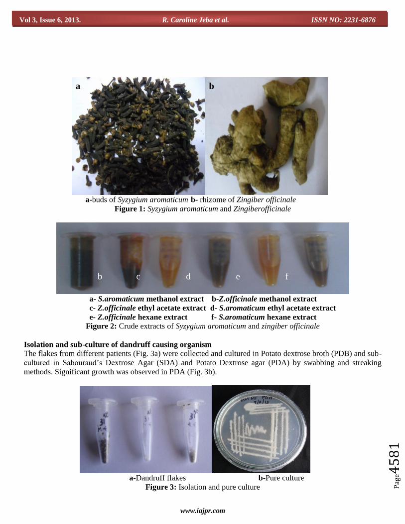
a-buds of Syzygium aromaticum b- rhizome of Zingiber officinale
Figure 1: Syzygium aromaticum and Zingiberofficinale
a- S.aromaticum methanol extract b-Z.officinale methanol extract
c- Z.officinale ethyl acetate extract d- S.aromaticum ethyl acetate extract
e- Z.officinale hexane extract f- S.aromaticum hexane extract
Figure 2: Crude extracts of Syzygium aromaticum and zingiber officinale
Isolation and sub-culture of dandruff causing organism
The flakes from different patients (Fig. 3a) were collected and cultured in Potato dextrose broth (PDB) and sub-
cultured in Sabouraud’s Dextrose Agar (SDA) and Potato Dextrose agar (PDA) by swabbing and streaking
methods. Significant growth was observed in PDA (Fig. 3b).
a-Dandruff flakes b-Pure culture
Figure 3: Isolation and pure culture
a b
a b c d e f
a b
Page 9
www.iajpr.com
Pag
e45
82
Vol 3, Issue 6, 2013. R. Caroline Jeba et al. ISSN NO: 2231-6876
Biochemical identification of the isolate The various identification tests were performed to confirm the presence of Malassezia furfur.
Catalase test
The catalase enzyme catalyzes the decomposition of hydrogen peroxide during oxidation. The effervescence
caused by liberation of free oxygen indicates a positive test (Fig. 4).
a- Control (without effervescence) b- Test (with effervescence)
Figure 4: Catalase test
Lactophenol cotton blue test
Grown isolates of Malassezia furfur smeared with lactophenol cotton blue were viewed in the microscope. The
bottle shaped cells of Malassezia furfur were observed (Fig 5).
Figure 5: Microscopic view of Malassezia furfur showing bottle shaped cells
Nitrate reduction test
1ml of sample was inoculated in nitrate broth and 5 drops of Sulfanilic acid and α-Naphthalamine reagent were
added. The solution gave a distinct light red colour which indicates a positive test (Fig. 6).
Figure 6: Nitrate reduction test
a b
a b
a- Control (Nitrate negative)
b- Test (Nitrate positive)
Page 10
www.iajpr.com
Pag
e45
83
Vol 3, Issue 6, 2013. R. Caroline Jeba et al. ISSN NO: 2231-6876
Urease test
Malassezia furfur culture was inoculated in Christensen’s urea broth. Then, it was incubated for24hrs. Pink
colour appeared which denotes the presence of urease positive organism (Fig. 7).
Figure 7: Urease test
Gelatin hydrolysis test
Pure culture of Malassezia furfur was inoculated into a sterile tube containing nutrient gelatin and incubated for
24hrs at 35-37ºC. The tube was placed on ice for few minutes and it was found that the media failed to solidify
which indicates a positive test (Fig. 8).
Figure 8: Gelatin hydrolysis test
Anti-dandruff assay
From the results it is clear that the methanol extracts of S.aromaticum and Z.officinale had significant inhibitory
effect on M. furfur when compared with ethyl acetate and hexane extracts (Fig. 9). The methanol extract of S.
aromaticum showed maximum inhibition with Zone of Inhibition 23mm. While, that for Z. officinale was
observed to be 22mm. ZOI was observed for the methanol extract of S. aromaticum from 250µg/ml while it was
500µg/ml for the methanol extract of Z. officinale (Table1).The activity was found to be effective in the
methanol extract of Syzygium aromaticum hence it was taken for further assay.
a b
a b
a- Test (Urease positive)
b- Control (Urease negative)
a-Test (semi-solid state)
b- Control (solidified state)
Page 11
www.iajpr.com
Pag
e45
84
Vol 3, Issue 6, 2013. R. Caroline Jeba et al. ISSN NO: 2231-6876
a- Syzygium aromaticum hexane b- Syzygium aromaticum ethyl acetate
c-Syzygium aromaticum methanol d- Zingiber officinale hexane
e-Zingiber officinale ethyl acetate f- Zingiber officinale methanol
1 – Standard (Ketaconazzole 100µg/ml)
Figure 9: Effect of extracts of S.aromaticum and Z.officinale on M. furfur
Table 1: Effect of plant extracts on Malassezia furfur
S.
n
o
Concent
ration
(µg/ml)
Zone of inhibition(mm)
Syzygium aromaticum Zingiber officinale Standard
(100 µg/ml) Hexane Ethyl
acetate Methanol Hexane
Ethyl
acetate Methanol
1 250 - 10.66±2.08 15±1 - - -
33.33±1.52 2 500 - 19±2.64 21.33±1.52 - - 16.33±1.52
3 750 11.33±1.52 20±1 22.33±1.52 10.66±1.5
2
- 19.33±2.51
4 1000 13.33±1.52 21±2 22.66±1.52 12±1 11.66±1.2 22.33±2.51
Values are expressed in mean±SEM
Broth dilution assay
Broth dilution assay was performed to know the minimum inhibitory concentration of Syzygium aromaticum.
The MIC was 100µg/ml and IC50 value was found to be 850µg/ml (Table 2). The inhibitory effect was studied
to be linear with the concentration of the sample (Fig. 10).
a b c
d e f
1 1 1
1 1 1
Page 12
www.iajpr.com
Pag
e45
85
Vol 3, Issue 6, 2013. R. Caroline Jeba et al. ISSN NO: 2231-6876
Table 2: Determination of IC50 of methanol extract of S. aromaticum
S.No Concentration(µg/ml) Inhibition (%)
1 100 0.68
2 200 2.18
3 300 2.73
4 400 5.32
5 500 15.98
6 600 21.72
7 700 27.32
8 800 32.92
9 900 60.24
10 1000 64.48
Inh
ibit
ion
(%
)
Concentration (µg/ml)
Figure 10: Determination of IC50 of methanol extract of S. aromaticum
Qualitative Phytochemical analysis
Qualitative phytochemical tests for alkaloids (a), flavonoids (b), phenols (c), tannins (d), glycosides (e),
saponins (f), reducing sugars (g) and proteins (h)
Figure 11: Qualitative phytochemical analysis
In the qualitative analysis of Syzygium aromaticum the presence of flavonoids, tannins, glycosides, reducing
sugars and phenolic compounds were determined. Qualitative analysis of Zingiber officinale showed the
presence of flavonoids, glycosides, reducing sugars and proteins (Fig. 11).
a b c d
h g f e
Page 13
www.iajpr.com
Pag
e45
86
Vol 3, Issue 6, 2013. R. Caroline Jeba et al. ISSN NO: 2231-6876
Table 3: Qualitative phytochemical analysis
S.No Phytochemical Syzygium
aromaticum Zingiber officinale
1 Alkaloids - -
2 Flavonoids +++ +
3 Phenols +++ -
4 Tannins +++ -
5 Glycosides + -
6 Saponins - -
7 Reducing sugars +++ ++
8 Proteins - +
- Not detectable using the assay followed + present in minor amount
++ present in moderate amount +++ present in higher amount
Quantitative estimation of phytochemicals
In the quantitative analysis of Syzygium aromaticum the total concentration of phenol was estimated to be
1052µg of GAE/g of extract, the total concentration of flavonoids was found to be 1299µg of QE/g of extract,
the total concentration of tannins was found to be 1255µg of GAE/g of extract and the amount of reducing
sugars was found to be 1052mg of Glucose/g sample.
Table 4: Quantitative estimation of phytochemicals in S.aromaticum
S .No Phytochemical Amount
1 Total phenolics 1052.66±1.52µg GAE/g sample
2 Total flavonoids 1299.33±2.08µg QE/g sample
3 Total tannins 1254.66±1.52µg GAE/g sample
4 Reducing sugars 1051.66±1.53mg Glucose/g sample
Values are expressed in mean±SEM
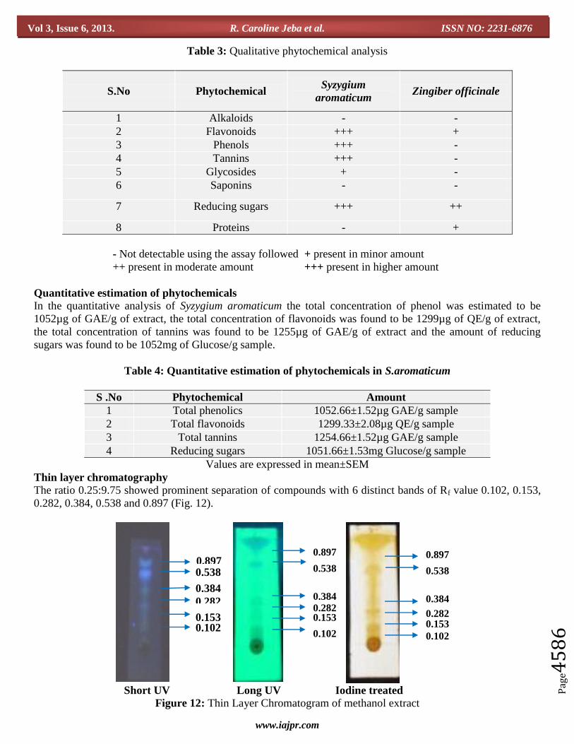
Thin layer chromatography
The ratio 0.25:9.75 showed prominent separation of compounds with 6 distinct bands of Rf value 0.102, 0.153,
0.282, 0.384, 0.538 and 0.897 (Fig. 12).
Short UV Long UV Iodine treated
Figure 12: Thin Layer Chromatogram of methanol extract
0.897 0.538
0.282
0.153 0.102
0.384
0.897
0.538
0.384 0.282 0.153
0.102
0.897
0.538
0.384
0.153 0.102
0.282
Page 14
www.iajpr.com
Pag
e45
87
Vol 3, Issue 6, 2013. R. Caroline Jeba et al. ISSN NO: 2231-6876
Control Treated
Bio-autography
In the bio-autography, the zone of inhibition was found in the compound possessing the Rf value 0.153.
Figure 13: Bio-autography showing clear zone around compound with Rf value 0.153
Compound inhibition by TLC
In this method the band formed by the compounds in the Malassezia furfur was inhibited when treated with the
extract of Syzygium aromaticum.
Figure 14: Compound inhibition by TLC
CONCLUSION
Nowadays, more individuals are susceptible to dandruff, which leads to both physiological and psychological
problem. The treatment is essential to control the severity and harmful effects of dandruff. The currently
available commercial agents such as Zinc pyrithione, Ketoconazole, Piroctone olamine, Cipropirox olamine, etc
though are effective lead to certain side effects such as hair loss and irritation. Herbal products help in
preventing dandruff without side effects.
Results of the present investigation reveal the antidandruff activity of the extracts of Syzygium aromaticum and
Zingiber officinale against Malassezia furfur, the dandruff causing organism. Among the three solvents used the
0.153
Page 15
www.iajpr.com
Pag
e45
88
Vol 3, Issue 6, 2013. R. Caroline Jeba et al. ISSN NO: 2231-6876
methanol extract of Syzygium aromaticum shows higher antidandruff activity and can be used to formulate a
potential therapeutic agent for dandruff.
The future research of the work could be to purify and isolate the compound that is responsible for the
inhibition of the dandruff causing organism and develop it into a potential herbal product. This work will have
importance in the field of cosmetics since it will be cost effective and there will be no side effects.
REFERENCES
1) Sirkar, N.N. (1989). Pharmacological basis of Ayurvedic therapeutics. In: Atal, C.K., Kapoor, B.M.
(Eds.), Cultivation and utilization of medicinal plants. Published by PID CSIR.
2) Gomez-Flores. R., Verástegui-Rodríguez, L., Quintanilla-Licea, R., Tamez-Guerra1, R., Tamez-
Guerra1, R., Rodríguez-Padilla, C. (2008). In vitro rat lymphocyte proliferation induced by Ocinum
basilicum, Persea americana, Plantago virginica, and Rosa spp. Extracts. Journal of Medicinal Plants
Research, 2(1), 005–010.
3) Prakash, P., Gupta, N. (2005). Therapeutic uses of Ocimum sanctum Linn (Tulsi) with a note on eugenol
and its pharmacological actions: A short review. Indian J Physiol Pharmocol, 49(2), 125–131.
4) Borchers, A.T., Sakai, S., Henderson, G.L., Harkey, M.R., Keen, C.L., Stern, J.S., Terasawa, K.,
Gershwin, M.E. (2000). Shosaiko-to and other Kampo (Japanese herbal) medicines: a review of their
immunomodulatory activities. J Ethnopharmacol 73, 1–13.
5) Prabhamanju, M., Shankar, SG.,Babu, k and Ranjith, MS,2009. Herbal vs. Chemical substances as
antidandruff ingredients: which are more effective in the management of dandruff?-An overview.
Egyptian Dermatology Online Journal 5(2):8.
6) Sibi, G., Gurmeetkaur, Devi, G., Dhananjaya, K., Ravikumar, KR and Mallesha, H, 2012. Anti-dandruff
activity of Ricinus communis L.leaf extracts. International journal of current pharmaceutical research
4(3):74-76.
7) Berenji, F., Rakhshandeh, H and Ebrahimipour, H, 2010.In vitro study of the effects of henna extracts
(Lawsonia inermis) on Malassezia species.Jundishapur J Microbiol 3(3): 125-128.
8) Naeini, A., Eidi, S and Shokri H, 2011.Fungitoxicity of Zataria multiflora essential oil against various
Malassezia species isolated from cats and dogs with Malassezia dermatitis. African Journal of
Microbiology Research 5(9):1057-1061.
9) Charles, W., Saunders, Scheynius, A and Heitman, J, 2012. Malassezia fungi are specialized to live on
skin and associated with dandruff, Eczema, and Other Skin Diseases. PLoS Pathogens 8(6): e1002701.
10) Ranasinghe, L., Jayawardena, B and Abeywickrama, K, 2002. Fungicidal activity of essential oils of
Cinnamomum zeylanicum (L.) and Syzygium aromaticum (L.) MerretL.M.Perry against crown rot and
anthracnose pathogens isolated from banana. Letters in Applied Microbiology 35:208-211.
11) Pinto, E., Vale-Silva, L., Cavaleiro, C and Salgueiro, L, 2009. Antifungal activity of the clove essential
oil from Syzygium aromaticum on Candida, Aspergillus and dermatophyte species. Journal of Medical
Microbiology 58:1454-1462.
12) Onyenekwe, PC and Hashimoto, S, 1999. The composition of the essential oil of dried Nigerian ginger
(Zingiber officinale Roscoe). Eur Food Res Technol 209:407-410.
13) Eloff, JN, 1998. J. Ethnopharmacology 60:1-8.
14) Gehan, S., El-Hadidy, Nahed, IM.,Gomaa, Abo bakr, RAE., Lobna, A and Metwally, 2007. Direct
molecular identification of Malassezia species from skin scales of patients with Seborrheic dermatitis by
Nested Terminal Fragment Length Polymorphism analysis. Egyptian Journal of Medical Microbiology
16(3):437-444.
15) Naveen, S., Karthika, S., Sentila, R., Mahenthiran, R and Michael, A, 2012. In-vitro evaluation of herbal
and chemical agents in the management of dandruff. Journal of Microbiology and Biotechnology
Research 2(6):916-921.
16) Vijayakumar, R., Muthukumar, C., Kumar, T and Saravanamuthu, R, 2006. Characterization of
Malassezia furfur and its control by using plant extracts. Indian J dermatol 51(2):145-148.
Page 16
www.iajpr.com
Pag
e45
89
Vol 3, Issue 6, 2013. R. Caroline Jeba et al. ISSN NO: 2231-6876
17) Buick, SF, 1997. Development of an In Vitro diagnostic technique for Malassezia furfur.A thesis
submitted in partial fulfillment of the requirement for the degree of Master of Science in Microbiology
in the university of Canterbury.
18) Yang, C., Chang, F., Chang, H., Wang, S., Hsieh, M and Chuang, L, 2012.Investigation of the
antioxidant activity of Illicium verum extracts. Journal of Medicinal Plants Research 6(2):314-324.
19) Sulieman, AME.,Issa, FM and Elkhalifa, EA, 2007. Quantitative determination of tannin content in
some Sorghum cultivars and evaluation of its antimicrobial activity. Research Journal of Microbiology
2(3):284-288.
20) Yemm, EW and Willis, AJ, 1954. The estimation of carbohydrates in plant extracts by Anthrone 57:508-
514.
21) Verma, A., Singh, N and Kumar A, 2013. Phytochemical investigation and thin layer chromatography of
Asparagus Racemosus (Asparagaceae) methanolic leaves extract. International journal of Biology,
Pharmacy and Allied sciences 2(1):187-191.
22) Choma, IM and Grzelak, EM, 2010. Bioautography detection in thin-layer chromatography. Journal of
Chromatography A :1-8.
23) Pawar, NK and Arumugam, N, 2011. Leaf extract of Centratherum Punctatum exhibits antimicrobial,
antioxidant and anti proliferative properties. Asian Journal of Pharmaceutical and Clinical Research
4(3):71-76.
54878478451001335
Submit your next manuscript to IAJPR and take advantage of: • Access Online first • Double blind peer review policy • No space constraints • Rapid publication • International recognition Submit your manuscript at: [email protected]