A Unique Palindromic Element Mediates Gamma InterferonInduction of mig Gene Expression
PHILIP WONG,' CHRISTOPHER W. SEVERNS,1 NANETTE B. GUYER,' AND
TIMOTHY M. WRIGHT' 2,3*
Division ofRheumatology and Clinical Immunology, Department ofMedicine, 1 Department ofMolecularGenetics and Biochemistry,2 and Pittsburgh Cancer Institute,3 University ofPittsburgh School ofMedicine,
Pittsburgh, Pennsylvania 15261
Received 20 August 1993/Returned for modification 25 September 1993/Accepted 5 November 1993
To define the molecular mechanisms involved in the action of gamma interferon (IFN-y), we have analyzedthe transcriptional regulation of the mig (monokine induced by gamma interferon) gene, a member of theplatelet factor 4-interleukin-8 cytokine family that is expressed in murine macrophages specifically in response
to IFN-y. Analysis of mig/CAT chimeric constructs transiently transfected into the RAW 264.7 mousemonocytic cell line revealed a unique IFN-y-responsive element ('yRE-1). The sequence of this cis regulatoryelement defined by deletion analysis contains an imperfect inverted repeat extending 27 bp. Examination ofmigICAT constructs with mutations in yRE-1 revealed that the palindromic positions in the element wereessential for activity. Consistent with its function as an enhancer, a single copy of 'yRE-1 conferred IFN-yinducibility to a heterologous (herpes simplex virus thymidine kinase) promoter. Exonuclease III protectionassays demonstrated symmetrical protection of a mig promoter fragment centered about the 'yRE-1 palin-dromic sequence. Using the gel electrophoretic mobility shift assay, we identified a factor (-yRF-1) present innuclear extracts prepared from IFN-y-stimulated RAW 264.7 cells which binds to -yRE-1. The activation of'yRF-1 occurred rapidly (within 1 min) in response to IFN-y and was independent of protein synthesis. Similarto the expression ofmig mRNA, the formation of yRF-1 was selectively induced by IFN-y and not IFN-c. Theregulation of gene expression through 'yRF-1 and -yRE-1 may explain the preferential activation of a subset ofinterferon-inducible genes by IFN-y.
The interferons (IFNs) are an important group of cyto-kines which share many biologic effects including the induc-tion of antiviral activity, the regulation of cell growth anddifferentiation, and the modulation of the immune response(20, 30). Similar to other cytokines, the IFNs induce many oftheir biologic effects through the regulation of specific RNAand protein expression in the responding cells (31). There-fore, knowledge of the ways in which the IFNs regulate genetranscription is essential to our understanding of the basicmechanisms of their action.Although type I (IFN-a and IFN-P) and type II (IFN-y)
IFNs bind to different cell surface receptors, they induce theexpression of an overlapping set of genes (4, 13, 31). Thestudy of genes activated by both type I and II interferons hasled to the identification of two well-characterized promoterelements, namely, the interferon-stimulated response ele-ment (ISRE) (15, 24) and the IFN-y activation site (GAS)(26). The ISRE is present in the promoter regions of anumber of IFN-stimulated genes including the class I majorhistocompatibility complex genes, guanylate-binding pro-tein, and metallothionein II (15, 24, 26). The transcriptionfactor which binds to the ISRE is a multimeric complexknown as ISGF3 and has been extensively characterized byseveral groups of investigators (1, 6, 16, 22, 25). The GASelement was identified as a region overlapping with an ISREsequence in the guanylate-binding protein promoter andcontributing to the responsiveness of the guanylate-bindingprotein gene to IFN-,y. It was subsequently found to bind
* Corresponding author. Mailing address: Division of Rheumatol-ogy and Clinical Immunology, University of Pittsburgh School ofMedicine, 985 Scaife Hall, Pittsburgh, PA 15261. Phone: (412)624-9028. Fax: (412) 624-4901.
914
factors (IFN--y activation factor [GAF] and IFN-ao activationfactor [AAF]) activated by both types of IFNs (7, 8).
It has long been recognized that IFN--y induces the ex-pression of unique mRNAs and proteins not induced byIFN-a and IFN-, (21, 38). Our understanding of the regula-tion of the genes encoding these unique mRNAs and proteinshas lagged behind the advances described above for genesinduced by both type I and II IFNs. This is in part becauseonly relatively recently have the cDNA and genomic clonescorresponding to this subset of IFN-y-inducible genes beenobtained (5, 11, 12, 29, 43). For some time, investigation intothe mechanism by which IFN-y preferentially activates genetranscription has focused primarily on the promoters of theclass II major histocompatibility complex genes (2, 3, 9, 14,32, 44). This has proven to be a complicated system withevidence for cell-type-specific, constitutive, and cytokine-regulated expression. Work in several laboratories has de-fined a complex array of cis elements (H, W, X, Y, and Zboxes and y-IRE) within the class II major histocompatibil-ity complex promoters which bind constitutive and inducedtranscription factors, and interactions between these multi-ple elements are reported to be necessary for regulation byIFN--y (9, 14, 44). Whether these elements will prove to beimportant in the regulation of other genes by IFN--y remainsto be determined.More recently, additional IFN--y-responsive sequences
have been identified in the promoters of genes rapidlyinduced by IFN-,y (29, 37, 41). Sims et al. (37) found that anelement (IR1) consisting of an imperfect inverted repeat oftwo GAAANN motifs, which are also present in the ISRE(24), is essential for responsiveness of the interferon regula-tory factor 1 (IRF-1) gene promoter to IFN (37). In addition,they identified a DNA-binding factor (IRFi) which is acti-
vated by IFN-y and to a lesser extent by IFN-a. IRFi bindsto IR1, but its binding is not inhibited by the ISRE (37).Similarly, analysis of the promoter region of the gene encod-ing the high-affinity Fc receptor for immunoglobulin G(FcyRI) has revealed an element (GRR) which is necessaryfor IFN-y-inducible expression and capable of conferringIFN-y responsiveness to a heterologous promoter (29).Interestingly, GRR contains a motif shared with the GASelement, and it has been shown that the two electrophoret-ically distinct GRR-protein complexes (FcRFy1 andFcRF-y2) observed by gel shift assay contain the p91 com-ponent of GAF and at least one additional protein responsi-ble for DNA binding (19, 23).The mig (monokine induced by IFN--y) cDNA was isolated
by differential screening of a cDNA library prepared fromlymphokine-stimulated RAW 264.7 cells (mouse macroph-age-like cell line) and encodes a member of the platelet factor4-interleukin-8 family of low-molecular-weight cytokines(12). Transcription of the mig gene occurs specifically inresponse to IFN--y (but not type I IFN) and occurs in theabsence of new-protein synthesis (41). These propertiesmake it a particularly suitable system for the examination ofthe IFN--y-specific transcriptional control. In a previousreport we demonstrated that an 88-bp mig promoter frag-ment was capable of conferring IFN--y inducibility to aheterologous promoter (41). We now present further analysisof mig gene regulation, including the identification of a novelpalindromic cis element responsive to IFN--y, and prelimi-nary characterization of the IFN--y-activated transcriptionfactor which binds to this element.
MATERIALS AND METHODS
Cells and transient transfections. The RAW 264.7 cell linewas obtained from the American Type Culture Collectionand was maintained in RPMI 1640 medium supplementedwith 10% fetal bovine serum and 2 mM L-glutamine. Tran-sient transfection of RAW 264.7 cells was performed by amodification of the DEAE-dextran method as describedpreviously (41). Briefly, cells were plated at 2 x 10⁶ per60-mm tissue culture dish 2 days prior to transfection. Afterbeing washed twice with 3 ml of OPTIMEM (GIBCO/LifeTechnologies), the cells were incubated with the DNA/DEAE-dextran solution (4 pg of plasmid DNA, 250 ,g ofDEAE-dextran per ml) in OPTIMEM for 1 h; this wasfollowed by the addition of chloroquine (final concentration,100 ,uM) and further incubation for 1.5 to 2 h. The DNA/DEAE-dextran solution was then aspirated, and the cellswere treated with 10% dimethyl sulfoxide in 2 ml of phos-phate-buffered saline (PBS) for 1 min and given three washeswith 5 ml of PBS. Culture medium (5 ml) was added, and thedishes were incubated at 37°C for 18 to 20 h. The contents ofeach dish were then scraped into 8 ml of fresh culturemedium and divided between two (or in some experimentsthree) 60-mm culture dishes. After incubation for 2 h at 37°Cto allow the cells to adhere, one dish from each transfectiongroup received medium alone, one received recombinantmurine IFN-y (1,000 U/ml; GIBCO/Life Technologies), andin some experiments a third dish received recombinantmurine IFN-a (1,000 U/ml; GIBCO/Life Technologies), asindicated. The cells were harvested for analysis of chloram-phenicol acetyltransferase (CAT) expression after an 18- to24-h period of stimulation.
Preparation of mig/CAT reporter constructs. A miggenomic fragment extending from position -1117 to +43relative to the transcription start site was ligated into the
SmaI site of the promoterless CAT reporter plasmidpUMSVOCAT (33). By using available EcoRV and HindIIIsites in the mig promoter (41), two plasmids, p(-704/+43)CAT and p(-358/+43)CAT (also referred to aspl19HIIICAT), containing major 5' deletions were prepared.A series of 5' deletion mutants was prepared fromp119HIIICAT by using exonuclease III (Erase-a-Base;Promega). The inserts of the 3' deletion mutants and theIFN-y-responsive element (-yRE-1) point mutants were pre-pared by PCR, using as primers the oligonucleotides corre-sponding to the indicated mig genomic sequences (or mutantsequences) with the addition of either BamHI or BglIIrestriction sites at the 5' ends of the oligonucleotides. Afterthe PCRs, the products were digested with BamHI and BgIIIand subjected to gel purification of the DNA fragments. TheDNA fragments containing 3' deletions of the mig promoterwere ligated into the BamHI site of pBLCAT2, whichcontains the herpes simplex virus thymidine kinase (tk)promoter upstream of the CAT gene (27). The DNA frag-ments containing point mutations in the -yRE-1 sequencewere ligated into pUMSVOCAT, which had been modified tocontain a BglII site in place of the SmaI cloning site.Additional plasmids containing the -yRE-l sequence or theindicated duplications of the left and right halves of -yRE-1were prepared by annealing complementary oligonucleotidesand ligating them into the BamHI site of pBLCAT2. Thesequences of the inserts were determined by the dideoxychain termination method.CAT assays. Preparation of cell lysates and determination
of CAT activity were performed as described previously (17)with the modification that butyryl coenzyme A (Pharmacia)was used in lieu of acetyl coenzyme A. CAT activities wereadjusted for differences in protein concentrations of theextracts. Relative CAT activity was determined by subtract-ing the value for mock-transfected cultures (arbitrarily set ata value of zero) and expressing the result as a percentage ofthe positive control plasmid (e.g., containing the indicatedwild-type sequence or p119HIIICAT).
Nuclear and cytosol extracts. Nuclear and cytosol extractswere prepared according to the method of Dignam et al. (10)with the modifications that 0.1% Triton X-100 was includedin all buffers and extracts were diluted 1:4 in buffer D(storage buffer) to reduce the salt concentration rather thanbeing dialyzed.
Exonuclease HI protection assay. DNA fragments radiola-beled at the 5' terminus of one strand and containing the miggenomic sequence from -235 to -148 were prepared by firstradiolabeling the oligonucleotide primers 5'-CGGGATCCAATTlTCTGCATGTTCTAC-3' and 5'-GAAGATCTCAGGTAGGAACTTGCCT-3', which correspond to the mig pro-moter sequences from -235 to -218 and from -148 to -165,respectively (to which have been added sequences forBamHI and BglII sites for cloning purposes), with T4polynucleotide kinase and [32P]ATP (6,000 Ci/mmol; NewEngland Nuclear). Two PCRs (labeling either the upper orlower strand) were performed with the above primer pair(one radiolabeled and one unlabeled primer) and a miggenomic fragment (-358 to +43) cloned into pBluescript asthe template. After amplification for 25 cycles, the radiola-beled products were purified by ion-exchange chromatogra-phy (Qiagen). Exonuclease III protection was based on themethod of Wu (42). The reaction mixtures contained 40 ,ug ofcrude nuclear extracts, 2 pg of denatured salnion spermDNA, end-labeled DNA fragment (3 x 10⁴ cpm), and bindingbuffer (50 mM KCl, 0.5 mM EDTA, 0.1% Triton X-100, 1.5mM MgCl2, 2 mM dithiothreitol, 12.5% glycerol, 10 mM
Tris-HCl [pH 7.5]) in a volume of 60 pl. After incubation for20 min at 25°C, exonuclease III (400 U; New EnglandBiolabs) was added and digestion was allowed to proceed for15 min at 25°C. The reactions were terminated by theaddition of 170 ,u of chilled (-60°C) stop solution (20 ,g oftRNA per ml, 4.5 mM EDTA, 0.9 M ammonium acetate,87% ethanol) and the samples were precipitated in a dryice-ethanol bath for 15 min. The precipitates were pelleted(15,000 x g for 15 min at 4°C), washed twice with 70%ethanol, dried for 15 min in a vacuum centrifuge, anddissolved in 10 R1 of formamide loading buffer. After beingheated at 90°C for 5 min, 5 ,ul of each sample was loaded ona 6% acrylamide-7.8 M urea sequencing gel. Thermocyclesequencing reaction mixtures (CircumVent; New EnglandBiolabs) with the same oligonucleotides and template used inpreparation of the radiolabeled fragments (see above) wereloaded in adjacent lanes to identify the termini of theprotected fragments.
Gel electrophoretic mobility shift assay. Double-strandedoligonucleotides containing the yRE-1 sequence (-200 to-167) of the mig promoter or the GAS sequence (-126 to-101) of the guanylate-binding protein promoter (26) wereradiolabeled with [32P]dGTP and [32P]dATP (both at 6,000Ci/mmol) by using the Klenow fragment ofDNA polymeraseI. Binding reaction mixtures (25 ,ul) contained the indicatedamount of nuclear or cytosol extract, 5 fmol (approximately105 cpm) of radiolabeled DNA fragment, and 2 ,g ofdenatured salmon sperm DNA in binding buffer. Reactionmixtures were incubated for 20 min at room temperature, 5,ul of 0.1% bromphenol blue in binding buffer was thenadded, and the mixture was immediately loaded on a 6%polyacrylamide gel. Electrophoresis was performed in 0.25 xTBE buffer (TBE buffer is 89 mM Tris base, 89 mM boricacid, and 2 mM EDTA [pH 8]) at 175 V with bufferrecirculation. Where indicated, unlabeled double-strandedoligonucleotides (specific competitors) were added to thebinding reaction mixture before the addition of radiolabeledfragment.
RESULTS
Mapping the 5' borders of IFN--responsive elementswithin the mig promoter. To define the region(s) of the miggene responsible for transcriptional activation by IFN-y, weprepared a series of constructs containing 5' deletions of themig promoter coupled to the CAT reporter gene in theplasmid pUMSVOCAT. These constructs were analyzed forIFN-,y inducibility in transient-transfection experiments withthe mouse macrophage cell line RAW 264.7. Transfectedcultures were treated with medium alone or medium supple-mented with IFN--y (1,000 U/ml) for 18 to 24 h, and then thecells were harvested and cell extracts were prepared foranalysis of CAT activity. The results of these transfectionswith constructs containing successive 5' deletions of the migpromoter region are shown in Fig. 1A. The basal activity ofthese constructs was extremely low, with a mean percentconversion of 0.07%, which was at least in part due to thelow background of plasmid pUMSVOCAT (33). The resultsare expressed as IFN-y-inducible CAT activity relative toplasmid p119HIIICAT (100%), which contains the miggenomic sequence from -358 to +43. This plasmid wasincluded in the transfection experiments as a positive controland internal standard. As shown in Fig. 1A, there wasmarked induction of CAT expression (250- to 1,000-fold) byIFN-y in cells transfected with plasmids containing thedeletions spanning the region from -1117 to -198. Consis-
RELATIVE CAT ACTIVITYFIG. 1. Mapping enhancer elements in the 5' regulatory region of
the mig gene. (A) IFN--y-induced CAT activity in RAW 264.7 cellstransiently transfected with plasmids containing 5' deletions of migpromoter region upstream of the CAT reporter gene in the plasmidpUMSVOCAT. The data are the composite from eight experiments(n 4 for each plasmid) and are expressed relative to the activity ofplasmid p119HIIICAT (set equal to 100) which contains the migsequence from -358 to +43. Shown are the values for relative CATactivity in IFN-y-treated cultures. Control (unstimulated) samplesdemonstrated uniformly minimal basal activity, with a mean con-version of 0.07% and relative activity of 0.19 (n = 69). (B) CATactivity of RAW 264.7 cells transfected with plasmids containing 3'deletions of the mig promoter region upstream of the herpes simplexvirus tk promoter in the vector pBLCAT2. The data are the meansof results from three experiments performed in duplicate and areexpressed as relative CAT activity with the value for plasmidp119HIIICAT set equal to 100.
tent with the selective stimulation of mig mRNA by IFN--yand not IFN-a, the activation of these constructs was totallydependent on treatment with IFN-,y (40, 41). Although therewas some variability (approximately two- to fourfold) inIFN-y-inducible CAT expression among the 5' deletionmutants with mutations spanning the region from -1117 to-198, possibly because of the influence of minor regulatoryelements, total loss of activity was observed in extracts fromcells transfected with the construct containing the mig genefragment from -196 to +43. The analysis of these deletionmutants, therefore, mapped the 5' border of a potent IFN-
y-responsive element to within 2 nucleotides (-198 to-197).Further deletions revealed a second IFN--y-responsive
element in the region of position -84. CAT activity inextracts from RAW 264.7 cells transfected with the con-struct containing the mig sequence from -84 to +43 wasstimulated approximately 40-fold by IFN--y. In contrast, theCAT activity was not induced by treatment with 1,000 U ofIFN-ot per ml (40). The absence of activity of this element inconstructs containing adjacent 5' sequence suggests thepresence of an overlapping negative regulatory element. Inthis regard, it is interesting that the sequence immediatelyupstream of position -84 (41) has a high degree of homology(12-of-15 match with the consensus) to the ISRE, a ciselement previously shown to be important in the positiveregulation of IFN-inducible genes (15, 24) and recently foundto suppress IFN-inducible transcription through the bindingof the interferon consensus sequence-binding protein (28).
Identification of the 3' border of the -yRE-1. Because of therelative potency of the two positive regulatory regionsidentified by 5' deletion mutants, we focused our efforts oncharacterizing the element(s) whose 5' border was defined asposition - 198 or -197 (yRE-1). Taking advantage of the factthat a single copy of the mig genomic fragment extendingfrom positions -235 to -148 conferred IFN--y responsive-ness to a heterologous (tk) promoter (41), we used PCR toprepare a series of DNA fragments containing variousamounts of mig genomic sequence 3' of position -198 andinserted them upstream of the tk promoter in the CATreporter plasmid pBLCAT2 (27). Shown in Fig. 1B are theresults of experiments in which these 3' deletion mutantswere tested for IFN--y inducibility by transient transfectionof RAW 264.7 cells. Similar to the marked responsiveness ofthe constructs containing the homologous mig promoterregion, the CAT activity in extracts from cells transfectedwith plasmids p119.1CAT2, p119.3CAT2, and p119.4CAT2,containing one copy of the indicated mig genomic sequence,was induced more than 100-fold by IFN-y. This activity wasindependent of orientation of the inserted mig genomicfragments (40, 41). In contrast, there was no stimulation ofCAT activity in cells transfected with the deletion mutantp119.5CAT2, which contained the mig sequence from -235to - 176.On the basis of the data from the 5' and 3' deletion
mutants, it was predicted that the IFN-y-responsive ciselement -yRE-1 was present within the sequence from -198to - 167. To confirm this prediction, a pair of complementaryoligonucleotides corresponding to the mig genomic sequencefrom -200 to -167 (the 5' border was extended to position-200 by the addition of a restriction site for cloning pur-poses) were annealed and ligated into pBLCAT2 upstream ofthe tk promoter. As shown in Fig. 2, treatment of RAW264.7 cells transiently transfected with this construct,p(-200/-167)CAT2, with IFN--y stimulated CAT activitymarkedly (over 25-fold). Similar to the activity of largerfragments containing the -yRE-1 sequence (41), the IFN-y-inducible activity of this construct was independent of theorientation of the inserted fragment (40).
Role of palindromic positions in 'yRE-1. A comparison ofthe -yRE-1 sequence with the consensus sequences of previ-ously reported IFN-responsive elements including the ISRE(15, 24), GAS (26), and -y-IRE (44) did not identify thepresence of these elements, although partial homology (7-of-15 match) to GAS was noted. Careful scrutiny, however,did reveal an imperfect palindrome extending from positions-198 to -172. In order to address the importance of the
WTp(-200/.1 67)CAT2
IFN-y - + - +
I
pBLCAT2 p11 9HIIICAT
- + - +
0.3 0.5 0.5 0.09.1 12.1 0.3 16.7
CAT ACTIVITY (%CONVERSION)FIG. 2. A single copy of yRE-1 confers IFN-y inducibility to the
herpes simplex virus tk promoter. Shown are the CAT activities ofcell extracts prepared from RAW 264.7 cells transfected with theindicated plasmids. Cells were treated with medium alone or me-dium supplemented with IFN--y (1,000 U/ml) for 23 h prior to harvestand determination of CAT activity. WT, wild type.
palindromic bases within -yRE-1, DNA fragments containingthe mig promoter sequence from -200 to +43 with variouspoint mutations in each of the four palindromic segments orthe central nonpalindromic region were generated by PCRmutagenesis. These fragments were inserted upstream of theCAT reporter gene in the vector pUMSVOCAT. The resultsof transient transfections of RAW 264.7 cells with theseconstructs are shown in Fig. 3A. Mutations in each of thefour palindromic segments led to near-total loss of IFN--y-inducible activity, whereas the construct with mutations inthe central palindromic region retained approximately 25%of the activity of the wild-type sequence. These data sup-ported the hypothesis that the palindromic nature of -yRE-1is important for its responsiveness to IFN--y.To evaluate this hypothesis further and to examine the
importance of the central nonpalindromic spacer region, weprepared a series of CAT reporter constructs containingduplications of the 5' or 3' half of the imperfect palindromepresent in yRE-1 with various central regions (none, single,or double). These constructs were prepared by annealingcomplementary oligonucleotides corresponding to the migpromoter sequences indicated in Fig. 3B (with added 5'GATC overhangs to permit cloning into a BamHI site) andligating them into the BamHI site of pBLCAT2. RAW 264.7cells were transiently transfected with these constructs andanalyzed for IFN--y-inducible CAT expression. As shown inFig. 3B, duplicating either half of -yRE-1 resulted in afunctional IFN--y-responsive enhancer. The activity of themutant element containing a duplication of the 3' half of-yRE-1 was significantly greater than that of the wild-typesequence, whereas the activity of the duplicated 5' half of-yRE-1 was consistently lower than that of the wild-typeelement. Deletion or duplication of the central nonpalindro-mic region of -yRE-1 resulted in total loss of activity. Theseresults suggest that the imperfect palindrome in yRE-1 maycontain high-affinity (3' half) and low-affinity (5' half) bindingsites, perhaps for a homodimeric transcription factor. Thespacing of the two halves of the palindrome was critical for-yRE-1 activity.
Identification of a 'yRE-1-binding protein. Nuclear extracts
FIG. 3. Effect of mutations in the yRE-1 sequence on the inducibility by IFN--y. (A) RAW 264.7 cells were transfected with plasmids inwhich DNA fragments corresponding to the indicated wild-type (wt) or mutant mig sequences were inserted upstream of the CAT gene in thevector pUMSVOCAT. (B) RAW 264.7 cells were transfected with plasmids containing the wild-type (WT) yRE-1 sequence or plasmidscontaining the indicated duplications of the 5' or 3' portion of yRE-1 inserted upstream of the tk promoter in the vector pBLCAT2.Palindromic positions are shown in capital letters. The sequences corresponding to the 5' and 3' portions of the -yRE-1 inverted repeat areenclosed in open and shaded rectangles, respectively. For both panels A and B, transfected cultures were treated with medium alone (openbars) or 1,000 U of IFN-,y per ml (solid bars) prior to harvest and determination of CAT activity. Data are the means of results from twoexperiments performed in duplicate and are expressed as relative CAT activity with the value for the plasmid containing the wt -yRE-1sequence set equal to 100.
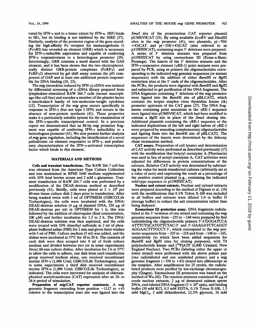
from RAW 264.7 cells were prepared and analyzed for thepresence of DNA-binding proteins capable of interactingwith a mig genomic DNA fragment (-235 to -148) contain-ing -yRE-1 by using the exonuclease III protection assay. Asshown in Fig. 4A, nuclear extracts from IFN-y-stimulatedcells but not control cells protected the end-labeled DNAfragments at positions -204 and -203 on the bottom strandand position -167 on the top strand. The protection by anIFN-y-inducible factor, referred to as -yRF-1, extended in anearly symmetrical manner 5 to 6 bases upstream and 5bases downstream of the margins of the imperfect palin-drome contained within -yRE-1 (Fig. 4B). A likely explana-tion for the extended area of protection outside of thesequence identified by 5' deletion analysis is that stericinterference between exonuclease III and -yRF-1 prevented
more-proximal digestion. Of note, there was no protection ofthe sequence 5' to -yRE-1 (41) which has homology to theISRE (15, 24).
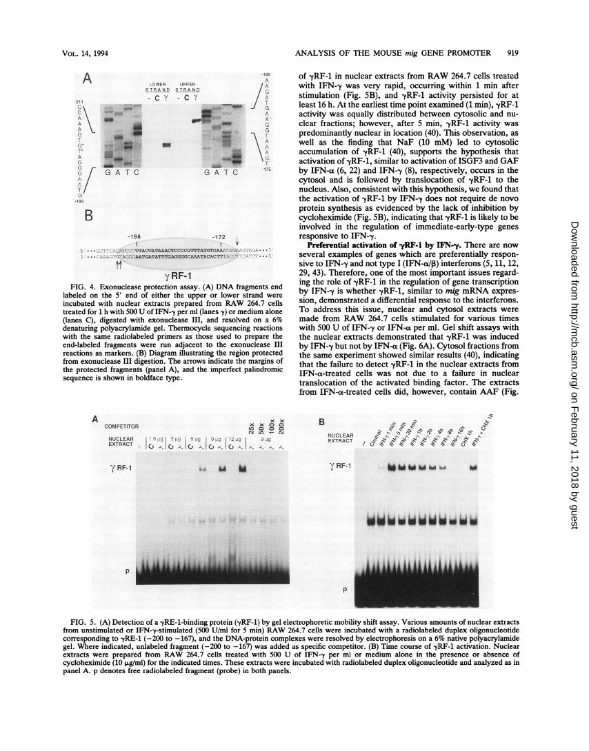
Analysis of -yRF-1 by gel shift assay. A DNA-proteincomplex corresponding to -yRF-1 was identified by the gelelectrophoretic mobility shift assay (gel shift) with a radio-labeled duplex oligonucleotide corresponding to positions-200 to -167 of the mig promoter. As shown in Fig. 5A,there was a single IFN-,y-inducible complex identified innuclear extracts from RAW 264.7 cells. Additional com-plexes with the labeled -yRE-1 fragment were formed, butthese complexes (present in control and stimulated cellextracts) were not inhibited by the addition of specificunlabeled fragment, indicating that they most likely repre-sent nonspecific DNA-protein interactions. The appearance
yRF-1FIG. 4. Exonuclease protection assay. (A) DNA fragments end
labeled on the 5' end of either the upper or lower strand were
incubated with nuclear extracts prepared from RAW 264.7 cellstreated for 1 h with 500 U of IFN--y per ml (lanes y) or medium alone(lanes C), digested with exonuclease III, and resolved on a 6%denaturing polyacrylamide gel. Thermocycle sequencing reactionswith the same radiolabeled primers as those used to prepare theend-labeled fragments were run adjacent to the exonuclease IIIreactions as markers. (B) Diagram illustrating the region protectedfrom exonuclease III digestion. The arrows indicate the margins ofthe protected fragments (panel A), and the imperfect palindromicsequence is shown in boldface type.
A x xCOMPETITOR , 0
Csl LO _ CMNUCLEAR 5 !.ig 3 io 6 Lg 3L9g| 12 LiC 9 Llg
C\Io-\ Io-\ -\-\I\
7 RF-1
of yRF-1 in nuclear extracts from RAW 264.7 cells treatedwith IFN--y was very rapid, occurring within 1 min afterstimulation (Fig. SB), and yRF-1 activity persisted for atleast 16 h. At the earliest time point examined (1 min), -yRF-1activity was equally distributed between cytosolic and nu-clear fractions; however, after 5 min, -yRF-1 activity waspredominantly nuclear in location (40). This observation, aswell as the finding that NaF (10 mM) led to cytosolicaccumulation of -yRF-1 (40), supports the hypothesis thatactivation of -yRF-1, similar to activation of ISGF3 and GAFby IFN-a (6, 22) and IFN--y (8), respectively, occurs in thecytosol and is followed by translocation of -yRF-1 to thenucleus. Also, consistent with this hypothesis, we found thatthe activation of yRF-1 by IFN-y does not require de novoprotein synthesis as evidenced by the lack of inhibition bycycloheximide (Fig. SB), indicating that -yRF-1 is likely to beinvolved in the regulation of immediate-early-type genesresponsive to IFN--y.
Preferential activation of -yRF-1 by IFN-y. There are nowseveral examples of genes which are preferentially respon-sive to IFN--y and not type I (IFN-a113) interferons (5, 11, 12,29, 43). Therefore, one of the most important issues regard-ing the role of -yRF-1 in the regulation of gene transcriptionby IFN-y is whether -yRF-1, similar to mig mRNA expres-sion, demonstrated a differential response to the interferons.To address this issue, nuclear and cytosol extracts were
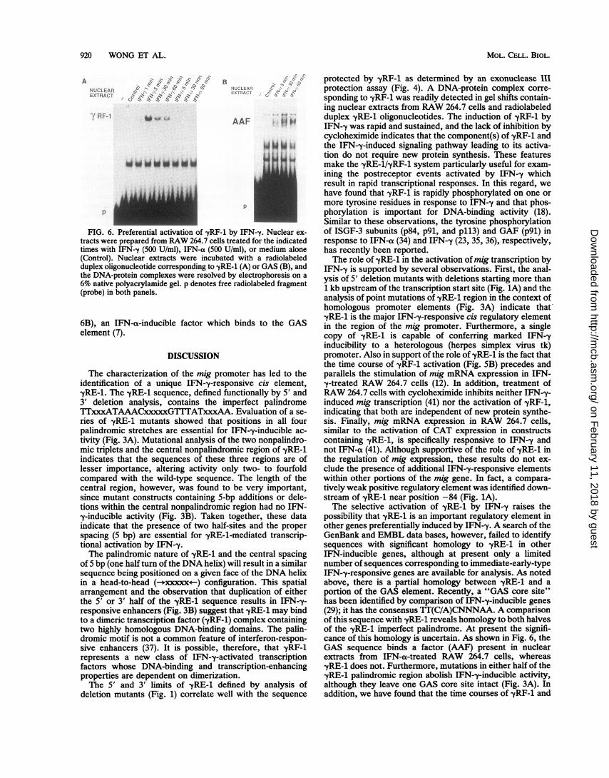
made from RAW 264.7 cells stimulated for various timeswith 500 U of IFN-y or IFN-a per ml. Gel shift assays withthe nuclear extracts demonstrated that -yRF-1 was inducedby IFN-y but not by IFN-a (Fig. 6A). Cytosol fractions fromthe same experiment showed similar results (40), indicatingthat the failure to detect -yRF-1 in the nuclear extracts fromIFN-a-treated cells was not due to a failure in nucleartranslocation of the activated binding factor. The extractsfrom IFN-a-treated cells did, however, contain AAF (Fig.
B .:" ~:.,z-"NUCLEAR -10 -" -, -, - \ " -\,K"'
EXTRACT 1-~ 11.Z Z.-zl-~ "
"l RF-1
p
p
FIG. 5. (A) Detection of a yRE-1-binding protein (yRF-1) by gel electrophoretic mobility shift assay. Various amounts of nuclear extractsfrom unstimulated or IFN--y-stimulated (500 U/ml for 5 min) RAW 264.7 cells were incubated with a radiolabeled duplex oligonucleotidecorresponding to -yRE-1 (-200 to -167), and the DNA-protein complexes were resolved by electrophoresis on a 6% native polyacrylamidegel. Where indicated, unlabeled fragment (-200 to -167) was added as specific competitor. (B) Time course of yRF-1 activation. Nuclearextracts were prepared from RAW 264.7 cells treated with 500 U of IFN-,y per ml or medium alone in the presence or absence ofcycloheximide (10 ,g/ml) for the indicated times. These extracts were incubated with radiolabeled duplex oligonucleotide and analyzed as inpanel A. p denotes free radiolabeled fragment (probe) in both panels.
ji|i."FIG. 6. Preferential activation of -yRF-1 by IFN-y. Nuclear ex-
tracts were prepared from RAW 264.7 cells treated for the indicatedtimes with IFN-y (500 U/ml), IFN-a (500 U/ml), or medium alone(Control). Nuclear extracts were incubated with a radiolabeledduplex oligonucleotide corresponding to -yRE-1 (A) or GAS (B), andthe DNA-protein complexes were resolved by electrophoresis on a6% native polyacrylamide gel. p denotes free radiolabeled fragment(probe) in both panels.
6B), an IFN-a-inducible factor which binds to the GASelement (7).
DISCUSSION
The characterization of the mig promoter has led to theidentification of a unique IFN-y-responsive cis element,yRE-1. The yRE-1 sequence, defined functionally by 5' and3' deletion analysis, contains the imperfect palindromeTIxxxATAAACxxxxxGT''ITATxxxAA. Evaluation of a se-ries of yRE-1 mutants showed that positions in all fourpalindromic stretches are essential for IFN-,y-inducible ac-tivity (Fig. 3A). Mutational analysis of the two nonpalindro-mic triplets and the central nonpalindromic region of yRE-1indicates that the sequences of these three regions are oflesser importance, altering activity only two- to fourfoldcompared with the wild-type sequence. The length of thecentral region, however, was found to be very important,since mutant constructs containing 5-bp additions or dele-tions within the central nonpalindromic region had no IFN-y-inducible activity (Fig. 3B). Taken together, these dataindicate that the presence of two half-sites and the properspacing (5 bp) are essential for yRE-1-mediated transcrip-tional activation by IFN-y.The palindromic nature of yRE-1 and the central spacing
of 5 bp (one half turn of the DNA helix) will result in a similarsequence being positioned on a given face of the DNA helixin a head-to-head (-*xxxxx<--) configuration. This spatialarrangement and the observation that duplication of eitherthe 5' or 3' half of the yRE-1 sequence results in IFN-y-responsive enhancers (Fig. 3B) suggest that yRE-l may bindto a dimeric transcription factor (yRF-1) complex containingtwo highly homologous DNA-binding domains. The palin-dromic motif is not a common feature of interferon-respon-sive enhancers (37). It is possible, therefore, that -yRF-1represents a new class of IFN--y-activated transcriptionfactors whose DNA-binding and transcription-enhancingproperties are dependent on dimerization.The 5' and 3' limits of yRE-1 defined by analysis of
deletion mutants (Fig. 1) correlate well with the sequence
protected by -yRF-1 as determined by an exonuclease IIIprotection assay (Fig. 4). A DNA-protein complex corre-sponding to yRF-1 was readily detected in gel shifts contain-ing nuclear extracts from RAW 264.7 cells and radiolabeledduplex yRE-1 oligonucleotides. The induction of -yRF-1 byIFN-y was rapid and sustained, and the lack of inhibition bycycloheximide indicates that the component(s) of yRF-1 andthe IFN-y-induced signaling pathway leading to its activa-tion do not require new protein synthesis. These featuresmake the yRE-1/yRF-1 system particularly useful for exam-ining the postreceptor events activated by IFN-,y whichresult in rapid transcriptional responses. In this regard, wehave found that -yRF-1 is rapidly phosphorylated on one ormore tyrosine residues in response to IFN--y and that phos-phorylation is important for DNA-binding activity (18).Similar to these observations, the tyrosine phosphorylationof ISGF-3 subunits (p84, p91, and p113) and GAF (p91) inresponse to IFN-a (34) and IFN-y (23, 35, 36), respectively,has recently been reported.The role of -yRE-1 in the activation of mig transcription by
IFN--y is supported by several observations. First, the anal-ysis of 5' deletion mutants with deletions starting more than1 kb upstream of the transcription start site (Fig. 1A) and theanalysis of point mutations of -yRE-1 region in the context ofhomologous promoter elements (Fig. 3A) indicate thatyRE-1 is the major IFN-y-responsive cis regulatory elementin the region of the mig promoter. Furthermore, a singlecopy of yRE-1 is capable of conferring marked IFN-yinducibility to a heterologous (herpes simplex virus tk)promoter. Also in support of the role of -yRE-1 is the fact thatthe time course of yRF-1 activation (Fig. SB) precedes andparallels the stimulation of mig mRNA expression in IFN-y-treated RAW 264.7 cells (12). In addition, treatment ofRAW 264.7 cells with cycloheximide inhibits neither IFN--y-induced mig transcription (41) nor the activation of -yRF-1,indicating that both are independent of new protein synthe-sis. Finally, mig mRNA expression in RAW 264.7 cells,similar to the activation of CAT expression in constructscontaining yRE-1, is specifically responsive to IFN--y andnot IFN-a (41). Although supportive of the role of -yRE-1 inthe regulation of mig expression, these results do not ex-clude the presence of additional IFN-y-responsive elementswithin other portions of the mig gene. In fact, a compara-tively weak positive regulatory element was identified down-stream of yRE-1 near position -84 (Fig. 1A).The selective activation of -yRE-1 by IFN-,y raises the
possibility that yRE-1 is an important regulatory element inother genes preferentially induced by IFN-,y. A search of theGenBank and EMBL data bases, however, failed to identifysequences with significant homology to yRE-1 in otherIFN-inducible genes, although at present only a limitednumber of sequences corresponding to immediate-early-typeIFN--y-responsive genes are available for analysis. As notedabove, there is a partial homology between yRE-1 and aportion of the GAS element. Recently, a "GAS core site"has been identified by comparison of IFN--y-inducible genes(29); it has the consensus TT(C/A)CNNNAA. A comparisonof this sequence with yRE-1 reveals homology to both halvesof the -yRE-1 imperfect palindrome. At present the signifi-cance of this homology is uncertain. As shown in Fig. 6, theGAS sequence binds a factor (AAF) present in nuclearextracts from IFN-a-treated RAW 264.7 cells, whereas-yRE-1 does not. Furthermore, mutations in either half of the,yRE-1 palindromic region abolish IFN--y-inducible activity,although they leave one GAS core site intact (Fig. 3A). Inaddition, we have found that the time courses of yRF-1 and
GAF activation by IFN-y are similar in RAW 264.7 cells butdiffer by several hours in HeLa cells (18). We are currentlyfurther investigating the potential relationship between theseelements and their respective binding factors.
Since the mig mRNA was originally identified in cells ofthe macrophage-monocyte lineage (12, 13), another possiblerole for -yRE-1 could be to regulate the tissue-specific ex-pression of genes by IFN--y. Arguing against this hypothesis,however, is the finding that yRF-1 is present in a widevariety of cell types after stimulation with IFN--y, includingmouse and human myeloid cells (RAW 264.7, human periph-eral blood monocytes, THP-1, U937, and HL-60), humanepidermoid carcinoma cell lines (HeLa and A431), humanfibroblasts (MRC-5 and primary dermal fibroblasts), a mouseB-lymphoma cell line (X16C8.5), a human melanoma cellline (Hs 294T), and the Hep-G2 human hepatoma cell line(40). Whether yRE-1 and yRF-1 are involved in the regula-tion of a specific subset of cellular responses to IFN--y (e.g.,induction of antiviral activity or inhibition of cell growth) inthese varied cell types awaits further identification of theIFN-y-responsive genes containing this new cis element.
ACKNOWLEDGMENTS
We thank J. Farber, H. Shin, and D. Nathans for their thoughtfuladvice; D. Honig and J. Gilmer for their excellent technical assis-tance; C. Feghali for her critical reading of the manuscript; and B.Knasko for her assistance in preparation of the manuscript.
This work was supported by Public Health Service grant CA55333from the National Cancer Institute and an Arthritis InvestigatorAward from the Arthritis Foundation.
REFERENCES1. Bandyopadhyay, S. K., D. V. R. Kalvakolanu, and G. C. Sen.
1990. Gene induction by interferons: functional complementa-tion between trans-acting factors induced by alpha interferonand gamma interferon. Mol. Cell. Biol. 10:5055-5063.
2. Basta, P. V., P. A. Sherman, and J. P.-Y. Ting. 1987. Identifi-cation of an interferon--y response region 5' of the humanhistocompatibility leukocyte antigen DRa chain gene which isactive in human glioblastoma multiforme lines. J. Immunol.138:1275-1280.
3. Collins, T., A. J. Korman, C. T. Wake, J. M. Boss, D. J. Kappes,W. Fiers, K. A. Ault, M. A. Gimbrone, Jr., J. L. Strominger, andJ. S. Pober. 1984. Immune interferon activates multiple class IImajor histocompatibility complex genes and the associatedinvariant chain gene in human endothelial cells and dermalfibroblasts. Proc. Natl. Acad. Sci. USA 81:4917-4921.
4. Colonno, R. J., and R. H. L. Pang. 1982. Induction of uniquemRNAs by human interferons. J. Biol. Chem. 257:9234-9237.
5. Dai, W., and S. L. Gupta. 1990. Regulation of indoleamine2,3-dioxygenase gene expression in human fibroblasts by inter-feron--y. J. Biol. Chem. 265:19871-19877.
6. Dale, T. C., A. M. A. Imam, I. M. Kerr, and G. R. Stark 1989.Rapid activation by interferon a of a latent DNA-binding proteinpresent in the cytoplasm of untreated cells. Proc. Natl. Acad.Sci. USA 86:1203-1207.
7. Decker, T., D. J. Lew, and J. E. Darnell, Jr. 1991. Two distinctalpha-interferon-dependent signal transduction pathways maycontribute to activation of transcription of the guanylate-bindingprotein gene. Mol. Cell. Biol. 11:5147-5153.
8. Decker, T., D. J. Lew, J. Mirkovitch, and J. E. Darnell, Jr. 1991.Cytoplasmic activation of GAF, an IFN--y-regulated DNA-binding factor. EMBO J. 10:927-932.
9. Dedrick, R. L., and P. F. Jones. 1990. Sequence elementsrequired for activity of a murine major histocompatibility com-plex class II promoter bind common and cell-type-specificnuclear factors. Mol. Cell. Biol. 10:593-604.
10. Dignam, J. D., R. M. Lebovitz, and R. G. Roeder. 1983.Accurate transcription initiation by RNA polymerase II in asoluble extract from isolated mammalian nuclei. Nucleic Acids
Res. 11:1475-1489.11. Fan, X., G. R. Stark, and B. R. Bloom. 1989. Molecular cloning
of a gene selectively induced by gamma interferon from humanmacrophage cell line U937. Mol. Cell. Biol. 9:1922-1928.
12. Farber, J. M. 1990. A macrophage mRNA selectively inducedby y-interferon encodes a member of the platelet factor 4 familyof cytokines. Proc. Natl. Acad. Sci. USA 87:5238-5242.
13. Farber, J. M. 1992. A collection of mRNA species that areinducible in the RAW 264.7 mouse macrophage cell line bygamma interferon and other agents. Mol. Cell. Biol. 12:1535-1545.
14. Finn, P. W., C. J. Kara, J. Douhan Ill, T. T. Van, V. Folsom,and L. H. Glimcher. 1990. Interferon -y regulates binding of twonuclear protein complexes in a macrophage cell line. Proc. Natl.Acad. Sci. USA 87:914-918.
15. Friedman, R. L., and G. R. Stark. 1985. a-Interferon-inducedtranscription of HLA and metallothionein genes containinghomologous upstream sequences. Nature (London) 314:637-639.
16. Fu, X.-Y., D. S. Kessler, S. A. Veals, D. E. Levy, and J. E.Darnell, Jr. 1990. ISGF3, the transcriptional activator inducedby interferon a, consists of multiple interacting polypeptidechains. Proc. Natl. Acad. Sci. USA 87:8555-8559.
17. Gorman, C. M., L. F. Moffat, and B. H. Howard. 1982.Recombinant genomes which express chloramphenicol acetyl-transferase in mammalian cells. Mol. Cell. Biol. 2:1044-1051.
18. Guyer, N. B., and T. M. Wright. Unpublished data.19. Igarashi, K.-I., M. David, A. C. Larner, and D. S. Finbloom.
1993. In vitro activation of a transcription factor by gammainterferon requires a membrane-associated tyrosine kinase andis mimicked by vanadate. Mol. Cell. Biol. 13:3984-3989.
20. Ijzermans, J. N. M., and R. L. Marquet. 1989. Interferon-gamma: a review. Immunobiology 179:456-473.
21. Kelley, V. E., W. Fiers, and T. B. Strom. 1984. Cloned humaninterferon--y, but not interferon-p or -a, induces expression ofHLA-DR determinants by fetal monocytes and myeloid leuke-mic cell lines. J. Immunol. 132:240-245.
22. Kessler, D. S., S. A. Veals, X.-Y. Fu, and D. E. Levy. 1990.Interferon-a regulates nuclear translocation and DNA-bindingaffinity of ISGF3, a multimeric transcriptional activator. GenesDev. 4:1753-1765.
23. Larner, A. C., M. David, G. M. Feldman, K.-I. Igarashi, R. H.Hackett, D. S. A. Webb, S. M. Sweitzer, E. F. Petricoin III, andD. S. Finbloom. 1993. Tyrosine phosphorylation of DNA bind-ing proteins by multiple cytokines. Science 261:1730-1733.
24. Levy, D. E., D. S. Kessler, R. Pine, N. Reich, and J. E. Darnell,Jr. 1988. Interferon-induced nuclear factors that bind a sharedpromoter element correlate with positive and negative transcrip-tional control. Genes Dev. 2:383-393.
25. Levy, D. E., D. J. Lew, T. Decker, D. S. Kessler, and J. E.Darnell, Jr. 1990. Synergistic interaction between interferon-aand interferon-y through induced synthesis of one subunit of thetranscription factor ISGF3. EMBO J. 9:1105-1111.
26. Lew, D. J., T. Decker, I. Strehlow, and J. E. Darnell. 1991.Overlapping elements in the guanylate-binding protein genepromoter mediate transcriptional induction by alpha and gammainterferons. Mol. Cell. Biol. 11:182-191.
27. Luckow, B., and G. Schuitz. 1987. CAT constructions withmultiple unique restriction sites for the functional analysis ofeukaryotic promoters and regulatory elements. Nucleic AcidsRes. 15:5490.
28. Nelson, N., M. S. Marks, P. H. Driggers, and K. Ozato. 1993.Interferon consensus sequence-binding protein, a member ofthe interferon regulatory factor family, suppresses interferon-induced gene transcription. Mol. Cell. Biol. 13:588-599.
29. Pearse, R. N., R. Feinman, and J. V. Ravetch. 1991. Character-ization of the promoter of the human gene encoding the high-affinity IgG receptor: transcriptional induction by Y-interferon ismediated through common DNA response elements. Proc. Natl.Acad. Sci. USA 88:11305-11309.
30. Pestka, S., J. A. Langer, C. K. Zoon, and C. E. Samuel. 1989.Interferons and their actions. Annu. Rev. Biochem. 56:727-777.
31. Revel, M., and J. Chebath. 1986. Interferon-activated genes.
Trends Biochem. Sci. 11:166-170.32. Rosa, F. M., and M. Felious. 1988. Regulation of HLA-DR gene
by IFN-y transcriptional and post-transcriptional control. J.Immunol. 110:1660-1662.
33. Salier, J.-P., and K. Kurachi. 1989. A CAT vector with virtuallyno background: pUMSVOCAT. BioTechniques 7:30-31.
34. Schindler, C., K. Shuai, V. R. Prezioso, and J. E. Darnell, Jr.1992. Interferon-dependent tyrosine phosphorylation of a latentcytoplasmic transcription factor. Science 257:809-813.
35. Shuai, K., C. Schindler, V. R. Prezioso, and J. E. Darnell, Jr.1992. Activation of transcription by IFN-y: tyrosine phospho-rylation of a 91-kD DNA binding protein. Science 258:1808-1812.
36. Shuai, K., G. Stark, I. M. Kerr, and J. E. Darnell, Jr. 1993. Asingle phosphotyrosine residue of Stat9l required for geneactivation by interferon-y. Science 261:1744-1746.
37. Sims, S. H., Y. Cha, M. F. Romine, P.-Q. Gao, K. Gottlieb, andA. B. Deisseroth. 1993. A novel interferon-inducible domain:structural and functional analysis of the human interferon reg-ulatory factor 1 gene promoter. Mol. Cell. Biol. 13:690-702.
38. Weil, J., C. J. Epstein, and L. B. Epstein. 1983. A unique set ofpolypeptides is induced by -y interferon in addition to thoseinduced in common with a and interferons. Nature (London)301:437-439.
39. Wong, P., C. W. Severns, C. A. Feghali, and T. M. Wright.Unpublished data.
40. Wong, P., and T. M. Wright. Unpublished data.41. Wright, T. M., and J. M. Farber. 1991. 5' regulatory region of a
novel cytokine gene mediates selective activation by interferon-y. J. Exp. Med. 173:417-422.
42. Wu, C. 1985. An exonuclease protection assay reveals heatshock and TATA box DNA-binding proteins in crude nuclearextracts. Nature (London) 317:84-87.
43. Wynn, T. A., C. M. Nicolet, and D. M. Paulnock. 1991.Identification and characterization of a new gene family inducedduring macrophage activation. J. Immunol. 147:4384-4392.
44. Yang, Z., M. Sugawara, P. D. Ponath, L. Wessendorf, J.Banerji, Y. Li, and J. L. Strominger. 1990. Interferon -y responseregion in the promoter of the human DPA gene. Proc. Natl.Acad. Sci. USA 87:9226-9230.