Kyowa Hakko Kogyo Co. Ltd., Tokyo Research Laboratories, 3-6-6 Asahimachi Machida, Tokyo, 1 andTsukuba Research Laboratories, Tsukuba-City, Ibarak 2 Japan
Received 3 April 1992/Accepted 11 September 1992
Plumbagin and shikonin, plant metabolites which have naphthoquinone structures, induced mammaliantopoisomerase II-mediated DNA cleavage in vitro. Treatment of a reaction mixture containing thesenaphthoquinones and topoisomerase II at an elevated temperature (65°C) resulted in a great reduction in DNAcleavage, suggesting that the mechanism of the topoisomerase II-mediated DNA cleavage induced by thesenaphthoquinones is through formation of a cleavable complex, as seen with antitumor agents such as
4'-(9-acridinylamino)methanesulfon-m-anisidide and demethylepipodophyllotoxin ethylidene-13-glucoside.Lawson and lapacol, which are structurally related plant metabolites with naphthoquinone moieties, could notinduce topoisomerase U-mediated DNA cleavage. Plumbagin and shikonin induced a similar DNA cleavagepattern with topoisomerase II which was different from the cleavage patterns induced with other knowntopoisomerase II-active drugs. A DNA-unwinding assay with T4 DNA ligase showed that shikonin, lawson, andlapacol did not intercalate into DNA, while plumbagin and 2-methyl-1,4-naphthoquinone intercalate into DNA,but to a lower degree than 4'-(9-acridinylamino)methanesulfon-m-anisidide does.
DNA topoisomerases are a class of enzymes that alterDNA conformation through a concerted breaking and rejoin-ing of the DNA molecule, thereby controlling the topologicalstate of DNA. They are reported to be involved in manyimportant processes of DNA metabolism including replica-tion, transcription, recombination, and chromosome segre-gation (38). In addition, topoisomerases have been potentialtargets for chemotherapy. Bacterial topoisomerase II (DNAgyrase) is well known as the primary target of quinoloneantibacterial agents. Mammalian topoisomerase II has alsobeen identified as the primary cellular target for a number ofclinically important antitumor agents which include interca-lating agents [e.g., 4'-(9-acridinylamino)methanesulfon-m-anisidide {m-AMSA}, adriamycin, and ellipticine] as well asnonintercalating agents (e.g., VP16 and VM26 [epipodophyl-lotoxin]) (6, 29, 34, 35). All of these drugs trap topoisomer-ase II in an intermediary conformation with DNA, termedthe "cleavable complex," which can be detected as DNAdouble-strand breaks upon treatment of the complex withprotein denaturants. Structure-activity studies of a largenumber of acridine derivatives, epipodophyllotoxin conge-ners, and antitumor quinolones have shown a strong corre-lation between antitumor activity and the ability to inducethe cleavable complex (23, 31, 39). In addition, there is nowgood evidence that mammalian topoisomerase I is the cellu-lar target of camptothecin, an alkaloid with antitumor activ-ity which was isolated from the Chinese tree Camptothecaacuminata. Camptothecin derivatives have shown promisingactivities in clinical studies (12, 16).Thus, important antitumor drugs, epipodophyllotoxins
and camptothecin derivatives, have been developed fromplant metabolites, and now their cellular target has beenfound to be topoisomerase. In order to identify new plantmetabolites with antitumor activity, we screened plant ex-tracts for their ability to induce a cleavable complex with
* Corresponding author.
purified mammalian topoisomerases. We found that the plantnaphthoquinones plumbagin and shikonin are potent induc-ers of the cleavable complex formation with topoisomeraseII in vitro.
In this report, we describe the nature of the cleavablecomplex induced by plant naphthoquinones and discuss thepossible relations of these activities in vitro to their cytotox-icities and antimicrobial activities.
MATERIALS AND METHODS
Topoisomerase II isolation and nucleic acids. DNA topo-isomerase II was isolated from calf thymus as describedpreviously (13) and was partially purified with Bio-Rex 70and hydroxylapatite (Bio-Gel HTP) and by P-11 phosphocel-lulose column chromatography. The topoisomerase II usedin the present study was functionally pure, in that it con-tained neither topoisomerase I activity (topoisomerase Iactivity was determined by the relaxation assay by a previ-ously described method [40]) nor endonucleolytic activity(inasmuch as there was no production of nicked or linearDNA in the relaxation assay). Topoisomerase II was kept at-20°C in a storage buffer containing 50% (vol/vol) glycerol,0.5 mM dithiothreitol, 0.1 mM EDTA, and 30 mM potassiumphosphate (pH 7.5). One unit of activity was the amount oftopoisomerase II that relaxed half of the 0.4 ,ug of super-coiled pUL402 DNA, which contains the scaffold-associatedregions from the Drosophila histone gene cluster (1) andsequences related to the topoisomerase II cleavage consen-sus (7, 11). Supercoiled pULA02 DNA was purified fromEscherichia coli as described previously (24).
Drugs and biochemicals. Plumbagin (5-hydroxy-2-methyl-1,4-naphthalenedione) was isolated from Dionaea muscip-ula. Plants derived from seedlings of D. muscipula weresubcultured every 4 weeks on half-strength Murashige-Skoog medium (27) containing 3% sucrose and 0.7% agaroseat 25°C under a 16-h light, 8-h dark photoperiod. Plumbaginwas extracted from plant materials with ethanol and was
2589
Dow
nloa
ded
from
http
s://j
ourn
als.
asm
.org
/jour
nal/a
ac o
n 16
Nov
embe
r 20
21 b
y 12
6.92
.170
.174
.
ANTIMICROB. AGENTS CHEMOTHER.
purified by two stages of silica gel chromatography by usingn-hexane-ethyl acetate-methanol (50:5:1) and chloroform-methanol (100:1). The topoisomerase II-mediated DNAcleavage activity was monitored throughout the purificationsteps by the topoisomerase II-mediated DNA cleavage assaydescribed below. The active fractions were concentrated andcrystallized at 4°C and were identified by spectroscopicanalysis. VP16 was obtained from the National CancerInstitute, and m-AMSA was a gift from the Warner-LambertCo. Shikonin and lawson (naphthoquinones) and genistein(isoflavone) were purchased from Funakoshi Co. The othernaphthoquinones, lapacol and 2-methyl-1,4-naphthoquinone(VK3), were purchased from Aldrich Chemical Co., andquercetin (flavone) was from Kanto Chemical Co. For stud-ies in vitro, stock solutions of the drugs were dissolved inmethanol containing 20% dimethyl sulfoxide, stored at-20°C, and diluted in the same solvent before use. Protein-ase K was obtained from Sigma Chemical Co., Bio-Rex 70and hydroxylapatite (Bio-Gel HTP) were from Bio-Rad, andP-11 phosphocellulose was from Whatman. HindIII and T4DNA ligase were purchased from Takara Shuzo Co., Ltd.Agarose gel assay for topoisomerase II-mediated DNA cleav-
age. Reactions (20 RI) containing 50 mM Tris-HCl (pH 7.5),100 mM KCl, 10 mM MgCl2, 1 mM ATP, 0.5 mM dithiothre-itol, 0.5 mM EDTA, 30 ,g of bovine serum albumin per ml,0.4 ,g of supercoiled pUL402 DNA, and calf thymus topo-isomerase II with or without drug were incubated at 37°C.After 30 min, reactions were terminated by the addition of 2,u of solution containing 5% sodium dodecyl sulfate (SDS)and 2.5 mg of proteinase K per ml. Following an additionalincubation at 37°C for 30 min, the samples were electro-phoresed through a 1.2% (wt/vol) agarose gel in 89 mMTris-borate (pH 8.3)-2 mM EDTA buffer containing 0.1%SDS. A total of 100 U of topoisomerase II was used in theDNA cleavage assay. After electrophoresis, gels werestained with ethidium bromide and photographed over UVillumination. To determine the amount of linear DNA pro-duced, the negatives were scanned with a Shimazu micro-densitometer (CS-930). The area of the Gaussian peak oftotal DNA in each lane was measured, and the percentage oflinear DNA was calculated.Comparisons of the major cleavage sites were as follows.
A total of 0.6 ,g of pUL402 DNA linearized with HindIII (assubstrate) and 150 U of topoisomerase II were used in theDNA cleavage assay. Other conditions of the topoisomeraseII-mediated DNA cleavage assay were the same as thosedescribed above.DNA-unwinding assay with T4 DNA ligase. The DNA-
unwinding effects of the drugs were assayed by the methoddescribed by Camilloni et al. (4), with minor modifications.Plasmid pUL402 DNA was linearized with HindIll restric-tion endonuclease and recovered by phenol extraction andethanol precipitation. Reaction mixtures (200 pl) containing66 mM Tris-HCl (pH 7.6), 6 mM MgCl2, 10 mM dithiothre-itol, 0.7 mM ATP, 0.6 ,ug of linearized DNA, and drug wereequilibrated at 15°C for 15 min; this was followed by incu-bation with an excess amount of T4 DNA ligase at 15°C for60 min. The reactions were stopped by the addition of EDTAat a final concentration of 20 mM. Two-dimensional electro-phoresis was performed as described previously (15), aftertreatment to remove the drug from the reaction mixture byextraction with phenol-ether and precipitation with ethanol.
Biological activities. (i) MIC determination. The in vitroantimicrobial activities of the naphthoquinones were deter-mined on nutrient agar (0.1% glucose, 0.3% Bacto Tryptone,0.3% meat extract, 1.6% agar) by a twofold serial dilution
method by using final inocula of approximately 3.0 x 10⁵(Staphylococcus aureus), 6 x 10⁴ (Enterococcus faecium),8.0 x 104 (Bacillus subtilis), 1.0 x 10⁴ (Klebsiella pneumo-niae), 2.0 x 10⁴ (E. coli), 1.2 x 10⁴ (Pseudomonas aerugi-nosa), 2.0 x 10" (Salmonella typhi), 3.0 x 10⁴ (Proteusvulgaris), 1.0 x 10⁴ (Shigella sonnei), and 5.8 x 10⁴ (Can-dida albicans) CFU per spot. The lowest concentration thatinhibited growth of a bacterial strain after 18 h of incubationat 37°C was recorded as the MIC.
(ii) Cytotoxic activity. H-ras-transfected mouse BALB/c3T3 (BALB/c 3T3 H-ras) cells were cultured in Dulbecco'smodified Eagle's medium containing 10% fetal calf serum,penicillin (100 U/ml), and streptomycin (100 ,g/ml). Fordetermination of the cytotoxic activities of the drugs,BALB/c 3T3 H-ras cells (1.5 x 10⁵ cells per well) werepreincubated for 24 h at 37°C in 96-well plastic plates andwere then treated with different dilutions of drug for 3 days.After washing the monolayer cells with phosphate-bufferedsaline, the concentrations of drugs required for 50% inhibi-tion of cell growth were determined by a Giemsa stainmethod described previously (26).
RESULTS
Topoisomerase 11-mediated DNA cleavage by naphthoqui-nones. In the course of screening plant extracts for com-pounds that stimulate topoisomerase II-mediated DNAcleavage activity, we found such an activity in the ethanolextract of D. muscipula grown on nutrient agar medium asdescribed in Materials and Methods. D. muscipula Ellis. is acarnivorous herb with a restricted range in the bogs ofcoastal North and South Carolina (2). The purified activecomponent has been shown to be identical to plumbagin,5-hydroxy-2-methyl-1,4-naphthalenedione (Table 1), by nu-clear magnetic resonance and mass spectrometric spectro-scopic analyses. Plumbagin is found in the roots and aerialparts of members of the tribe Plumbaginaceae (36) and hasalso been reported to be a constituent of members of thetribe Droseraceae (28).On the basis of the findings presented above, we screened
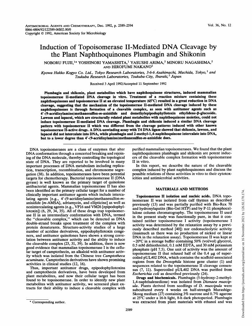
plant metabolites with a naphthoquinone structure like thatof plumbagin for their ability to induce topoisomerase II-mediated DNA cleavage by using purified calf thymus topo-isomerase II and supercoiled pUL402 DNA as substrates.Figure 1 shows a photograph of agarose gels comparingtopoisomerase TI-mediated DNA cleavage activity in thepresence of various amounts of naphthoquinones. In thecase of plumbagin and shikonin, the linear-form DNA pro-gressively appeared as the concentration of drug increased(from 0.5 ,uM in lanes c and h to 250 ,M in lanes g and 1 ofFig. 1). For comparison, the antitumor drugs VP16 andm-AMSA, which have been shown to promote topoisomer-ase II-mediated DNA cleavage, were included (Fig. 1, lanesm to q and K to 0, respectively).To obtain more quantitative data, the amount of linearized
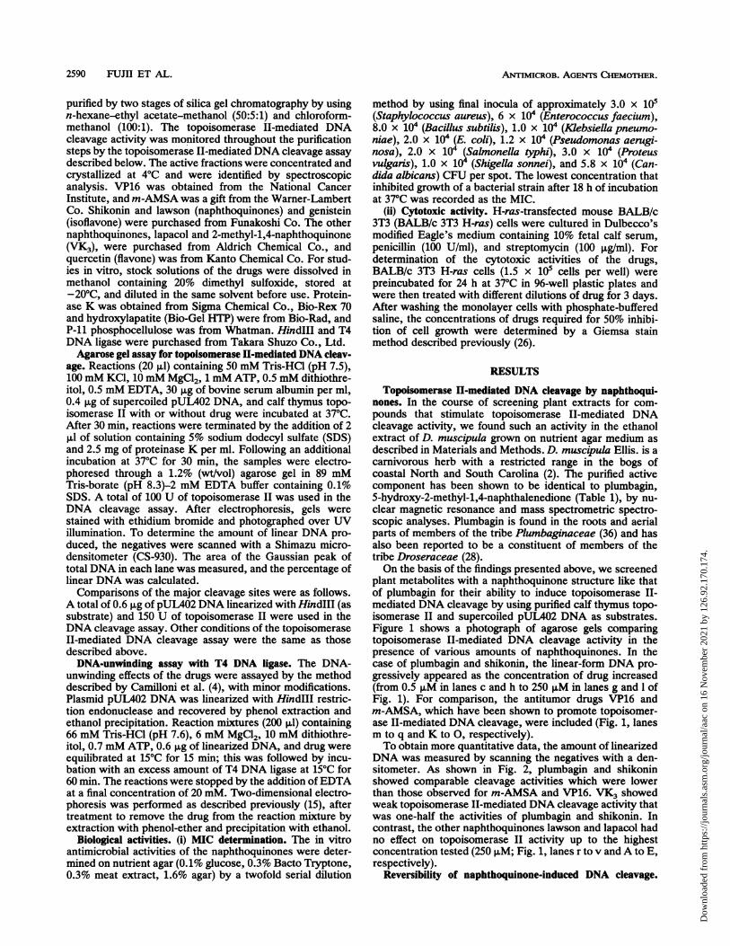
DNA was measured by scanning the negatives with a den-sitometer. As shown in Fig. 2, plumbagin and shikoninshowed comparable cleavage activities which were lowerthan those observed for m-AMSA and VP16. VK3 showedweak topoisomerase II-mediated DNA cleavage activity thatwas one-half the activities of plumbagin and shikonin. Incontrast, the other naphthoquinones lawson and lapacol hadno effect on topoisomerase II activity up to the highestconcentration tested (250 ,uM; Fig. 1, lanes r to v and A to E,respectively).
Reversibility of naphthoquinone-induced DNA cleavage.
2590 FIJJII ET AL.
Dow
nloa
ded
from
http
s://j
ourn
als.
asm
.org
/jour
nal/a
ac o
n 16
Nov
embe
r 20
21 b
y 12
6.92
.170
.174
.
TOPOISOMERASE II-MEDIATED DNA CLEAVAGE 2591
TABLE 1. Comparison of biological activities ofnaphthoquinone compoundsa
plumbagin shikonin
RI CH3 KH0 H
R2 H
plumbagin shikonin VP16a b cdef gh i j k I m nopq
lawson lapacol VK3
OH g-,--- CH3
H H OH H
R3 OH OH H H H
R4 H
Relative topoisomerase 11-mediated DNA cleavage 5.64activity (Itg/ml)
RelativeIntercalation activity
OH H H H
6.34 >100 >100 18.58
+
Cytotoxicity ICSe (tg/ml) 0.26 0.063 18.5
1_4 0.1+
1.4 0.11
1.6 2.1 >100 >100
3.1 4.2 >100 >100
0.2 0.3 12.5 2.9
6.3 >100 >100 >100
12.5 >100 >100 >100
12.5 >100 >100 >100
3.1 >100 >100 >100
0.4 >100 12.5 2.9
8.3
8.3
0.1
>100
>100
>100
>100
2.1
6.3 >100 >100 >100 >100
0.2 >100 >100 >100 1.0
a Topoisomerase II-mediated DNA cleavage activities were compared bydetermining the concentration (in micrograms per milliliter) of drug thatinduced 10% linear DNA from total DNA. As a positive control, the relativetopoisomerase II-mediated DNA cleavage activity of VP16 was 1.06 ,ug/ml(data not shown). Intercalating activity is signified as the presence of a drugconcentration-dependent DNA band shift in the DNA-unwinding assay. Theintercalation activities of naphthoquinones (250 p,M) were shown equal to thatobtained with m-AMSA at the following concentrations: 2.5 to 12.5 P.M (+),12.5 to 50 p.M (+ +), and .50 pM (+ + +); -, no effect. For cytotoxicity, thevalues indicated the concentration (in micrograms per milliliter) of drugrequired for 50% inhibition of cell growth (IC5e) of BALB/c 3T3 H-ras over 72h of exposure. Antimicrobial activity was shown by the MIC determined by an
agar dilution method.
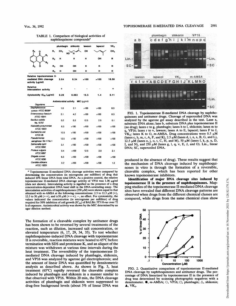
The formation of a cleavable complex by antitumor drugshas been shown to be reversed by several treatments of thereaction, such as dilution, increased salt concentration, or
elevated temperature (6, 17, 29, 34, 35). To test whethernaphthoquinone-induced DNA cleavage with topoisomeraseII is reversible, reaction mixtures were heated to 65°C beforetermination with SDS and proteinase K, and an aliquot of themixture was withdrawn at various time intervals during theheat treatment. The reversibility of the topoisomerase II-mediated DNA cleavage induced by plumbagin, shikonin,and VP16 was analyzed by agarose gel electrophoresis; andthe amount of linear DNA was quantified by densitometricanalysis as described above. As shown in Fig. 3, heattreatment (65°C) rapidly reversed the cleavable complexinduced by plumbagin and shikonin in a manner similar tothat observed with VP16. Within 20 min, the DNA cleavageactivities of plumbagin and shikonin were suppressed todrug-free background levels (about 5% of linear DNA was
FIG. 1. Topoisomerase II-mediated DNA cleavage by naphtho-quinones and antitumor drugs. Cleavage of supercoiled DNA was
analyzed by the agarose gel assay described in the text. Lane a,substrate DNA alone; lane b, substrate DNA plus topoisomerase II(no drug); lanes c to g, plumbagin; lanes h to 1, shikonin; lanes m toq, VP16; lanes r to v, lawson; lanes A to E, lapacol; lanes F to J,VK3; lanes K to 0, m-AMSA. Drug concentrations were 0.5 p,M(lanes c, h, m, r, A, F, and K), 2.5 p,M (lanes d, i, n, s, B, G, and L),12.5 pM (lanes e, j, o, t, C, H, and M), 50 p,M (lanes f, k, p. u, D,I, and N), and 250 p,M (lanes g, 1, q, v, E, J, and 0). Lin., linearDNA; SC, supercoiled DNA.
produced in the absence of drug). These results suggest thatthe mechanism of DNA cleavage induced by naphthoqui-nones in vitro is through the formation of a reversible,cleavable complex, which has been reported for otherknown topoisomerase inhibitors.
Identification of major DNA cleavage sites induced bytopoisomerase H in the presence of naphthoquinones. Map-ping studies of the topoisomerase II-mediated DNA cleavagesites have revealed that different DNA cleavage patterns are
observed when drugs from the different chemical classes are
compared, while drugs from the same chemical class show
50
z040co0c
30
0-
20
10
0
1 10 100Concentration (pM)
1000
FIG. 2. Quantitative comparison of topoisomerase II-mediatedDNA cleavage by naphthoquinones and antitumor drugs. The per-centage of DNA linearized by topoisomerase II in the presence ofdrug was determined by scanning photographic negatives with a
densitometer. *, m-AMSA; 0, VP16; Cl, plumbagin; A, shikonin;A, VK3.
ATCC 26Pseudomonasaeruginosa Bin H No.1Salmonella typhi
ATCC 9992Proteus vulgaris
ATCC 6897Shigella sonnei
ATOC 9290Candida albicans
ATOC 10231
VOL. 36, 1992
Dow
nloa
ded
from
http
s://j
ourn
als.
asm
.org
/jour
nal/a
ac o
n 16
Nov
embe
r 20
21 b
y 12
6.92
.170
.174
.
ANTIMICROB. AGENTS CHEMOTHER.
50
z0 40
300
*20
10
00 10 20
Time of treatment (min.)FIG. 3. Heat reversal of drug-induced topoisomerase II-medi-
ated DNA cleavage. DNA cleavage assays were done as describedin the text. A reaction mixture (100 pl) containing either naphtho-quinone or VP16 (250 pM for each compound) was incubated at370C for 30 min. The reaction mixture was then heated to 65°C, andan aliquot (20 p,l) was withdrawn at various times during the heattreatment. SDS and proteinase K treatment were done as describedin the text. The percentage of DNA linearized by topoisomerase IIin the presence of drugs was determined by scanning the photo-graphic negatives with a densitometer. 0, VP16; El, plumbagin; A,shikonin.
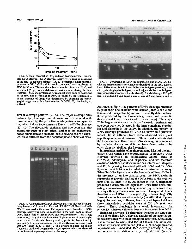
similar cleavage patterns (5, 35). The major cleavage sitesinduced by plumbagin and shikonin were compared withthose induced by the plant flavonoids genistein and querce-tin, which induce topoisomerase II-mediated DNA cleavage(25, 41). The flavonoids genistein and quercetin are alsonatural products of plant origin, similar to the naphthoqui-nones plumbagin and shikonin, while flavonoids are a chem-ical class different from the naphthoquinone chemical class.
FIG. 4. Comparison ofDNA cleavage patterns induced by naph-thoquinones and flavonoids. Plasmid pUL402 DNA linearized withHindIII was used in the assay. The topoisomerase II-mediated DNAcleavage assay was done as described in the text. Lane a, linearDNA alone; lane b, linear DNA plus topoisomerase II (no drug);lanes c to j, drug plus topoisomerase II (lanes c and d, plumbagin;lanes e and f, shikonin; lanes g and h, genistein; lanes i and j,quercetin). Drug concentrations were 50 pM (lanes c, e, g, and i) and250 p.M (lanes d, f, h, and j). The arrows indicate the majorfragments produced by genistein and/or quercetin but not detectedin the lanes of naphthoquinones in the assay.
FIG. 5. Unwinding of DNA by plumbagin and m-AMSA. Un-winding measurements were made as described in the text. Lane a,linear DNA alone; lane b, linear DNA plus T4 ligase (no drug); lanesc to e, plumbagin plus T4 ligase; lanes f to j, m-AMSA plus T4 ligase.Drug concentrations were 0.5 p.M (lane f), 2.5 p.M (lane g), 12.5 p.M(lanes c and h), 50 p.M (lanes d and i), and 250 pLM (lanes e and j).
As shown in Fig. 4, the patterns of DNA cleavage producedby plumbagin and shikonin were similar (lanes c and d andlanes e and f, respectively) and were distinctly different fromthose produced by the flavonoids genistein and quercetin(lanes g and h and lanes i and j, respectively). The majorDNA fragments observed with the flavonoids genistein andquercetin were not detected in the lanes containing plumba-gin and shikonin in the assay. In addition, the pattern ofDNA cleavage produced by VP16 as shown in a previousreport (40) is different from those observed with plantnaphthoquinones and flavonoids. These results indicate thatthe topoisomerase II-mediated DNA cleavage sites inducedby naphthoquinones are different from those induced byother plant metabolites, the flavonoids.
Intercalation activity of naphthoquinone. Most of the anti-tumor drugs which have topoisomerase II-mediated DNAcleavage activities are intercalating agents, such asm-AMSA, adriamycin, and ellipticine, and we thereforeexamined whether naphthoquinone can intercalate into plas-mid DNA by using linearized pUL402 DNA and T4 DNAligase (4). m-AMSA was included as a control in the assay.When T4 DNA ligase rejoins the free ends of linear DNA inthe presence of an intercalating drug, the DNA moleculesupercoils negatively, shifting the DNA band to the negativeform (Fig. 5, lanes f to j). As shown in Fig. 5, plumbaginproduced a concentration-dependent DNA band shift, indi-cating a decrease in the linking number (Fig. 5, lanes c to e),although their potencies were an order of magnitude lowerthan that ofm-AMSA in this regard (Fig. 5, lanes f to j). VK3showed a weak intercalation activity (one-fifth that of plum-bagin). In contrast, shikonin, lawson, and lapacol did notshow intercalation activities even at 250 puM (data notshown). Thus, plumbagin is a weak intercalator likem-AMSA, whereas shikonin does not intercalate into DNA.
Biological activities. To determine whether the topoisom-erase II-mediated DNA cleavage activity of the naphthoqui-nones correlated with other biological activities, cytotoxicitywas measured by using BALB/c 3T3 H-ras, and antimicro-bial activities were determined (Table 1). Plumbagin (relativetopoisomerase TI-mediated DNA cleavage activity, 5.64 ,ug/ml; relative intercalation activity, +), shikonin (relative
2592 FUJII ET AL.
Dow
nloa
ded
from
http
s://j
ourn
als.
asm
.org
/jour
nal/a
ac o
n 16
Nov
embe
r 20
21 b
y 12
6.92
.170
.174
.
TOPOISOMERASE II-MEDIATED DNA CLEAVAGE 2593
topoisomerase II-mediated DNA cleavage activity, 6.34 ,ug/ml; relative intercalation activity, -), and VK3 (relativetopoisomerase II-mediated DNA cleavage activity, 18.58,g/ml; relative intercalation activity, +) were cytotoxic with50% inhibitory concentrations of 0.26, 0.063, and 0.11 ,ug/ml,respectively. In contrast, the cytotoxicities of lapacol andlawson (relative topoisomerase II-mediated DNA cleavageactivity, both >100 ,ug/ml; relative intercalation activities,both -) were lower (50% inhibitory concentrations, 1.4 and18.5 ,ug/ml, respectively) than that of plumbagin. Plumbaginexhibited a broad spectrum of antimicrobial activity againstboth gram-positive and -negative bacteria and C. albicans.Shikonin and VK3 showed antimicrobial activities againstonly gram-positive bacteria. Lapacol and lawson, which didnot show topoisomerase II-mediated DNA cleavage activityor intercalation activity, exhibited marginal activity onlyagainst B. subtilis and P. vulgans. The topoisomerase II-active drugs plumbagin, shikonin, and VK3 showed signifi-cantly greater cytotoxic and antimicrobial activities com-pared with those of the non-topoisomerase II-active drugslapacol and lawson. However, the relative topoisomeraseII-mediated DNA cleavage activity did not correlate quanti-tatively with cytotoxicity among the topoisomerase II-activedrugs.
DISCUSSION
Topoisomerases are viewed as the primary cellular targetsof many drugs that have been used extensively in cancerchemotherapy. Among the antitumor drugs which induce aDNA-cleavable complex with topoisomerases, epipodophyl-lotoxins and camptothecin derivatives have been developedfrom plant metabolites (6, 16). In the present study, weshowed that the plant naphthoquinones plumbagin andshikonin also induce topoisomerase II-mediated DNA cleav-age in vitro.Plumbagin is one of the well-known plant naphthoqui-
nones which is reported as a constituent of several generawithin the plant kingdom and has been studied for itsbiological activity (10, 14, 19). Krishnaswamy and Pu-rushothaman (21) reported that plumbagin isolated fromPlumbago zeylanica causes a regression of tumor growth inmethylcholanthrene-induced fibrosarcomas in Wistar rats. Itwas also active against P388 lymphocytic leukemia in vivo ata dose of 4 mg/kg of body weight but was not active againstan L-1210 lymphoid leukemia (21). Shikonin is a well-knownpigment from the root of Lithospermum erythrorhizon andhas been used as a material to prepare an ointment which isused for the treatment of wounds and burns (18). It has beensuggested that the antimicrobial activity of shikonin is one ofthe mechanisms responsible for the effects (18). In addition,Sankawa et al. (32) reported that shikonin has antitumoractivity against a murine tumor model with intraperitoneallyinoculated sarcoma 180 ascites cells at a dose of 5 to 10mg/kg/day.However, the critical biochemical targets of plumbagin
and shikonin have yet to be identified. Here we presenteddata showing that the plant naphthoquinones plumbagin andshikonin induce topoisomerase II-mediated DNA cleavage.These topoisomerase II-active drugs showed significantlygreater cytotoxicity in comparison with those of the non-topoisomerase II-active plant naphthoquinones lapacol andlawson. Although the relative topoisomerase II-mediatedDNA cleavage activity did not correlate quantitatively withcytotoxicity among the topoisomerase II-active drugs, it isconsidered that permeability and metabolic inactivation in
cells may be different among these compounds with differentsubstituents (e.g., R1, CH3 and 4-oxo-2-hexeneyl; R3 and R4,OH). From these results, we suggest that topoisomerase II isan important cellular target of plumbagin and shikonin,leading to their antitumor and cytotoxic activities.
It has been reported that the mechanism of action of drugstargeting mammalian topoisomerase II is very similar tothose of nalidixic acid and the related quinolone antibacterialagents which induce a DNA-cleavable complex with DNAgyrase, a bacterial topoisomerase II; both types of drugs trapthe topoisomerase II-DNA-cleavable complex in very simi-lar manners (3, 9, 30). In agreement with these commonproperties, it has been previously shown that VP16, which isan antitumor drug which enhances mammalian topoisomer-ase II-mediated DNA cleavage activity, is also a poison ofbacterial DNA gyrase (22). Furthermore, several new deriv-atives of quinolone antibiotics have recently been shown toinduce a DNA-cleavable complex with mammalian topo-isomerase II and possess antitumor activities in vivo (39).Taking these findings together, it is likely that the antibac-terial activities of plumbagin and shikonin might be mediatedthrough the action of bacterial topoisomerases. This hypoth-esis should be determined in vitro by using purified DNAgyrase and the new, recently identified bacterial topoi-somerases (8, 20, 33, 37).
ACKNOWLEDGMENTS
We thank M. Matsumoto and M. Kusunoki for skillful technicalassistance.
REFERENCES1. Adachi, Y., E. Kas, and U. K. Laemmli. 1989. Preferential,
cooperative binding of DNA topoisomerase II to scaffold-associated regions. EMBO J. 8:3997-4006.
2. Bailey, L. H., and E. Z. Bailey. 1976. Hortus third, a concisedictionary of plants cultivated in the United States and Canada.MacMillan Publishing Co., New York.
3. Barrett, J. F., T. D. Gootz, P. R. McGuirk, C. A. Farrell, andS. A. Sokolowski. 1989. Use of in vitro topoisomerase II assaysfor studying quinolone antibacterial agents. Antimicrob. AgentsChemother. 33:1697-1703.
4. Camilloni, G., F. D. Seta, R. Negri, A. G. Ficca, and E. D.Mauro. 1986. Structure of RNA polymerase II promoters.Conformational alterations and template properties of circular-ized Saccharomyces cerevisiae GALl-GAL10 divergent pro-moters. EMBO J. 5:763-771.
5. Capranico, G., F. Zunino, K. W. Kohn, and Y. Pommier. 1990.Sequence-selective topoisomerase II inhibition by anthracyclinederivatives in SV40 DNA: relationship with DNA bindingaffinity and cytotoxicity. Biochemistry 29:562-569.
6. Chen, G. L., L. Yang, T. C. Rowe, B. D. Halligan, K. M. Tewey,and L. F. Liu. 1984. Nonintercalative antitumor drugs interferewith the breakage-reunion reaction of mammalian DNA topo-isomerase II. J. Biol. Chem. 259:13560-13566.
7. Cockerrill, P. N., and W. T. Garrard. 1986. Chromosomal loopanchorage of the kappa immunoglobulin gene occurs next to theenhancer in a region containing topoisomerase II sites. Cell44:273-282.
8. DiGate, R. J., and K. J. Marians. 1989. Molecular cloning andDNA sequence analysis of Eschenchia coli topB, the geneencoding topoisomerase III. J. Biol. Chem. 264:17924-17930.
9. Drlica, K., and R. J. Franco. 1988. Inhibitors of DNA topo-isomerases. Biochemistry 27:2253-2259.
10. Fetterer, R. H., and M. W. Fleming. 1991. Effects of plumbaginon development of the parasitic nematodes Haemonchus con-tortus and Ascaris suum. Comp. Biochem. Physiol. 100C:539-542.
11. Gasser, S. M., and U. K. Laemmli. 1986. Cohabitation of
VOL. 36, 1992
Dow
nloa
ded
from
http
s://j
ourn
als.
asm
.org
/jour
nal/a
ac o
n 16
Nov
embe
r 20
21 b
y 12
6.92
.170
.174
.
ANTIMICROB. AGENTS CHEMOTHER.
scaffold binding regions with upstream/enhancer elements ofthree developmentally regulated genes of D. melanogaster. Cell46:521-530.
12. Giovanelia, B. C., J. C. Stehlin, M. E. Wall, M. C. Wani, A. W.Nicholas, L. F. Liu, R. Silber, and M. Potmesil. 1989. DNAtopoisomerase I-targeted chemotherapy of human colon cancerin xenografts. Science 246:1046-1048.
13. Hallipn, B. D., K. A. Edwards, and L. F. Lu. 1985. Purificationand characterization of a type II DNA topoisomerase frombovine calf thymus. J. Biol. Chem. 260:2475-2482.
14. Hassan, H. M., and I. Fridovich. 1979. Intracellular productionof superoxide radical and of hydrogen peroxide by redox activecompounds. Arch. Biochem. Biophys. 196:385-395.
15. Hirose, S., and Y. Suzuki. 1988. In vitro transcription of eukary-otic genes is affected differently by the degree of DNA super-coiling. Proc. Natl. Acad. Sci. USA 85:718-722.
16. Hsiang, Y. H., R. Hertzberg, S. Hecht, and L. F. Uu. 1985.Camptothecin induced protein-linked DNA breaks via mamma-lian DNA topoisomerase I. J. Biol. Chem. 260:14873-14878.
17. Hsiang, Y. H., and L. F. Uu. 1989. Evidence for the reversibilityof cellular DNA lesion induced by mammalian topoisomerase IIpoisons. J. Biol. Chem. 264:9713-9715.
18. Hus, H. Y., and W. G. Peacher. 1976. Chinese herb medicineand therapy, p. 150. Autra Publishers Inc., Nashville, Tenn.
19. Joshi, N. K., and F. Sehnal. 1989. Inhibition of ecdysteroidproduction by plumbagin in Dydercus cingulatus. J. InsectPhysiol. 35:737-741.
20. Kato, J., Y. Nishimura, R. Imamura, H. Nild, S. Hirata, and H.Suzuki. 1990. New topoisomerase essential for chromosomesegregation in E. coli. Cell 63:393-404.
21. Krishnaswamy, M., and K. K. Purushothaman. 1980. Plumba-gin: a study of its anticancer, antibacterial and antifungalproperties. Indian J. Exp. Biol. 18:876-877.
22. Liu, L. F. 1989. DNA topoisomerase poisons as antitumordrugs. Annu. Rev. Biochem. 58:351-375.
23. Long, B. H. 1987. Structure-activity relationships of podophyl-lotoxin congeners that inhibit topoisomerase II. NCI Monogr.First Conference on DNA Topoisomerases in Cancer Chemo-therapy, 1986, New York. NCI Monogr. 4:123-127.
24. Maniatis, T., E. F. Fritsch, and J. Sambrook. 1982. Molecularcloning: a laboratory manual. Cold Spring Harbor Laboratory,Cold Spring Harbor, N.Y.
25. Markovits, J., C. Linassier, P. Fosse, J. Couprie, J. Pierre, A.Jacquemin-Sablon, J. M. Saucier, J. B. Le Pecq, and A. K.Larsen. 1989. Inhibitory effects of tyrosine kinase inhibitorgenistein on mammalian DNA topoisomerase II. Cancer Res.49:5111-5117.
26. Mirabelli, C. K., H. Bartus, J. 0. Bartus, R. Johnson, S. M.Mong, C. P. Sung, and S. T. Crooke. 1985. Application of atissue culture microtiter test for the detection of cytotoxicagents from natural products. J. Antibiot. 38:758-766.
27. Murashige, T., and F. Skoog. 1962. A revised medium for rapidgrowth and bio assays with tobacco tissue cultures. Physiol.Plant 15:473-497.
28. Nahrstedt, A. 1980. Absence of cyanogenesis from Droseraceae.Phytochemistry 19:2757-2758.
29. Nelson, E. M., K. M. Tewey, and L. F. Uu. 1984. Mechanism ofantitumor drug action: poisoning of mammalian DNA topoisom-erase II on DNA by 4'-(9-acridinylamino)-methanesulfon-m-anisidide. Proc. Natl. Acad. Sci. USA 81:1361-1365.
30. Robinson, M. J., B. A. Martin, T. D. Gootz, P. R. McGuirk, M.Moynihan, J. A. Sutcliffe, and N. Osheroff. 1991. Effects ofquinolone derivatives on eukaryotic topoisomerase II. J. Biol.Chem. 266:14585-14592.
31. Rowe, T. C., G. L. Chen, Y. H. Hdang, and L. F. LUu. 1986.DNA damage by antitumor acridines mediated by mammalianDNA topoisomerase II. Cancer Res. 46:2021-2026.
32. Sankawa, U., Y. Ebizuka, T. Miyazald, Y. Isomura, H. Otsuka,S. Shibata, M. Inomata, and F. Fukuoka. 1977. Antitumoractivity of shikonin and its derivatives. Chem. Pharm. Bull.25:2392-2395.
33. Slesarev, A. I., D. A. Zaitzev, V. M. Kopylov, K. 0. Stetter, andS. A. Kozyavkin. 1991. DNA topoisomerase III from extremelythermophilic archaebacteria. J. Biol. Chem. 266:12321-12328.
34. Tewey, K. M., G. L. Chen, E. M. Nelson, and L. F. Liu. 1984.Intercalative antitumor drugs interfere with the breakage-re-union reaction of mammalian DNA topoisomerase II. J. Biol.Chem. 259:9182-9187.
35. Tewey, K. M., T. C. Rowe, L. Yang, B. D. Halligan, and L. F.Liu. 1984. Adriamycin-induced DNA damage mediated bymammalian DNA topoisomerase II. Science 226:466-468.
36. van der Vijver, L. M. 1972. Distribution of plumbagin in thePlumbaginaceae. Phytochemistry 11:3247-3248.
37. Walis, J. W., G. Chrebet, G. Brodsky, M. Rolfe, and R.Rothstein. 1989. A hyper-recombination mutation in S. cerevi-siae identifies a novel eukaryotic topoisomerase. Cell 58:409-419.
38. Wang, J. C. 1985. DNA topoisomerases. Annu. Rev. Biochem.54:665-697.
39. Yamashita, Y., T. Ashizawa, M. Morimoto, J. Hosomi, and H.Nakano. 1992. Antitumor quinolones with mammalian topo-isomerase II mediated DNA cleavage activity. Cancer Res.52:2818-2822.
40. Yamashita, Y., S. Kawada, N. Fujii, and H. Nakano. 1991.Induction of mammalian DNA topoisomerase I and II mediatedDNA cleavage by saintopin, a new antitumor agent from fungus.Biochemistry 30:5838-5845.
41. Yamashita, Y., S. Kawada, and H. Nakano. 1990. Induction ofmammalian topoisomerase II dependent DNA cleavage bynonintercalative flavonoids, genistein and orobol. Biochem.Pharmacol. 39:737-744.