*>.*bs,i.B"*.,',. , ,.,. ,/. 4 ,I ; ,,,", ...................... ...................... I1 e. ..................... -.-. ^ ~ . ~ : ,,,? ,. 1 ................ ::il j .*.b".. ............... ................. ... ...................... Anna Siromoney', 1. Raghuram2, Arul Siromone$, I. Korah2, G.N.S. Prasad ' Doportment of Physics, Wones's Clirisiioii College, Cheniioi, Iiitlio Deportment of Rodiodiagiiosis, Cliriitioii Medico1 College ond IHospitol, Vellore, lndin Sthool of Computer Science ond Engineering, Anno Uiiiversiiy, Chenntii, lndin ' Deportnient of Physics, Aiinn Uiiiversity, Cliennoi, Indio ..................... 1 I 72 Inductive Logic Programming for Knowledge Discovery from MRI Data A loo/ for Quanfitafively Discriminating Between Meningioma and Asfrocyfoma Tumors ne of the uses of iiiagnetic resonance 0. Imaging (MKl) in clinical diagnosis is in-vivo i1iscrimin;ition bctwccn tumor and normal tissue and between tumor types in the brain. There is much intcrcst in in- creasing the qiialilative and quantilative incormation available from tliese images. This article presents a study that uses lhc inductivc logic programming tool Progol on measurements of signal intensities in clinical scan images or 28 patients (18 with meningiomas and 10 with astrocytomas) to attempl to discover knowledge that quantitatively discrim- nates hetwcen thc two types ol'tiimors. Overview Magnetic Resonance Imaging Magnelic resonance imaging is based on the physical phenomenon OS nuclear magnctic rcsoiiaiice (NMR) [I]. NMR is the resonance transition between nuclear spin statcs of certain nuclei (e.g., the hy- drogen nucleus) in lhe presence of an ex- ternal magnetic field. The return of the irucleus to thc cquilibriuin state is charac- terized by lwo parameters-the spin-lat- tice relaxation lime (TJ and the spill-spin relaxation time (TJ, which, along with the promn density, are characteristic of thc tissuc bcing imagcd. Thccncilation is by ii radio frequency (RF) pulse at the Lxmor frequency and is related to the external magnetic field. In MRI, thc local iiiagnetic field in thc sample is varied in aprescribed maimer to produce a planncd variation in Larmor frequencicr, thus producing a "map" of the spatial locations of thc nuclei and their dcnsitics. The measuremeiil of [lie ouliiut forms to coinpute the MR image. The T I and T2 timcs for discascd tissues is well known lo he elongaled compared to those of normal tissues 121. The intensities of the resultant image on the screcn dcpciid on the physical paramcters or the scaii and on the relaxation times of the tissnes ac- cording to thc solution of thc Rloch equa- tions l I Cor the given pulse sequence 13 l, Intcnsitics in scans pcrformcd using iden- tical scan paramelers caii be compared. Application of MRI in Medicine MRimagesiifpatientsiireused inradi- ology for noninvasive diagnosis of vari- ous diseases, including brain tumors. The radiologist qoalitatively analyzes a se1 of' images to distinguish the tumor from thc normal tissne as well as to ohtain knowl- edge ahoul the probable tumor type. MRI is considered safe for biological systems as there are at prcsent no known harmkil ellects ollhe high slalic magnetic field or the RF as used in normal clinical practicc. MRI also does no1 use any ionizing radia- tion-unlike, for example, computcd to- mography (CT) or posilron emissioii tomography (PET) scanning. 'Thc MR im- age also possesses fairly good spatial res- olution and contrast resolution, and it is useful for imaging difficult-to-acccss ar- eas such as the brain. There is much interest in incrcasing thc diagnostic inrormation available to the ra- diologist from thc MR imagcs, both quali- tatively and quantitatively. One of the ultimate goals is to distinguish among all types of tumors accuratcly, without having lo resort lo invasive procedures 12,4,51. It two-dimensional (2-D) Fouricr trans- IEEE ENGINEERING IN MEDI[INEAND BIOLOGY mors FroinquantitativedalaofMRimagcs. 0739-51 75/00/$10.00020001EEE July/Augwf 2000
Transcript
* > . * b s , i . B " * . , ' , . , ,. , . ,/. 4 ,I ; ,,,", ...................... ...................... I 1 e . ..................... -.-. ^ ~ . ~ : , , , ? ,. 1 ................ ::il j
Deportment of Rodiodiagiiosis, Cliriitioii Medico1 College ond IHospitol, Vellore, lndin
Sthool of Computer Science ond Engineering, Anno Uiiiversiiy, Chenntii, lndin
'' Deportnient of Physics, Aiinn Uiiiversity, Cliennoi, Indio
..................... 1
I
72
Inductive Logic Programming for Knowledge Discovery from MRI Data A loo/ for Quanfitafively Discriminating Between Meningioma and Asfrocyfoma Tumors
ne of the uses of iiiagnetic resonance 0. Imaging (MKl) in clinical diagnosis is in-vivo i1iscrimin;ition bctwccn tumor and normal tissue and between tumor types in the brain. There is much intcrcst in in- creasing the qiialilative and quantilative incormation available from tliese images. This article presents a study that uses lhc inductivc logic programming tool Progol on measurements of signal intensities in clinical scan images or 28 patients (18 wi th m e n i n g i o m a s and 10 wi th astrocytomas) to attempl to discover knowledge that quantitatively discrim- nates hetwcen thc two types ol'tiimors.
Overview Magnetic Resonance Imaging Magnelic resonance imaging is based
on the physical phenomenon OS nuclear magnctic rcsoiiaiice (NMR) [I]. NMR is the resonance transition between nuclear spin statcs of certain nuclei (e.g., the hy- drogen nucleus) in lhe presence of an ex- ternal magnetic field. The return of the irucleus to thc cquilibriuin state is charac- terized by lwo parameters-the spin-lat- tice relaxation lime (TJ and the spill-spin relaxation time (TJ, which, along with the promn density, are characteristic of thc tissuc bcing imagcd. Thccncilation is by ii radio frequency (RF) pulse at the Lxmor frequency and is related to the external magnetic field.
In MRI, thc local iiiagnetic field i n thc sample is varied in aprescribed maimer to produce a planncd variation in Larmor frequencicr, thus producing a "map" of the spatial locations of thc nuclei and their dcnsitics. The measuremeiil of [lie ouliiut
forms to coinpute the MR image. The T I and T2 timcs for discascd tissues is well known lo he elongaled compared to those of normal tissues 121. The intensities of the resultant image on the screcn dcpciid on the physical paramcters or the scaii and on the relaxation times of the tissnes ac- cording to thc solution of thc Rloch equa- tions l I Cor the given pulse sequence 13 l , Intcnsitics in scans pcrformcd using iden- tical scan paramelers caii be compared.
Application of MRI in Medicine MRimagesiifpatientsiireused inradi-
ology for noninvasive diagnosis of vari- ous diseases, including brain tumors. The radiologist qoalitatively analyzes a se1 of' images to distinguish the tumor from thc normal tissne as well as to ohtain knowl- edge ahoul the probable tumor type. MRI is considered safe for biological systems as there are at prcsent no known harmkil ellects ollhe high slalic magnetic field or the RF as used in normal clinical practicc. MRI also does no1 use any ionizing radia- tion-unlike, for example, computcd to- mography (CT) or posilron emissioii tomography (PET) scanning. 'Thc MR im- age also possesses fairly good spatial res- olution and contrast resolution, and it is useful for imaging difficult-to-acccss ar- eas such as the brain.
There is much interest in incrcasing thc diagnostic inrormation available to the ra- diologist from thc MR imagcs, both quali- tatively and quantitatively. One of the ultimate goals is to distinguish among all types of tumors accuratcly, without having lo resort lo invasive procedures 12,4,51. It
two-dimensional (2-D) Fouricr trans-
I E E E E N G I N E E R I N G IN MEDI[ INEAND BIOLOGY
mors FroinquantitativedalaofMRimagcs.
0739-51 75/00/$10.00020001EEE July/Augwf 2000
Knowledge Discovery from Data The term knowledge discovery from
data, or knowlcdgcdiscovcry i n databases (KDD), was coined in 1989 lo refer to the hroiid process of finding kiiowlcdgc iii
dala. KDD is the nontrivial process ot identifying valid, novel, potentially use- ful, and ultimately undcrstandahlc pat- lerns i n data. The KDD process is a multistep process that involves the use of data mining methods lo exlract knowl- edge from a data set. Data mining is 21 step i n the KDD process, and it involves fitting models to or determining patterns from observed dala 161.
Inductive logic prograinming (ILP) 171 uses first-order logic (usually Horn clauses, a subset of first-order logic, and often cxpresscd in the logic programming language Prolog) Lo represent the knowl- cdgc discovered from the databases. The use of first-order logic allows the repre- sentation of complex structured objects and relations among objects or their coni- poncnts [XI. A survcy of ILP tcchniqucs and KDD applications of ILP is discusscd in [Xl.ILPhasheenappliedtoinedicaliin- age data for thc study ofgl;nacoma images [91 using thcILPsystcni GKS. (GKS is an abbreviation of "GaKuShu," which iiieans "learning" in Japanese). The spa- tial relalionships OS the dillcrcnt Stxitiires i n the ocular fundus image are specified, and thcsc rclationships arc iiscd to Icarn whether an cyc is gkiocomatous or not.
A study that successfiilly discrim- natcd between tlie two types of normal brain tissues (i.e., gray and white malter) inMRI human brain scan images by using the ILP systcin Progol has been reported elsewhere [I01 hy soine of the authors of this article. It was found that it was possi- ble to distinguish belwcen these iioriilal tissues by using their signal intensities alone, to a certain exlent, without invok- ing llic rclationships bctwecn the s i p 1 Ntensities ol cliCCerenL tissue types.
Astrocytomas and Meningiomas The tcrinprimai;y brain tiifnor cncum-
passes neoplasms atid relaled inass le- sions that arise from the brain and its linings. Primary brain tumors are again
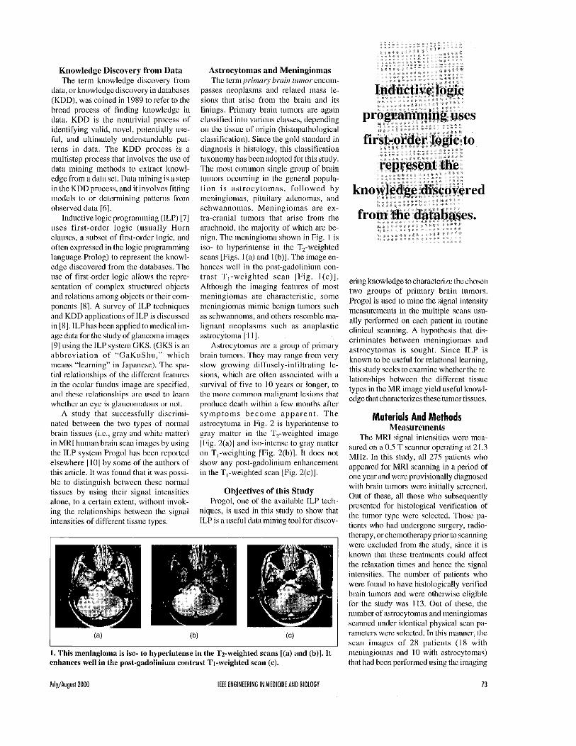
on tlic tissue of origin (histopathological classifiaition). Since the gold standard i n diagnosis is histology, this classification taixunuiny has been adopled Sor this study. The inlost coininon single group of brain tumors occurring in the general popule- l ion i s as t rocytomas, fol lowed by meningiomas, pituitary adenomas, and schwannomas. Meningiomas are ex- tra-cranial tumors that arise from the arachnoid, lhe majority OS which are bc- nign. The meningioma shown in Fig. I is iso- Lo hyperinlense iii Ihc T,-weightcd scatis [Figs. I (a) and I(b)]. The image en- hanccs wel l i n the pnst-gadolinium con- trast T,-weighted scan IFig. I (c) l . Although the imaging features of most incningiomas are characlcrislic, some meningiomas iiiimic benign luinors such as schw"mna , and othcrs rcscinblc ma- lignant iieoplesins such as anaplestic astrocytoma I I l l .
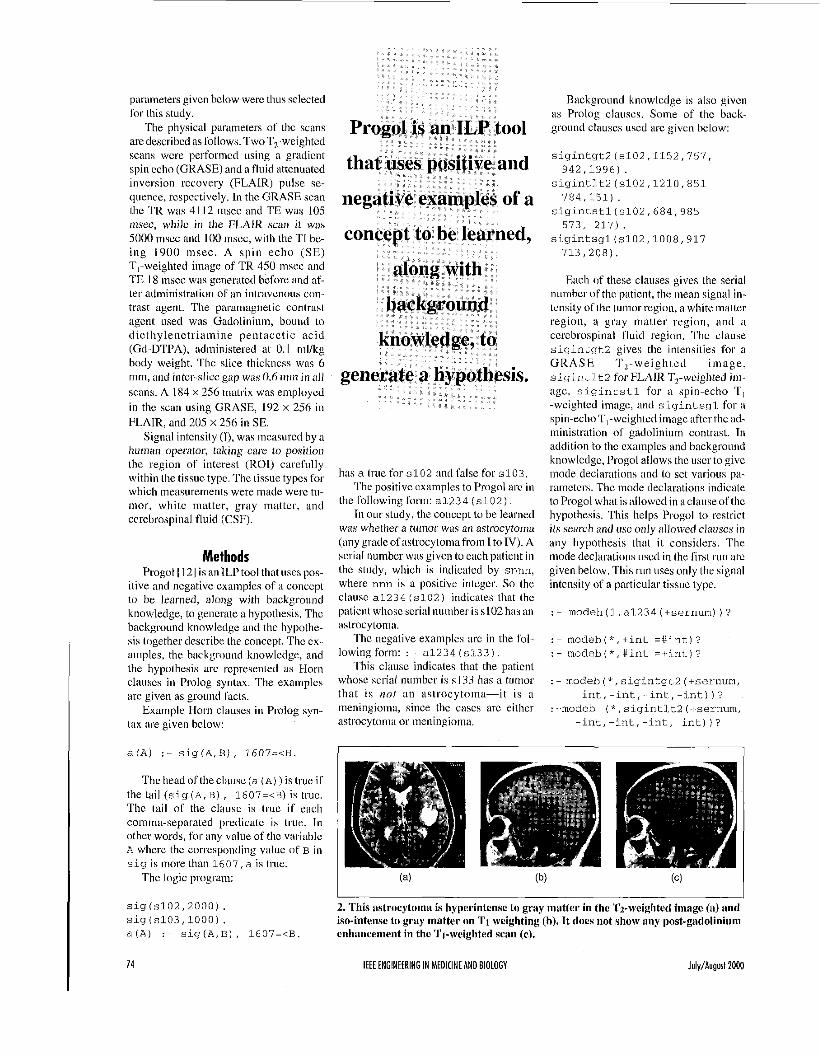
Astrocytomas are ;I group of primary brain tuniurs. 'They may range from very slow growing diffusely-iiifiltrating le- sions, which are o k n associaled with a siirvivial of five tu I O years or longer, to lhc inore common malignant lesions that produce dcalh within a fcw months after s y m p t o m s b e c o m e a p p a r e n t . T h e astrocytoma in Fig. 2 is hyperintense to gray nlaltcr in the T,-wcighled image (Fig. Z(a)l and iso-intense to gray matter on T,-weighting [Fig. 2(b)l. It docs not show any post-gadolinium enhancemenl io the TI-weighted scan IFig. 2(cj].
assitled . . ' " into various classes, depending
Objectives of this Stndy Progol, one or Lhe available ILP tech-
niqucs, is iiscd i n this study to show that ILP is a useful dala mining tool fordiscov-
. This meningioma is iso- to hyperintense in the Tz-weighted scans [(a) and (b)]. I t nnhnnnnr ..mll in the nn~. t -nnAnl :~: . . .n ,.,._ t*...t 7.. -... u:nLtarl m.rn In) C.... "..%Cm I..,.. ... L1.L p"".-$j'.""".l.Y... L"I.I.'IDL . ,-*rr.&l.'c" IIC'.., IC,.
ering knowledge to characlcrizc the chosen two groups of primary brain tumors. Progol is used lo mine the signal intensity measureinelits in the multiple scans usu- ally perlbrmed on eacli patient in routine clinical scanning. A hypothesis thal dis- criminates between meningiomas and astrocytomas is sought. Since ILP is known lo bc useful for relational learning, this study seeks to examine whether the re- lationships hctwccn the different tissue types in the MI< image yield useful knowl- cdgc that characterizes these tunior tissues.
Materials And Methods Measurements
The MRI signal inlensilies were mea- sured on a 0.5 T scanner operating at 21.3 MHz. In this study, all 275 paticnts who appcarcd fur MRI scanning in a period of one year and were provisionally diagnosed with brain tumors were initially screened. Out of these, all those who subsequently presented for histological verification of the tunior type were selected. Thosc pa- tients who had undergone surgery, radio- therapy, orchciiiotherapy prior lo scanning were cxcludcd from the study, since it is known lhal lhese treliltnents could affect the relaxation times and hence the signal intensilies. The number o l patients who were found to have histologically verified brain luinors and were othcrwise eligible Cor the study was 113. Out of lhese, tlie number of astrocytomas and meningiomas scanned under identical physical scan pa- rameters were selected. In this manner, the scan images of 28 patients ( I 8 with mcniiigioinas and 10 with astrocytomas) that had been perforined using the imaging
Julq/Augurt 2000 IEEE ENGINEERING I N MEDICINE AND BIOLOGY 73
pal-ameters givcn below were thus selected for this study.
The physical parsmeters o f lhc scans aredescribcd as Ibllows. TwoT,-weighted scans wcre perforincd using a griidicnt spin ccho (GRASE) and a fluid attcnuated inversion rccovery (FLAIR) pulse se- quence, rcspcctively. In thc GRASE scan the 1'11 was 4 I I2 inscc and TI? was 105 insec, whilc in the FLAIR scan i t was 5000 mscc and 100 mscc, with the TI he- in& 1900 nisec. A spin echo (SE) T,-weighted image of TR 450 nisec atid TE 18 niscc was genenitcd hefore and nf- ter administration 01 an intravcnous cow mast agent. The parainagnetic contrast ageiir used was Gadolinium, bound to dietl iylenctriarnine pentaccl ic acid (Gd-DTPA), administered at 0. I ml/kg body weight. The slice thickncss was 6 mm, and inter-slice gap wiis 0.6 m n i n all scans. A I84 x 256 matrix was employcd in thc scan using GRASE, 192 x 256 i n FLAIR, and 205 x 256 in SE.
Signal intensily (I), was Ineasurcd by a hurnan operator, taking care to position the region of interest (1101) ca rd i~ l ly within h e tissuc typc. The tissue types for which measurements were made werc til- mor, whitc mattcr, gray mattcr, and ccrchrospinal fluid (CSF).
Methods Progol I 121 is an ILP tool that uses pos-
itive and negativc cxamples of ii concept to bc learned, along with background knowledge, to gencrate a hypothesis. Thc background knowledge and thc hypothe- sis together dcscribe the concept. Thc cx- amples, the hackgroiind knowlcdge, and the hypothesis arc rcpresented as Horn cleuscs in Prolog syntax. The examples are given as ground facts.
Example Horn clauses in Prolog syn- tax are given below:
a ( A ) : - sig(A,B), 1607=<B
has a true for s102 and rake for s103 The positivc examples to Progol arc in
thc following form: a1.234 (~102) In our study, the conccpt tu he learned
was whether it tumor was nil astrocytoma (anygl.adcofaslrocytoinafi.om I toIV). A scriiil number was givcn to each patient i n lhc study, which is indicaled by snnn, where nnn is a positive intcgcr. So the clause a1234 ( ~ 1 0 2 ) indicates that the patient whoscscrial numher iss102h;is an 1IstrocytoIlIa.
Thc ncgative examples arc in the Ihl- lowing form: : - a1234 ( ~ 1 3 3 1 .
This clause indicates that the patient whose serial nuriibcr is s13.1 has a totnor that is nof an astrocytoma-it is a meningioma, since the casts arc either astrocytoma or meningioiiiii.
Background knowledge is also givcn as Prolog clauses. Some of thc back- ground clauses uscd arc given helow:
s i g i n t g t l (~102,1152,757,
sigintI.t2(~102,1210,851
sigintstl(s102.684,985
sigintsg1(~102,1008,91.7
942,1996).
784,151) .
573, 217).
713,200).
Bach of these clauses gives the serial numbcr of the paticnt, the mean signal in- tcnsily of the tilmor region, awhitematter region, a gmy matter region, i d ii ccrcbrospinal fluid region. 'L'hc clause sigintgtl gives the intensities for a G RA S E Tz- w e i g h tcd i in a g e , sigintlt2 for FLAIR T,-wcighted iin- age, sigintstl for a spin-echo T I -wcightcd image, and sigintsgl for ii spin~cchoT,-weiglited image after the ad- ininistizition of gadolinium contIpst. In addition to thc cxainplcs and background knowlcdge, Progol allows the user to give
itions and lo set vaious pa- ramctcl's. Thc mode cleclarations indicate toProgol wh;itis;illowcdin aclauseoftlie hypothesis. This helps Progol to restrict its search and usc only allowed clauscs i n any hypothesis that it considers. The mode declarations U S C ~ i n Ihe first run arc given below. This ruii uses only the signal intcnsily or a particular tissuc typc.
: -modeh ( ~ k , sigintlt2 (+sernum, -int,-int,-int,-int)l?
-int,-int,-int,-int)]?
I comma-separated predicatc is true. In other words, Ibl- any value of thc variahlc A where thc corrcsponding velue of B i n s ig is tnorethall 1607,a is truc.
The logic program:
s i g ( s 1 0 2 . 2 0 0 0 ) . sig(s103,10001. a ( A ) :- sig(A,B], 1 6 0 7 = i B . enhancement in the Ti-weighted scan (c).
74 IEEE ENGINEERING IN MEDICINE AND BIOLOGY Iuly/Augurf 2000
2. This astrocytoma is hyperintense to gray matler in the '1'2-weigbted imagc (a) and iso-intense to gray matter on T I weighting (1)). It does not show any post-gadolinium
: - modebl*,siqintsgll+sernum, -int, - i n t , -int, -j.nt) ) ?
Progol that the hcad o fa hypothesis clause can use a1234. In lither words, the hy- Ipolhesis w i l l decide if a ~i in ior i s an astmcytiima. (This also implies that tlic p s i l i v e and negative examples wi l l usc the Same a1234.)
The 1 in (he modeh tlcclalation indi- cates that a1.234 ciui occtir only oncc iii the head of a cluti\c in the hypothesis. The sernum indicales that tlic term used in a1234 has to be o f type sernum. All the serial iiuinbcrs used in the study are de- clared LO he o f type sernum by using clauses of the lorin given below.
sernumls102 1 . sernum ( s1.53 1
'l'hc modeb dcclar;iliuns indicale to Progol what can bc used i n the body US a hypothesis clause. 'She :Ii indicales that there can he up lo a deSaault number 0 1 oc- curreiiccs. (A specific value can also he used to help redocc the time taken by Progol. For cx;implc, if 5 is used, Progol knows tllat a name can occiir only up to l ive times iii the body of a liypothcsis clausc.)
So, thcmodeb declarations liere indicale Ilia1 =<, sigintqt2, sigintlt2,
Predicted Positive Examples (Astrocytomas)
(Meningiomas) Predicted Negative Examples
sigintstl, and sigintsgl caii occur in the body oca hypothesis clause.
111 the modc declarations, the # indi- cales that term to he constant. The + and - indicatc whether that twin i s an input or an output, which is used to coiincct terms be- lwceii the prcdicales. In the decl;iralions used above, the vane variable can he used as the second tern1 in sigintgt2 and as
't term in =<, to say that the signal ty of the luinor i s less tlian a partic-
We give an example o f ii hypothesis ular constant value.
clause that has beeii generated by Progol:
a1234 (A) : ~
sigintgtl (A,B,C,D,E) , 1607=<B, C=<708.
This clinisc says that a patient his astrocytoma i l the inean signal intensity iii the C R A W T,-weighted image for the tti- inor region i s grcatcr llian or eqii i i l lo 1607, while Ibr a white inattcr region i t i s less Illall 708.
The set command sets the given Progol parameter:
: - set(inflaLe,100000)? : ~ set (nodes, 10000) ? : - set (verbose, 0 ) ?
The paramctcr infla?e i s set io 100000, nodes tu 10000, and verbose to 0. inf ].ate specifics the amount 01 iii- Elatioii allowcd in (he inlorination con-
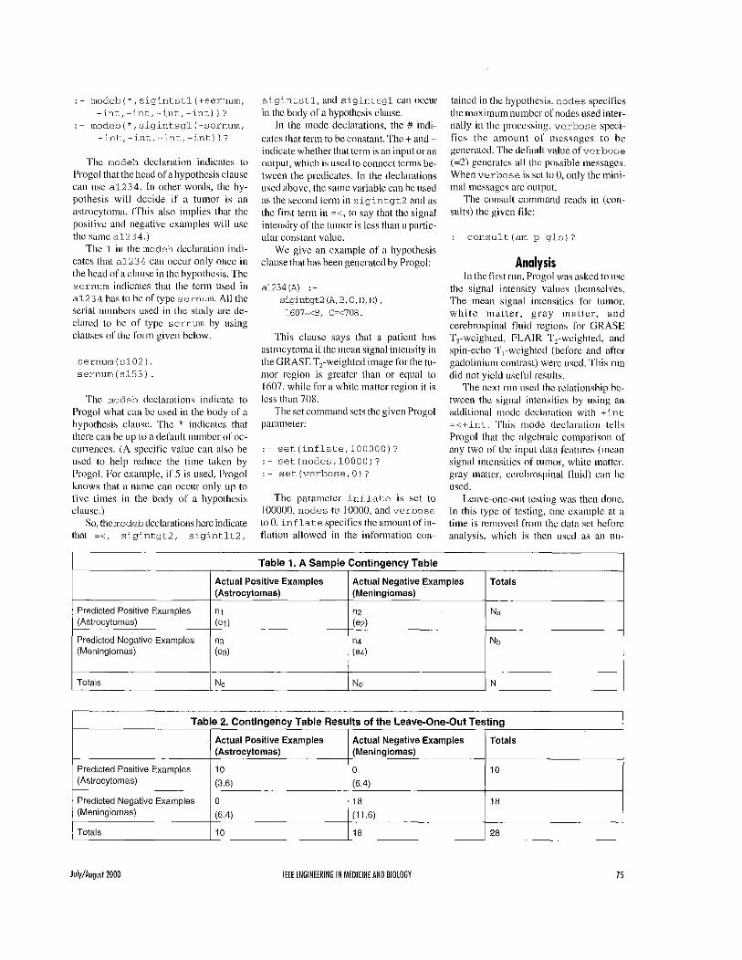
Actual Positive Examples Actual Negative Examples Totals (Astrocytomas) (Meningiomas)
n, n2 Na (e i ) (ez)
n3 n4 Nb (e31 (e41
~~~
laincd in the hypothesis. nodes specilics the inaximum number ofnodes used inter- nally ill the processing. verbose speci- f ies the aniotinl oC incssagcs to he gcncratcd. The default valiie o f verbose (=2) gcncl-atcs iill l l ie pnssihle messages. When verbose i s set lu 0, only the mini- inal incssages are output.
The consult command reads in (con- sul ts) lhc given file:
:- consultlamg-gls)?
Analysis In thc i i rs l run, Progol was asked to use
lhc signal intensity values themsclves. The mean signel iiilcnsities lor tumor, w h i t e ma t le r , g ray inat tcr , and cerebrospinal f lu id regions for GRASE '12-wcighlcd, P I A I R 'I,-wcighled, and spin-echo 'I',-wcighlcd (before and iiftcr gadolinium conlrasl) were used. T h i s run did not yield usefiil results.
The inext riin used the rclationship bc- twccii the signal intensities by iising an addilional iiiode dc =<+int. 'This inode declaralion tells Progol that tlic ;ilgcbraic comparison of any two nf tlic input daki Ieattires (incan signal iiileiisilies of tumor, white iiiatter, gray matter, cerebrospinal Cluid) can hc used.
Leave-one-out testing was theii done. In this type of testing, one cx;imple at a lime is rcniovccl froin [he data set before analysih, which i s then used as an 1111-
Totals NC Nd N
Table 2. Contingency Table Results of the Leave-One-Out Testing
Actual Positive Examples Actual Negative Examples Totals (Astrocytomas) (Meningiomas)
July/Augurt 2000 IEEE ENGINEERING IN MEOICINE AN0 BIOLOGY 75
known test case. The hypothesis is ii i- cover airy useful knowledge. (This is in coii- duccd from the remaining examples i n the trasl 10 the simpler knowledge discovery ap. data set. The induced hypothesis is then plication of iiurma1 tissue discri~i i ioa~ioi i used on tlie unknown test case to predict using signal inlciisitics alonc, descrihed in the tumor type. This i s repeated for eiich 110l.) However, tlic iipplic:itiiin ofl'riigol to example i n tlie ciata set. The contingency kn rY the relationships iimong Ihe signal iiitcnsity table and the overall percentage (II. accii- 11l' iliflcrciit regions (tunior, while inlitter,
gray inaller, and ccrcbruspinel h i d ) gave a very clear resiill.
racy in predicting the tumor type of the unknown test cases are then ciilciiliitcd.
A samplc contingeiicy lablc I I .i l i s showninTable I .Thevaluen is tlheiium- ber of actual astrocytomas (positive cx- An ILP loo1 was used for data iriinirig amplcs) correctly predicted, whereas ii., i s of medical imagiiig diita to qoantitatively the number OS actual astrocytomas iiicor- discriminate bctwccii mcn ing iom;~~ and rectly predicted as meningiomiis. S i m - ;~strocytiimas, It was Ibund that air I1.P larly, the values nd and ii2 w e the actiiiil i l ly suitcd fiir this type (if
meningiomiis (negative exwnplcs) cor- knowledge iliscovcry l'rom inctliciil iiii- agcs, where the relationships among d e ~ a frnni i l i l lcrcnt regions i n (tic ~i iedici i l i n -
rectly and incorrcctly predicted, rcspcc- tively. 'She value N, = n, + n2 is tlie total number predicted as i~strocylomas a n d N, age ;ire osed. The 1I.P tool Progril was = iil+ii, i s ( l ie IoIaI iiumher 4' actual i l ly iiseful wlieii theen- astrocytomas. Similarly, tlie viilucs NI, viroimieiit was OIIC uf rclatioiliil Icarning. and N,, are Ibr nieningiomas. The value el Priigol iihlaiocd ii singlc rule that gave = N,N,IN i s the expected value for the ac- 100%: accur i i t c r e s u l t s iii the tual as t~~cy tumas as search, since tlie resoiirces availatile were
leave-one-out testing. This rule uses Ihe rclatiiinsliip hetween different regions of aslrocylolllas, Illlder the llypothesis tllat exceeded 111 other words, by Llsillg the
is indepelldellt of tile pre. sigiial intensity or ;iny pirt icul i i r region, brain Lissuc. Mc;isurement of t l ie M R sig- llictcd ollc, Silllilar,y, c2 ~ N,,NdN, ci = Pmgi iuld iiot detcriiiiiic m y iisefiil hy- i ia l iiitciisity (71' [lie IUIIIO~ region iilniie i s
iiot ciiough to ~ ) r i~v idc quanti tal i~~e [lis- c r i i i i i~ ia t io~ i bctwccii astrucylom;is and isestirnatedasp= (Il,+n,j,N a,nd astrocytoiiia I'roni those will1 mciiiii-
iiieiiiiigiomas, whercas (lie releliimsliip t l l C error ill this eStilnatc is gio~na.l l~us,t l i isri i i idid i iotyieldsignif i-
betwccn l l ic intensities 111' the tiimiir rc- c;mt rcsLIIIs. +x/(P(~ - P) 1 N.
quickly gencratcd a mcaningful hyputhc- l,ostt-g~l~~,,~~n~urn scilll ,jrcjvilles a clear dis- s is when i t was asked to niake a n algebraic crilllillilliun betwecl, ;Istrocytornas Progol generated the Sollowing hy-
ties. The hypothesis generated was thc mLlk vei{;lblc ollc l]cfilliti"c f,lc. alone. 'The rol lowing lhree cbuses were generated:
single clanse: tor to (lie diagiiostic radiulogist, and i t i s thus uscful in arriving at a differential di-
a1234(A) : - sigintgt2(A,B,C,D,E), a1234(A) ' - agnosis in specific situations where astriicytomas ~ m i l d iniiii ic iiieningiomas.
a1234(A) : - sigintgt2(A,B,C,D,E), Tlius, l l ie automated knowledge discuv- process ill ilnages lnenirlgiolnas
a1234 (A, :- si,gintgtl (A ,B ,C ,D,E) , and astrocylomis is facilitated by the q -
of
Conclusion
ilctllal
N,,N,IN and c'I = N,,N,,,N, prccdictivc pot11 to i l ist i i igi i ish Ipaticiits w i th
-~ ~ ~ ~ -~
In the ~ " 1 run, how 1 Progcll gion an[[ white nlatter ill the spin-ccho
Results
pothesis ill the run using signal intensity colrlI)arison hetweet' the intcnsi- Illell ioiiias. This soccessf'ul result
1607=<B, C=<708.
1194=<B, 1953-<E, C=<764.
1380=<B, 1863=<E, C=<769.
sigintscil (A, B, C, D, E) 8 B=<C.
This clause ineaiis that the tumor i s an astrucYtol1Ya if the l l iei l l i signal ilitelisitY ol'thc tumor i s Icss than that of white mat- ter in the stiii-echo v o s t - ~ ~ ~ i l i i l i ~ i i i ~ ~ i i SCLIII.
plication o i ILP.
The clauses mean that, in l l i i s dala set, the tumor i s an astrocytoma il; Ihr the GRASE TZ-weighted scans, thc n i w i til-
mor signal intensity i s =>I607 A N D the white matter signal illtensity =<708, O R themean tumor signal intensity is>=l 194 A N D the white matter signal intensity =<764 A N D the cerebrospinal fluid signal intensily>=1953, OR themean tuiiirxsig- nal intensity i s >=I380 A N D tlie white matler signal intcnsity ~ 7 6 9 A N D tlic cerebrospinal f l u i d signal intensi ty >=1863.
Progol took inore tliaii 14 iiiiii aiitl couldnot completcall possihlcpathsin its
. I Progol took only around IO sec to deter- ininc this.
The results ofthe leave-onc-out tcstiiig are shown i n the contingency table (Table 2). The iweraII accuracy = 100.00% + 0.00%. The hypothesis cliiiisc i s the saiiic:
a1234(A) :-
sigintsgl(A,B,C,D,E), B=<C
Progol took I0:46 min to do the entirc Icavc-ooc-out tcsting mil achieved 100% accurate results.
Amilysis ol'llie signal inlensity ofoiie re- gion alone (tlie tmior tissue) did not dis-
111 1993 sliecom
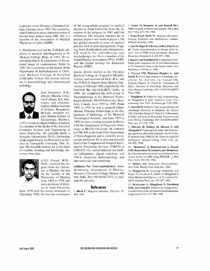
A II , I O Si roin on e y , M.Sc., B.Url., M.l'liil., Ph.D. , r e c e i v e d the M.Sc. degree in physics in 1979 from the U n - versity c i ( M;all-as. In 19x3, she reccivcd the R.Ed. degree froin tlie Univcrsity 01' Mysnre.
uleted tlie M.Phil. deeree I
i n physics Sr1~niAnna University, Cheniiai (Madl;is), wilh ii Faculty Iinprovcmcnt Program grant lrom the Lliiivcrsity Grants Commission of Ihe Governinent oS India, iiiid she completed her Ph.D. i n I999 at the same [Jniversity, She has heen a lecturer
76 IEEE ENGINEERING IN MEDICINE AND BIOLOGY lulq/Augurl2000
i n physics at (lie Wnmen’s Christ iai Col- lege, Cliciiiiai, since 19x3, Hcr ciirrc111 re- search iiitcrcst i s lissuecharactcriz;iliiiii o f hui i iai i brain luiiiors using MRI. She i s a ineiiiber nf tlic Associalion of Medical Physicisls of Iiidiii (AMPI).
I,. RoxIiwciin rcccivcd tlic I).M.R.Il. ld i - ploma i n iiicdical radioihgnosis) a1 I31 Mcdical College, Punc, in 1993. He ID-
ceivctl thc Ui1i.N.B. (diploinatc nf l l ic nil- tional board of examinations. India) iii 1997. He i s currcnlly on the faculty of the Dcprlnienr or Rsdiodi;tgnosis a1 Chris- liiiii M e d i c a l C o l l c g c & H o s p i t a l (CMCaH), Vellore. His ciirrciit iiitcrcsls are iii neuroradioiogy and inlervenlioiial radiology.
A w l Siroinoiicj’, U.Sc. physics (Miidras Chris- lian College), 13.11. elec- tronics aiid coiiiiiiiiii-
icatinns (Indian Iiislilure of Science, H;lngalr~re), M.Tcc1i. compulcr sci- ence (Indian lristilulc of Technology. Madl-;is),
LI‘CL (l‘rinily College ofMusic, Loiiduiij, i s ii incmber of the hcul ly of tlic Sc la~o l OS Coinpiiter Science mil Phgineeriiig at Aiiiia University. t l c currcnlly l i d t l s a Kniipiiku (disserlalion Ph.11.) Fcllnwship 01 the Japan Snciety fnr Promixion of Sci- ence ill Yaniaguchi Univcrsily, tJ1ie. Ja- paii. His research iiitcrcsls arc in llic are;is III‘ iiiiicliini. Iexi i ing and kiiowlcdgc dis- covcl-y frnm dal;i.
C. N.S. Prrrsail, M.Sc., Ph.D., received l i i s de- grees froin the Univcr- sity o f Madras. He was on lhc fecully o f llie Uii ivcrsi ly of Madras from I963 111 I978 ilnd was a processor ofpliys- ics iit Anna l i i i ivcrsi iy
from 1978 iiiilil h i s recent retirement in Dcccnthcr I W 8 , He was the cnorilii lat~ir
nf tlic postgnidii;ilc program in iiietlical physics a1 Aiina University from the i i i -
ception of thc prngraiii in 1981 until h i s
physics si icl i a s pain iiian;ige.menl. imag- ing, hone ~lcnsiloiiielry and ostcuporo. LQ i i i o d c l s for r a d i o t h c r a p y i i i i d hr;shytlier;ipy, cic. I l c i s il member of lhc Indian Physics Aasircietioii (IPA), AMPI, and llie Indian Socicty for Radial ion Physics (ISRPj.
Ipesoi i Kwrih sludied a1 the Christian Medical College & Ilospital (CMC&H), Vcllorc, ani1 rcceived llie M.B., B.S., a id llic D.M.R.1) degrees I‘rom M u h i s Uni- versity in I982 and 1988,rcspectively. He received tlic Uip.N.H.(RDj, India, iii 1990. FIc coiiipleled the I’ellowship iii Neuror;idiology a1 l l ie Montrciil Neuro- logical Inslilulc, McGi l l University, Moii- Lreal, C;in;al;i, frirm I993 L o 199s. Proin 1992 lo I993 lie was a resciircli fcllow (Jeanne l i n i i i i i n s Fcllowsliipj at llie De- pirlincnl of Radiology nf the Montrc;il Ncumlogical Insl i l i i le, and from I993 lo 1995, l ie was ii visiling assistant profcssm wi l l i the 1)cp;irtiiicnt o f Diiignoslic Riidi- ology a1 McOi l l llnivcrsity. He reluriieil to CMC&H iis lhc liead of llic Ikparlmcnt of Iladiotliagiiosis ;ind i s curreiilly an as- sociate [irofcssiir. He is also concurrcnlly hciid of [lie Compulerised Ilospilal Iiifirr- inatinii Pruccssiiig Scrviccs CHIPS) at CMC&H. His ciirrenl iiitere. c d ioforoiatics, digital r;idinlogy mil PACS, diagnostic iiciirorailiology, aiitl iiite.rventional neororialk~logy.
Address for Cerrespondcnce: Anna Siroinoi icy, 1)cp;irtmcnl cif Physics, Womcn’s Christian College, Madliis 601) 006, Iiitliii. ‘Scl: +9 I 44 642 147 I. E-inail: asiroB vsiiI.coni.