JOURNAL OF ORAL & MAXILLOFACIAL RESEARCH Shavit and Juodzbalys Inferior Alveolar Nerve Injuries Following Implant Placement - Importance of Early Diagnosis and Treatment: a Systematic Review Ilana Shavit 1 , Gintaras Juodzbalys 1 1 Department of Maxillofacial Surgery, Lithuanian University of Health Sciences, Kaunas, Lithuania. Corresponding Author: Ilana Shavit Sukilėlių pr. 89-35, LT 49233, Kaunas Lithuania Phone: +370 67004611 E-mail: [email protected]ABSTRACT Objectives: The purpose of this article is to systematically review diagnostic procedures and risk factors associated with inferior alveolar nerve injury following implant placement, to identify the time interval between inferior alveolar nerve injury and its diagnosis after surgical dental implant placement and compare between outcomes of early and delayed diagnosis and treatment given based on case series recorded throughout a period of 10 years. Material and Methods: We performed literature investigation through MEDLINE (PubMed) electronic database and manual search through dental journals to find articles concerning inferior alveolar nerve injury following implant placement. The search was restricted to English language articles published during the last 10 years, from December 2004 to March 2014. Results: In total, we found 33 articles related to the topic, of which 27 were excluded due to incompatibility with established inclusion criteria. Six articles were eventually chosen to be suitable. The studies presented diagnostic methods of inferior alveolar nerve sensory deficit, and we carried out an assessment of the proportion of patients diagnosed within different time intervals from the time the injury occurred. Conclusions: Various diagnostic methods have been developed throughout the years for dealing with 1 quite frequent complication in the implantology field - inferior alveolar nerve injury. Concurrently, the importance of early diagnosis and treatment was proved repeatedly. According to the results of the data analysis, a relatively high percentage of the practitioners successfully accomplished this target and achieved good treatment outcomes. Keywords: dental implants; inferior alveolar nerve; fifth cranial nerve injury; mandibular nerve; paraesthesia. Accepted for publication: 2 December 2014 To cite this article: Shavit I, Juodzbalys G. Inferior Alveolar Nerve Injuries Following Implant Placement - Importance of Early Diagnosis and Treatment: a Systematic Review. J Oral Maxillofac Res 2014;5(4):e2 URL: http://www.ejomr.org/JOMR/archives/2014/4/e2/v5n4e2ht.pdf doi: 10.5037/jomr.2014.5402 http://www.ejomr.org/JOMR/archives/2014/4/e2/v5n4e2ht.htm J Oral Maxillofac Res 2014 (Oct-Dec) | vol. 5 | No 4 | e2 | p.1 (page number not for citation purposes)
Transcript
JOURNAL OF ORAL & MAXILLOFACIAL RESEARCH Shavit and Juodzbalys
Inferior Alveolar Nerve Injuries Following Implant Placement - Importance of Early Diagnosis and Treatment: a Systematic Review
Ilana Shavit1, Gintaras Juodzbalys1
1Department of Maxillofacial Surgery, Lithuanian University of Health Sciences, Kaunas, Lithuania.
Objectives: The purpose of this article is to systematically review diagnostic procedures and risk factors associated with inferior alveolar nerve injury following implant placement, to identify the time interval between inferior alveolar nerve injury and its diagnosis after surgical dental implant placement and compare between outcomes of early and delayed diagnosis and treatment given based on case series recorded throughout a period of 10 years.Material and Methods: We performed literature investigation through MEDLINE (PubMed) electronic database and manual search through dental journals to find articles concerning inferior alveolar nerve injury following implant placement. The search was restricted to English language articles published during the last 10 years, from December 2004 to March 2014.Results: In total, we found 33 articles related to the topic, of which 27 were excluded due to incompatibility with established inclusion criteria. Six articles were eventually chosen to be suitable. The studies presented diagnostic methods of inferior alveolar nerve sensory deficit, and we carried out an assessment of the proportion of patients diagnosed within different time intervals from the time the injury occurred.Conclusions: Various diagnostic methods have been developed throughout the years for dealing with 1 quite frequent complication in the implantology field - inferior alveolar nerve injury. Concurrently, the importance of early diagnosis and treatment was proved repeatedly. According to the results of the data analysis, a relatively high percentage of the practitioners successfully accomplished this target and achieved good treatment outcomes.
Accepted for publication: 2 December 2014To cite this article:Shavit I, Juodzbalys G. Inferior Alveolar Nerve Injuries Following Implant Placement - Importance of Early Diagnosis and Treatment: a Systematic Review.J Oral Maxillofac Res 2014;5(4):e2URL: http://www.ejomr.org/JOMR/archives/2014/4/e2/v5n4e2ht.pdfdoi: 10.5037/jomr.2014.5402
http://www.ejomr.org/JOMR/archives/2014/4/e2/v5n4e2ht.htm J Oral Maxillofac Res 2014 (Oct-Dec) | vol. 5 | No 4 | e2 | p.1(page number not for citation purposes)
http://www.ejomr.org/JOMR/archives/2014/4/e2/v5n4e2ht.htm J Oral Maxillofac Res 2014 (Oct-Dec) | vol. 5 | No 4 | e2 | p.2(page number not for citation purposes)
JOURNAL OF ORAL & MAXILLOFACIAL RESEARCH Shavit and Juodzbalys
INTRODUCTION
Despite considerable development of technology and techniques used in dentistry throughout the last few decades, tooth loss is still a common problem. Dental implants are used in such incidences as a solution. As practitioners gained experience in the implantology field, they became exposed to various complications arising as a result of the surgical procedure. One of the most common and serious complications faced by the clinician and the patient following implant placement in the mandible is injury to the inferior alveolar nerve (IAN). It ranges from 0% to 40% of implant-related IAN injuries [1-4]. These injuries may occur during preparation or placement of an implant. They may be directly related to the depth of preparation or implant length or width [5] and may result from local anaesthetic application as well [6,7]. Different degrees of nerve injury are available. According to Seddon [8], nerve injuries are divided into 3 types based upon the severity of tissue injury, prognosis, and time for recovery-neurapraxia, axonotmesis, and neurotmesis. Neurapraxia is the mildest form, with the best prognosis, while neurotmesis is the most severe [8].Sensory disturbances resulting from the injury will provide an unpleasant experience for both the doctor and the patient. Effective management of those cases is based on providing treatment-conservative or surgical, immediately upon diagnosis of damage. That is to say, early diagnosis is the key for successful treatment. Renton and coworkers [9] state that peripheral sensory nerve injuries are more likely to be persistent when there is an increased duration between injury and reviewing of the patient. The problem is that occasionally the diagnosis is delayed, which may have a crucial effect on treatment results.Another possible sequel of late diagnosis is the development of tunnel syndrome. Its underlying pathophysiology is ascribed to an increased pressure on peripheral nerves, which leads to impaired neural microcirculation followed by focal demyelination [10]. For example, an unnoticeable, minor IAN injury may develop due to haematoma, and the practitioner may not diagnose it on time because of its mildness. Later, oedema will develop, and the injury will worsen. This is due to the fact that the IAN is bounded within the limits of the mandibular canal (MC). Thus, if oedema rapidly develops, subsequent nerve compression is expected. MC width depends on the patient’s age and gender-females and elderly patients thus are at higher risk to develop tunnel syndrome-related nerve degeneration [1].
Using long-acting local anaesthetics and the difficulty of evaluating the extent of the damage are some of the factors preventing early disclosure of the complication.Literature regarding this topic suggests diagnostic methods such as neurosensory testing, including objective and subjective methods, but those tests can be performed only after fading of the local anaesthetic effect. The presence of continuous anaesthesia, dysaesthesia, or spontaneous pain indicates poor prospects for recovery, even with surgical intervention. Overall, 25% of patients with iatrogenic paraesthesia suffer permanent effects [5].To make it easier for readers, we presented the following important aspects regarding IAN injury separately: risk factors, diagnostic methods, adequate time for diagnosis, and treatment outcome as a function of diagnosis time.
Diagnosis of IAN injury according to early signs and symptoms reported by patients
Signs and symptoms, any sensation described by the patient, should be taken into consideration since radiography is not always helpful. There are cases were the implant does not seem to damage the nerve, but in fact, it does [11].One of the most important things the practitioner should check after the operation is completed is whether the patient experienced a return of normal sensation. Six hours after operation (after the local anaesthesia effect wears off), the patient should be contacted. In cases of the patient reporting persistent numbness or anaesthesia, it would be the first sign for the surgeon that something went wrong and nerve damage probably occurred [1]. Numbness usually will be felt on the side of implant placement, including the lower lip and chin. Another symptom that may indicate nerve damage is the feeling of tingling, tickling, or burning skin in other words, paraesthesia. In some cases, numbness may not appear immediately but later on. The patient does feel improvement in sensation in the beginning, although some discomfort is present, and later numbness appears. A case study of 4 females, which recorded their sensations following implant-associated IAN damage, concluded that all patients suffered from numbness of the affected inferior alveolar dermatome [5].Renton et al. [12] found, in 2012, in their study regarding post-implant neuropathy of the trigeminal nerve, that over 50% of patients suffered constant pain and/or discomfort. Paraesthesia was the main feature for 47% of cases.
http://www.ejomr.org/JOMR/archives/2014/4/e2/v5n4e2ht.htm J Oral Maxillofac Res 2014 (Oct-Dec) | vol. 5 | No 4 | e2 | p.3(page number not for citation purposes)
JOURNAL OF ORAL & MAXILLOFACIAL RESEARCH Shavit and Juodzbalys
However, there are various alterations of sensation patients can report, such as dysaesthesia, allodynia, hypoaesthesia, hyperaesthesia, and more [1].
Diagnosis of IAN injury based on subjective sensory tests
A study in the year 2009 by Poort et al. [13] reveals an interesting fact - subjective evaluation has been found to be the most commonly used method (64% of all methods used) to detect implant-associated IAN injury. Every alteration in sensation reported by the patient should be carefully taken into consideration, both intra- and postoperatively. That is because the patient’s own feeling is the best evaluation for change in sensation, as sometimes the alteration may be not big enough to be detected by any diagnostic tools, which are less sensitive than the human him/herself [14]. Some patients get an “electric-shock-type” sensation during administration of local anaesthetic, which can give a hint about further nerve injury, but it is not a definitive sign since injuries can occur without any intraoperative demonstration. After the fading of the local anaesthetic effect, the doctor should ascertain with the patient if any changes occurred and if some suspicion for neuropathy is present. A basic neurosensory examination should be performed. Basically, subjective sensory tests are divided into 2 major categories: mechanoceptive and nociceptive.
Mechanoceptive methods
Under the mechanoceptive category, several methods are available. First is static light touch (SLT) detection, in which the patient closes his eyes and says “yes” whenever he feels a light touch with cotton to the face [15]. The second one is brush directional discrimination (brush stroke direction [BSD]) in which the patient tells if any sensation is detected and in which direction the filament or brush moved. The third is two-point discrimination (TPD), in which the patient closes his eyes, and the clinician, using an object having two sharp edges, touches the face in different areas [15]. The distance between the sharp edges should be changed in order to define the minimal distance in which the patient is still able to discriminate two points. The average value of this test is about 5 mm, but it is very individual and varies considerably [16]. Generally, it depends on the size of receptive fields in the tested area and on the density of the fields. Some investigators found that the TPD test is unreliable because it has great variability between measurements even on the control side and does not
add information to the SLT test [13]. Another method is sharp/blunt discrimination (SBD), in which a sharp right-angle dental probe is applied to the area, with indentation but with no breach of the mucosa/skin. The patient is then asked to compare this sensation to that produced by a blunt region of the probe. The test is considered positive if the patient recognises 3 out of 5 of each stimulus correctly.
Nociceptive methods
The nociceptive category suggests 2 methods: pin pressure nociception and thermal discrimination [17]. The former compares the pain threshold of affected and unaffected areas and allows defining the response as hyperaesthesia, anaesthesia, or hypoaesthesia. The aim is to establish the sensation threshold for the unaffected side and compare it to the affected side. Usually, the normal value to initiate a response is about 15 gm [18]. Thermal tests are also suggested. This type of test checks whether the patient feels normal cold or heat sensation by applying ethyl chloride and a mirror handle warmed to 43 °C to affected and unaffected areas. This test is useful but not essential.
Diagnosis of IAN injury based on objective sensory tests
A study conducted in June 2011 by Biasiotta et al. [19] states that the masseter inhibitory reflex (MIR) test is the most used neurophysiological tool for the functional assessment of the trigeminal mandibular division. It consists of a reflex inhibition of the jaw-closing muscles elicited by peri- or intraoral electrical stimulations. Their study revealed that MIR testing shows an almost absolute specificity (99%) and reliably demonstrates nerve damage beyond doubt, whereas the relatively low sensitivity (51%) makes the finding of a normal MIR by no means sufficient to exclude nerve damage [19]. In other words, this test principle is that abnormality does prove nerve damage, and normality does not disprove it. It is important to remark that in that study, all patients (160) were examined at least 2 months after injury, so its efficiency as an early diagnostic method cannot yet be discussed.The recording of somatosensory evoked potentials (SEPs) is a noninvasive, routine clinical testing procedure in neurology. The idea is to stimulate the peripheral nerves and then record the electric potential occurring in the brain through the scalp [20]. Electrodes are applied to the skin of the area to be tested as well as on the scalp. For trigeminal
http://www.ejomr.org/JOMR/archives/2014/4/e2/v5n4e2ht.htm J Oral Maxillofac Res 2014 (Oct-Dec) | vol. 5 | No 4 | e2 | p.4(page number not for citation purposes)
JOURNAL OF ORAL & MAXILLOFACIAL RESEARCH Shavit and Juodzbalys
nerve stimulation, however, SEPs have not received widespread clinical attention. In addition, this technique has a lot of disadvantages: the reliability of the recorded signals has been questioned because, rather than reflecting genuine brain activity, they probably result from volume-conducted muscle signals, as they disappear in the curarized subject [19,21]. Thus, more researches should be conducted in order to establish some general guidelines for using this method.Orthodromic sensory nerve action potential is another method used to record the function of IAN [22,23]. However, it was not used by the clinicians to evaluate IAN function after implant-related injury. Recently, the International Federation of Clinical Neurophysiology has recommended investigating the trigeminal function with reflex rather than evoked potential studies. Unlike the above techniques, the blink reflex after mental [24] or lingual stimulation seems a promising alternative to the MIR. The blink reflex responses are recorded simultaneously on both sides. Blink reflex proved to be a sensitive test in detecting IAN lesions within 2 to 3 months from injury [25].
Diagnosis of IAN injury using alternative methods
Several researches have proposed that early implant removal may lead to better healing with the return of sensation [5,26]. However, in most cases, by the time diagnosis is made, it may lose its efficiency. The problem in making an immediate diagnosis is that usually, by the end of the surgery, the patient is still found under the effect of local anaesthetics. That occurs especially when surgeons use long-acting anaesthetics in order to delay the pain as much as possible. Phentolamine mesylate (PM) is an anti-hypertensive reversal agent of local anaesthesia. A study conducted by Froum et al. in 2010 [27] checked the possibility of its utilisation immediately after implant placement.Without using PM, the local anaesthetic effect fades away in the range of 3 to 5 hours after injection. Thus, using a reversing agent will significantly increase the chances of full recovery, as its application permits making the earliest detection of nerve damage currently available.The purpose of the present article is to identify the time interval of inferior alveolar nerve injury diagnosis after dental implant placement operation and compare between outcomes of early and delayed diagnosis and treatment given after inferior alveolar nerve injury based on case series recorded throughout the years.
MATERIAL AND METHODSProtocol and registration
The review is registered in an international prospective register of systematic reviews, ‘PROSPERO’ [28]. Registration number: CRD42014014724.
Types of publication
The review included studies on humans published in English.
Types of studies
The review included any published observational studies and case series.
Information sources
The information source was the electronic database MEDLINE (PubMed).
Population
The population consisted of adult patients who sustained IAN injury following surgical implant placement.
Inferior alveolar nerve injury (IAN) definition
IAN injury includes damage or severance of nervous tissue as a result of trauma transaction, cutting (laceration), compression, shearing, or crushing, which results in sensory disturbances.
Literature search strategy
According to the PRISMA guidelines [29], we conducted an electronic search using MEDLINE (PubMed) database to locate articles concerning IAN injury following implant placement. The keywords used to find related articles were “ INFERIOR -ALVEOLAR -NERVE - INJURY-ASSOCIATED-WITH-IMPLANT-SURGERY”, “ I N F E R I O R - A LV E O L A R - N E RV E - I N J U RY -IMPLANT”,-“INFERIOR-ALVEOLAR-NERVE-I N J U RY- D I A G N O S I S ” , - a n d - “ I N F E R I O R -ALVEOLAR-NERVE-INJURY-MANAGEMENT”. The search was restricted to English language articles published during the last 10 years, from December 2004 to March 2014. The quest yielded a high amount of articles regarding IAN injury. However, most of the articles were excluded due to irrelevancy of their titles
http://www.ejomr.org/JOMR/archives/2014/4/e2/v5n4e2ht.htm J Oral Maxillofac Res 2014 (Oct-Dec) | vol. 5 | No 4 | e2 | p.5(page number not for citation purposes)
JOURNAL OF ORAL & MAXILLOFACIAL RESEARCH Shavit and Juodzbalys
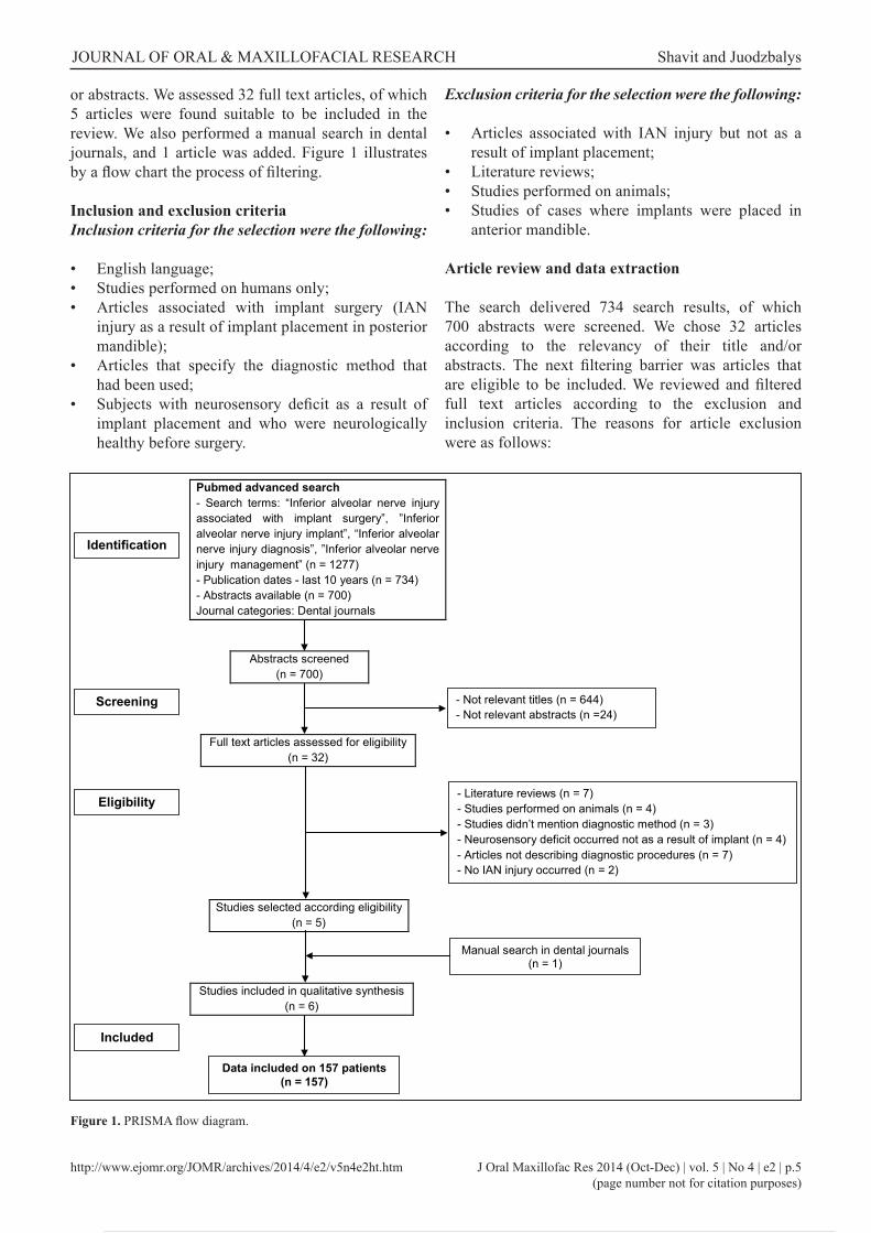
or abstracts. We assessed 32 full text articles, of which 5 articles were found suitable to be included in the review. We also performed a manual search in dental journals, and 1 article was added. Figure 1 illustrates by a flow chart the process of filtering.
Inclusion and exclusion criteriaInclusion criteria for the selection were the following:
• English language;• Studies performed on humans only;• Articles associated with implant surgery (IAN
injury as a result of implant placement in posterior mandible);
• Articles that specify the diagnostic method that had been used;
• Subjects with neurosensory deficit as a result of implant placement and who were neurologically healthy before surgery.
Exclusion criteria for the selection were the following:
• Articles associated with IAN injury but not as a result of implant placement;
• Literature reviews;• Studies performed on animals;• Studies of cases where implants were placed in
anterior mandible.
Article review and data extraction
The search delivered 734 search results, of which 700 abstracts were screened. We chose 32 articles according to the relevancy of their title and/or abstracts. The next filtering barrier was articles that are eligible to be included. We reviewed and filtered full text articles according to the exclusion and inclusion criteria. The reasons for article exclusion were as follows:
Full text articles assessed for eligibility (n = 32)
Studies selected according eligibility (n = 5)
Studies included in qualitative synthesis (n = 6)
Identification
- Not relevant titles (n = 644) - Not relevant abstracts (n =24)
Screening
Eligibility - Literature reviews (n = 7) - Studies performed on animals (n = 4) - Studies didn’t mention diagnostic method (n = 3) - Neurosensory deficit occurred not as a result of implant (n = 4) - Articles not describing diagnostic procedures (n = 7) - No IAN injury occurred (n = 2)
http://www.ejomr.org/JOMR/archives/2014/4/e2/v5n4e2ht.htm J Oral Maxillofac Res 2014 (Oct-Dec) | vol. 5 | No 4 | e2 | p.6(page number not for citation purposes)
JOURNAL OF ORAL & MAXILLOFACIAL RESEARCH Shavit and Juodzbalys
• Literature reviews (n = 7);• Studies performed on animals (n = 4);• Studies did not mention diagnostic method
(n = 3);• Neurosensory deficit occurred not as a result of
implant (n = 4);• Articles not describing diagnostic procedures
(n = 7);• No IAN injury occurred (n = 2);• The data was included on 157 patients.
Risk of bias across studies
We assessed the risk of bias (for example, operation was not performed by authors themselves, missing information regarding exact time of diagnosis, postoperative neurosensory examination performed by single examiner, sex scission, and low objectives number) that could affect the cumulative evidence across the studies. We used the Cochrane Collaboration’s tool for assessing the risk of bias [30].
RESULTS
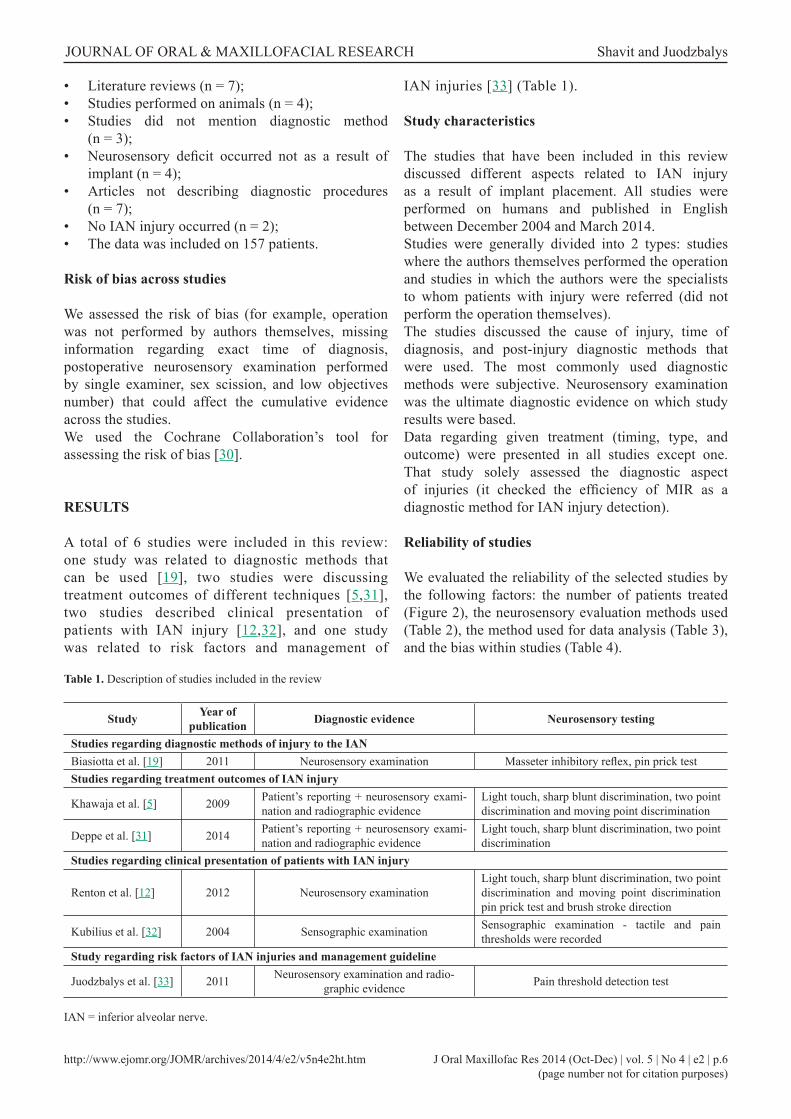
A total of 6 studies were included in this review: one study was related to diagnostic methods that can be used [19], two studies were discussing treatment outcomes of different techniques [5,31], two studies described clinical presentation of patients with IAN injury [12,32], and one study was related to risk factors and management of
IAN injuries [33] (Table 1).
Study characteristics
The studies that have been included in this review discussed different aspects related to IAN injury as a result of implant placement. All studies were performed on humans and published in English between December 2004 and March 2014.Studies were generally divided into 2 types: studies where the authors themselves performed the operation and studies in which the authors were the specialists to whom patients with injury were referred (did not perform the operation themselves).The studies discussed the cause of injury, time of diagnosis, and post-injury diagnostic methods that were used. The most commonly used diagnostic methods were subjective. Neurosensory examination was the ultimate diagnostic evidence on which study results were based.Data regarding given treatment (timing, type, and outcome) were presented in all studies except one. That study solely assessed the diagnostic aspect of injuries (it checked the efficiency of MIR as a diagnostic method for IAN injury detection).
Reliability of studies
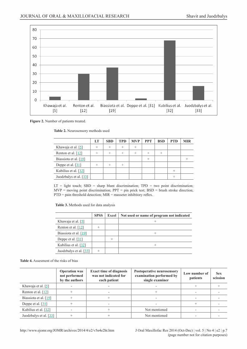
We evaluated the reliability of the selected studies by the following factors: the number of patients treated (Figure 2), the neurosensory evaluation methods used (Table 2), the method used for data analysis (Table 3), and the bias within studies (Table 4).
Table 1. Description of studies included in the review
Study Year of publication Diagnostic evidence Neurosensory testing
Studies regarding diagnostic methods of injury to the IANBiasiotta et al. [19] 2011 Neurosensory examination Masseter inhibitory reflex, pin prick testStudies regarding treatment outcomes of IAN injury
Khawaja et al. [5] 2009 Patient’s reporting + neurosensory exami-nation and radiographic evidence
Light touch, sharp blunt discrimination, two point discrimination and moving point discrimination
Deppe et al. [31] 2014 Patient’s reporting + neurosensory exami-nation and radiographic evidence
Light touch, sharp blunt discrimination, two point discrimination
Studies regarding clinical presentation of patients with IAN injury
Renton et al. [12] 2012 Neurosensory examinationLight touch, sharp blunt discrimination, two point discrimination and moving point discrimination pin prick test and brush stroke direction
Kubilius et al. [32] 2004 Sensographic examination Sensographic examination - tactile and pain thresholds were recorded
Study regarding risk factors of IAN injuries and management guideline
Juodzbalys et al. [33] 2011 Neurosensory examination and radio-graphic evidence Pain threshold detection test
http://www.ejomr.org/JOMR/archives/2014/4/e2/v5n4e2ht.htm J Oral Maxillofac Res 2014 (Oct-Dec) | vol. 5 | No 4 | e2 | p.7(page number not for citation purposes)
JOURNAL OF ORAL & MAXILLOFACIAL RESEARCH Shavit and Juodzbalys
Figure 2. Number of patients treated.
Table 2. Neurosensory methods used
LT SBD TPD MVP PPT BSD PTD MIRKhawaja et al. [5] + + + +Renton et al. [12] + + + + + +Biassiota et al. [19] + +Deppe et al. [31] + + +Kubilius et al. [32] +Juodzbalys et al. [33] +
LT = light touch; SBD = sharp blunt discrimination; TPD = two point discrimination; MVP = moving point discrimination; PPT = pin prick test; BSD = brush stroke direction; PTD = pain threshold detection; MIR = masseter inhibitory reflex.
Table 3. Methods used for data analysis
SPSS Excel Not used or name of program not indicatedKhawaja et al. [5] +Renton et al. [12] +Biassiota et al. [19] +Deppe et al. [31] +Kubilius et al. [32] +Juodzbalys et al. [33] +
Table 4. Assesment of the risks of bias
Operation was not performed by the authors
Exact time of diagnosis was not indicated for
each patient
Postoperative neurosensory examination performed by
single examiner
Low number of patients
Sex scission
Khawaja et al. [5] - - - + +Renton et al. [12] + - + - -Biassiota et al. [19] + + - - -Deppe et al. [31] + - - + -Kubilius et al. [32] - + Not mentioned - -Juodzbalys et al. [33] + + Not mentioned - -
http://www.ejomr.org/JOMR/archives/2014/4/e2/v5n4e2ht.htm J Oral Maxillofac Res 2014 (Oct-Dec) | vol. 5 | No 4 | e2 | p.8(page number not for citation purposes)
JOURNAL OF ORAL & MAXILLOFACIAL RESEARCH Shavit and Juodzbalys
Risk of bias within studies
All studies included exhibited at least 1 bias. We grouped the risks of bias that were indicated within other studies and presented as a lack of information value as follows: operation was not performed by the authors, exact time of diagnosis was not indicated for each patient, postoperative neurosensory examination performed by single examiner, low number of patients, and sex scission (Table 4).
Risk factors associated with IAN injury
Risk factors may be classified into general, intraoperative, and postoperative, as described by Juodzbalys et al. [1]. Possible risk factors are summarised in Table 5 [1,7,33].
Diagnosis of IAN injury
Diagnosis of IAN injury should be done based on the patient’s complaints and clinical symptoms, in combination with different techniques. Patients’ sensations vary from case to case. Nerve injury can lead to anaesthesias, paraesthesias, and dysaesthesias. As has been already mentioned, timing in such circumstances is of high importance. Moreover, Khawaja and Renton [5] made a conclusion based on 4 case studies: early removal of implants associated with IAN injury (less than 36 hours post-injury) may assist in minimising or even resolving IAN neuropathy, while delayed diagnosis and removal
may result in no improvement at all [5,12,19,31,32].
Time interval between nerve injury and its diagnosis and treatment
Kim et al. [26] found in their study regarding clinical outcomes of conservative treatment in cases of IAN injury that patients who visited the hospital within 9 months after injury exhibited greater improvement in their symptoms than those visiting after 9 months. These results reinforce the already proven fact that early treatment is important in cases of nerve damage. For treatment to be as early as possible, timely diagnosis should be done.Case series were reviewed and the time interval between the time of injury and the diagnosis and treatment was recorded. In some of the articles that were analysed, the exact time of diagnosis was not mentioned. Thus, we based our determination of diagnosis time upon the assumption that the practitioner realised the injury was present when it was revealed that the subject continued to feel numbness after the effect of local anaesthetic wore off, since it’s the first postoperative alarming sign.In addition, it is important to mention that we divided the studies mainly into 2 types. The first type of articles described operations performed by the authors themselves. However, the second type of articles described reports by specialists to whom patients were referred after nerve injury for consultation (surgical implant placement was not performed by them). Results are presented in Table 6.
Table 5. Possible risk factors associated with IAN injury following implant placement
General risk factors
- Patient’s unrealistic expectations;- Pre-existing altered sensation;- Improper selection of site for implant placement;- Anatomical and radiological risk factors related to mandibular vital structures;- Female patients;- Increased age of patients.
Intraoperative risk factors
- Protrusion through lingual or buccal plate;- Perforation of mandibular canal;- Direct mechanical injury (manifests by “sudden give” type of feeling);- Extensive bleeding;- Extrusion of preparative debris into canal;- Slippage of the drill, implant placement deeper than planned, or bigger diameter implant placement;- Excessive force using implant drill (density and thickness of the bone surrounding the mandibular canal is not able to resist it);- Repeated IAN blocks.
Postoperative risk factors
- The fact that the IAN is contained within the bony canal (compression, ischemia);- Severity of injury;- Interval between time of injury and diagnosis, treatment.
http://www.ejomr.org/JOMR/archives/2014/4/e2/v5n4e2ht.htm J Oral Maxillofac Res 2014 (Oct-Dec) | vol. 5 | No 4 | e2 | p.9(page number not for citation purposes)
JOURNAL OF ORAL & MAXILLOFACIAL RESEARCH Shavit and Juodzbalys
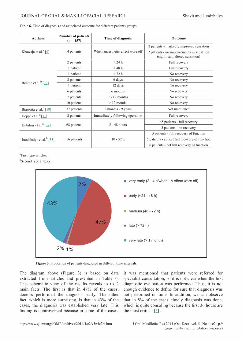
The diagram above (Figure 3) is based on data extracted from articles and presented in Table 6. This schematic view of the results reveals to us 2 main facts. The first is that in 47% of the cases, doctors performed the diagnosis early. The other fact, which is more surprising, is that in 43% of the cases, the diagnosis was established very late. This finding is controversial because in some of the cases,
it was mentioned that patients were referred for specialist consultation, so it is not clear when the first diagnostic evaluation was performed. Thus, it is not enough evidence to define for sure that diagnosis was not performed on time. In addition, we can observe that in 8% of the cases, timely diagnosis was done, which is quite consoling because the first 36 hours are the most critical [5].
Table 6. Time of diagnosis and associated outcome for different patients groups
Authors Number of patients (n = 157) Time of diagnosis Outcome
Khawaja et al.a [5] 4 patients When anaesthetic effect wore off2 patients - markedly improved sensation2 patients - no improvements in sensation
(significant altered sensation)
Renton et al.b [12]
2 patients < 24 h Full recovery1 patient < 48 h Full recovery1 patient < 72 h No recovery2 patients 6 days No recovery1 patient 12 days No recovery6 patients 6 months No recovery7 patients 7 - 12 months No recovery10 patients > 12 months No recovery
Biasiotta et al.b [19] 37 patients 2 months - 9 years Not mentioned
Deppe et al.a [31] 2 patients Immediately following operation Full recovery
Kubilius et al.a [32] 68 patients 2 - 48 hours65 patients - full recovery3 patients - no recovery
Juodzbalys et al.b [33] 16 patients 10 - 52 h5 patients - full recovery of function
5 patients - almost full recovery of function6 patients - not full recovery of function
aFirst type articles.bSecond type articles.
Figure 3. Proportion of patients diagnosed in different time intervals.
http://www.ejomr.org/JOMR/archives/2014/4/e2/v5n4e2ht.htm J Oral Maxillofac Res 2014 (Oct-Dec) | vol. 5 | No 4 | e2 | p.10(page number not for citation purposes)
JOURNAL OF ORAL & MAXILLOFACIAL RESEARCH Shavit and Juodzbalys
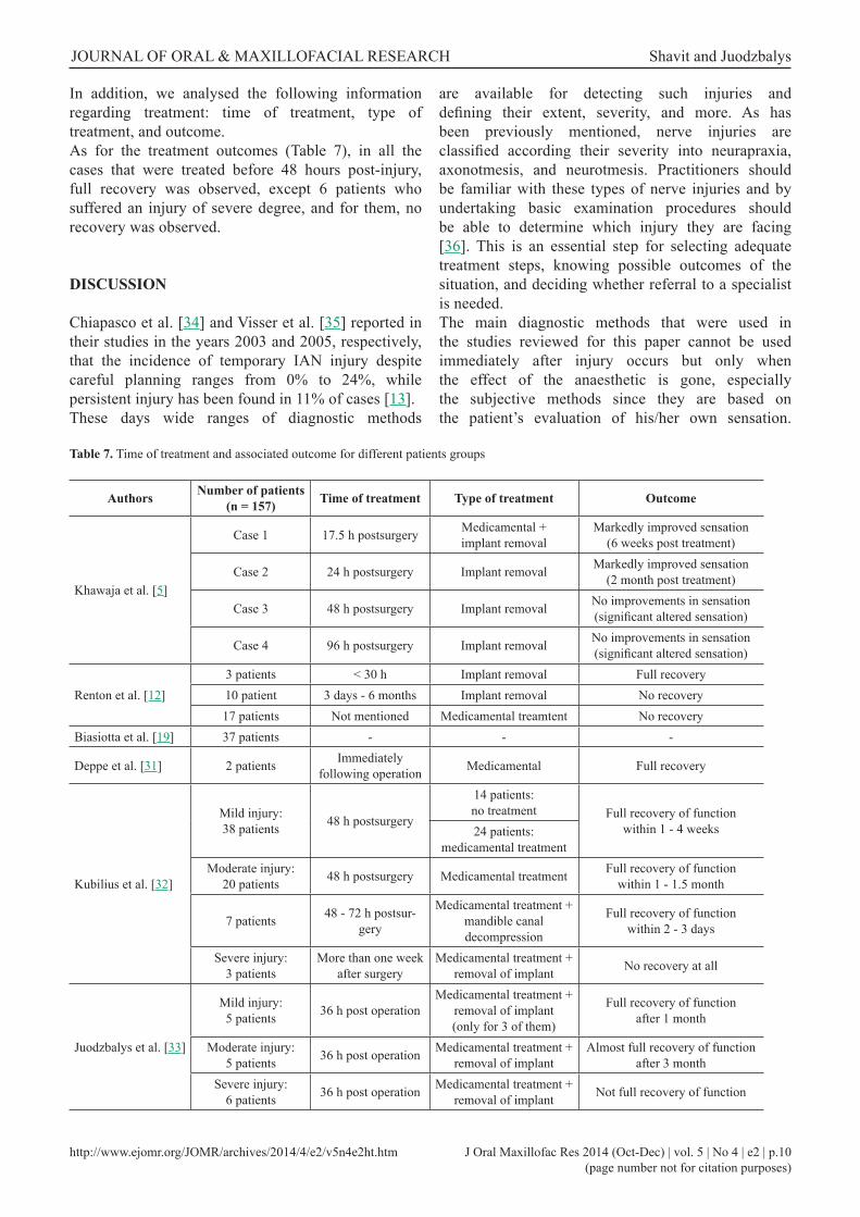
In addition, we analysed the following information regarding treatment: time of treatment, type of treatment, and outcome. As for the treatment outcomes (Table 7), in all the cases that were treated before 48 hours post-injury, full recovery was observed, except 6 patients who suffered an injury of severe degree, and for them, no recovery was observed.
DISCUSSION
Chiapasco et al. [34] and Visser et al. [35] reported in their studies in the years 2003 and 2005, respectively, that the incidence of temporary IAN injury despite careful planning ranges from 0% to 24%, while persistent injury has been found in 11% of cases [13].These days wide ranges of diagnostic methods
are available for detecting such injuries and defining their extent, severity, and more. As has been previously mentioned, nerve injuries are classified according their severity into neurapraxia, axonotmesis, and neurotmesis. Practitioners should be familiar with these types of nerve injuries and by undertaking basic examination procedures should be able to determine which injury they are facing [36]. This is an essential step for selecting adequate treatment steps, knowing possible outcomes of the situation, and deciding whether referral to a specialist is needed. The main diagnostic methods that were used in the studies reviewed for this paper cannot be used immediately after injury occurs but only when the effect of the anaesthetic is gone, especially the subjective methods since they are based on the patient’s evaluation of his/her own sensation.
Table 7. Time of treatment and associated outcome for different patients groups
Authors Number of patients (n = 157) Time of treatment Type of treatment Outcome
Khawaja et al. [5]
Case 1 17.5 h postsurgery Medicamental + implant removal
Markedly improved sensation (6 weeks post treatment)
Case 2 24 h postsurgery Implant removal Markedly improved sensation (2 month post treatment)
Case 3 48 h postsurgery Implant removal No improvements in sensation (significant altered sensation)
Case 4 96 h postsurgery Implant removal No improvements in sensation (significant altered sensation)
Renton et al. [12]3 patients < 30 h Implant removal Full recovery10 patient 3 days - 6 months Implant removal No recovery17 patients Not mentioned Medicamental treamtent No recovery
Biasiotta et al. [19] 37 patients - - -
Deppe et al. [31] 2 patients Immediately following operation Medicamental Full recovery
Kubilius et al. [32]
Mild injury: 38 patients 48 h postsurgery
14 patients: no treatment Full recovery of function
within 1 - 4 weeks24 patients: medicamental treatment
Moderate injury: 20 patients 48 h postsurgery Medicamental treatment Full recovery of function
http://www.ejomr.org/JOMR/archives/2014/4/e2/v5n4e2ht.htm J Oral Maxillofac Res 2014 (Oct-Dec) | vol. 5 | No 4 | e2 | p.11(page number not for citation purposes)
JOURNAL OF ORAL & MAXILLOFACIAL RESEARCH Shavit and Juodzbalys
the area together with subjective evaluation. However, 2 additional methods were added to the process this time: a pin prick test and brush stroke direction test. Those tests also fall into the subjective category. Finally, Juodzbalys et al. [33] have chosen the pain threshold detection test.It is clearly seen that most of the practitioners chose to deal with IAN injury using subjective methods of examination. This fact does not correlate with Biasiotta et al. [19] statement that the masseter muscle inhibitory reflex (objective method) is the most used neurophysiologic tool for the functional assessment of the trigeminal mandibular division. Surprisingly, not even 1 clinician from the reviewed studies has used this method or any other objective test. The muscle inhibitory reflex method has been proven to be very accurate, and maybe its efficiency is somehow underestimated in general practice.As the importance of early diagnosis for better treatment outcomes is well documented in previous studies, the objective was to find how prevalent early diagnosis is among practitioners. It is important to mention that treatment outcome is influenced not just by time of diagnosis but by several other factors, for example, severity of injury - in some cases, the damage extent is so wide that the timing plays a minor role in recovery, if any. Thus, we can see that occasionally, although patients are diagnosed within a period of 48 hours, which is considered early, they experience persistent neurosensory deficit because the injury was severe. However, when the damage has a better prognosis, time is a crucial factor, and satisfactory treatment results are achieved. In case of IAN injury, early implant removal is suggested for resolution of neuropathy if there is radiographic evidence of contact between the MC and implant. It is important to understand the idea of this treatment option. Sometimes, it does not seem
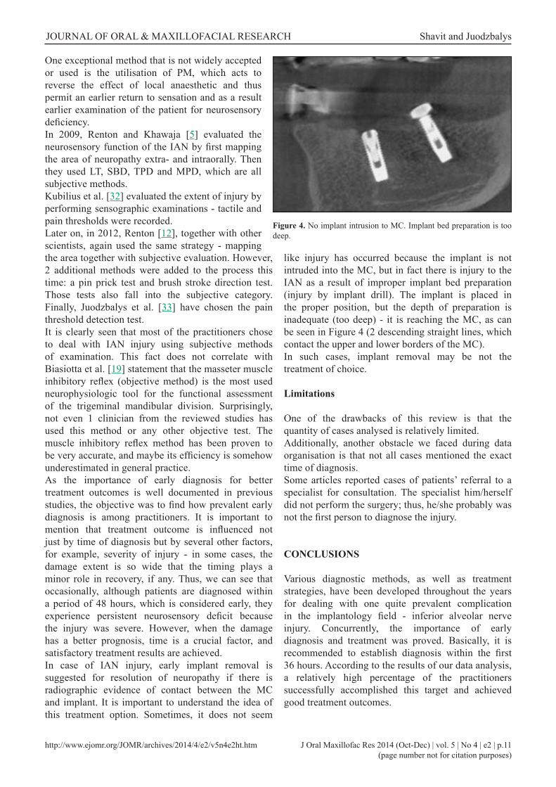
Figure 4. No implant intrusion to MC. Implant bed preparation is too deep.
like injury has occurred because the implant is not intruded into the MC, but in fact there is injury to the IAN as a result of improper implant bed preparation (injury by implant drill). The implant is placed in the proper position, but the depth of preparation is inadequate (too deep) - it is reaching the MC, as can be seen in Figure 4 (2 descending straight lines, which contact the upper and lower borders of the MC).In such cases, implant removal may be not the treatment of choice.
Limitations
One of the drawbacks of this review is that the quantity of cases analysed is relatively limited.Additionally, another obstacle we faced during data organisation is that not all cases mentioned the exact time of diagnosis.Some articles reported cases of patients’ referral to a specialist for consultation. The specialist him/herself did not perform the surgery; thus, he/she probably was not the first person to diagnose the injury.
CONCLUSIONS
Various diagnostic methods, as well as treatment strategies, have been developed throughout the years for dealing with one quite prevalent complication in the implantology field - inferior alveolar nerve injury. Concurrently, the importance of early diagnosis and treatment was proved. Basically, it is recommended to establish diagnosis within the first 36 hours. According to the results of our data analysis, a relatively high percentage of the practitioners successfully accomplished this target and achieved good treatment outcomes.
One exceptional method that is not widely accepted or used is the utilisation of PM, which acts to reverse the effect of local anaesthetic and thus permit an earlier return to sensation and as a result earlier examination of the patient for neurosensory deficiency. In 2009, Renton and Khawaja [5] evaluated the neurosensory function of the IAN by first mapping the area of neuropathy extra- and intraorally. Then they used LT, SBD, TPD and MPD, which are all subjective methods. Kubilius et al. [32] evaluated the extent of injury by performing sensographic examinations - tactile and pain thresholds were recorded.Later on, in 2012, Renton [12], together with other scientists, again used the same strategy - mapping
http://www.ejomr.org/JOMR/archives/2014/4/e2/v5n4e2ht.htm J Oral Maxillofac Res 2014 (Oct-Dec) | vol. 5 | No 4 | e2 | p.12(page number not for citation purposes)
JOURNAL OF ORAL & MAXILLOFACIAL RESEARCH Shavit and Juodzbalys
REFERENCES
1. Juodzbalys G, Wang HL, Sabalys G. Injury of the Inferior Alveolar Nerve during Implant Placement: a Literature Review. J Oral Maxillofac Res. 2011 Apr 1;2(1):e1. [URL: http://www.ejomr.org/JOMR/archives/2011/1/e1/v2n1e1ht.pdf] [Medline: 24421983] [doi: 10.5037/jomr.2011.2101]
2. Kiyak HA, Beach BH, Worthington P, Taylor T, Bolender C, Evans J. Psychological impact of osseointegrated dental implants. Int J Oral Maxillofac Implants. 1990 Spring;5(1):61-9. [Medline: 2202671]
3. Bartling R, Freeman K, Kraut RA. The incidence of altered sensation of the mental nerve after mandibular implant placement. J Oral Maxillofac Surg. 1999 Dec;57(12):1408-12. [Medline: 10596660] [doi: 10.1016/S0278-2391(99)90720-6]
4. Ellies LG, Hawker PB. The prevalence of altered sensation associated with implant surgery. Int J Oral Maxillofac Implants. 1993;8(6):674-9. [Medline: 8181830]
5. Khawaja N, Renton T. Case studies on implant removal influencing the resolution of inferior alveolar nerve injury. Br Dent J. 2009 Apr 11;206(7):365-70. [Medline: 19357667] [doi: 10.1038/sj.bdj.2009.258]
6. Renton T, Adey-Viscuso D, Meechan JG, Yilmaz Z. Trigeminal nerve injuries in relation to the local anaesthesia in mandibular injections. Br Dent J. 2010 Nov;209(9):E15. [Medline: 21072069] [doi: 10.1038/sj.bdj.2010.978]
7. Renton T, Janjua H, Gallagher JE, Dalgleish M, Yilmaz Z. UK dentists’ experience of iatrogenic trigeminal nerve injuries in relation to routine dental procedures: why, when and how often? Br Dent J. 2013 Jun;214(12):633-42. [Medline: 23787854] [doi: 10.1038/sj.bdj.2013.583]
8. Kaya Y, Sarikcioglu L. Sir Herbert Seddon (1903-1977) and his classification scheme for peripheral nerve injury. Childs Nerv Syst. 2014 Oct 1. [Epub ahead of print] [Medline: 25269543] [doi: 10.1007/s00381-014-2560-y]
9. Renton T, Yilmaz Z. Managing iatrogenic trigeminal nerve injury: a case series and review of the literature. Int J Oral Maxillofac Surg. 2012 May;41(5):629-37. Epub 2012 Feb 10. [Medline: 22326447] [doi: 10.1016/j.ijom.2011.11.002]
10. Schmid AB, Bland JD, Bhat MA, Bennett DL. The relationship of nerve fibre pathology to sensory function in entrapment neuropathy. Brain. 2014 Dec;137(Pt 12):3186-99. Epub 2014 Oct 27. [Medline: 25348629] [PMC free article: 4240296] [doi: 10.1093/brain/awu288]
11. Al-Ouf K, Salti L. Postinsertion pain in region of mandibular dental implants: a case report. Implant Dent. 2011 Feb;20(1):27-31. [Medline: 21278524] [doi: 10.1097/ID.0b013e3182096c94]
12. Renton T, Dawood A, Shah A, Searson L, Yilmaz Z. Post-implant neuropathy of the trigeminal nerve. A case series. Br Dent J. 2012 Jun 8;212(11):E17. [Medline: 22677874] [doi: 10.1038/sj.bdj.2012.497]
13. Poort LJ, van Neck JW, van der Wal KG. Sensory testing of inferior alveolar nerve injuries: a review of methods used in prospective studies. J Oral Maxillofac Surg. 2009 Feb;67(2):292-300. Review. [Medline: 19138602] [doi: 10.1016/j.joms.2008.06.076]
14. Loescher AR, Smith KG, Robinson PP. Nerve damage and third molar removal. Dent Update. 2003 Sep;30(7):375-80, 382. Review. [Medline: 14558203]
15. Ylikontiola L, Vesala J, Oikarinen K. Repeatability of 5 clinical neurosensory tests used in orthognathic surgery. Int J Adult Orthodon Orthognath Surg. 2001;16(1):36-46. [Medline: 11563394]
16. Kawamura P, Wessberg GA. Normal trigeminal neurosensory responses. Hawaii Dent J. 1985 Apr;16(4):8-11. [Medline: 3864769]
17. Ylikontiola L, Kinnunen J, Oikarinen K. Comparison of different tests assessing neurosensory disturbances after bilateral sagittal split osteotomy. Int J Oral Maxillofac Surg. 1998 Dec;27(6):417-21. [Medline: 9869278] [doi: 10.1016/S0901-5027(98)80028-3]
18. Walter JM Jr, Gregg JM. Analysis of postsurgical neurologic alteration in the trigeminal nerve. J Oral Surg. 1979 Jun;37(6):410-4. [Medline: 220400]
19. Biasiotta A, Cascone P, Cecchi R, Cruccu G, Iannetti G, Mariani A, Spota A, Truini A. Iatrogenic damage to the mandibular nerves as assessed by the masseter inhibitory reflex. J Headache Pain. 2011 Aug;12(4):485-8. [Medline: 21660431] [PMC free article: 3139056] [doi: 10.1007/s10194-011-0354-0]
20. Nakagawa K, Ueki K, Takatsuka S, Takazakura D, Yamamoto E. Somatosensory-evoked potential to evaluate the trigeminal nerve after sagittal split osteotomy. Oral Surg Oral Med Oral Pathol Oral Radiol Endod. 2001 Feb;91(2): 146-52. [Medline: 11174589] [doi: 10.1067/moe.2001.112331]
21. Jones DL, Wolford LM, Hartog JM. Comparison of methods to assess neurosensory alterations following orthognathic surgery. Int J Adult Orthodon Orthognath Surg. 1990;5(1):35-42. [Medline: 2373911]
ACKNOWLEDGMENTS AND DISCLOSURE STATEMENTS
The authors report no conflicts of interest related to this study.
http://www.ejomr.org/JOMR/archives/2014/4/e2/v5n4e2ht.htm J Oral Maxillofac Res 2014 (Oct-Dec) | vol. 5 | No 4 | e2 | p.13(page number not for citation purposes)
JOURNAL OF ORAL & MAXILLOFACIAL RESEARCH Shavit and Juodzbalys
22. Jääskeläinen SK, Peltola JK, Forssell K, Vähätalo K. Evaluating function of the inferior alveolar nerve with repeated nerve conduction tests during mandibular sagittal split osteotomy. J Oral Maxillofac Surg. 1995 Mar;53(3):269-79. [Medline: 7861277] [doi: 10.1016/0278-2391(95)90223-6]
23. Jääskeläinen SK, Teerijoki-Oksa T, Forssell K, Vähätalo K, Peltola JK, Forssell H. Intraoperative monitoring of the inferior alveolar nerve during mandibular sagittal-split osteotomy. Muscle Nerve. 2000 Mar;23(3):368-75. [Medline: 10679713] [doi: 10.1002/(SICI)1097-4598(200003)23:3<368::AID-MUS8>3.0.CO;2-0]
24. Jääskeläinen SK, Peltola JK. Clinical application of the blink reflex with stimulation of the mental nerve in lesions of the inferior alveolar nerve. Neurology. 1994 Dec;44(12):2356-61. [Medline: 7991126] [doi: 10.1212/WNL.44.12.2356]
25. Jääskeläinen SK. Blink reflex with stimulation of the mental nerve. Methodology, reference values, and some clinical vignettes. Acta Neurol Scand. 1995 Jun;91(6):477-82. [Medline: 7572043] [doi: 10.1111/j.1600-0404.1995.tb00449.x]
26. Kim YT, Pang KM, Jung HJ, Kim SM, Kim MJ, Lee JH. Clinical outcome of conservative treatment of injured inferior alveolar nerve during dental implant placement. J Korean Assoc Oral Maxillofac Surg. 2013 Jun;39(3):127-33. Epub 2013 Jun 25. [Medline: 24471030] [PMC free article: 3858167] [doi: 10.5125/jkaoms.2013.39.3.127]
27. Froum SJ, Froum SH, Malamed SF. The use of phentolamine mesylate to evaluate mandibular nerve damage following implant placement. Compend Contin Educ Dent. 2010 Sep;31(7):520, 522-8. [Medline: 20879205]
28. Chien PF, Khan KS, Siassakos D. Registration of systematic reviews: PROSPERO. BJOG. 2012 Jul;119(8):903-5. [Medline: 22703418] [doi: 10.1111/j.1471-0528.2011.03242.x]
29. Moher D, Liberati A, Tetzlaff J, Altman DG; PRISMA Group. Preferred reporting items for systematic reviews and meta-analyses: the PRISMA statement. PLoS Med. 2009 Jul 21;6(7):e1000097. Epub 2009 Jul 21. [Medline: 19621072] [PMC free article: 2707599] [doi: 10.1371/journal.pmed.1000097]
30. Higgins JPT, Green S. Cochrane Handbook for Systematic Reviews of Interventions. [URL: http://www.cochrane.org/cochrane-interventions-handbook]
31. Deppe H, Mücke T, Wagenpfeil S, Kesting M, Linsenmeyer E, Tölle T. Trigeminal nerve injuries after mandibular oral surgery in a university outpatient setting-a retrospective analysis of 1,559 cases. Clin Oral Investig. 2014 Mar 16. [Epub ahead of print] [Medline: 24633652] [doi: 10.1007/s00784-014-1222-5]
32. Kubilius R. Sabalys G, Juodzbalys G, Gedrimas V. Traumatic Damage to the Inferior Alveolar Nerve Sustained in Course of Dental Implantation. Possibility of Prevention. Stomatologija, Baltic Dent Maxillofac J.2004; 6:106-10.
33. Juodzbalys G, Wang HL, Sabalys G, Sidlauskas A, Galindo-Moreno P. Inferior alveolar nerve injury associated with implant surgery. Clin Oral Implants Res. 2013 Feb;24(2):183-90. Epub 2011 Nov 1. [Medline: 22092662] [doi: 10.1111/j.1600-0501.2011.02314.x]
34. Chiapasco M, Gatti C. Implant-retained mandibular overdentures with immediate loading: a 3- to 8-year prospective study on 328 implants. Clin Implant Dent Relat Res. 2003;5(1):29-38. [Medline: 12831726] [doi: 10.1111/j.1708-8208.2003.tb00179.x]
35. Visser A, Raghoebar GM, Meijer HJ, Batenburg RH, Vissink A. Mandibular overdentures supported by two or four endosseous implants. A 5-year prospective study. Clin Oral Implants Res. 2005 Feb;16(1):19-25. [Medline: 15642027] [doi: 10.1111/j.1600-0501.2004.01085.x]
36. Alhassani AA, AlGhamdi AS. Inferior alveolar nerve injury in implant dentistry: diagnosis, causes, prevention, and management. J Oral Implantol. 2010;36(5):401-7. Epub 2010 Jun 14. [Medline: 20545547] [doi: 10.1563/AAID-JOI-D-09-00059]
To cite this article:Shavit I, Juodzbalys G. Inferior Alveolar Nerve Injuries Following Implant Placement - Importance of Early Diagnosis and Treatment: a Systematic Review.J Oral Maxillofac Res 2014;5(4):e2URL: http://www.ejomr.org/JOMR/archives/2014/4/e2/v5n4e2ht.pdfdoi: 10.5037/jomr.2014.5402
This is an open-access article, first published in the JOURNAL OF ORAL & MAXILLOFACIAL RESEARCH, distributed under the terms of the Creative Commons Attribution-Noncommercial-No Derivative Works 3.0 Unported License, which permits unrestricted non-commercial use, distribution, and reproduction in any medium, provided the original work and is properly cited. The copyright, license information and link to the original publication on (http://www.ejomr.org) must be included.