Inferior frontal and insular cortical thinning is related to dysfunctional brain activation/deactivation during working memory task in schizophrenic patients Núria Pujol a,b,d , Rafael Penadés a,b,c,d,n , Giuseppina Rametti a , Rosa Catalán a,b,c,d , Didac Vidal-Piñeiro a , Eva Palacios a,b , Núria Bargallo a,b , Miquel Bernardo a,b,c,d , Carme Junqué a,b a Department of Psychiatry and Clinical Psychobiology, Faculty of Medicine, University of Barcelona, C/Casanova 143, 08036 Barcelona, Spain b Institut d'Investigacions Biomèdiques August Pi i Sunyer (IDIBAPS), C/ Villarroel 170, 08036 Barcelona, Spain c Centre for Biomedical Research on Mental Health (CIBERSAM), Instituto de Salud Carlos III, Barcelona, Spain d Clinical Institute of Neurosciences (ICN), Hospital Clinic, C/ Villarroel 170, 08036 Barcelona, Spain article info Article history: Received 16 July 2012 Received in revised form 27 May 2013 Accepted 14 June 2013 Keywords: Schizophrenia Independent component analysis Cortical thickness Default mode network Central executive network Salience network abstract Although working memory is known to be impaired in schizophrenia the anatomical and functional relationships underlying this deficit remain to be elucidated. A combined imaging approach involving functional and structural magnetic resonance techniques was used, applying independent component analysis and surface-based morphometry to 14 patients with schizophrenia and 14 healthy controls. Neurocognitive functioning was assessed by a neuropsychological test battery that measured executive function. It was hypothesized that working memory dysfunctional connectivity in schizophrenia is related to underlying anatomical abnormalities. Patients with schizophrenia showed cortical thinning in the left inferior frontal gyrus and insula, which explained 57% of blood oxygenation level-dependent signal magnitude in functional magnetic resonance imaging in the central executive network (lateral prefrontal and parietal cortex) over-activation and default mode network (anterior and posterior cingulate) deactivation. No structure-function relationship emerged in the healthy control group. The study provides evidence to suggest that dysfunctional activation/deactivation patterns in schizophrenia may be explained in terms of underlying gray matter deficits. & 2013 Elsevier Ireland Ltd. All rights reserved. 1. Introduction Working memory is a critical ability that is required for many higher cognitive functions such as problem-solving, decision- making or planning, and it has been shown to be impaired in schizophrenia. Therefore, a main body of research has attempted to elucidate the underlying neural substrates (Barch et al., 2009; Bledowski et al., 2010). Working memory performance usually involves the recruit- ment of the lateral prefrontal cortex (dorsolateral and ventrolater- all prefrontal cortex), bilateral anterior cingulate and the inferior parietal lobe, cerebral regions that make up what is known as the central executive network (CEN) (Seeley et al., 2007; Bressler and Menon, 2010; Palaniyappan et al., 2011). The CEN has consistently been shown to be dysfunctional in patients with schizophrenia and in their first-degree relatives (Callicott et al., 2003; Barch and Csernansky, 2007). The dysfunctional prefrontal/parietal pattern in schizophrenia includes both hyper- and hypo-activation (Callicott et al., 2003; Tan et al., 2006), depending on modulating variables such as the information load (Metzak et al., 2012), task perfor- mance (Callicott et al., 2003), specific components (encoding/ maintenance) (Anticevic et al., 2013) or heterogeneity of the samples (Manoach, 2003). In the context of low accuracy in schizophrenia the prefrontal over-activation pattern has been interpreted as reflecting the underlying psysiological inefficiency (greater degree of activation is required to perform at the same or lower level) of the neural system. This overactivation is then considered compensatory in nature (Callicott et al., 2000; Callicott et al., 2003; Tan et al., 2006). Recently, working memory performance has also been related to the default mode network (DMN), a rest-related network that comprises a set of brain regions including the anterior and posterior cingulate, medial prefrontal gyrus, parahippocampus, Contents lists available at ScienceDirect journal homepage: www.elsevier.com/locate/psychresns Psychiatry Research: Neuroimaging 0925-4927/$ - see front matter & 2013 Elsevier Ireland Ltd. All rights reserved. http://dx.doi.org/10.1016/j.pscychresns.2013.06.008 Abbreviations: DMN, default mode network; CEN, central executive network. n Corresponding author at: Clinical Institute of Neurosciences (ICN), Hospital Clinic, C/ Villarroel 170, 08036 Barcelona, Spain. Tel.: +34 93 227 54 00; fax: +34 93 403 52 94. E-mail addresses: [email protected], [email protected] (R. Penadés). Psychiatry Research: Neuroimaging 214 (2013) 94–101
Inferior frontal and insular cortical thinning is related to dysfunctionalbrain activation/deactivation during working memory task inschizophrenic patients
Núria Pujol a,b,d, Rafael Penadés a,b,c,d,n, Giuseppina Rametti a, Rosa Catalán a,b,c,d,Didac Vidal-Piñeiro a, Eva Palacios a,b, Núria Bargallo a,b,Miquel Bernardo a,b,c,d, Carme Junqué a,b
a Department of Psychiatry and Clinical Psychobiology, Faculty of Medicine, University of Barcelona, C/Casanova 143, 08036 Barcelona, Spainb Institut d'Investigacions Biomèdiques August Pi i Sunyer (IDIBAPS), C/ Villarroel 170, 08036 Barcelona, Spainc Centre for Biomedical Research on Mental Health (CIBERSAM), Instituto de Salud Carlos III, Barcelona, Spaind Clinical Institute of Neurosciences (ICN), Hospital Clinic, C/ Villarroel 170, 08036 Barcelona, Spain
a r t i c l e i n f o
Article history:Received 16 July 2012Received in revised form27 May 2013Accepted 14 June 2013
Although working memory is known to be impaired in schizophrenia the anatomical and functionalrelationships underlying this deficit remain to be elucidated. A combined imaging approach involvingfunctional and structural magnetic resonance techniques was used, applying independent componentanalysis and surface-based morphometry to 14 patients with schizophrenia and 14 healthy controls.Neurocognitive functioning was assessed by a neuropsychological test battery that measured executivefunction. It was hypothesized that working memory dysfunctional connectivity in schizophrenia isrelated to underlying anatomical abnormalities. Patients with schizophrenia showed cortical thinning inthe left inferior frontal gyrus and insula, which explained 57% of blood oxygenation level-dependentsignal magnitude in functional magnetic resonance imaging in the central executive network (lateralprefrontal and parietal cortex) over-activation and default mode network (anterior and posteriorcingulate) deactivation. No structure-function relationship emerged in the healthy control group. Thestudy provides evidence to suggest that dysfunctional activation/deactivation patterns in schizophreniamay be explained in terms of underlying gray matter deficits.
& 2013 Elsevier Ireland Ltd. All rights reserved.
1. Introduction
Working memory is a critical ability that is required for manyhigher cognitive functions such as problem-solving, decision-making or planning, and it has been shown to be impaired inschizophrenia. Therefore, a main body of research has attemptedto elucidate the underlying neural substrates (Barch et al., 2009;Bledowski et al., 2010).
Working memory performance usually involves the recruit-ment of the lateral prefrontal cortex (dorsolateral and ventrolater-all prefrontal cortex), bilateral anterior cingulate and the inferiorparietal lobe, cerebral regions that make up what is known as thecentral executive network (CEN) (Seeley et al., 2007; Bressler and
Menon, 2010; Palaniyappan et al., 2011). The CEN has consistentlybeen shown to be dysfunctional in patients with schizophreniaand in their first-degree relatives (Callicott et al., 2003; Barch andCsernansky, 2007). The dysfunctional prefrontal/parietal pattern inschizophrenia includes both hyper- and hypo-activation (Callicottet al., 2003; Tan et al., 2006), depending on modulating variablessuch as the information load (Metzak et al., 2012), task perfor-mance (Callicott et al., 2003), specific components (encoding/maintenance) (Anticevic et al., 2013) or heterogeneity of thesamples (Manoach, 2003). In the context of low accuracy inschizophrenia the prefrontal over-activation pattern has beeninterpreted as reflecting the underlying psysiological inefficiency(greater degree of activation is required to perform at the same orlower level) of the neural system. This overactivation is thenconsidered compensatory in nature (Callicott et al., 2000;Callicott et al., 2003; Tan et al., 2006).
Recently, working memory performance has also been relatedto the default mode network (DMN), a rest-related network thatcomprises a set of brain regions including the anterior andposterior cingulate, medial prefrontal gyrus, parahippocampus,
N. Pujol et al. / Psychiatry Research: Neuroimaging 214 (2013) 94–101 95
inferior parietal lobule and precuneus (Harrison et al., 2007).Previous studies have shown that working memory performanceis accompanied by deactivation of the DMN in healthy population.In schizophrenia, there are several studies reporting altered DMNdeactivation during a wide range of working memory tasks, itbeing suggested that these alterations might actively interferewith patients' performance (Harrison et al., 2007; Pomarol-Clotetet al., 2008; Kim et al., 2009; Whitfield-Gabrieli et al., 2009;Anticevic et al., 2011).
There is also a growing body of research focused on thestructure-function relationship underlying impaired cognition inschizophrenia. This is a crucial question, since gray matterabnormalities are a core feature of schizophrenia, and someevidence suggests that they may progress over time (Olabi et al.,2011), especially in poor outcome patients (Hulshoff Pol and Kahn,2008). Moreover, structural abnormalities may be the cause ofcertain deficits in brain activation. For instance, DMN dysfunc-tional connectivity has been related to gray matter volumereductions in medial cortical regions that overlapping withseveral nodes of this network (Salgado-Pineda et al., 2011). Inaddition, altered functional and anatomical connectivity in medialfrontal and anterior cingulate gyrus have been related to cognitiveimpairments in schizophrenia (Pomarol-Clotet et al., 2010;Camchong et al., 2011). Volume reductions may also be due toabnormalities in the cortical surface area and/or cortical thick-ness, which are not only driven by different cellular mechanismsbut also have distinct genetic aetiologies (Panizzon et al., 2009;Fischl et al., 2009). Previous studies using surface-based morpho-metry (SBM), a method that allows the independent measure-ment of cortical area and thickness, have found that patients withschizophrenia showed prefrontal cortical thinning (van Harenet al., 2011; Schultz et al., 2012), which in turn has been linkedto impaired executive functions (Crespo-Facorro et al., 2011).
Findings of these studies suggest that structural abnormalitiesmay be one explanation for the differences in the dysfunctionalactivation/deactivation pattern associated to working memory inpatients with schizophrenia. However, to our knowledge, theassociation between cortical thickness and functional CEN/DMNhas not yet been investigated. The present study combined corticalthickness and functional magnetic resonance imaging in a homo-geneous sample of young adult patients with severe negativesymptoms and age/gender matched healthy controls. The study isbased on two main hypotheses. First, we expect that patients willshow a dysfunctional CEN and DMN during the N-back task.Second, we expect that the activation/deactivation alteration willbe related to cortical thinning within working memory relevantfrontal areas.
Table 1Demographic and clinical variables of the sample
Demographic and clinical variables Schizophrenia group
Mean (S.D.)
Sample size 14Age 29.92 (7.17)Years of education 11.14 (2.742)Gender (F/M) 3/11Handedness (R/L) 14/0Length of illness (years) 8.64 (5.03)Number of hospitalizations 2.21 (1.57)Clorpromazine equivalents (mgr) 255.71 (142.36)PANSS
The study was approved by the Ethics Committee at the Hospital Clinic,Barcelona. All participants gave their written informed consent following acomprehensive description of the study.
2.1. Subjects
The demographic characteristics of schizophrenic patients (n¼14) and healthycomparison subjects (n¼14) are shown in Table 1. There were no significantdifferences between the groups in age or gender. All patients were recruited fromthe Psychiatry Department at the Hospital Clinic (Barcelona) and met DSM-IV-TRcriteria for schizophrenia. Psychotic exacerbation or changes in medication dosesduring the previous month were both exclusion criteria. All patients were takingantipsychotic medication, with the mean daily dose (in chlorpromazine equiva-lents) being 255.71 mg. In the patient group positive symptoms were assessedusing the Spanish adaptation of the Positive and Negative Syndrome Scale (PANSS)(Kay et al., 1990). The corresponding scores are summarized in Table 1. The healthycomparison subjects were screened for the presence of lifetime Axis I psychotic ormood disorders using the SCID non-patient version and for the presence of a first-degree relative with schizophrenia. None of the patients or controls had a history ofneurological or significant medical illness, or recent substance abuse.
2.2. Neuropsychological assessment
Patients and controls underwent a neuropsychological assessment in order toestimate their pre-morbid IQ and establish their cognitive status in global executivefunctioning. All subjects completed the neuropsychological battery (Table 2).
2.3. Procedure
All subjects underwent functional and structural MRI scanning in a singlesession, using the same 3 T TIM TRIO scanner (Siemens, Germany) at the HospitalClinic (Barcelona).
2.3.1. Experimental setup and n-back and resting-state paradigmWe used a block-design n-back working memory fMRI paradigm, including two
levels of cognitive load (2-back and 0-back). Short resting periods (in which a whitecross was shown on a black screen) were introduced after each 2-back condition.Each block consisted of 0-back/2-back/rest (90-s duration). The sequence consistedof a total of six blocks (see supplementary material for a detailed description ofthe task).
2.3.2. MRI dataFor each participant, functional and structural imaging data were acquired on a
3TMRI scanner (Magnetom Trio Tim, Siemens Medical Systems, Germany). DuringfMRI a T2n-weighted GE-EPI sequence depicting blood-oxygenation-level-dependent (BOLD) contrast was used, and 280 volumes were acquired(TR¼2000 ms, TE¼29 ms, flip angle¼901, slice thickness¼3 mm, distance factor¼25%, FOV¼240 mm, matrix size¼128�128) providing whole brain coverage.
A T1-weighted structural image was also acquired for each subject withMPRAGE 3D protocol (TR¼2300 ms, TE¼2.98 ms, TI¼900 ms, 240 slices, slicethickness¼1 mm, FOV¼256 mm, matrix size¼256�256).
WAIS-III: Wechsler Adult Intelligence Scale, Third Edition. WMS-III: Wechsler Memory Scale, Third Edition. TMT-A: Trail Making Test, part A.TMT-B: Trail Making Test, part B. WCST: Wisconsin Card Sorting Test. TOL: Tower of London.
N. Pujol et al. / Psychiatry Research: Neuroimaging 214 (2013) 94–10196
2.3.3. fMRI data pre-processing and analysisThe following data pre-processing was carried out on the fMRI data set using
the FMRIB Software Library (http://www.fmrib.ox.ac.uk/fsl): motion correctionusing MCFLIRT (Jenkinson et al., 2002), removal of non-brain structures from theechoplanar imaging volumes using BET (Smith, 2002), spatial smoothing using aGaussian kernel of 5 mm FWHM, and mean-based intensity normalization of allvolumes by the same factor (4D grand-mean). Task-activation functional data werefiltered using a high-pass filter of 90 s. The functional scans were registered toMNI152 standard space (4 mm resample resolution) by using affine registrationwith FLIRT (Jenkinson and Smith, 2001).
After this pre-processing, fMRI analysis was carried out using TensorialIndependent Component Analysis (TICA) as implemented in the MultivariateExploratory Linear Decomposition into Independent Components (MELODIC Ver-sion 3.1) (Beckmann and Smith, 2004) tool, part of FSL software. TICA is a data-driven approach that decomposes the data into a set of Independent Components(ICs) where each IC is composed of a spatial map, a time-course and a vector ofsubject modes, which quantifies the strength of both activations and deactivations.Higher subject mode values indicate higher activations and higher deactivations ofthe positive and negative parts of an IC, respectively. Model order was estimatedusing the Laplace approximation to the Bayesian evidence for a probabilistic PCAmodel (Beckmann and Smith, 2005). Estimated Component maps were divided bythe standard deviation of the residual noise and thresholded by fitting a mixturemodel to the histogram of intensity values. Z (Gaussianised T/F) statistic imageswere thresholded using clusters determined by Z≥2.3 and a cluster significancethreshold of p≤0.05 corrected for multiple comparisons.
2.3.4. Measurement of cortical thicknessAdvances in MR image analysis algorithms have led to the development of
automated parcellation tools which can segment the whole brain into anatomicregions and quantify the features of each region (Desikan et al., 2006). TheFreeSurfer software package (version 4.3.1, available at: http://surfer.nmr.harvard.edu; Fischl et al., 1999; Fischl and Dale, 2000) was applied to each participant's pre-processed scan. The implemented processing stream involved: removal of non-brain tissue; transformation to the Talairach reference space; segmentation intoGM and WM (Dale et al., 1999); correction of topological defects (Fischl et al.,2001); intensity normalization (Dale et al., 1999); tissue segmentation (subcorticalstructures, brain stem, cerebellum and cerebral cortex) (Fischl et al., 2002; Fischlet al., 2004); automated correction of topology defects; surface deformation toform the gray/white matter boundary and gray matter/CSF boundary (Fischl et al.,2004); and parcellation of the cerebral cortex (Desikan et al., 2006). All surfacemaps models in our study were visually inspected and corrected by adjusting inputparameters to the skull stripping step for accuracy. Surface maps were smoothedwith a full-width-half-maximum Gaussian kernel of 15 mm for the entire cortexanalysis. An ROI was automatically outlined on the cortical surface template of theFreesurfer including all the vertices in which we find statistical differences below po0.005.
2.4. Statistical procedures
The general linear model (GLM) was used to identify brain regions whereschizophrenia patients showed differences in cortical thickness compared tocontrols. The left and right hemisphere cortical surface was analyzed separately.All cortical thickness results were corrected for multiple comparisons using aMonte-Carlo simulation. The initial cluster forming threshold was set at po0.05.Clusters were then tested against an empirical null distribution of maximumcluster size built using synthesized Z distributed data across 10,000permutations.
2.4.1. MRI correlation analysisThe most important goal of this study was to determine how cortical
thickness deficits might be related to dysfunctional patterns of brain activationin schizophrenia. To this end we performed a simple regression analysis. First,we extract the mean values of the ROI defined on the contiguous areas ofsignificant cortical thinner in the schizophrenia group compared to healthycontrols. These values were entered as a predictor for the subject modes (themean BOLD response for each subject within the IC-1, including both, activationand deactivation). To avoid the age confounding effect, the regression model wasweighted by this variable.
Since the small sample of the present study, the effect size and power analysiswere also reported.
2.4.2. Correlation analysis between MRI and cognitive and clinical variablesIn order to test the relationship between cognitive deficits and cortical
thinning in schizophrenia patients, a study of neuropsychological performance(Similarities, Digit span, Logical memory I, II and Verbal fluency), and corticalthickness values extracted from the values provided by FreeSurfer subjectsparcelations was carried out by correlation analyses using non-parametricstatistics (Spearman's rho). Bonferroni correction was applied (po0.05/5) (i.e.po0.01).In the same way, an exploratory study of the relationships betweenclinical variables (age of onset/duration of the illness, number of hospitalizations,antipsychotic dosage, PANSS) and cortical it was carried out to test if clinicalseverity is associated with strong brain alterations. Also, Bonferroni correctionwas used (po0.05/5, i.e. po0.01).
2.4.3. Non-imaging statistical analysesNon-imaging statistical analyses were conducted using the Student's t-test for
normally-distributed quantitative variables and the Pearson χ2 test for categoricalvariables. All statistical analyses were carried out using SPSS v. 18.0 (SPSS Inc.,Chicago, IL).
N. Pujol et al. / Psychiatry Research: Neuroimaging 214 (2013) 94–101 97
3. Results
3.1. Demographic findings
Demographic findings are summarized in Table 1. There wereno significant differences between patients and controls in termsof sex or age. However, and as expected, patients had a lowereducational level (years of education) than did healthy controls.
3.2. Behavioral data
3.2.1. N-back taskThere were no significant differences between the groups on
the 0-back and 2-back tasks (Table 2).
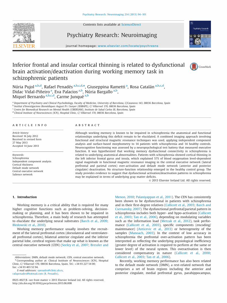
Fig. 1. A 3D view of the inflated (top) and pial surface (down) is shown in thisfigure. Compared to the healthy control group, patients with schizophrenia showabnormally reduced cortical thickness in the left inferior frontal gyrus (parsopercularis/BA 44), insular and precentral cortex (po0.005).
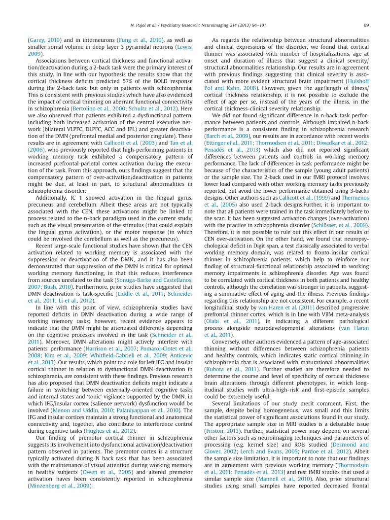
Fig. 2. Independent Component (IC)-1 specific component of 2 back task evidences a signnetwork) represent activation pattern during 2 back task (overactive in patients). Dark to(and increased deactivation in patients) during task.
3.2.2. Executive functionThe schizophrenic group performed significantly worse than
controls on all the tests administered (po0.005) (Table 2).
3.3. Anatomical results
Cortical thickness analyses revealed that patients showedthinner cortex in the left inferior frontal gyrus (pars opercularis/BA 44) compared with controls. In the same hemisphere, we alsoobserved that patients showed a thinner insular cortex andthinner cortex in the precentral gyrus than controls (po0.005).Results are shown in Fig. 1.
No significant results were identified in the right hemisphere.
3.4. fMRI results: independent component analysis
3.4.1. Between-groups comparison3.4.1.1. Differences between patients and controls during taskcondition. Compared to controls, patients with schizophreniashowed IC1 task-related over-activation in the bilateral middleand inferior frontal gyrus (IFG), left medial frontal gyrus, bilateralinferior and superior parietal lobule, bilateral precuneus and bilateralmiddle occipital gyrus, which together comprise the central executivenetwork (CEN). Patients also showed significantly increased DMN hubdeactivation (bilateral anterior and posterior cingulate) in comparisonwith controls (po0.011) (see Fig. 2, Table 3 and e-results).
3.5. Correlation between IFG structural deficits and ICA data
To examine further the relationship between brain activation/deactivation and the anatomical data, regression analyses wereperformed on the IC 1 subject mode (mean BOLD response of theIC-1 activations and deactivations of each subject) using thecortical thickness values (mean values of the ROI) as a predictorand age as a covariate. The results showed that the magnitude ofthe IC1 BOLD signal was modulated by the structural abnormal-ities. In patients, 57% of fMRI BOLD signal magnitude in the IC1CEN activation and DMN deactivation was predicted by corticalthickness deficits (R¼0.577; t¼2.235; po0.04), (f²¼0.499; and0.43 chance of Type II error) (Fig. 3). This structural/functionalcorrelation was not found in the healthy control group (R¼0.23;t¼0.762; p¼0.464) (See Table 4). Furthermore, we also carried outan ANOVA analysis in the combined sample (schizophrenia andhealthy control) to test the group x cortical thickness interactioneffect. This model (F¼2146, p¼0,123) showed not significantgroup effect (F¼1295, p¼0,267) and a tendency towards
ificant group difference (Po0.001). Red and yellow colored areas (central executivelight blue colors (default mode network) represent a deactivation pattern of the IC 1
Table 3Anatomical specifications are provided for significant differences of IndependentComponent (IC-1) between groups.
nts with schizophrenia showed the IC-1 over-activation/deactivation pattern during then-back task compared with healthy control subjects. Peak activations of each cluster arespecified by their anatomical site, Talairach coordinate and z-value.
Fig. 3. Scatter plot of subject mode IC-1 (mean BOLD response for each subject,including both activation and deactivation) and cortical thickness (mean values ofthe ROI defined on the contiguous areas of significant cortical thinner) forschizophrenic patients.
Table 4Summary of simple regression analyses for cortical thickness predicting centralexecutive network activation and default mode network deactivation in schizo-phrenia and healthy control groups weighted by age
Variable Schizophrenia group Healthy control group
N. Pujol et al. / Psychiatry Research: Neuroimaging 214 (2013) 94–10198
significance in the group x cortical thickness interaction effect(F¼3191, p¼0.061), which suggest the specificity of functional-anatomical relationship in the schizophrenia group.
3.6. Correlation between IFG structural deficits andneuropsychological, clinical and demographic variables
Correlations between cognitive performance and structuralmeasures were investigated for schizophrenia sample. After apply-ing Bonferroni correction (Po0.01), we found that thinner cortexwas correlated with deficits in Digit span (rho¼�0,679, p¼0,008)(see Supplementary material, e-Figure).
We evaluated the association between the anatomical data andclinical variables (number of hospitalizations, PANSS, age of onset/duration of the illness), and found that thinner cortex wascorrelated with number of hospitalizations (rho¼�0.733; po0.001), age at onset (rho¼�0.517 p¼0.029) and duration of theillness (rho¼�0.776; po0.001) (corrected for multiple compar-isons), suggesting that clinical severity is associated with greaterstructural abnormalities. However, a relationship between age andlength of illness (rho¼0,800, p¼0.001) as well as age and corticalthickness (rho¼�0.824; p¼0.001) were also found. Therefore, itis not possible to exclude the effect of age per se, instead of theduration of the illness, in the cortical thickness-clinical severityrelationship.
Since antipsychotic agents might contribute to anatomicalchanges found in schizophrenia we evaluated this potential con-founding variable, the results showing a non-significant correlationbetween cortical thinning and antipsychotic dosage (in chlorpro-mazine equivalents) (rho¼�0.078; po0.396).
Regarding the role of demographic variables (age, years ofeducation and gender) on the functional/structural relationship,the effect of age on cortical thickness was observed in bothpatients (rho¼�0.824; po0.001) and healthy controls (rho¼�0.510; p¼0.037). Correlation between cortical thickness andyears of education was not significant in any groups schizophrenicpatients (rho¼�0.060, p¼0.837) and healthy control group(rho¼0.058, p¼0.850). Furthermore, to exclude the role of thesepotential confounding variables, we carried out a partial correla-tion between smode-IC1 CEN and cortical thickness values withgender and years of education as covariates in schizophrenia andhealthy control group. The results corroborated the functional-structural relationship in schizophrenia (r¼0.705, p¼0.010) aswell as its absence in healthy control group (r¼0.511, p¼0.131).
4. Discussion
The present study is the first to report evidence of thinnercortex in the left inferior frontal gyrus (IFG), insula and precentralgyrus, which is associated with the dysfunctional pattern ofCentral executive network (CEN) activation and Default modenetwork (DMN) deactivation during working memory.
Patients with schizophrenia showed specific thinner cortex inthe left IFG (pars opercularis/BA 44), insular cortex and precentralgyrus. This finding is in line with growing evidence of corticalthickness alterations in these regions, which have also beenreported in first-episode psychosis (Takayanagi et al., 2011;Schultz et al., 2012), along with the gray matter reductions observedin classical VBM studies (Honea et al., 2005; Glahn et al., 2008).
Cortical thickness is a highly heritable structural brain mea-surement that is assumed to reflect the size, arrangement anddensity of neurons, inhibitory interneurons, glial cells and unmye-linated neuron processes (dendrites, dendritic spines and axons),referred to as neuropil (Panizzon et al., 2009). Therefore, the leftIFG/insular/precentral cortical thinner observed here may imply adisruption of the anatomical configuration in one or a combinationof these components. In this context, previous research hasevidenced prefrontal loss of dendritic spines in chronic patients
N. Pujol et al. / Psychiatry Research: Neuroimaging 214 (2013) 94–101 99
(Garey, 2010) and in interneurons (Fung et al., 2010), as well assmaller somal volume in deep layer 3 pyramidal neurons (Lewis,2009).
Associations between cortical thickness and functional activa-tion/deactivation during a 2-back task were the primary interest ofthis study. In line with our hypothesis the results show that thecortical thickness deficits predicted 57% of the BOLD responseduring the 2-back task, but only in patients with schizophrenia.This is consistent with previous studies which have also evidencedthe impact of cortical thinning on aberrant functional connectivityin schizophrenia (Bertolino et al., 2000; Schultz et al., 2012). Herewe also observed that patients exhibited a dysfunctional pattern,including both increased activation of the central executive net-work (bilateral VLPFC, DLPFC, ACC and IPL) and greater deactiva-tion of the DMN (prefrontal medial and posterior cingulate). Theseresults are in agreement with Callicott et al. (2003) and Tan et al.(2006), who previously reported that high-performing patients inworking memory task exhibited a compensatory pattern ofincreased prefrontal-parietal cortex activation during the execu-tion of the task. From this approach, ours findings suggest that thecompensatory pattern of over-activation/deactivation in patientsmight be due, at least in part, to structural abnormalities inschizophrenia disorder.
Additionally, IC 1 showed activation in the lingual gyrus,precuneus and cerebellum. Albeit these areas are not typicallyassociated with the CEN, these activations might be linked toprocess related to the n-back paradigm used in the current study,such as the visual presentation of the stimulus (that could explainthe lingual gyrus activation), or the motor response (in whichcould be involved the cerebellum as well as the precuneus).
Recent large-scale functional studies have shown that the CENactivation related to working memory is associated with thesuppression or deactivation of the DMN, and it has also beendemonstrated that suppression of the DMN is critical for optimalworking memory functioning, in that this reduces interferencefrom sources unrelated to the task (Sonuga-Barke and Castellanos,2007; Bush, 2010). Furthermore, prior studies have suggested thatDMN deactivation is task-specific (Liddle et al., 2011; Schneideret al., 2011; Li et al., 2012).
In line with this point of view, schizophrenia studies havereported deficits in DMN deactivation during a wide range ofworking memory tasks; however, recent evidence appears toindicate that the DMN might be attenuated differently dependingon the cognitive processes involved in the task (Schneider et al.,2011). Moreover, DMN alterations might actively interfere withpatients' performance (Harrison et al., 2007; Pomarol-Clotet et al.,2008; Kim et al., 2009; Whitfield-Gabrieli et al., 2009; Anticevicet al., 2013). Our results, which point to a role for left IFG and insularcortical thinner in relation to dysfunctional DMN deactivation inschizophrenia, are consistent with these findings. Previous researchhas also proposed that DMN deactivation deficits might indicate afailure in ‘switching' between externally-oriented cognitive tasksand internal states and ‘tonic’ vigilance supported by the DMN, inwhich IFG/insular cortex (salience network) dysfunction would beinvolved (Menon and Uddin, 2010; Palaniyappan et al., 2010). TheIFG and insular cortices maintain a strong functional and anatomicalconnectivity and, together, also contribute to interference controlduring cognitive tasks (Hughes et al., 2012).
Our finding of premotor cortical thinner in schizophreniasuggests its involvement into dysfunctional activation/deactivationpattern observed in patients. The premotor cortex is a structuretypically activated during N back task that has been associatedwith the maintenance of visual attention during working memoryin healthy subjects (Owen et al., 2005) and altered premotoractivation haves been consistently reported in schizophrenia(Minzenberg et al., 2009).
As regards the relationship between structural abnormalitiesand clinical expressions of the disorder, we found that corticalthinner was associated with number of hospitalizations, age atonset and duration of illness that suggest a clinical severity/structural abnormalities relationship. Our results are in agreementwith previous findings suggesting that clinical severity is asso-ciated with more evident structural brain impairment (HulshoffPol and Kahn, 2008). However, given the age/length of illness/cortical thickness relationship, it is not possible to exclude theeffect of age per se, instead of the years of the illness, in thecortical thickness-clinical severity relationship.
We did not found significant difference in n-back task perfor-mance between patients and controls. Although impaired n-backperformance is a consistent finding in schizophrenia research(Barch et al., 2009), our results are in accordance with recent works(Ettinger et al., 2011; Thormodsen et al., 2011; Diwadkar et al., 2012;Penadés et al., 2013) which also did not reported significantdifferences between patients and controls in working memoryperformance. The lack of differences in task performance might bebecause of the characteristics of the sample (young adult patients)or the sample size. The 2-back used in our fMRI protocol involveslower load compared with other working memory tasks previouslyreported, but avoid the lower performance obtained using 3-backsdesigns. Other authors such as Callicott et al., (1999) and Thermenoset al., (2005) also used 2-back designs.Further, it is important tonote that all patients were trained in the task immediately before tothe scan. It has been suggested activation changes (over-activation)with the practice in schizophrenia disorder (Schlösser, et al., 2009).Therefore, it is not possible to rule out this effect in our results ofCEN over-activation. On the other hand, we found that neuropsy-chological deficit in Digit span, a test classically associated to verbalworking memory domain, was related to fronto-insular corticalthinner in schizophrenia patients, which help to reinforce ourfinding of structural-functional relationship associated to workingmemory impairments in schizophrenia disorder. Age was foundto be correlated with cortical thickness in both patients and healthycontrols, although the correlation was stronger in patients, suggest-ing a summative effect of aging and the illness. Previous findingsregarding this relationship are not consistent. For example, a recentlongitudinal study by van Haren et al. (2011) described progressiveprefrontal thinner cortex, which is in line with VBM meta-analysis(Olabi et al., 2011), in indicating a different pathologicalprocess alongside neurodevelopmental alterations (van Harenet al., 2011).
Conversely, other authors evidenced a pattern of age-associatedthinning without differences between schizophrenia patientsand healthy controls, which indicates static cortical thinning inschizophrenia that is associated with maturational abnormalities(Kubota et al., 2011). Further studies are therefore needed todetermine the course and level of specificity of cortical thicknessbrain alterations through different phenotypes, in which long-itudinal studies with ultra-high-risk and first-episode samplescould be extremely useful.
Several limitations of our study merit comment. First, thesample, despite being homogeneous, was small and this limitsthe statistical power of significant associations found in our study.The appropriate sample size in MRI studies is a debatable issue(Friston, 2013). Further, statistical power may depend on severalother factors such as neuroimaging techniques and parameters ofprocessing (e.g. kernel size) and ROIs studied (Desmond andGlover, 2002; Lerch and Evans, 2005; Pardoe et al., 2012). Albeitthe sample size limitation, it is important to note that our findingsare in agreement with previous working memory (Thormodsenet al., 2011; Penadés et al., 2013) and rest fMRI studies that used asimilar sample size (Mannell et al., 2010). Also, prior structuralstudies using small samples have reported decreased frontal
N. Pujol et al. / Psychiatry Research: Neuroimaging 214 (2013) 94–101100
volume related to attentional impairment (Salgado-Pineda et al.,2003) as well as frontal white matter abnormalities associatedwith dysfunctional networks (Jeong et al., 2009) in schizophrenia.Second, we were unable to control for potential medication effectsin patients with schizophrenia, and although we found no sig-nificant relationship between antipsychotic dosage (in chlorpro-mazine equivalents) and structural and functional MRI data, wecannot rule out the influence of medication (e.g., lifetime use ofantipsychotics) on the observed brain alterations or the functional/structural relationship found here. Third, although we report apositive correlation between structure and function in schizo-phrenia, the causal relationship remains to be demonstrated. Insum, because this is the first functional CEN/DMN-cortical thick-ness study of schizophrenia and the exploratory nature of it,replication of these findings in a larger sample is necessary inorder to validate these results.
5. Conclusions
In summary, we found that dysfunctional connectivity inschizophrenia during working memory tasks is directly relatedto underlying anatomical abnormalities. Functional networks aredynamic, and therefore over-activation/deactivation probablyreflects a compensatory brain mechanism in response to structuralcortical abnormalities. Finally, in patients with schizophrenia theIFG and insular cortical thinner appears to be involved not only intask-related activation but also in the dysfunctional DMN deacti-vation. Given the small sample size of our study, these resultsshould be considered preliminary and studies with larger samplesare needed to replicate our findings.
Acknowledgments
We thank the patients for their participation in the study. Thisstudy was partially supported by a grant from the Instituto deSalud Carlos III (FIS no PI 07/0258) and the N. Pujol grant RioHortega IDIBAPS.
Appendix A. Supporting information
Supplementary data associated with this article can be found inthe online version at http://dx.doi.org/10.1016/j.pscychresns.2013.06.008.
References
Anticevic, A., Repovs, G., Barch, D.M., 2013. Working memory encoding andmaintenance deficits in schizophrenia: neural evidence for activation anddeactivation abnormalities. Schizophrenia Bulletin 39 (1), 168–178.
Anticevic, A., Repovs, G., Corlett, P.R., Barch, D.M., 2011. Negative andnonemotional interference with visual working memory in schizophrenia.Biological Psychiatry 70 (12), 1159–1168.
Barch, D.M., Csernansky, J.G., 2007. Abnormal parietal cortex activation duringworking memory in schizophrenia: verbal phonological coding disturbancesversus domain-general executive dysfunction. American Journal of Psychiatry64 (7), 1090–1098.
Barch, D.M., Berman, M.G., Engle, R., Jones, J.H., Jonides, J., Macdonald, A., Nee, D.E.,Redick, T.S., Sponheim, S.R., 2009. CNTRICS final task selection: workingmemory. Schizophrenia Bulletin 35 (1), 136–152.
Beckmann, C.F., Smith, S.M., 2004. Probabilistic independent component analysisfor functional magnetic resonance imaging. IEEE Transactions on MedicalImaging 23 (2), 137–152.
Beckmann, C.F., Smith, S.M., 2005. Tensorial extensions of independent componentanalysis for multisubject FMRI analysis. NeuroImage 25 (1), 294–311.
Bertolino, A., Esposito, G., Callicott, J.H., Mattay, V.S., Van Horn, J.D., Frank, J.A.,Berman, K.F., Weinberger, D.R., 2000. Specific relationship between prefrontalneuronal N-acetylaspartate and activation of the working memory corticalnetwork in schizophrenia. American Journal of Psychiatry 157 (1), 26–33.
Bledowski, C., Kaiser, J., Rahm, B., 2010. Basic operations in working memory:contributions from functional imaging studies. Behavioural Brain Research 214(2), 172–179.
Bressler, S.L., Menon, V., 2010. Large-scale brain networks in cognition: emergingmethods and principles. Trends in Cognitive Science 14 (6), 277–290.
Callicott, J.H., Mattay, V.S., Bertolino, A., Finn, K., Coppola, R., Frank, J.A., Goldberg,TE, Weinberger, DR., 1999. Physiological characteristics of capacity constraintsin working memory as revealed by functional MRI. Cerebral Cortex 9 (1), 20–26.
Callicott, J.H., Bertolino, A., Mattay, V.S., Langheim, F.J., Duyn, J., Coppola, R.,Goldberg, T.E., Weinberger, D.R., 2000. Physiological dysfunction of the dorso-lateral prefrontal cortex in schizophrenia revisited. Cerebral Cortex 10 (11),1078–1092.
Callicott, J.H., Mattay, V.S., Verchinski, B.A., Marenco, S., Egan, M.F., Weinberger, D.R., 2003. Complexity of prefrontal cortical dysfunction in schizophrenia: morethan up or down. American Journal of Psychiatry 60 (12), 2209–2215.
Crespo-Facorro, B., Roiz-Santiáñez, R., Pérez-Iglesias, R., Rodriguez-Sanchez, J.M.,Mata, I., Tordesillas-Gutierrez, D., Sanchez, E., Tabarés-Seisdedos, R., Andreasen,N., Magnotta, V., Vázquez-Barquero, J.L., 2011. Global and regional corticalthinning in first-episode psychosis patients: relationships with clinical andcognitive features. Psychological Medicine 41 (7), 1449–1460.
Dale, A.M., Fischl, B., Sereno, M.I., 1999. Cortical surface-based analysis. I. Segmen-tation and surface reconstruction. NeuroImage 9, 179–194.
Desikan, R.S., Segonne, F., Fischl, B., Quinn, B.T., Dickerson, B.C., Blacker, D., Buckner,R.L., Dale, A.M., Maguire, R.P., Hyman, B.T., Albert, M.S., Killiany, R.J., 2006. Anautomated labeling system for subdividing the human cerebral cortex on MRIscans into gyral based regions of interest. NeuroImage 31, 968–980.
Desmond, J.E., Glover, G.H., 2002. Estimating sample size in functional MRI (fMRI)neuroimaging studies: statistical power analyses. Journal of NeuroscienceMethods 118 (2), 115–128.
Diwadkar, V.A., Pruitt, P., Zhang, A., Radwan, J., Keshavan, M.S., Murphy, E., Rajan, U.,Zajac-Benitez, C., 2012. The neural correlates of performance in adolescents atrisk for schizophrenia: inefficiently increased cortico-striatal responses mea-sured with fMRI. Journal of Psychiatric Research 46, 12–21.
Ettinger, U., Williams, S.C., Fannon, D., Premkumar, P., Kuipers, E., Möller, H.J.,Kumari, V., 2011. Functional magnetic resonance imaging of a parametricworking memory task in schizophrenia: relationship with performance andeffects of antipsychotic treatment. Psychopharmacology 216 (1), 17–27.
Fischl, B., Sereno, M.I., Tootell, R.B., Dale, A.M., 1999. High-resolution intersubjectaveraging and a coordinate system for the cortical surface. Human BrainMapping 8, 272–284.
Fischl, B., Dale, A.M., 2000. Measuring the thickness of the human cerebral cortexfrom magnetic resonance images. Proceedings of the National Academy ofSciences of the United States of America 97, 11050–11055.
Fischl, B., Liu, A., Dale, A.M., 2001. Automated manifold surgery: constructinggeometrically accurate and topologically correct models of the human cerebralcortex. IEEE Transactions on Medical Imaging 20, 70–80.
Fischl, B., Salat, D.H., Busa, E., Albert, M., Dieterich, M., Haselgrove, C., van derKouwe, A., Killiany, R., Kennedy, D., Klaveness, S., Montillo, A., Makris, N., Rosen,B., Dale, A.M., 2002. Whole brain segmentation: automated labeling ofneuroanatomical structures in the human brain. Neuron 33, 341–355.
Fischl, B., Salat, D.H., van der Kouwe, A.J., Makris, N., Segonne, F., Quinn, B.T., Dale, A.M., 2004. Sequence-independent segmentation of magnetic resonance images.NeuroImage 23 (1), S69–S84.
Fischl, B., Seidman, L., Dale, A., Kremen, W.S., 2009. Distinct genetic influenceson cortical surface area and cortical thickness. Cerebral Cortex 19 (11),2728–2735.
Friston, K., 2013. Sample size and the fallacies of classical inference. Neuroimage 81,503–504.
Fung, S.J, Webster, M.J., Sivagnanasundaram, S, Duncan, C, Elashoff, M, Weickert,CS., 2010. Expression of interneuron markers in the dorsolateral prefrontalcortex of the developing human and in schizophrenia. American Journal ofPsychiatry 167 (12), 1479–1488.
Garey, L., 2010. When cortical development goes wrong: schizophrenia as aneurodevelopmental disease of microcircuits. Journal of Anatomy 217 (4),324–333.
Glahn, D.C., Laird, A.R., Ellison-Wright, I., Thelen, S.M., Robinson, J.L., Lancaster, J.L.,Bullmore, E., Fox, P.T., 2008. Meta-analysis of gray matter anomalies inschizophrenia: application of anatomic likelihood estimation and networkanalysis. Biological Psychiatry 64 (9), 774–781.
Harrison, B.J., Yücel, M., Pujol, J., Pantelis, C., 2007. Task-induced deactivation ofmidline cortical regions in schizophrenia assessed with fMRI. SchizophreniaResearch 91 (1–3), 82–86.
Honea, R., Crow, T.J., Passingham, D., Mackay, C.E., 2005. Regional deficits in brainvolume in schizophrenia: a meta-analysis of voxel-based morphometry studies.American Journal of Psychiatry 162 (12), 2233–2245.
Hulshoff Pol, H.E., Kahn, R.S., 2008. What happens after the first episode? A reviewof progressive brain changes in chronically ill patients with schizophrenia.Schizophrenia Bulletin 34 (2), 354–366.
N. Pujol et al. / Psychiatry Research: Neuroimaging 214 (2013) 94–101 101
Jenkinson, M., Smith, S.M., 2001. A global optimisation method for robust affineregistration of brain images. Medical Image Analysis 5 (2), 143–156.
Jenkinson, M., Bannister, P.R., Brady, S.M., Smith, S.M., 2002. Improved optimisationfor the robust and accurate linear registration and motion correction of brainimages. NeuroImage 17 (2), 825–841.
Jeong, B., Wible, C.G., Hashimoto, R., Kubicki, M., 2009. Functional and anatomicalconnectivity abnormalities in left inferior frontal gyrus in schizophrenia.Human Brain Mapping 30 (12), 4138–4151.
Kay, S.R., Fiszbein, A., Vital-Herne, M., Fuentes, LS., 1990. The positive and negativesyndrome scale—Spanish adaptation. The Journal of Nervous and MentalDisease 178, 510–517.
Kim, D.I, Manoach, D.S, Mathalon, D.H, Turner, J.A, Mannell, M., Brown, G.G., et al.,2009. Dysregulation of working memory and default-mode networks inschizophrenia using independent component analysis, an fBIRN and MCICstudy. Human Brain Mapping 30, 3795–3811.
Kubota, M., Miyata, J., Yoshida, H., Hirao, K., Fujiwara, H., Kawada, R., Fujimoto, S.,Tanaka, Y., Sasamoto, A., Sawamoto, N., Fukuyama, H., Murai, T., 2011. Age-related cortical thinning in schizophrenia. Schizophrenia Research 125 (1),21–29.
Lerch, J.P., Evans, A.C., 2005. Cortical thickness analysis examined through poweranalysis and a population simulation. Neuroimage 24 (1), 163–173.
Lewis, D.A., 2009. Neuroplasticity of excitatory and inhibitory cortical circuits inschizophrenia. Dialogues in Clinical Neuroscience 11 (3), 269–280.
Li, B., Wang, X., Yao, S., Hu, D., Friston, K., 2012. Task-dependent modulation ofeffective connectivity within the default mode network. Frontiers in Psychology3, 206.
Liddle, E.B., Hollis, C., Batty, M.J., Groom, M.J., Totman, J.J., Liotti, M., Scerif, G., Liddle,PF., 2011. Task-related default mode network modulation and inhibitory controlin ADHD: effects of motivation and methylphenidate. Journal of Child Psycho-lology and Psychiatry 52 (7), 761–771.
Mannell, M.V., Franco, A.R., Calhoun, V.D., Cañive, J.M., Thoma, R.J., Mayer, A.R.,2010. Resting state and task-induced deactivation: a methodological compar-ison in patients with schizophrenia and healthy controls. Human BrainMapping 31 (3), 424–437.
Manoach, D.S., 2003. Prefrontal cortex dysfunction during working memoryperformance in schizophrenia: reconciling discrepant findings. SchizophreniaResearch 60 (2-3), 285–298.
Menon, V., Uddin, L.Q., 2010. Saliency, switching, attention and control: a networkmodel of insula function. Brain Structure and Function 214, 655–667.
Metzak, P.D., Riley, J.D., Wang, L., Whitman, J.C., Ngan, E.T., Woodward, T.S., 2012.Decreased efficiency of task-positive and task-negative networks during work-ing memory in schizophrenia. Schizophrenia Bulletin 38 (4), 803–813.
Minzenberg, M.J., Laird, A.R., Thelen, S., Carter, C.S., Glahn, D.C., 2009. Meta-analysisof 41 functional neuroimaging studies of executive function in schizophrenia.Archives of General Psychiatry 66 (8), 811–822.
Olabi, B., Ellison-Wright, I., McIntosh, A.M., Wood, S.J., Bullmore, E., Lawrie, S.M.,2011. Are there progressive brain changes in schizophrenia? A meta-analysis ofstructural magnetic resonance imaging studies. Biological Psychiatry 70 (1),88–96.
Owen, A.M., McMillan, K.M., Laird, A.R., Bullmore, E., 2005. N-back workingmemory paradigm: a meta-analysis of normative functional neuroimagingstudies. Human Brain Mapping 25 (1), 46–59.
Palaniyappan, L., Mallikarjun, P., Joseph, V., White, T.P., Liddle, P.F., 2010. Realitydistortion is related to the structure of the salience network in schizophrenia.Psychological Medicine 13, 1–8.
Palaniyappan, L., Mallikarjun, P., Joseph, V., White, T.P., Liddle, P.F., 2011. Regionalcontraction of brain surface area involves three large-scale networks inschizophrenia. Schizophrenia Research 129 (2-3), 163–168.
Panizzon, M.S., Fennema-Notestine, C., Eyler, L.T., Jernigan, T.L., Prom-Wormley, E.,Neale, M., Jacobson, K., Lyons, M.J., Grant, M.D., Franz, C.E., Xian, H., Tsuang, M.,2009. Distinct genetic influence on cortical surface area and cortical thickness.Cerebral Cortex 19 (11), 2728–2735.
Pardoe, H.R., Abbott, D.F., Jackson, G.D., 2012. The Alzheimer's Disease Neuroima-ging Initiative, 2012. Sample size estimates for well-powered cross-sectional
cortical thickness studies. Human Brain Mapping, electronic publication 17 July;http://dxdoi.org/10.1002/hbm.22120.
Penadés, R., Pujol, N., Catalán, R., Massana, G., Rametti, G., García-Rizo, C., Bargalló,N., Gastó, C., Bernardo, M., Junqué, C., 2013. Brain effects of cognitive remedia-tion therapy in schizophrenia: a structural and functional neuroimaging study.Biological Psychiatry 73 (10), 1015–1023.
Pomarol-Clotet, E., Salvador, R., Sarró, S., Gomar, J., Vila, F., Martínez, A., et al., 2008.Failure to deactivate in prefrontal cortex in schizophrenia: dysfunction ofdefault mode network? Psychological Medicine 38, 1185–1193.
Pomarol-Clotet, E., Canales-Rodríguez, E.J., Salvador, R., Sarró, S., Gomar, J.J., Vila, F.,Ortiz-Gil, J., Iturria-Medina, Y., Capdevila, A., McKenna, P.J., 2010. Medialprefrontal cortex pathology in schizophrenia as revealed by convergent find-ings from multimodal imaging. Molecular Psychiatry 15 (8), 823–830.
Salgado-Pineda, P., Baeza, I., Pérez-Gómez, M., Vendrell, P., Junqué, C., Bargalló, N.,Bernardo, M., 2003. Sustained attention impairment correlates to gray matterdecreases in first episode neuroleptic-naive schizophrenic patients. Neuro-image 19 (2 Pt 1), 365–375.
Salgado-Pineda, P., Farra, E., Delaveau, P., McKenna, P.J., Pomarol-Clotet, E., Blin, O.,2011. Correlated structural and functional brain abnormalities in the defaultmode network in schizophrenia patients. Schizophrenia Research 125 (2-3),101–109.
Schneider, FC, Royer, A, Grosselin, A, Pellet, J, Barral, FG, Laurent, B, Brouillet, D,Lang, F., 2011. Modulation of the default mode network is task-dependant inchronic schizophrenia patients. Schizophrenia Research 125 (2–3), 110–117.
Schultz, CC., Koch, K., Wagner, G., Nenadic, I., Schachtzabel, C., Güllmar, D.,Reichenbach, J.R., Sauer, H., Schlösser, R.G., 2012. Reduced anterior cingulatecognitive activation is associated with prefrontal-temporal cortical thinning inschizophrenia. Biological Psychiatry 71 (2), 146–153.
Seeley, W.W., Menon, V., Schatzberg, A.F., Keller, J., Glover, G.H., Kenna, H., Reiss, A.L., Greicius, M.D., 2007. Dissociable intrinsic connectivity networks for salienceprocessing and executive control. The Journal of Neuroscience 27 (9),2349–2356.
Smith, S.M., 2002. Fast robust automated brain extraction. Human Brain Mapping17 (3), 143–155.
Sonuga-Barke, E.J., Castellanos, F.X., 2007. Spontaneous attentional fluctuations inimpaired states and pathological conditions: a neurobiological hypothesis.Neuroscience and Biobehavioral Reviews 31 (7), 977–986.
Schlösser, R., Koch, K., Wagner, G., Schultz, C., Röbel, M., Schachtzabel, C., Reich-enbach, JR., Sauer, H., 2009. Intensive practice of a cognitive task is associatedwith enhanced functional integration in schizophrenia. Psychological Medicine39 (11), 1809–1819.
Takayanagi, Y., Takahashi, T., Orikabe, L., Masuda, N., Mozue, Y., Nakamura, K.,Kawasaki, Y., Itokawa, M., Sato, Y., Yamasue, H., Kasai, K., Okazaki, Y., Suzuki, M.,2011. Volume reduction and altered sulco-gyral pattern of the orbitofrontalcortex in first-episode schizophrenia. Schizophrenia Research 121 (1-3), 55–65.
Tan, HY., Sust, S., Buckholtz, J.W., Mattay, V.S., Meyer-Lindenberg, A., Egan, M.F.,Weinberger, D.R., Callicott, J.H., 2006. Dysfunctional prefrontal regional specia-lization and compensation in schizophrenia. American Journal of Psychiatry163 (11), 1969–1977.
Thermenos, HW, Goldstein, JM, Buka, S.L., Poldrack, R.A., Koch, J.K., Tsuang, M.T.,Seidman, L.J., 2005. The effect of working memory performance on functionalMRI in schizophrenia. Schizophrenia Research 74 (2-3), 179–194.
Thormodsen, R., Jensen, J., Holmèn, A., Juuhl-Langseth, M., Emblem, K.E., Andreas-sen, O.A., Rund, B.R., 2011. Prefrontal hyperactivation during a working memorytask in early-onset schizophrenia spectrum disorders: an fMRI study. Psychia-try Research: Neuroimaging 194 (3), 257–262.
van Haren, N.E., Schnack, H.G., Cahn, W., van den Heuvel, M.P., Lepage, C., Collins, L.,Evans, A.C., Hulshoff Pol, H.E., Kahn, R.S., 2011. Changes in cortical thicknessduring the course of illness in schizophrenia. Archives of General Psychiatry 68(9), 871–880.
Whitfield-Gabrieli, S., Thermenos, H.W., Milanovic, S., Tsuang, M.T., Faraone, S.V.,McCarley, R.W. , 2009. Hyperactivity and hyperconnectivity of default modenetwork in schizophrenia and in first-degree relatives of persons with schizo-phrenia. Proceedings of the National Academy of Sciences of the United Statesof America 106, 1279–1284.