60
Acute and Chronic Acute and Chronic Inflammation Inflammation
| Date post: | 29-Dec-2015 |
| Category: |
Documents |
| Upload: | munawirsyam91 |
| View: | 97 times |
| Download: | 0 times |

Acute and Chronic Acute and Chronic InflammationInflammation

INFLAMMATIONINFLAMMATION
► Is a complex reaction in vascularized tissue to Is a complex reaction in vascularized tissue to injurious agents such as microbes and injurious agents such as microbes and damages, usually necrotic, cells that consist damages, usually necrotic, cells that consist vascular responses, migration and activation vascular responses, migration and activation of leucocytes and systemic reactionsof leucocytes and systemic reactions
► Unique feature is reaction of blood vessels Unique feature is reaction of blood vessels accumulation of fluid and leucocytes in accumulation of fluid and leucocytes in extravascular tissueextravascular tissue
► Inflammatory is fundamentally a protective Inflammatory is fundamentally a protective response but potentially harmful response but potentially harmful (lifethreatening hypersensitivity, rhematoid (lifethreatening hypersensitivity, rhematoid arthritis) arthritis)

The inflammatory response The inflammatory response consists of two main consists of two main components :components :
►a vascular reaction a vascular reaction ►a cellular reactiona cellular reaction many tissues and cells are involved in many tissues and cells are involved in these reactions, including the fluid andthese reactions, including the fluid and proteins of plasma, circulating cells,proteins of plasma, circulating cells, blood vessels, and cellular and blood vessels, and cellular and extracellular constituents of connective extracellular constituents of connective tissue tissue

The components of acute and chronic inflammatory responses: circulating cells and proteins, cells of blood vessels, and cells and proteins of the extracellular
matrix

Inflammation is divided into :Inflammation is divided into :
► Acute inflammationAcute inflammation isis rapid in onsetrapid in onset (seconds (seconds or minutes) and is of relatively short duration, or minutes) and is of relatively short duration, lasting for minutes, several hours, or a few lasting for minutes, several hours, or a few days; its main characteristics are the days; its main characteristics are the exudation of fluid and plasma proteinsexudation of fluid and plasma proteins ((edemaedema)) and the emigration of leukocytes, and the emigration of leukocytes, predominantly neutrophils. predominantly neutrophils.
► Chronic inflammationChronic inflammation is ofis of longer durationlonger duration and is associated histologically with the and is associated histologically with the presence ofpresence of lymphocytes and macrophages, lymphocytes and macrophages, the proliferation of blood vessels, fibrosis, and the proliferation of blood vessels, fibrosis, and tissue necrosistissue necrosis. .

► The vascular and cellular reactions of both The vascular and cellular reactions of both acute and chronic inflammation are acute and chronic inflammation are mediated by mediated by chemical factorschemical factors that are that are derived from plasma proteins or cells and derived from plasma proteins or cells and are produced in response to or activated by are produced in response to or activated by the inflammatory stimulusthe inflammatory stimulus
► Inflammation is terminated when the Inflammation is terminated when the offending agent is eliminated and the offending agent is eliminated and the secreted mediators are broken down or secreted mediators are broken down or dissipated dissipated

acute inflammation :acute inflammation :
a rapid response to an injurious agent a rapid response to an injurious agent that serves to deliver mediators of host that serves to deliver mediators of host defense-leukocytes and plasma proteins-defense-leukocytes and plasma proteins-to the site of injuryto the site of injury3 major component3 major component• Alteration in vascular caliber Alteration in vascular caliber blood flow blood flow• structural changes in microvasculature structural changes in microvasculature plasma protein and leucocyte leave the plasma protein and leucocyte leave the circulationcirculation• Emigration of leucocyte from the Emigration of leucocyte from the microcirculation,accumualtion in focus of injury, microcirculation,accumualtion in focus of injury, activation to eliminate offendingactivation to eliminate offending

The major local manifestations of acute
inflammation, compared to normal. (1) Vascular
dilation and increased blood flow (causing
erythema and warmth), (2) extravasation and
deposition of plasma fluid and proteins (edema), and
(3) leukocyte emigration and accumulation in the
site of injury

TERMINOLOGI TERMINOLOGI ► Eksudasi. Eksudasi.
Keluarnya cairan, protein, dan sel darah dari sis tem vaskuler ke jaringan Keluarnya cairan, protein, dan sel darah dari sis tem vaskuler ke jaringan interstisial atau rongga tubuh.interstisial atau rongga tubuh.
► Eksudat. Eksudat. Cairan radang ekstraseluler yang mengandung protein berkonsentrasi tinggi, Cairan radang ekstraseluler yang mengandung protein berkonsentrasi tinggi, banyak debris sel, dan gravitas (berat jenis) spesifik di atas 1,020. banyak debris sel, dan gravitas (berat jenis) spesifik di atas 1,020.
► Transudat. Transudat. Cairan yang berkadar protein rendah dan gravitas (berat jenis) spesifik di Cairan yang berkadar protein rendah dan gravitas (berat jenis) spesifik di bawah 1,012. Pada hakekatnya cairan ini adalah ultrafiltrat plasma darah dan bawah 1,012. Pada hakekatnya cairan ini adalah ultrafiltrat plasma darah dan terjadi akibat ketidakseimbangan tekanan hidrostatik di endotel vaskuler.terjadi akibat ketidakseimbangan tekanan hidrostatik di endotel vaskuler.
► Edema. Edema. kelebihan cairan di jaringan interstisial atau rongga serosa; dapat berupa kelebihan cairan di jaringan interstisial atau rongga serosa; dapat berupa eksudat atau transudat.eksudat atau transudat.
► Pus Pus Eksudat radang purulen yang kaya akan leukosit dan debris sel parenkimal.Eksudat radang purulen yang kaya akan leukosit dan debris sel parenkimal.

STIMULI FOR ACUTE INFLAMMATION STIMULI FOR ACUTE INFLAMMATION
► Infections (bacterial, viral, parasitic) and microbial toxins Infections (bacterial, viral, parasitic) and microbial toxins ► Trauma (blunt and penetrating) Trauma (blunt and penetrating) ► Physical and chemical agents (thermal injury, e.g., burns Physical and chemical agents (thermal injury, e.g., burns
or frostbite; irradiation; some environmental chemicals) or frostbite; irradiation; some environmental chemicals) ► Tissue necrosis (from any cause) Tissue necrosis (from any cause) ► Foreign bodies (splinters, dirt, sutures) Foreign bodies (splinters, dirt, sutures) ► Immune reactions (also called hypersensitivity Immune reactions (also called hypersensitivity
reactions) reactions)

VASCULAR CHANGESVASCULAR CHANGES
Perubahan diameter dan arus vaskulerPerubahan diameter dan arus vaskuler
► Pada awalnya terjadi vasokonstriksi arteriol yang sementara Pada awalnya terjadi vasokonstriksi arteriol yang sementara dan singkat dan singkat
► Kemudian terjadi vasodilatasi sehingga arus bertambah; ini Kemudian terjadi vasodilatasi sehingga arus bertambah; ini yang menyebabkan panas dan warna kemerahanyang menyebabkan panas dan warna kemerahan
► Peningkatan permeabilitas vaskuler Peningkatan permeabilitas vaskuler cairan kaya protein ke cairan kaya protein ke jaringan ekstravaskulerjaringan ekstravaskuler
► Konsentrasi sel darah merah di pembuluh kecil Konsentrasi sel darah merah di pembuluh kecil viskositas viskositas darah naik darah naik dilatasi pembuluh kecil terisi eritrosit dan arus dilatasi pembuluh kecil terisi eritrosit dan arus darah turun darah turun stasis stasis
► Dengan adanya perlambatan, terjadi marginasi Dengan adanya perlambatan, terjadi marginasi leukosit,terakumulasi sekitar endotelium dan menempel, ini leukosit,terakumulasi sekitar endotelium dan menempel, ini yang merupakan awal dari peristiwa seluleryang merupakan awal dari peristiwa seluler
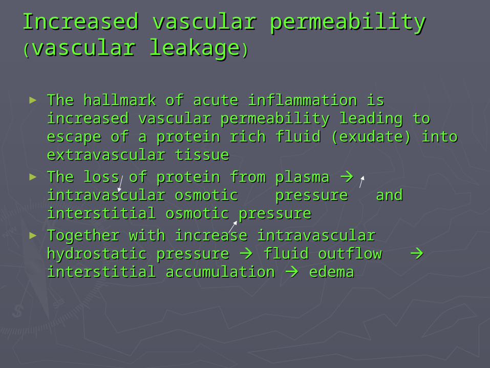
Increased vascular permeability Increased vascular permeability ((vascular leakagevascular leakage))
► The hallmark of acute inflammation is increased The hallmark of acute inflammation is increased vascular permeability leading to escape of a vascular permeability leading to escape of a protein rich fluid (exudate) into extravascular protein rich fluid (exudate) into extravascular tissuetissue
► The loss of protein from plasma The loss of protein from plasma intravascular intravascular osmotic pressure and interstitial osmotic osmotic pressure and interstitial osmotic pressure pressure
► Together with increase intravascular hydrostatic Together with increase intravascular hydrostatic pressure pressure fluid outflow fluid outflow interstitial interstitial accumulation accumulation edema edema

Blood pressure and plasma colloid Blood pressure and plasma colloid osmotic forces in normal and osmotic forces in normal and inflamed microcirculation. inflamed microcirculation. AA, Normal , Normal hydrostatic pressure (hydrostatic pressure (red arrowsred arrows) is ) is about 32 mm Hg at the arterial end about 32 mm Hg at the arterial end of a capillary bed and 12 mm Hg at of a capillary bed and 12 mm Hg at the venous end; the mean colloid the venous end; the mean colloid osmotic pressure of tissues is osmotic pressure of tissues is approximately 25 mm Hg (approximately 25 mm Hg (green green arrowsarrows), which is equal to the mean ), which is equal to the mean capillary pressure. Although fluid capillary pressure. Although fluid tends to leave the precapillary tends to leave the precapillary arteriole, it is returned in equal arteriole, it is returned in equal amounts via the postcapillary venule, amounts via the postcapillary venule, so that the net flow (so that the net flow (black arrowsblack arrows) in ) in or out is zero. or out is zero. BB, Acute inflammation. , Acute inflammation. Arteriole pressure is increased to 50 Arteriole pressure is increased to 50 mm Hg, the mean capillary pressure mm Hg, the mean capillary pressure is increased because of arteriolar is increased because of arteriolar dilation, and the venous pressure dilation, and the venous pressure increases to approximately 30 mm increases to approximately 30 mm Hg. At the same time, osmotic Hg. At the same time, osmotic pressure is reduced (averaging 20 pressure is reduced (averaging 20 mm Hg) because of protein leakage mm Hg) because of protein leakage across the venule. The net result is across the venule. The net result is an excess of extravasated fluid.an excess of extravasated fluid.

Diagrammatic Diagrammatic representation of representation of five mechanisms of five mechanisms of increased vascular increased vascular permeability in permeability in inflammation inflammation

In acute inflammation, fluid loss from vessels In acute inflammation, fluid loss from vessels with increased permeability occurs in distinct with increased permeability occurs in distinct phases :phases :
1.1. An immediate transient response lasting for 30 An immediate transient response lasting for 30 minutes or lessminutes or less
2.2. A delayed response starting at about 2 hours and A delayed response starting at about 2 hours and lasting for about 8 hours mediated by kinins, lasting for about 8 hours mediated by kinins, complement products etccomplement products etc
3.3. A prolonged response that is most noticeable A prolonged response that is most noticeable after direct endothelial injuryafter direct endothelial injury

Cellular events: leukocyte extravasation Cellular events: leukocyte extravasation and phagocytosisand phagocytosis
The sequence of events in the journey of leukocytes The sequence of events in the journey of leukocytes from the vessels lumen to the interstitial tissue from the vessels lumen to the interstitial tissue (extravasation): (extravasation):
1. in the lumen : margination, rolling, and1. in the lumen : margination, rolling, and
adhesion to the endotheliumadhesion to the endothelium
2. transmigration across the endothelium2. transmigration across the endothelium
(diapedesis)(diapedesis)
3. migration in interstitial tissues toward a3. migration in interstitial tissues toward a
chemotactic stimuluschemotactic stimulus

The multistep process of leokocyte migration through blood vessels, shown here for neutrophils
process-selectins in rolling; chemokines in activating the neutrophils to increase avidity of integrins (in green); integrins in firm adhesion; and CD31 (PECAM-1) in transmigration

Leukocyte adhesion and transmigrationLeukocyte adhesion and transmigration
► Are regulated largely by the binding ofAre regulated largely by the binding of complementary adhesion moleculescomplementary adhesion molecules on the on the leukocyte and endothelialleukocyte and endothelial surfaces, andsurfaces, and chemical chemical mediators-chemoattractantsmediators-chemoattractants and certainand certain cytokinescytokines--affect these processes by modulating the surface affect these processes by modulating the surface expression or avidity of such adhesion moleculesexpression or avidity of such adhesion molecules
► The adhesion receptors involved belong to 4 The adhesion receptors involved belong to 4 molecular families :molecular families :- selectin- selectin- immunoglobulin superfamily- immunoglobulin superfamily- integrins- integrins- mucin;like glycoproteins - mucin;like glycoproteins

Endothelial/Leukocyte Adhesion MoleculesEndothelial/Leukocyte Adhesion Molecules
Endothelial Endothelial MoleculeMolecule
Leukocyte Leukocyte ReceptorReceptor
Major RoleMajor Role
P-selectinP-selectin Sialyl-Lewis XSialyl-Lewis XPSGL-1PSGL-1
Rolling (neutrophils, monocytes, Rolling (neutrophils, monocytes, lymphocytes)lymphocytes)
E-selectinE-selectin Sialyl-Lewis XSialyl-Lewis X Rolling, adhesion to activated endothelium Rolling, adhesion to activated endothelium (neutrophils, monocytes, T cells)(neutrophils, monocytes, T cells)
ICAM-1ICAM-1 CD11/CD18 CD11/CD18 (integrins)(integrins)(LFA-1, Mac-(LFA-1, Mac-1)1)
Adhesion, arrest, transmigration (all Adhesion, arrest, transmigration (all leukocytes)leukocytes)
VCAM-1VCAM-1 αα44ββ1 (VLA4) 1 (VLA4) (integrins)(integrins)αα44ββ7 (LPAM-7 (LPAM-1)1)
Adhesion (eosinophils, monocytes, Adhesion (eosinophils, monocytes, lymphocytes)lymphocytes)
GlyCam-1GlyCam-1 L-selectinL-selectin Lymphocyte homing to high endothelial Lymphocyte homing to high endothelial venulesvenules
CD31 CD31 (PECAM)(PECAM)
CD31CD31 Leukocyte migration through endotheliumLeukocyte migration through endothelium

< & £?>
Regulation of endothelial and leukocyte adhesion molecules. A, Redistribution of P-selectin. B, Cytokine activation of endothelium. C, Increased binding avidity of integrins

ChemotaxisChemotaxis
► After extravasation, leukocytes emigrate in tissues After extravasation, leukocytes emigrate in tissues toward the site of injurytoward the site of injury chemotaxischemotaxis
► All granulocytes, monocytes, lymphocytes respond All granulocytes, monocytes, lymphocytes respond to chemotactic stimulito chemotactic stimuli
► Both exogenous and endogenous substances can Both exogenous and endogenous substances can act as chemoattrsactantsact as chemoattrsactants
► Exogenous agents : bacterial productExogenous agents : bacterial product► Endogenous agentsEndogenous agents :: complement system (C5a), complement system (C5a),
lipoxigenase pathway (leukotriene B4), cytokines lipoxigenase pathway (leukotriene B4), cytokines (IL-8)(IL-8)

Leukocyte activationLeukocyte activation
The functional responses that are induced The functional responses that are induced on leukocyte activation include :on leukocyte activation include :
- - production ofproduction of arachidonic acidarachidonic acid
metabolites from phospholipidsmetabolites from phospholipids
- degranulation and secretion of- degranulation and secretion of lysosomallysosomal
enzymes and activation of the oxidative burstenzymes and activation of the oxidative burst
- secretion of- secretion of cytokinescytokines
-- modulation ofmodulation of leukocyte adhesion moleculesleukocyte adhesion molecules

Figure: Leukocyte activation. Different classes of cell surface receptors of leukocytes recognize different stimuli. The receptors initiate responses that mediate the functions of the leukocytes. Only some receptors are depicted

PhagocytosisPhagocytosis
Involves 3 distinct but interrelated steps Involves 3 distinct but interrelated steps ::
1.1. Recognition and attachmentRecognition and attachment
2.2. Engulfment, with subsequent Engulfment, with subsequent formation of phagocytic vacuoleformation of phagocytic vacuole
3.3. Killing or degradation of the ingested Killing or degradation of the ingested material material

Figure : A, Phagocytosis of a particle (e.g., bacterium) involves attachment and binding of Fc and C3b to receptors on the leukocyte membrane, engulfment, and fusion of lysosomes with phagocytic vacuoles, followed by destruction of ingested particles within the phagolysosomes. Note that during phagocytosis, granule contents may be released into extracellular tissues. B, Production of microbicidal reactive oxygen intermediates within phagocytic vesicles

Release of leukocyte products and Release of leukocyte products and leukocyte-induced tissue injuryleukocyte-induced tissue injury
► During activation & phagocytosis, leukocytes During activation & phagocytosis, leukocytes release microbial and other productsrelease microbial and other products
► The most important of these substances in The most important of these substances in neutrophils & macrophages are :neutrophils & macrophages are : lysosomal lysosomal enzymesenzymes, , reactive oxygen intermediatesreactive oxygen intermediates,, products products ofof arachidonic acid metabolismarachidonic acid metabolism (Pg(Pg and and leukotrienes)leukotrienes)

Table 2-2. Clinical Examples of Table 2-2. Clinical Examples of Leukocyte-Induced InjuryLeukocyte-Induced Injury
AcuteAcute ChronicChronic
Acute respiratory distress Acute respiratory distress syndromesyndrome
ArthritisArthritis
Acute transplant rejectionAcute transplant rejection AsthmaAsthma
AsthmaAsthma AtherosclerosisAtherosclerosis
GlomerulonephritisGlomerulonephritis Chronic lung diseaseChronic lung disease
Reperfusion injuryReperfusion injury Chronic rejectionChronic rejection
Septic shockSeptic shock
VasculitisVasculitis

Defects in Leukocyte FunctionDefects in Leukocyte Function
► defects in leukocyte function, both genetic defects in leukocyte function, both genetic and acquired, lead to increased vulnerability and acquired, lead to increased vulnerability to infectionsto infections
► the existence of clinical genetic deficiencies the existence of clinical genetic deficiencies in each of the critical steps in the process in each of the critical steps in the process has been described :has been described :1. defects in leukocyte adhesion 1. defects in leukocyte adhesion 2. defects in phagolysosome function 2. defects in phagolysosome function 3. defects in microbicidal activity 3. defects in microbicidal activity

Table 2-3. Defects in Leukocyte FunctionsTable 2-3. Defects in Leukocyte Functions
DiseaseDisease DefectDefect
GeneticGenetic
Leukocyte adhesion deficiency 1Leukocyte adhesion deficiency 1 β chain of CD11/CD18 integrinsβ chain of CD11/CD18 integrins
Leukocyte adhesion deficiency 2Leukocyte adhesion deficiency 2 Fucosyl transferase required for synthesis of Fucosyl transferase required for synthesis of sialylated oligosaccharide (receptor for selectin)sialylated oligosaccharide (receptor for selectin)
Chronic granulomatous diseaseChronic granulomatous disease Decreased oxidative burstDecreased oxidative burst
X-linkedX-linked NADPH oxidase (membrane component)NADPH oxidase (membrane component)
Autosomal recessiveAutosomal recessive NADPH oxidase (cytoplasmic components)NADPH oxidase (cytoplasmic components)
Myeloperoxidase deficiencyMyeloperoxidase deficiency Absent MPO-HAbsent MPO-H22OO22 system system
Chédiak-Higashi syndromeChédiak-Higashi syndrome Protein involved in organelle membrane docking Protein involved in organelle membrane docking and fusionand fusion
AcquiredAcquired
Thermal injury, diabetes, Thermal injury, diabetes, malignancy, sepsis, malignancy, sepsis, immunodeficienciesimmunodeficiencies
ChemotaxisChemotaxis
Hemodialysis, diabetes mellitusHemodialysis, diabetes mellitus AdhesionAdhesion
Leukemia, anemia, sepsis, Leukemia, anemia, sepsis, diabetes, neonates, malnutritiondiabetes, neonates, malnutrition
Phagocytosis and microbicidal activityPhagocytosis and microbicidal activity

TERMINATION OF THE ACUTE TERMINATION OF THE ACUTE INFLAMMATORY RESPONSE INFLAMMATORY RESPONSE
► a switch in the production of pro-a switch in the production of pro-inflammatory leukotrienes toinflammatory leukotrienes to anti-anti-inflammatory lipoxinsinflammatory lipoxins from arachidonic acid from arachidonic acid
► thethe liberation of an anti-inflammatory liberation of an anti-inflammatory cytokine, transforming growth factor-βcytokine, transforming growth factor-β (TGF-(TGF-β), from macrophages and other cellsβ), from macrophages and other cells
► neural impulsesneural impulses (cholinergic discharge) that (cholinergic discharge) that inhibit the production of TNF in inhibit the production of TNF in macrophages.macrophages.

Chemical mediators of
inflammation

General Principles of the Mediators
1. M. originate either from plasma or cells are present in plasma in precursors form that must be activated. Major source platelets, neutrophyl, monocytes etc
2. Active M is triggered by microbial products or by host proteins, such as complement, kinin and coagulation systems
3. Perform their biologic activity by binding to specific receptor on target cell, direct enzymatic activity or mediate oxidative damage
4. Can stimulate the release of M. by target cell themselves
5. Can act on one of few target cell type6. Short-lived (quickly decay, inactivated, scavenged
or inhibited)7. Have the potential to cause harmful effects

Figure: Chemical mediators of inflammation. EC, endothelial cells

Vasoactive Amines
The mast cells (also found in basophills & platelets)The mast cells (also found in basophills & platelets)► Stimulated by :Stimulated by : - physical injury- physical injury
- immune reaction ( ab-mast - immune reaction ( ab-mast cells)cells)
- anaphylatoxins (C3a & C5a)- anaphylatoxins (C3a & C5a)- histamine-releasing proteins - histamine-releasing proteins derived from leucocytesderived from leucocytes- neuropeptides (substance P)- neuropeptides (substance P)- cytokines (IL-1, IL-8)- cytokines (IL-1, IL-8)
► It acts on the microcirculation mainly via H1 It acts on the microcirculation mainly via H1 receptorsreceptors
Histamin

Serotonin (5-OH tryptamine)
• Present in platelets & enterochromaffin cells
• Actions = histamine
• Release of S is stimulated when platelets aggregate
• Platelet aggregation & release are also stimulated by PAF derived from mast cell during IGE-mediated reactions increased permeability during immunologic reactions

Plasma Proteins
A variety of phenomena in the inflammatory response are mediated by 3 interrelated plasma-derived factors :
1. Complement system
2. Kinin system
3. Clotting system

Figure : Interrelationships between the four plasma mediator systems triggered by activation of factor XII (Hageman factor). Note that thrombin induces inflammation by binding to protease-
activated receptors (principally PAR-1) on platelets, endothelium, smooth muscle cells, and other cells

Complement System
• Consist of 20 component proteins, greatest cons in plasma
• As inactive forms in plasma are numbered C1-C9 (C3 & C5 are the most important inflam-mediators)
• The biologic functions :1. Cells lysis by the MAC (Membrane Attack
Complex)2. Proteolytic
• The most critical step is the activation of the 3 rd compl, C3, by the classic pathway, the alternative pathway or lectin pathway.

Overview of Complement Activation Pathways

Complement-Derived Factors Affect a Variety of Phenomena in AcuteInflammation
1. Vascular phenomena : C3a, C5a & to a small extent, C4a (anaphylatoxins) vasc. permeab. & vasodilation (histamin from mast cells). C5a also activates the lipoxygenase pathway of AA metab. in neutrophils & monocytes release of inflam med.
2. Leucocyte adhe, chemotaxis and activation. C5a is powerful chemotaxis agent for neutro, mono, eosino & basophyls
3. Phagocytosis : C3b & C3bi, when fixed to the bact. cell wall, act as opsonins & favor phagocytosis by neutro & macrophages
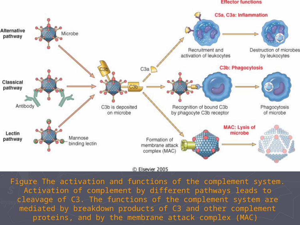
Figure The activation and functions of the complement system. Activation of complement by different pathways leads to cleavage of C3. The functions of the
complement system are mediated by breakdown products of C3 and other complement proteins, and by the membrane attack complex (MAC)

Regulation of Complement Activation
Complement activation can be controlled at several steps :
• Regulation of C3 & C5 convertases– Decay acceleration of convertase
complex (DAF)– Proteolytically cleaving C3b
• Binding of active complement components by specific proteins in the plasma :
– C1-inhibitor– Membrane inhibitor of reactive lysis to
inhibit MAC formation

Disorders of the Complement System
• Deficiency of C3 : results in increased susceptibility to infections
• Deficiency of C2 & C4 : are associated with auto immune disease (exp. SLE)
• Deficiency of C1 inhibitor is associated with the syndrome of hereditary angioneurotic edema

A Few General Conclusion
• B. kinin, C3a & C5a : mediators of vasc permeab
C5a : mediator of chemotaxis
Thrombin : are likely to be the most important in vivo,
also leuco adhesion & fibroblast proliferation
• C3 & C5 can be generated by : classic pathway, alternative pathway, lectin pathway & agents with little in immuno. specificity (plasmin, kallikrein)
• Activated Hageman factor (f.XIIa) initiated 4 system involved in inflamm. response : the kinin, the clotting, the fibrinolytic & the complement system

Coagulation & Inflammation are Tightly Linked
• Acute inflammation, by activating or
damaging the endothelium can trigger
coagulation and induce thrombus formation
• Conversely, the coagulation cascade induces
inflammation, primarilly via the action of
thrombus
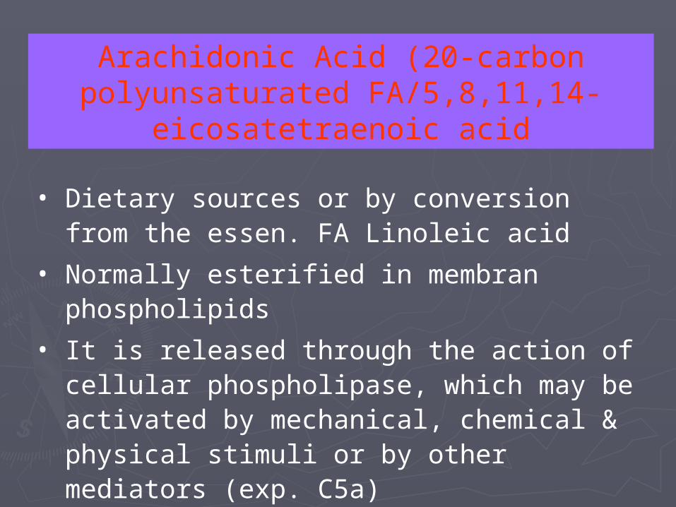
Arachidonic Acid (20-carbon polyunsaturated FA/5,8,11,14-
eicosatetraenoic acid
• Dietary sources or by conversion from the essen. FA Linoleic acid
• Normally esterified in membran phospholipids
• It is released through the action of cellular phospholipase, which may be activated by mechanical, chemical & physical stimuli or by other mediators (exp. C5a)

Arachidonic Acid Metabolites (Eicosanoids)
1. Prostaglandins2. Leukotrienes3. Lipoxinsare synthesized by 2 major classes of enzymes :
1. Cyclooxygenases (prostaglandins & thromboxanes)2. Lipoxygenases (leukotrienes & lipoxins)
Eico. bind to G protein-coupled receptors on many cell types and can mediate virtually every step of inflammation

Figure: Generation of arachidonic acid metabolites and their roles in inflammation. The molecular targets of action of some anti-inflammatory drugs are indicated by a
red X. COX, cyclooxygenase; HETE, hydroxyeicosatetraenoic acid; HPETE, hydroperoxyeicosatetraenoic acid

Table : Inflammatory Actions of EicosanoidsTable : Inflammatory Actions of Eicosanoids
ActionAction MetaboliteMetabolite
VasoconstrictionVasoconstriction Thromboxane AThromboxane A22, ,
leukotrienes Cleukotrienes C44, D, D44, E, E44
VasodilationVasodilation PGIPGI22, PGE, PGE11, PGE, PGE22, PGD, PGD22
Increased vascular Increased vascular permeabilitypermeability
Leukotrienes CLeukotrienes C44, D, D44, E, E44
Chemotaxis, leukocyte Chemotaxis, leukocyte adhesionadhesion
Leukotriene BLeukotriene B44, HETE, , HETE,
lipoxinslipoxins

Anti-inflammatory Therapy
• Aspirin and NSAIDs (indomethacin or ibuprofen) inhibit cyclooxygenase thus inhibit prostaglandin syntesis.
• Glucocorticoids which are powerful anti-inflammatory agents, may act by down-regulating the expression of spec target genes, including COX2, genes encoding proinflammatory cytokines (such as IL-1 and TNF-), and nitric oxide synthase (iNOS). Also up-regulate genes that encode potent anti-inflammatory proteins, such as lipocortin 1 inhibits release of AA from memb. Phospho.
• Leukotrienes from fatty acids found in fish oil are less potent than those derived from the Arach. Acid found in most animal or vegetable fat.

Platelet Activating Factor
• Is acetyl-glyceryl-ether-phosphorylcholine (AGEPC)
• A phospholipid, mediates its effects via a single G protein-coupled receptor
• Its effects are regulated by a family of inactivating PAF acetylhydrolases
• A variety of cell types, including platelets, basophils (& mast cells), neutro, mono, macrophages and endoth. can elaborate PAF

The Effect of PAF
• Vasoconstriction, bronchoconstriction.
• At extremely low concentration it induces. vasodilation and venular permeability with a potency 100 – 10.000 x greater than that of histamine.
leuco adhesion to endothelium, chemotaxis, degranulation, oxidative hurst.
• Also boosts the synthesis of other mediators, particularly Eicosanoids by leuco and other cells

Structure, Source and main inflammatory action of PAF
SourceSource Major inflammatory actionsMajor inflammatory actions
Mast cell/basophilsMast cell/basophils
NeutrophilsNeutrophils
Monocyte/macrophMonocyte/macroph
EndotheliumEndothelium
PlateletsPlatelets
OthersOthers
Increased vasc. permeabIncreased vasc. permeab
Leucocyte aggregationLeucocyte aggregation
Leucocyte adhesionLeucocyte adhesion
L. priming/chemotaxisL. priming/chemotaxis
Platelet activationPlatelet activation
Stimulation of other Stimulation of other mediators (leucotrienes)mediators (leucotrienes)

Cytokines and Chemokines• Cytokines are proteins produced by many cells
types (activated lympho, macroph, also endo, epithel & conn. tissue cells)
• To be involved in cellular immune responses
• Colony stimulating factors (CSFs) stimulate the growth of immature peuco in the BM.
• Interleukins, a broad family of cytokines, are made by hematopoetic cells and act primarily on leucocytes.
• Chemokines are cytokines that share the ability to stimulate leukocyte movement (chemokinesis) and directed movement (chemotaxis)

General Properties of Cytokines
• Are produced during immune & inflamm. responses
• Secretion is transient and closely regulated
• Many cell types produce multiple cytokines
• The protein are pleotropic in that they can act on different cell types
• C can influence the synthesis or action of other C
• Are multifunctional in that an individual C may have both pos. and neg. regulatory actions

Cytokines can be grouped into 5 classess, depending on their major function as the
nature of the target cells
1. C that regulate lymphocyte function (IL-2, IL-4, IL-10 & TGF-)
2. C involved with natural immunity (TNF & IL-1; type 1 interferons (IFN-, IFN- ) and IL-6
3. C that active inflammatory cells
4. Chemokines
5. C that stimulate hematopoiesis

Biosynthesis of leukotrienes and lipoxins by cell-cell interaction. Activated neutrophils generate LTB4 from arachidonic acid-derived LTA4 by the action of 5-lipoxygenase, but they do not possess LTC4-synthase activity and consequently do not produce LTC4. In contrast, platelets cannot form LTC4 from endogenous substrates, but they can generate LTC4 and lipoxins from neutrophil-derived LTA4. (Courtesy of Dr. C. Serhan, Brigham and Women's Hospital, Boston, MA.)
Biosynthesis of leukotrienes and lipoxins by cell-cell interaction

Figure : Major effects of interleukin-1 (IL-1) and tumor necrosis factor (TNF) in inflammation

Figure : Functions of nitric oxide (NO) in blood vessels and macrophages, produced by two NO synthase enzymes. NO causes vasodilation, and NO free radicals are toxic to microbial and mammalian cells. NOS, nitric oxide synthase.

Table : Summary of Mediators of Acute Table : Summary of Mediators of Acute InflammationInflammation
ActionAction
MediatorMediator SourceSource Vascular Vascular LeakageLeakage
Chemotaxis OtherChemotaxis Other
Histamine and Histamine and serotoninserotonin
Mast cells, plateletsMast cells, platelets ++ --
BradykininBradykinin Plasma substratePlasma substrate ++ -- PainPain
C3aC3a Plasma protein via liverPlasma protein via liver ++ -- Opsonic fragment (C3b)Opsonic fragment (C3b)
C5aC5a MacrophagesMacrophages ++ ++ Leukocyte adhesion, Leukocyte adhesion, activationactivation
ProstaglandinsProstaglandins Mast cells, from membrane Mast cells, from membrane phospholipidsphospholipids
Potentiate other Potentiate other mediatorsmediators
-- Vasodilation, pain, feverVasodilation, pain, fever
Leukotriene BLeukotriene B44 LeukocytesLeukocytes -- ++ Leukocyte adhesion, Leukocyte adhesion, activationactivation
Leukotriene CLeukotriene C44, ,
DD44, E, E44
Leukocytes, mast cellsLeukocytes, mast cells ++ -- Bronchoconstriction, Bronchoconstriction, vasoconstrictionvasoconstriction
Oxygen Oxygen metabolitesmetabolites
LeukocytesLeukocytes ++ -- Endothelial damage, tissue Endothelial damage, tissue damagedamage
PAFPAF Leukocytes, mast cellsLeukocytes, mast cells ++ ++ Bronchoconstriction, Bronchoconstriction, leukocyte primingleukocyte priming
IL-1 and TNFIL-1 and TNF Macrophages, otherMacrophages, other -- ++ Acute-phase reactions, Acute-phase reactions, endothelial activationendothelial activation
ChemokinesChemokines Leukocytes, othersLeukocytes, others -- ++ Leukocyte activationLeukocyte activation
Nitric oxideNitric oxide Macrophages, endotheliumMacrophages, endothelium ++ ++ Vasodilation, cytotoxicityVasodilation, cytotoxicity


![KISA URt]N BiLGiSi - ilacweb.com filesemptomlann belirgin oldufu, erken dcinem psikozlar, akut gizofrenik alevlenmeler, kronik qizofieni ve diler psikotik durumlar dahil gizofreni](https://static.documents.pub/doc/80x56/5d5eb94a88c99301668bc30d/kisa-urtn-bilgisi-belirgin-oldufu-erken-dcinem-psikozlar-akut-gizofrenik-alevlenmeler.jpg)


