+ All Categories
Home > Documents > INFLAMMATION AND OXIDATIVE STRESS IN AN LIMBIC …

INFLAMMATION AND OXIDATIVE STRESS IN AN LIMBIC …

Date post: 21-Dec-2021
Category:
Upload: others
View: 1 times
Download: 0 times
Share this document with a friend
157
INFLAMMATION AND OXIDATIVE STRESS IN AN ANIMAL MODEL OF INFECTION-INDUCED LIMBIC EPILEPSY by Dipankumar C. Patel A dissertation submitted to the faculty of The University of Utah in partial fulfillment of the requirements for the degree of Doctor of Philosophy Department of Pharmacology and Toxicology The University of Utah December 2016
Transcript

INFLAMMATION AND OXIDATIVE STRESS IN AN

ANIMAL MODEL OF INFECTION-INDUCED

LIMBIC EPILEPSY

by

Dipankumar C. Patel

A dissertation submitted to the faculty of The University of Utah

in partial fulfillment of the requirements for the degree of

Doctor of Philosophy

Department of Pharmacology and Toxicology

The University of Utah

December 2016

Copyright © Dipankumar C. Patel 2016

All Rights Reserved

T h e U n i v e r s i t y o f U t a h G r a d u a t e S c h o o l

STATEMENT OF DISSERTATION APPROVAL

The dissertation of Dipankumar C. Patel

has been approved by the following supervisory committee members:

Karen S. Wilcox , Chair 08/08/2016

Date Approved

Donald K. Blumenthal II , Member 08/08/2016

Date Approved

H. Steve White , Member 08/08/2016

Date Approved

Robert S. Fujinami , Member 08/08/2016

Date Approved

Thomas E. Lane , Member 08/08/2016

Date Approved

and by Karen S. Wilcox , Chair/Dean of

the Department/College/School of Pharmacology and Toxicology

and by David B. Kieda, Dean of The Graduate School.

ABSTRACT

Central nervous system (CNS) infection can induce epilepsy that is often

refractory to established antiseizure drugs. The Theiler’s murine encephalomyelitis virus

(TMEV)-induced mouse model of limbic epilepsy is an important model in which to

study the mechanisms underlying epileptogenesis and to identify novel therapeutics.

Previous studies have demonstrated the importance of inflammation, especially mediated

by tumor necrosis factor-α (TNFα), in the development of TMEV-induced acute seizures.

TNFα is known to modulate glutamate receptor trafficking via TNF receptor 1 (TNFR1)

to cause increased excitatory synaptic transmission. Therefore, we hypothesized that an

increase in hippocampal TNFα following TMEV infection might contribute to

hyperexcitability and seizures by increasing excitatory postsynaptic strength through

TNFR1. Furthermore, inflammation is known to contribute to oxidative stress that in turn

can precipitate seizures. Therefore, we also investigated the occurrence of oxidative

stress in TMEV-infected mice.

We found a significant increase in the levels of oxidative stress markers and

TNFα in the hippocampus, a brain region known to be involved in seizure generation,

following TMEV infection. In addition, a significant increase in the protein expression

ratio of TNF receptors (TNFR1:TNFR2) in hippocampus suggests that TNFα signaling,

predominantly through TNFR1, may contribute to limbic hyperexcitability. Consistent

with increased TNFR1 signaling, increases in hippocampal cell surface glutamate

iv

receptor expression was also observed during the acute period. While pharmacological

inhibition of TNFR1-mediated signaling had no effect on acute seizures, several lines of

transgenic animals deficient in either TNFα or its receptors were found to have robust

changes in seizure incidence and severity following TMEV infection. TNFR2-/- mice

were highly susceptible to developing TMEV-induced acute seizures, suggesting that

signaling through the TNFR2 pathway may provide beneficial effects during the acute

seizure period. Moreover, cannabidiol (180 mg/kg), which exhibits antiinflammatory and

antiseizure properties, dramatically inhibited TMEV-induced acute seizures.

Taken together the present results suggest that oxidative stress and inflammation

in the hippocampus contribute to hyperexcitability and increase the probability of

seizures following TMEV infection and that the TNFα signaling pathway is involved in

this process. Pharmacotherapies designed to suppress inflammation and oxidative stress

may provide antiseizure and disease modifying effects following CNS infection.

TABLE OF CONTENTS

ABSTRACT ....................................................................................................................... iii LIST OF FIGURES .......................................................................................................... vii LIST OF TABLES ............................................................................................................. ix LIST OF ABBREVIATIONS ..............................................................................................x Chapters 1. POSTINFECTIOUS EPILEPSY .................................................................................. 1

Abstract ................................................................................................................... 1 Introduction ............................................................................................................. 2 Herpes simplex virus (HSV)-induced model of limbic seizures ............................ 5 West Nile virus (WNV)-induced limbic seizures ................................................. 10

Neurocysticercosis (NCC) model of limbic seizures ............................................ 12 TMEV-induced murine model of limbic epilepsy ................................................ 14 Conclusion ............................................................................................................ 37 References ............................................................................................................. 37

2. OXIDATIVE STRESS IN MURINE THEILER’S VIRUS-INDUCED TEMPORAL

LOBE EPILEPSY ....................................................................................................... 47

Abstract ................................................................................................................. 47 Introduction ........................................................................................................... 49

Methods................................................................................................................. 52 Results ................................................................................................................... 54 Discussion ............................................................................................................. 57

References ............................................................................................................. 63 3. HIPPOCAMPAL TNFα SIGNALING CONTRIBUTES TO

HYPEREXCITABILITY IN AN INFECTION- INDUCED MOUSE MODEL OF LIMBIC EPILEPSY ................................................................................................... 70

Introduction ........................................................................................................... 70

Methods................................................................................................................. 73

vi

Results ................................................................................................................... 81 Discussion ............................................................................................................. 93 References ............................................................................................................. 99

4. CANNABIDIOL TREATMENT PREVENTS SEIZURES FOLLOWING CNS

INFECTION WITH THEILER’S MURINE ENCEPHALOMYELITIS VIRUS... 113

Introduction ......................................................................................................... 113 Methods............................................................................................................... 116 Results ................................................................................................................. 118 Discussion ........................................................................................................... 123 References ........................................................................................................... 126

5. SUMMARY, FUTURE DIRECTIONS, AND PERSPECTIVES ............................ 136

Summary and implications of findings ............................................................... 137 Future directions ................................................................................................. 139 Perspectives......................................................................................................... 141 References ........................................................................................................... 143

LIST OF FIGURES Figures 1.1 Schematic of the TMEV-infection mouse model of temporal lobe epilepsy. ............. 44 1.2 Electroencephalographic (EEG) recording from TMEV-infected mice. .................... 45 2.1 Acute behavioral seizures in TMEV-infected mice. ................................................... 66 2.2 Impaired GSH redox status in TMEV-infected mice. ................................................ 67 2.3 Increased levels of 3NT in TMEV-infected mice. ...................................................... 68 2.4 No oxidative stress in cerebellum of TMEV mice at 3 dpi. ........................................ 69 3.1 Increase in the levels of TNFα and in a ratio of the protein expression of TNFR1:TNFR2 in the hippocampus of TMEV-infected mice during acute seizure activity period. ............................................................................................................................. 105 3.2 CNS administration of XPro1595 does not affect TMEV-induced acute seizure frequency and intensity. .................................................................................................. 107 3.3 TMEV-induced acute behavioral seizure susceptibility in WT, TNFα-/-, TNFR2-/-, and TNFR1-/-TNFR2-/- mice. ................................................................................................. 108 3.4 Increase in the cell surface levels of GluA1 and GluA2 subunits of AMPARs in TMEV-infected WT C57BL/6J mice during acute seizures. .......................................... 109 3.5 Increase in the cell surface levels of GluA1 and GluA2 subunits of AMPARs in TMEV-infected TNFR2-/- mice during acute seizures. ................................................... 110 3.6 No difference in the properties of miniature excitatory postsynaptic currents (mEPSCs) of dentate granule cells (DGCs) between PBS-injected (control) and TMEV-infected mice during acute seizure activity period. ........................................................ 111 4.1 Prophylactic treatment with 180 mg/kg CBD reduces average frequency and severity of TMEV-induced acute seizures. ................................................................................... 129

viii

4.2 Therapeutic treatment with CBD (180 mg/kg) reduces average frequency and severity of TMEV-induced acute seizures. ................................................................................... 131 4.3 Only one dose of CBD (180 mg/kg; range of 22.5 to 180 mg/kg) reduces frequency and severity of TMEV-induced acute seizures. .............................................................. 133 4.4 Low doses of CBD have no effect on TMEV-induced acute seizures. .................... 134 4.5 CBD (150 mg/kg) administration decreases TMEV-induced seizures monitored at 4 h post-CBD treatment but not at 9 h post-CBD treatment. ................................................ 135

LIST OF TABLES Tables 1.1 Salient features of the TMEV model of limbic epilepsy. ........................................... 46 3.1 Significant increase in the protein levels of various inflammatory mediators in the hippocampus of TMEV-infected mice during acute seizure activity period. ................. 112

LIST OF ABBREVIATIONS 3NT, 3-nitrotyrosine

AMPAR, α-amino-3-hydroxy-5-methyl-4-isoxazole propionic acid

ANOVA, analysis of variance

ASD, antiseizure drug

CBD, cannabidiol

DG, dentate gyrus

DGCs, dentate granule cells

DI, during injection

dpi, days postinfection

GABA, gamma-aminobutyric acid

GSH, glutathione

GSSG, glutathione disulfide

HSV, herpes simplex virus

i.c.v., intracerebroventricular

i.p., intraperitoneal

IFNγ, interferon-γ

IL, interleukin

JEV, Japanese encephalitis virus

LTα3, lymphotoxin α3

xi

mEPSC, miniature excitatory postsynaptic current

mIPSC, miniature inhibitory postsynaptic current

NCC, neurocysticercosis

OD, optical density

PAG, phosphate-activated glutaminase

PFU, plaque forming units

RNS, reactive nitrogen species

ROS, reactive oxygen species

s.c., subcutaneous

SCI, spinal cord injury

SD, standard deviation

SEM, standard error of the mean

sTNFα, soluble TNFα

TGFβ, transforming growth factor-β

TLE, temporal lobe epilepsy

TMEV, Theiler’s murine encephalomyelitis virus

tmTNFα, transmembrane TNFα

TNFR, tumor necrosis factor receptor

TNFα, tumor necrosis factor-α

TPE, time to the peak effect

vEEG, video electroencephalography

WNV, West Nile virus

CHAPTER 1

POSTINFECTIOUS EPILEPSY1

Abstract

Central nervous system (CNS) infections are common risk factors for seizures and

the development of epilepsy. Various infectious agents including viruses, parasites,

bacteria, and fungi are clinically associated with seizures and epilepsy. The detailed

cellular and molecular mechanisms of pathological and physiological changes in the

brain due to acute infection are not clearly understood. Infection induces inflammation in

the brain which could contribute to the process of epileptogenesis in which normal

neuronal circuits transform into epileptic circuits which increase the probability of the

development of epilepsy. A detailed understanding of epileptogenesis following infection

is of utmost importance for the development of advanced therapies to treat the seizures as

well as to prevent the progression of disease. Several infection-induced animal models of

seizure and epilepsy have been described mainly using viral infection and parasitoses in

rodents. This chapter describes several of them, including those models using herpes

simplex virus-1, West Nile virus, neurocysticercosis, and Theiler’s virus. We will discuss

the methods of seizure generation, pathological and physiological changes in the brain,

1 This article was published in Models of Seizure and Epilepsy, Second Edition, in press, Dipan C. Patel, Karen S. Wilcox, Postinfectious epilepsy, Copyright Elsevier (2017). Reproduced with permission from Elsevier.

2

clinical relevance, advantages, and limitations of the models.

Introduction

Infections of the central nervous system are common risk factors for seizures and

the development of epilepsy. Infections can potentially cause encephalitis (inflammation

of the brain parenchyma) and encephalopathy (diffuse CNS disease manifested by altered

consciousness, a range of cognitive and motor neurological symptoms, and systemic

metabolic disturbances) by directly or indirectly damaging the brain. A broad range of

infectious agents including viruses (e.g., herpes viruses, enteroviruses, flaviviruses,

paramyxoviruses), parasites (e.g., Taenia solium, Plasmodium falciparum, Toxoplasma

gondii), bacteria (e.g., Haemophilus, Streptococcus, Neisseria, Mycobacterium), and

fungi (e.g., Candida, Cryptococcus) are clinically associated with seizures and epilepsy

(Vezzani et al., 2016). Among them, viral encephalitis and parasitic infections are the

most widely reported causes of infection-induced epilepsies in patients.

Over 100 neurotrophic viruses are known to cause encephalitis in humans and

many of them may subsequently contribute to seizures and epilepsy. The prevalence of

viral encephalitis which can be either sporadic or epidemic has been calculated ~7.5

persons per 100,000 people in the general population (Misra et al., 2008). Herpes simplex

virus type 1 (HSV-1) is the most common cause of sporadic encephalitis (Theodore,

2014). Early (acute) seizures can occur in 40-60% of cases of HSV-1 encephalitis which

is probably due to tropism of HSV-1 for mesial temporal lobe structures, especially

hippocampus, that are strongly involved in seizure generation and epileptogenesis. A

prospective study of the consequences of prolonged febrile seizures (FEBSTAT)

3

determined primary or prior human herpes virus-6B (HHV-6B) and HHV-7 viremia in

32% and 7% of pediatric patients with febrile status epilepticus, respectively, which is

associated with increased risk for hippocampal injury and temporal lobe epilepsy

(Epstein et al., 2012). Japanese encephalitis is the most common form of epidemic

encephalitis associated with acute seizures, which occur in 7-46% of patients (Misra et

al., 2008). Viral encephalitis also increases the risk for the development of late (chronic)

unprovoked/spontaneous seizures. As in acute symptomatic seizures, the development of

chronic seizures also varies among the types of viral encephalitis; for example, late

unprovoked seizures occur in 40-65% of patients following HSV encephalitis, while only

10-12% of La Crosse encephalitis patients go on to develop epilepsy (Misra et al., 2008).

Other noteworthy viral infections implicated in seizures and epilepsy are human

immunodeficiency virus (HIV), enterovirus, West Nile virus, measles,

cytomegaloviruses, influenza viruses, dengue, and chikungunya (Vezzani et al., 2016).

CNS infection due to parasites can also cause seizures. Major parasitoses

associated with increased incidence of seizures and epilepsy are neurocysticercosis

(NCC) and cerebral malaria (CM) notably in low income countries. Up to 10% of African

children suffering from CM develop epilepsy (Birbeck et al., 2010). CM-associated

seizures are often focal which may occasionally generalize and ~20% of children present

with subclinical seizures (Birbeck et al., 2010). 28% of CM patients may develop status

epilepticus, which is often resistant to antiseizure medications and associated with

increased neurological disability and mortality rate. NCC is caused by CNS infection of

the larval stage of tapeworm Taenia solium and it is the most common preventable risk

factor for adult-acquired epilepsy worldwide. About 80% of patients with symptomatic

4

NCC develop recurrent seizures and NCC accounts for up to 29% of all epilepsy cases in

some endemic regions (Ndimubanzi et al., 2010).

There are fewer clinical cases of epilepsies associated with bacterial infection

compared to viral infection and parasitoses. However, any bacterial infection of the CNS

can cause acute seizures and chronic epilepsy. Infections with Haemophilus influenzae

are more commonly associated with seizures compared to other bacterial infections

(Stringer, 2006). Acute infection of meninges by Neisseria meningitidis can result in

acute seizures. After a latent period ranging from several weeks to few years, chronic

spontaneous seizures, often drug resistant, develop in about 5-10% of survivors of

meningitis (Oostenbrink et al., 2002). Bacterial infections may induce formation of

intracranial empyemas and abscesses, and seizures occur in about 35% of patients with

brain cerebral abscesses and in >50% of patients with dural empyemas (Labar and

Harden, 1998). Although the cases of fungal infection-induced epilepsy are relatively

uncommon, many species of fungi including Candida, Cryptococcus, Aspergillus,

Blastomyces, and Histoplasma have been found to cause seizures, especially in

immunocompromised people (Vezzani et al., 2016).

Infection can cause damage to the brain parenchyma by directly infecting the cells

or by unleashing uncontrolled inflammatory reactions to surrounding tissue. CNS damage

and excessive inflammation may engender acute seizures and persistent neurological

abnormalities can result in epilepsy. The detailed mechanisms of neuropathological and

inflammatory changes due to acute infection and the mechanisms of epileptogenesis as a

consequence (a process of structural and physiological changes in brain parenchyma

following insult which transform normal neuronal circuits into epileptic circuits) are

5

unknown and it is an active area of research. A detailed understanding of epileptogenesis

following infection is of utmost importance as it will open the door for novel therapeutic

approaches to prevent the progression of disease.

Several infection-induced animal models of seizure and epilepsy have been

described mainly using viral infection and parasitoses in rodents. This chapter describes

several of them, including those models using HSV-1, West Nile virus, NCC, and

Theiler’s virus. The Theiler’s virus model of limbic epilepsy is the most extensively

studied among all the models with numerous studies published in the last few years

validating its usefulness for translational studies. We will discuss the method of seizure

generation, structural and functional changes in the brain, clinical relevance, advantages,

and limitations of the models.

Herpes simplex virus (HSV)-induced model of limbic seizures

HSV-1 is a neurotrophic virus capable of penetrating into brain from intranasal

route via retrograde axonal pathway and replicating into brain parenchymal cells. It is the

most common virus clinically associated with seizures and epilepsy. Attempts have been

made to develop animal models for HSV-1 encephalitis-induced seizures in rabbit

(Stroop and Schaefer, 1989), rat (Beers et al., 1993; Solbrig et al., 2006), mouse (Wu et

al., 2003; Wu et al., 2004), and an in vitro system (Chen et al., 2004).

Methods of generation

Intranasal or corneal inoculation methods have been used for generating HSV-

induced model of seizures. Animals are briefly anesthetized before infecting them.

6

Female New Zealand white (NZW) rabbits weighing 2-3 kg are inoculated in each nostril

with 0.1 ml solution containing 106 TCID50 (50% tissue culture infectious dose) of

neurovirulent +GC substrain of HSV-1. Similarly, female Lewis rats (around 225 g) are

infected intranasally with 1.4x10⁶ TCID50 (50% tissue culture infectious dose) of +GC. In

another rat study, 9 week-old male Lewis rats are infected with 3x10⁶ PFU (plaque

forming units) of McKrae HSV-1 administered as eye drops in the right eye and

conjunctival sac followed by closing and opening the eye. For the mouse model, 5-6

weeks old male BALB/c mice are inoculated with RE strain of HSV-1 ranging from

2x10⁵ to 2x10⁶ PFU into the right eye by corneal scarification. For in vitro system,

organotypic cultures of hippocampal slices from Sprague-Dawley rat pups (P10-12) are

infected with 1x10⁵ PFU of RE at 14 days in vitro for 1 h.

Characteristics and defining features Seizures

NZW rabbits infected with +GC resulted in severe motor seizures in about 59% of

rabbits (10/17) during 5-12 days postinfection (dpi) (Stroop and Schaefer, 1989). The

rabbits had the Jacksonial type of seizures lasting for several minutes during each

occurrence, beginning with muscle movements around nose and mouth which

sequentially spread to neck, forelimbs and hindlimbs followed by unnatural upright

posture with nose pointed almost vertically. Electroencephalographic (EEG)

abnormalities were observed during the first week of infection in two of three rabbits. Of

the 22 rabbits used overall, all except two rabbits eventually either died or had to be

euthanized due to moribund condition and chronic studies could not be conducted.

7

Lewis rats infected with +GC developed complex partial seizures which

secondarily generalized between 7-10 dpi (Beers et al., 1993). Compared to the rabbit

study, 39% of the rats (9/23) had acute seizures of 30-60 s in duration and only 12%

(3/26) of the rats died during acute infection. It was not determined if epilepsy developed

in those rats that survived the infection. Lewis rats infected with McKrae HSV-1

exhibited Racine stage 1 and 2 limbic focal seizures that did not generalize, although

long-term EEG recordings were not performed (Solbrig et al., 2006).

BALB/c mice infected with the RE strain developed progressively worsening

physical symptoms from ruffled fur, hunched posture, and loss of appetite to limb

weakness, ataxia, and seizures between 4 and 14 dpi (Wu et al., 2003). Neurological

deficits, seizures, and mortality varied among infected mice and were correlated with

viral titer. All the mice with severe encephalitis developed behavioral and EEG seizures

originating from hippocampus and died by 10 dpi. Only 21% of mice with moderate

encephalitis had seizures, whereas the mice with mild symptoms of infection did not

develop seizures. Behavioral seizures started with staring and chewing, and progressed to

forelimb clonus with/without rearing and generalized tonic-clonic extension. The

duration of EEG seizures varied from several to tens of seconds.

Neuropathology

HSV-1 invaded and replicated in the rabbit brain as viral antigens were detected

in cortical layers IV-VI, trigeminal and olfactory system, amygdala, nucleus accumbens,

locus ceruleus, and brainstem. The shedding of virus was also detected in ocular and

nasal secretions in the first 2 weeks of infection. The neuropathological changes such as

8

mild leptomeningitis, lymphocytic infiltration in medulla, and death of hippocampal

neurons were found at 6 dpi. The virulence of the virus correlated with its expression

levels in the brain.

In Lewis rats infected with +GC, the bilateral inflammatory and hemorrhagic

lesions and astrogliosis were colocalized with the presence of viral antigens and nucleic

acids in trigeminal ganglia, olfactory bulbs, piriform and entorhinal cortices,

hippocampus, and amygdala during the first week of infection. As in the rabbit study,

shedding of virus in ocular and nasal secretions was also observed. On the other hand,

inflammatory lesions, gliosis, and viral antigens were not detected in any brain structure

at 76 and 160 dpi. However, viral nucleic acids were present in the hippocampus and the

piriform and entorhinal cortices at 76 and 160 dpi suggesting that HSV-1 can establish

CNS latency in the rat.

In BALB/c mice infected with RE-HSV-1, the severity of neurological damage

was highest in the limbic region particularly CA3 of hippocampus, amygdala, entorhinal

and pyriform cortex, and correlated with the high expression level of viral antigens.

Infiltration of neutrophils and lymphocytes was detected in hippocampus, pyriform

cortex, and meninges. Patch-clamp recordings from the CA3 hippocampal neurons during

7-12 dpi showed more depolarized resting membrane potential, increased membrane

input resistance, and decreased threshold for bursting activity suggesting that

hyperexcitability in CA3 neuronal circuitry could facilitate the development of seizures.

These electrophysiological changes in CA3 neurons were also reported in RE-HSV-1-

infected organotypic hippocampal slice culture. HSV-1-infected mice had an increased

susceptibility to kainic acid-induced seizures (Wu et al., 2003) and HSV-1 infection also

9

caused neuronal death and a marked increase in mossy fiber sprouting in the

supragranular area in slice cultures (Chen et al., 2004). Neuronal loss was ameliorated by

treating the infected slices with antiviral agent, acyclovir (Chen et al., 2004). Similarly,

valacyclovir decreased pentylenetetrazole-induced seizure susceptibility in HSV-1-

infected mice (Wu et al., 2004). These results suggest that HSV-1 infection can cause

hyperexcitability in the hippocampal neuronal circuit leading to seizures and such

changes can be prevented by restricting the infection.

Limitations

These models are limited by high mortality during the acute infection, although

the rat model should be investigated for the development of unprovoked chronic seizures

and epilepsy. HSV-1 also poses other problems with respect to ease of use, as it is known

to cause diseases in humans and is classified under biosafety level 2 (BSL2) requiring

regulatory approvals for study.

Insight into human disorders

Both rat and mouse models could be useful to study herpes infection-induced

epilepsies in human as the rodents infected with HSV-1 develop behavioral seizures and

show clinically relevant neurological lesions and histological changes, especially in the

temporal region. Therefore, they could be valuable to investigate mechanisms of HSV-1-

mediated seizures. For example, the study investigating the mechanism of seizure

generation using male Lewis rats infected with McKrae HSV-1 found a decrease in the

expression of dynorphin A, an endogenous opioid molecule which can contribute to

10

anticonvulsant activity, in the hippocampus (Solbrig et al., 2006). A kappa opioid

receptor agonist, U50488, effectively blocked ictal activity confirming the antiseizure

functions of dynorphin. The slice culture model could also be important as it allows high-

throughput screening of potential antiseizure and antiepileptogenic compounds for future

detailed investigation in animal models (Dyhrfjeld-Johnsen et al., 2010). However, a

thorough validation of the in vitro system is essential given its drawbacks.

West Nile virus (WNV)-induced limbic seizures

WNV is a neurotrophic virus causing epidemic encephalitis often manifested with

seizures. One mouse model of WNV-induced limbic seizures is reported (Getts et al.,

2007).

Methods of generation

Adult female C57BL/6 mice aged 8-14 weeks are briefly anesthetized and

intranasally inoculated with 10 µl of WNV solution in sterile phosphate-buffered saline

containing 6x10⁴ PFU of virus.

Characteristics and defining features Acute seizures

Mice are monitored for seizures twice daily until 4 dpi and then every four hours.

Seizure intensity is recorded based on a summative scoring system ranging from 0 to 12.

Limbic seizures first appeared on 5 dpi and started with piloerection, tail stiffening,

hunching, excessive face washing, extreme wet dog shaking, and developed into

11

handling-induced seizures followed by rearing and falling. Seizure intensity gradually

increased and all the mice developed limbic seizures. The highest seizure scores were

coincided with advanced stages of disease on 7 dpi.

Neuropathology

Viral antigens first appeared in the olfactory bulb by 3 dpi and subsequently

spread into cortex and pyramidal neurons of CA1-3 regions of hippocampus on 5 dpi. By

6 dpi, virus was detected throughout the hippocampus. Thus, the extent of viral

infiltration and replication in the brain appear to correlate with the induction of seizures

and gradual increase in the seizure intensity. No histopathological abnormalities were

detected in the first 3 days of infection. Adhesion of leukocytes to vascular endothelium,

leukocyte infiltration into the brain parenchyma, and microgliosis were observed at 6 dpi.

The expression of tumor necrosis factor-α (TNFα) and interleukin-6 (IL-6) in the

brain were significantly elevated at 7 dpi. However, TNFα-/- mice intranasally infected

with WNV had limbic seizures comparable to WT mice. The role of interferon-γ (IFNγ)

was evaluated in detail in this model (Getts et al., 2007). IFNγ-/- mice infected with WNV

had a significant reduction in seizure intensity and did not have severe limbic seizures,

indicating that the presence of IFNγ may contribute to seizure progression. The pattern of

viral infiltration and replication in the brain, neuropathological and inflammatory changes

including the levels of TNFα and IL-6 were not different between WT and IFNγ-/- mice,

suggesting that additional factors might be implicated in causing different seizure

outcomes in the two strains.

12

Limitations

All the mice succumb to infection around 7-8 dpi, thus restricting the use of this

model to infection-induced acute seizures.

Neurocysticercosis (NCC) model of limbic seizures

General description

NCC is a common helminthic infection of the brain caused by the larval form of

the tapeworm Taenia solium and is a major cause of seizures and acquired epilepsy

worldwide. Pigs serve as intermediate reservoir for larval vesicles (metacestode or

cysticerci) and eggs (containing oncosphere – embryonic form of tapeworm) of T. solium.

Humans are infected by consumption of raw or undercooked pork or by ingesting food

and water contaminated with human or animal feces containing T. solium eggs. Eggs

hatch into larva in the intestines, penetrate the intestinal wall, and migrate to the brain

where they transform into cycticerci (Stringer, 2006). It can take several years to develop

clinical symptoms, including seizures, after infection. Altered blood-brain barrier and

inflammatory reaction around the cysts or associated with calcified granulomas appear in

patients with NCC-associated seizures, but the factors crucial in causing seizures are

unclear. Several rodent models of NCC have been developed using mice infected

intracranially with T. crassiceps (Matos-Silva et al., 2012) or Mesocestoides corti

(Mishra et al., 2013); however, seizures were not studied in these models. One study

reported epileptiform activity in a rat model of NCC using granulomas associated with T.

crassiceps infection (Stringer et al., 2003). In addition, seizures were observed in a

recently reported rat model of NCC using activated T. solium oncospheres (Verastegui et

13

al., 2015).

Methods of generation

As reported in the study by Stringer et al., 2003, female BALB/c mice are first

inoculated intraperitoneally with T. crassiceps. Live parasites do not cause much

inflammation but the dying parasites initiate chronic granulomatous reaction in the

peritoneal cavity. The development of granuloma can be histopathologically graded from

stage 1-4 with increasing levels of inflammation and tissue damage. Granulomas

indicative of each stage are isolated from the peritoneal cavity after 3 months of infection

and the extract of granulomas is injected into the hippocampus or amygdala or other brain

region of Sprague-Dawley rats (125-175 g) to model NCC. In another model (Verastegui

et al., 2015), Holtzman rats aged 10-26 days are infected intracranially with activated T.

solium oncospheres suspended in sterile physiologic salt solution.

Characteristics and defining features

Intrahippocampal electrodes are used to record epileptiform activity in the rats

treated with granuloma extract. All rats injected with the extract from stage 1-2, but not

stage 3-4, granulomas developed electrographic seizures of average 44 s within 3 min of

injection. However, subsequent epileptiform activity, spreading of seizures out of

hippocampus and behavioral seizures were not observed. Neuronal or glial cell loss was

not detected in the brain. Such findings question the usefulness of this model to study the

mechanism of epileptogenesis.

Epilepsy developed in 9% of rats (2/23) infected with activated T. solium

14

oncospheres at 5 months after infection. The seizures were characterized as generalized

tonic-clonic and the frequency was at least once a week before necropsy. The presence of

inflammatory cells and perivascular infiltrate were observed around cysts in cortical and

subcortical limbic regions. This recent model overcomes some limitations of the previous

models, but the numbers of rats developing epilepsy are very low. Future studies should

conduct EEG recordings so as to characterize the seizures that occur as a consequence of

the development of epilepsy in this model.

TMEV-induced murine model of limbic epilepsy

General description

Theiler’s murine encephalomyelitis virus (TMEV) is a single-stranded positive-

sense ribonucleic acid (RNA) virus belonging to Picornaviridae family and Cardiovirus

genus. Several strains of TMEV have been characterized based on their neurovirulence.

They are primarily grouped into two categories: 1) TO (Theiler’s original) and 2) GDVII.

The TO group contains less neurovirulent strains such as TO, DA (Daniels), BeAn8386

(BeAn), and WW; whereas the GDVII group contains highly neurovirulent strains

including GDVII and FA. Mice infected with the TO group of TMEV strains generally

survive the infection and exhibit a variety of pathological changes depending on the

strain of both virus and mice, and therefore, they are useful to model several different

diseases. C57BL/6J mice infected with the DA strain of TMEV develop acute seizures

during the first two weeks of infection and develop limbic epilepsy after about two

months of infection (Libbey et al., 2008; Stewart et al., 2010a).

15

Methods of generation

Five to seven weeks old C57BL/6J (B6) mice are briefly anesthetized with 3%

isoflurane and infected with 20 µl of DA-TMEV solution intracortically in the right

hemisphere by inserting the needle at a 90° angle to the skull. The injection region is

located slightly medial to the equidistant point on the imaginary line connecting the eye

and the ear. A sterilized syringe containing a plastic jacket on the needle exposing 2.5

mm of needle is used for infection to restrict the injection site to the somatosensory

cortex without damaging the hippocampus. The schematic in Figure 1.1 summarizes the

timeline of TMEV infection and the development of acute and chronic seizures.

Characteristics and defining features Age

Generally, 5-7 weeks old mice are used for TMEV infection. However, 3 months

old mice have also been observed to respond similarly to TMEV infection-induced

seizures (Libbey et al., 2011a).

Sex

TMEV-induced seizure patterns are similar among male and female mice (Libbey

et al., 2008).

Species

Development of seizures is C57BL/6J strain-specific. SJL/J (male and female),

BALB/c (male), and FVB/N (male) mice do not show seizures following intracortical

16

infection with DA-TMEV (Broer et al., 2016; Libbey et al., 2008).

Acute seizures

Mice develop acute behavioral seizures from 3-10 dpi. Handling the mice or

mildly agitating the cages induces seizures during the acute infection period. Seizures

follow a pattern of focal onset with secondarily generalized tonic-clonic seizures and the

seizure severity can be characterized by a modified Racine scale from stage 1 to 6 where

stage 1 – mouth and facial movement, stage 2 – head nodding, stage 3 – forelimb clonus,

stage 4 – forelimb clonus, rearing, stage 5 – forelimb clonus, rearing, and falling, and

stage 6 – intense running, jumping, repeated falling, and severe clonus (Racine, 1972).

Generally, mild seizures (stage 1-3) appear in about 25-50% of the infected mice by 3

dpi. As the infection progresses, additional mice develop seizures and the seizure

intensity and duration steadily increases (stage 4-6) by 5 dpi. Numbers of seizures

gradually decrease after 5 dpi without reduction in the intensity and most of the mice

cease to have seizures by 8 dpi (Bhuyan et al., 2015). Mice do not generally develop

status epilepticus during the acute infection period.

Seizures are afebrile in nature and their properties, including frequency, intensity,

duration, and latency to first appearance, vary among mice. The percentage of infected

mice developing seizures is positively correlated with the titer of DA-TMEV used for

infection. A dose-response study demonstrated that seizure incidence increased from 30%

with 3x10³ PFU, to 40% with 3x10⁴ PFU, to 65% with 3x10⁵ PFU, and to 80% with

3x10⁶ PFU (Libbey et al., 2011b). Mice experience 10-15% weight loss during acute

seizures and regain the weight slowly afterwards. Mice appear sick during the acute

17

infection period with varying symptoms of hunched posture, lethargy, decreased

locomotion, and ruffled fur; however, mortality is very rare. Mice clear the virus from the

brain by 2 weeks postinfection and survive the infection.

EEG seizures

A long-term continuous video encephalography (vEEG) study in mice surgically

implanted with a recording electrode on the cortical surface 10-14 days before infection

have been used to detect and monitor seizures (Figure 1.2) (Stewart et al., 2010a).

Electrographic seizures are characterized as rhythmic spikes or sharp-wave discharges

with amplitudes at least 2 times higher than baseline and lasting longer than 6 s and are

associated with convulsive seizures (Figure 1.2). In this study, 75% (12/16) of mice were

found to have seizures by vEEG monitoring compared to an incidence of only 52%

(54/103) by twice-daily observations during the first 2 weeks of infection with 2x10⁴

PFU per mouse (Libbey et al., 2008; Stewart et al., 2010a). This first vEEG study

reported average seizure incidence of 14.5 ± 10.5 (mean ± SD) seizures per mouse and an

average seizure duration that increased from 24 ± 13 to 50 ± 17 s (mean ± SD) from 3 to

5 dpi.

Chronic seizures and epilepsy

Chronic spontaneous seizures and the development of epilepsy have been studied

by vEEG up to 8 months post-TMEV infection (Stewart et al., 2010a). In this study, 14

mice which had acute behavioral seizures detected by visual observation in the first two

weeks of infection were implanted with cortical surface electrodes at 8 weeks

18

postinfection. The continuous vEEG recordings conducted at 2-months postinfection for

one week, and at 4 and 7 months postinfection for one month each detected

electrographic seizures accompanied by stage 4-5 behavioral seizures in 9/14 (64%), 4/7

(57%), and 2/5 (40%) mice, respectively. All mice had epileptiform activity defined as an

abnormal electrographic activity characterized by large amplitude, multispike

morphology and either associated with sudden cessation of activity or no distinct

behavioral correlate. Chronic seizure frequency was 2.1 seizures per mouse per week at 2

months and <1 seizure per mouse per week during the 5th- and 7th-month recording

periods. A more recent vEEG study also demonstrated that all infected mice exhibit

epileptiform EEG activity well after the infection has cleared and early seizures have

resolved (Broer et al., 2016). In addition, this group also found that DA-TMEV infection

results in spontaneous recurrent seizures in about 38% of the mice examined (Broer et al.,

2016) and, as previously described, the frequency of these seizures was very low.

Neuropathology Localization of TMEV in the brain

Many viruses associated with seizures exhibit tropism for neurons in limbic brain

structures (Misra et al., 2008). The DA-TMEV strain has also been shown to infect

primarily neurons in the limbic and temporal regions of B6 mice. TMEV antigens can be

detected bilaterally in the hippocampus, septal nuclei, periventricular thalamic nuclei, and

parietal, piriform, and entorhinal cortices of B6 mice during 3-7 dpi (Stewart et al.,

2010b). Pyramidal neurons of the CA2 and CA1 region of the hippocampus are highly

susceptible to TMEV infection, whereas CA3 and dentate gyrus of the hippocampus are

19

usually devoid of TMEV antigens. TMEV is largely undetectable in the brain by 14 dpi.

Antigens of DA-TMEV have also been found in the brainstem at 2 dpi (Buenz et al.,

2009). The cell surface receptor(s) that TMEV uses for insertion into the cells has not

been determined, but sialic acid (carbohydrate co-receptor) has been implicated in

cellular attachment of DA-TMEV (Lipton et al., 2006).

Inflammation following infection

Infection in the host is known to induce a variety of defense mechanisms that are

intended to fight the infectious agents. However, the immune defense is akin to a double-

edged sword. Optimal functions of the immune system are desirable; whereas excessive

and prolonged activation of immune mediators can cause autoimmune inflammatory

conditions and tissue damage. Several studies investigating the role of immune cells and

molecules have been conducted in the TMEV model of epilepsy. These studies were

aimed to address the following key important questions regarding this model: 1) do the

infection-induced inflammatory conditions in the brain primarily drive the seizure

activity and/or is the inflammation actually a consequence of seizure activity; 2) what is

the relative contribution of the cells of the innate and adaptive immune system in seizure

induction; and 3) do the neurological changes and seizure development accompanying

TMEV infection recapitulate the clinical findings from patients suffering from infectious

encephalitis-induced epilepsy? The outcome of these studies are summarized and

discussed here.

20

Role of innate immune system

The innate immune system is the first line of defense against tissue damage

caused by mechanical, chemical, or biological insult. A temporally and spatially

regulated well-coordinated response comprised of a variety of proteins including

cytokines, chemokines, complement proteins, growth factors, adhesion molecules, and

intracellular signaling components rapidly develops upon stimuli such as infection and

even seizures. The cell types implicated in driving the acute immune response in the CNS

are microglia, astrocytes, endothelial cells of the blood vessels, and the immune cells of

the peripheral compartment (macrophages, neutrophils, natural killer cells, and dendritic

cells) that can infiltrate into the central compartment due to compromised blood-brain

barrier and sometimes neurons.

TGFβ (transforming growth factor-β). Initial studies measured the level of TGFβ

in the brain after infection because activation of the TGFβ signaling pathway contributes

to seizure generation and epileptogenesis during and following CNS injury (Cacheaux et

al., 2009). A significant increase in the level of TGFβ was found in the hippocampus

from 5-7 days post-TMEV infection in seized mice (Libbey et al., 2008). TGFβ was

highly expressed in the pyknotic neurons in the pyramidal layer of hippocampus and less

extensively in the cortical neurons. Activation of TGFβ signaling in astrocytes in blood-

brain barrier injury models of epilepsy contributes to astrocyte dysfunction and the

impairment in the regulation of neuronal functions underlying the development of

epilepsy (Vezzani et al., 2011). Interestingly, losartan, which is known to inhibit

peripheral TGFβ signaling, significantly reduced the numbers of rats developing chronic

seizures and the average seizure frequency (Bar-Klein et al., 2014) in the models of

21

vascular injury. This study provides evidence that TGFβ signaling could be targeted as a

potential antiepileptogenic strategy. The validity of this strategy should be evaluated in

the TMEV infection-induced model of epilepsy.

Cytokines. IL-1, IL-6, and TNFα are among the most widely investigated

cytokines in various animal models of epilepsy as well as in clinical studies (Vezzani et

al., 2008). The constitutive protein levels of these cytokines in the brain are normally

barely detectable but they rapidly increase following seizure-inducing stimuli.

In the TMEV model, analysis of messenger RNA (mRNA) expression of

cytokines – IL-1, IL-6, and TNFα – in the whole brain lysate at 6 dpi showed an increase

in the levels of IL-1β in the seized mice (16-fold) and in the nonseized mice (9-fold),

whereas the levels of IL-1α was increased by 4.3-fold in the seized mice and by 4.7-fold

in the nonseized mice (Kirkman et al., 2010). TNFα level was dramatically increased by

128.5-fold in mice having acute seizures and by 13.5-fold in TMEV infected mice

without seizures. IL-6 levels were also significantly increased by 67-fold in the seized

mice and 44.5-fold in the nonseized mice. There was no difference in the mRNA levels

of IL-1 and TNFα at 2 dpi before the induction of the first seizure. These data suggest an

association between the mRNA levels of cytokines, especially TNFα and IL-6, and the

acute seizure activity. To confirm the contribution of these cytokines in the development

of seizures, mice strains deficient either in these cytokines or their receptors or signaling

protein were tested for their seizure response following TMEV infection (Kirkman et al.,

2010). Both IL-1α and IL-1β bind to IL-1R and initiate signaling via the adaptor protein

known as myeloid differentiation primary response gene 88 (MyD88). About 38% of IL-

1R1-/- mice (n = 16) and 47% of MyD88-/- mice (n = 45) had behavioral seizures in the

22

first two weeks of infection compared to 52% of WT mice (n = 103). These effects were

not statistically significant. In contrast, only 15% of IL-6-/- mice (n = 20) and 10% of

TNFα receptor 1-/- mice (TNFR1-/-, n = 20) developed seizures compared to the control

group. It is concluded from these experiments that IL-1 signaling via IL-1R and MyD88

may not be involved in seizure induction in TMEV model, whereas TNFα and IL-6

signaling contribute significantly in seizure generation during the acute infection period.

The fact that IL-1-mediated signaling is not strongly implicated in TMEV-

induced seizures is surprising, as IL-1β has been established by studies from multiple

groups as an important cytokine that underlies proconvulsant effects in various animal

models of epilepsy (Vezzani et al., 2011). The animal studies exploring the functions of

IL-6 in seizure generation present dichotomous results (Campbell et al., 1993; Penkowa

et al., 2001). Similarly, studies in other animal models of epilepsy have shown either

proconvulsive or anticonvulsive effects of TNFα. TNFα is a pleiotropic cytokine having

important functions in inflammation and immunoregulation. TNFα exerts its effects via

two receptors: TNFR1 and TNFR2. The effects of TNFα on neural circuit function

depends on various factors, including relative expression levels of TNFRs (TNFR1 and

2) in the tissue and the cell types expressing them (McCoy and Tansey, 2008). Indeed,

TNFR1-mediated signaling has been implicated in causing hyperexcitatory and ictogenic

effects of TNFα, whereas TNFR2 mediated antiseizure effects of TNFα in rodents treated

with kainic acid (Balosso et al., 2005; Weinberg et al., 2013). Further studies should

thoroughly investigate the roles of these cytokines in acute and chronic seizure

development in the TMEV model. The mRNA levels were measured in the whole brain

but there are currently no published data on the expressions of IL-1, IL-6, and TNFα in

23

the hippocampus, which is the epicenter of TMEV-induced damage and a likely region of

seizure initiation. The contributions of the TNFRs in seizure generation should also be

evaluated in the future.

In conclusion, inflammation mediated by cytokines, especially TNFα and IL-6,

appears to contribute significantly in the development of acute seizures in the TMEV

model. Cytokines can modulate neurotransmission by affecting the levels of excitatory

and inhibitory neurotransmitters and their receptors, enzymes involved in the metabolism

of neurotransmitters, and cellular signaling proteins (Wilcox and Vezzani, 2014).

Hyperexcitable conditions and seizures can cause neurotoxicity which could in turn fuel

the inflammatory process and contribute to the process of epileptogenesis. However,

inflammation is also crucial to clear the viral infection, and therefore, some functions of

cytokines in the TMEV model are likely to be beneficial and this needs further

investigation. There must be a tightly regulated threshold in the level of inflammation,

and violation of that threshold due to impaired regulatory processes could “prime” the

neuronal circuits for hyperexcitation and development of either acute or chronic

spontaneous seizures.

The complement system. The complement system is another component of the

innate immune system that recognizes and eliminates pathogens through direct cell lysis

and mobilizing innate and adaptive immunity (Ricklin et al., 2010). It is comprised of

several serum proteins synthesized in the CNS by neurons, glia, and blood endothelial

cells (Alexander et al., 2008). The expression of complement component proteins

increases after viral infection primarily in microglia and macrophages. The activation of

C3, the most abundant complement protein, can induce the release of TNFα and IL-6

24

(Zhang et al., 2007). Therefore, the role of the complement system in seizure

development was also recently evaluated in the TMEV model.

The mRNA level of C3 was increased over 100-fold at 6 dpi in the brain of

TMEV-infected mice, both with or without behavioral seizures (Kirkman et al., 2010).

Interestingly, only 17% of C3-/- C57BL/6J mice (n = 41) had behavioral seizures in the

first 3 weeks of TMEV infection compared to 52% in WT C57BL/6J mice (n = 103),

indicating that C3 is involved in seizure generation in this model (Kirkman et al., 2010).

About 40% of C57BL/6J mice (n = 22) pretreated with cobra venom factor to deplete C3

in the periphery developed behavioral seizures during acute TMEV infection, which was

comparable to WT C57BL/6J mice (Kirkman et al., 2010). These data along with

increased mRNA level of C3 in the brain indicate that the increased expression of C3 in

the CNS is implicated in seizure generation in the TMEV model. The contribution to

seizure induction by C3 could be either directly, by manipulating the synaptic functions

in the CNS, and/or indirectly by increasing the release of TNFα or IL-6.

Chemokines. Chemokines are specific types of cytokines that are important for

chemotaxis and recruitment of various immune cells in the blood to the sites of infection

or injury. The mRNA expression of several chemokine ligands and receptors involved in

the recruitment of polymorphonuclear granulocytes (PMNs), macrophages/monocytes,

and natural killer (NK) cells were found to be elevated in the brains of TMEV-infected

mice with seizures at 6 dpi compared to PBS-injected control mice (Libbey et al., 2011a).

This study did not clearly correlate the particular type of infiltrating cell with the

development of acute behavioral seizures because there are overlapping functions of

many chemokines in the attraction of various infiltrating cells; however, it underscores

25

that infiltrating immune cells from the peripheral compartment could be important in the

regulation of seizures.

Other inflammatory proteins. The functions of other inflammatory mediators

such as toll-like receptors, adhesion molecules, prostaglandins, interferons, and

inflammatory signaling proteins, for example, nuclear factor kappa-light-chain-enhancer

of activated B cells (NF-κB) and mammalian target of rapamycin (mTOR), which are

implicated in the development of seizures and epilepsy in some experimental animal

models and in some human epilepsies, are not clear in the TMEV model and warrant

future investigation.

Immune cells of innate immunity. Cells of the innate immune system include

PMNs (neutrophils, basophils, and eosinophils), macrophages, monocytes, NK cells, and

dendritic cells in the peripheral compartment. In the CNS, glial cells (microglia and

astrocytes) are particularly involved in innate immune response. These cells are

“activated” under pathogenic conditions and are known to release cytokines in order to

neutralize the threat or insult.

Gliosis, which is the activation of glial cells in response to CNS injury, has been

observed in mice during TMEV-infection (Loewen et al., 2016). Immunohistochemical

analysis has revealed a significant increase in Ricinus communis agglutinin-I-positive

cells (marker for activated microglia/macrophage) at 5, 7, 14, 21, and 35 days post-

TMEV infection and in glial fibrillary acidic protein (GFAP)-positive cells (marker for

activated astrocytes) at 7 and 14 days post-TMEV infection in the brains of mice with

seizures compared to nonseized and PBS-injected mice (Kirkman et al., 2010). Gliosis

was also confirmed by confocal microscopy which showed an increase in

26

immunoreactivity for ionized calcium-binding adapter molecule 1 (IBA-1, another

marker for activated microglia) and GFAP in the hippocampus of TMEV-infected mice

during and after behavioral seizures during the acute infection period (4 and 14 dpi)

(Loewen et al., 2016). Glial proliferation also occurs in the hippocampus of TMEV-

infected mice during seizures (4 dpi). Astrogliosis was also found in the hippocampus at

4-6 months post-TMEV infection (Stewart et al., 2010a), suggesting that although TMEV

is cleared from the CNS within 2 weeks of infection, chronic inflammatory conditions are

sustained in the brains of TMEV-infected mice that had acute seizures. Further studies

should elucidate the network consequences of long-term astrogliosis in the TMEV-

infected mice.

Activated microglia, astrocytes, and infiltrating immune cells in the TMEV-

infected brain could be involved in seizure development through increased production

and release of cytokines, especially TNFα and IL-6. Mice treated with minocycline

during the first week of TMEV infection to inhibit activation and recruitment of

monocytes/macrophages and activation of microglia were significantly less susceptible to

developing acute seizures compared to vehicle treated mice (Libbey et al., 2011a),

supporting the role of both CNS and peripheral immune cells in seizure development.

The relative contribution of resident CNS cells versus infiltrating cells in the

development of seizures was studied by inhibiting activation and/or infiltration of

particular peripheral immune cell in the CNS. The numbers of mice developing acute

seizures in the first 3 weeks of infection were not significantly different in mice

pretreated with either anti-Gr-1 antibody (targeting neutrophils, 50% seized, n = 20), or

anti-CXCR2 antibody (targeting PMNs, 58% seized, n = 19), or anti-NK1.1 antibody

27

(targeting NK cells, 40% seized, n = 15) compared to control mice (61% seized, n = 28)

indicating that PMNs especially neutrophils and NK cells may not be instrumental in the

development of seizures (Libbey et al., 2011a). These experiments along with gliosis

studies show that infiltrating macrophages/monocytes, microglia, and astrocytes may be

involved in TMEV-induced seizure development.

The contribution of infiltrating macrophages/monocytes and glial cells in

inducing the release of TNFα and IL-6 has been addressed using chimeric mice. Wildtype

and IL-6-/- C57BL/6J mice were lethally irradiated to destroy the bone marrow, and were

transplanted with bone marrow cells from either WT or IL-6-/- donor mice to generate

chimeric mice that were deficient in IL-6 specifically either in the CNS (WT→IL-6-/-) or

in the periphery (IL-6-/-→WT) (Libbey et al., 2011a). Only 25% of WT→IL-6-/- mice (n =

16) and 17% of IL-6-/-→WT mice (n = 18) developed behavioral seizures compared to

65% of age-matched WT control mice (n = 40), implicating IL-6 production by both

resident CNS cells and infiltrating cells in the development of seizures. TMEV infection

was conducted at 3 months of age for this experiment instead of the usual age of 4-7

weeks, suggesting that the older mice are also similarly susceptible to TMEV-induced

seizures. Similarly, the experiments using lethally irradiated WT C57BL/6J mice

transplanted with GFP+ bone marrow cells, in which the majority of infiltrating

macrophages, but not microglia, were GFP-labelled, showed a significant increase in the

infiltration of macrophages in the brain following TMEV infection (Cusick et al., 2013).

Flow cytometric analysis of the brain samples from TMEV-infected GFP+ chimeric mice

revealed significantly higher numbers of GFP+ macrophages labelled with IL-6, whereas

GFP- microglial cells were labelled with TNFα, indicating that significantly more TNFα

28

and IL-6 are produced by resident microglia and infiltrating macrophages, respectively

(Cusick et al., 2013).

Role of adaptive immune system

The roles of B cells and CD4+ T cells have not been elucidated in the TMEV

model. CD8+ cytotoxic T cells were detected around the perivascular cuffs in the

hippocampus of TMEV-infected mice with and without acute seizures during 5-7 dpi

(Libbey et al., 2008). The extent of perivascular cuffing and the infiltration of T cells

were similar between seized and nonseized mice, indicating that T cells may not be

implicated in seizure induction per se, but they could be involved in the clearance of

viral-infected cells. The contribution of cytotoxic T cells in viral clearance and in the

development of acute seizures was investigated using OT-I transgenic mice in which the

majority of CD8+ T cells are highly specific for detecting ovalbumin. The seizure

susceptibility of OT-I and WT C57BL/6J mice was similar during acute TMEV infection

which suggests that the TMEV-specific CD8+ T cells do not modulate seizure activity

(Kirkman et al., 2010). Similar to WT mice, acute seizures resolved in OT-I mice after 10

dpi. However, both RNA and protein of virus were detected in the brains of TMEV-

infected OT-I mice with and without seizures during 12-17 dpi, whereas WT mice clear

the virus from the brain by 14 dpi (Kirkman et al., 2010). This suggests that the

persistence of virus in the brain is not correlated with observable seizure activity. The

innate inflammatory immune response directed against viral infection, and subsequently

intensified due to seizures, is one of the most probable driving factors causing TMEV-

induced behavioral seizures. The experiments involving OT-I mice also indicate that

29

CD8+ T cells, along with C3 as mentioned above, are important for viral clearance from

the brain.

Oxidative stress

Increased levels of cytokines, chemokines and other inflammatory mediators in

response to TMEV infection can directly damage mitochondria, resulting in oxidative

stress. In addition, oxidative stress can facilitate seizure generation by damaging the

neurons and intensifying the inflammatory processes. Cross-talk between inflammation

and oxidative stress could be involved in the development of seizures following TMEV

infection. TMEV-infected mice have a significant reduction of reduced glutathione

(GSH) and a concomitant increase in oxidized glutathione (GSSG) in the hippocampus

during acute seizures that persists after cessation of seizures (Bhuyan et al., 2015). The

GSH and GSSG levels were similar between infected and control mice before the

induction of seizures. The ratio of 3-nitrotyrosine/tyrosine (3NT/Tyr) which is a marker

for reactive oxygen and nitrogen species was also increased at 3, 4 and 14 dpi (Bhuyan et

al., 2015). There was no change in these markers of oxidative stress in the cerebellum.

These data suggest that oxidative stress occurs in the TMEV model concurrently with

inflammation and acute seizures and may prove to be an important therapeutic target for

treatment of acute seizures and perhaps even the process of epileptogenesis.

Neural injury

TMEV exhibits tropism for limbic structures of the brain and, as mentioned

earlier, TMEV antigens are found in hippocampus, cortical structures, and thalamus.

30

Neuronal death occurs bilaterally in the neurons of the CA1/CA2 pyramidal layer of

hippocampus, entorhinal and parietal cortices, and periventricular thalamic nuclei during

acute infection (Stewart et al., 2010b), thus, overlapping in the areas with the presence of

virus. However, the cell death may not be exclusively due to virus itself. In fact, one

study found that majority of degenerating hippocampal neurons were not infected with

TMEV and the authors concluded that CA1 pyramidal neurons die as “bystanders”

(Buenz et al., 2009). Inflammation, oxidative stress, and seizures may all contribute to the

observed neuronal damage. Hippocampal neurodegeneration and sclerosis result in

significant bilateral hippocampal atrophy and coincident increase in the size of the lateral

ventricles as early as 1 month postinfection (Stewart et al., 2010a). Other

neuropathological changes that have been described in the hippocampus during acute

infection include perivascular cuffs (accumulation of leukocytes around vessels that is

commonly observed in viral encephalitis) and pyknosis (chromatin condensation in

necrotic cells) (Libbey et al., 2008). Demyelination in the spinal cord, which occurs in

SJL mice infected with DA-TMEV, does not occur in TMEV-infected C57BL/6J mice

(Stewart et al., 2010b).

Neurophysiological changes

Acute seizures that occur with viral encephalitis are associated with an increased

risk of developing chronic spontaneous seizures. The hyperexcitability in TMEV-infected

mice was assessed by seizure threshold tests and corneal kindling at 2 months

postinfection (Stewart et al., 2010b). The seizure thresholds for limbic and forebrain

seizures, but not hindlimb seizures, and the numbers of corneal stimulations required to

31

achieve the fully kindled state were significantly reduced in TMEV-infected mice which

indicate that TMEV-infected mice with acute seizures have a hyperexcitable neural

circuit in the forebrain and limbic structures which is associated with an increased

susceptibility to develop chronic seizures.

A significant increase in c-fos staining, a marker for increased neuronal activity,

was observed in CA1-3 and dentate gyrus regions of hippocampus of TMEV-infected

mice within 2 h of acute seizures (Smeal et al., 2012). These findings suggested that the

hippocampus of TMEV-infected mice was involved in seizure generation and/or spread.

Subsequent patch-clamp recordings of CA3 pyramidal cells were consistent with the

findings of c-fos expression by revealing a significant increase in the amplitude and

frequency of spontaneous excitatory postsynaptic currents (sEPSCs) during the acute

infection (3-7 dpi) as well as 2 months postinfection (Smeal et al., 2012). The amplitude

and frequency of miniature EPSCs (mEPSCs) in the CA3 cells were also significantly

increased during both time points, suggesting that the excitatory synaptic changes occur

early during the infection and can be sustained for a long time. CA3 cells receive

excitatory input from the mossy fibers from the dentate gyrus, recurrent collateral

connections of CA3 cells, and also via the perforant path from the entorhinal cortex. The

increases in the larger amplitudes of mEPSCs (>20 pA), which are known to have mossy

fiber origin (Henze et al., 1997), were similar between acute and chronic time points. The

proportion of mEPSCs of 11-20 pA, associated with CA3 recurrent collaterals, was

significantly increased during acute infection and decreased during 2 months

postinfection. The different pattern of changes in mEPSC amplitudes suggests that the

dynamic alterations in the hippocampal tri-partite circuit occur during epileptogenesis

32

and may reflect different seizure generating or sustaining circuits at the later time point.

A recent patch-clamp study in CA3 pyramidal cells also shows a reduction in the

amplitudes of spontaneous as well as miniature inhibitory postsynaptic currents (sIPSCs

and mIPSCs) during the acute infection period (3-7 dpi) but not at 2 months postinfection

(Smeal et al., 2015). From a mechanistic standpoint, TNFα seems to provide a key

contribution in acute seizure development in the TMEV model. TNFα signaling has been

shown to increase the membrane insertion of glutamate receptors and to decrease

membrane expression of GABAA receptor subunits (Stellwagen and Malenka, 2006).

Thus, it is possible that TNFα-mediated synaptic scaling might contribute to

hyperexcitation in the CA3 circuit during acute TMEV infection. However, it is crucial to

test this hypothesis at various time points after infection.

Comorbidities

TMEV-infected mice display impairment in motor functions and coordination.

TMEV-infected mice have a significant righting reflex deficiency during the acute

infection period (Libbey et al., 2008). Following the acute infection period, TMEV-

infected mice show anxiety-like behavior as observed by their impaired performances in

open field and light-dark box tests (Umpierre et al., 2014). While TMEV-infected mice

do not have apparent signs of depressive-like behavior in a saccharin preference test

(Umpierre et al., 2014), they were cognitively impaired in a novel object place

recognition task and in the Morris water maze test. Impairment in spatial memory in

TMEV-infected mice was also reported by another research group (Buenz et al., 2006)

and is consistent with the neural loss that is observed in the hippocampus.

33

Use in therapy and biomarker development

Efforts are currently ongoing to incorporate the TMEV model into the repertoire

of animal models used for the National Institute of Neurological Disorders and Stroke

(NINDS)-sponsored Epilepsy Therapy Screening Program. Recently, the efficacy and

disease-modifying potential of carbamazepine (CBZ) and valproic acid (VPA) was

assessed in the TMEV model (Barker-Haliski et al., 2015). VPA (200 mg/kg) and CBZ

(20 mg/kg) given twice daily during the first week of TMEV infection did not decrease

the proportion of mice developing acute seizures, although VPA reduced the seizure

burden. CBZ, in fact, increased the numbers of mice developing seizures and the

concomitant seizure burden, and decreased the latency to first seizure. Treatment with

either drug did not improve anxiety-like behavior associated with TMEV-infected mice.

However, this study did not assess the ability of early treatment with these drugs to

prevent the subsequent development of chronic spontaneous seizures. In addition, as

mentioned earlier, the mechanisms driving provoked and spontaneous seizures in this

model are likely to be different, and CBZ and VPA may have different effects on the

seizures that occur after epilepsy develops. Inflammation plays an instrumental role in

seizure development in the TMEV model. Indeed, minocycline and wogonin, which

decrease inflammation by suppressing activation of microglia/macrophages and

infiltration of macrophages in the CNS, were both found to reduce TMEV-induced acute

seizures (Cusick et al., 2013). Thus, this model provides the opportunity to study the

etiologically relevant epileptogenesis mechanisms to identify novel classes of therapies to

prevent seizures as well as the epileptogenesis process.

In contrast, the low frequency of chronic seizures during the epileptic phase in the

34

TMEV model highlights a challenge for using chronic seizures as an outcome measure

for drug development. Alternatively, predictive biomarkers such as reduced seizure

thresholds, behavioral comorbidities such as anxiety-like behavior and cognitive

performance, or changes in the levels of inflammatory proteins, may prove useful to

identify potential disease-modifying therapies in this model.

Advantages

The TMEV model is not technically challenging to establish and has been

successfully replicated (Broer et al., 2016). TMEV is a natural pathogen of mice, and it

does not infect humans and is not known to cause any adverse human health issues.

Therefore, the TMEV model does not pose specific challenges to the researchers. Finally,

because the strain of mouse used (C57BL/6) is the common background on which many

transgenic mouse models are generated, it is possible to use powerful genetic

manipulations to test hypotheses relevant to epileptogenesis in this model.

Limitations and model optimization considerations

While clearly this first model of infection-induced TLE is an important model of a

common cause of human TLE, the frequency and incidence of spontaneous seizures after

the virus clears are fairly low. Thus, the detailed studies of epileptogenesis and drug

treatment in the later time period would require a very large cohort of mice monitored by

24/7 vEEG for prolonged periods of time. However, the numbers of mice developing

seizures during the acute infection period are correlated with TMEV titer and the initial

chronic vEEG experiments were conducted in mice that had been infected using 2x10⁴

35

PFU of TMEV. Future experiments should investigate whether the development of

subsequent TLE following infection with a higher titer of TMEV increases the incidence

of seizures. In addition, focal seizures without a behavioral correlate may have been

missed in the original report. Therefore, subsequent studies using hippocampal depth

electrodes may provide additional outcome measures (e.g., interictal spiking,

hippocampal paroxysmal discharges, etc.) that can be quantified and associated with the

development of TLE.

Insight into human disorders

TMEV-infected mice exhibit seizures during the acute infection period, survive

the infection, clear the virus, develop spontaneous seizures, and show evidence of

inflammation, neuropathological abnormalities, and behavioral and cognitive

comorbidities (Table 1.1). Thus, the TMEV model mimics many aspects of seizures that

occur in patients during and following infection and provides a unique opportunity to

study mechanisms of infection-induced seizures, subsequent epileptogenesis, and

behavioral comorbidities.

The nature of neuropathological changes in the brain and the involvement of

numbers of inflammatory markers in seizure development following TMEV infection

substantiates that the TMEV model could be a relevant model for human mesial TLE.

First, the limbic region is a critical area of the brain in patients with TLE, as epileptiform

and ictal activity, as well as pathological damage, have been observed (Sharma et al.,

2007). Second, many studies in human epilepsy patients have reported increases in the

mRNA and protein levels of cytokines in the serum, cerebrospinal fluid, and sometimes

36

in surgically resected brain tissues (Vezzani et al., 2011). The complement system has

also been suspected to contribute to seizures and epileptogenesis in patients with mesial

TLE (Aronica et al., 2007). Third, there is evidence of oxidative stress in patients with

TLE (Rowley and Patel, 2013). Finally, activation of microglia, astrocytes, macrophages,

and PMNs has been reported in human epilepsy cases (Vezzani et al., 2011). Despite

these useful clinical reports, it is important to mention that the human studies are

constrained by the limited availability of sufficient brain samples and appropriate control

samples for biochemical and physiological analysis and therefore, they often involve

measuring the levels of inflammatory molecules in the serum. Thus, these data may not

be sufficient to establish a cause-and-effect relationship between the increased levels of

inflammatory molecules and the seizure activity. Nevertheless, it is evident that

inflammatory markers are present at high levels in epileptic patients, thus warranting in-

depth investigation where TMEV model could be useful to understand how excessive

inflammation affects neuronal functions to cause seizures.

The prevalence of neurological and psychiatric disorders such as anxiety,

depression, cognitive impairment, and migraine is significantly higher in patients with

epilepsy than in the general population (Brooks-Kayal et al., 2013). Antiseizure

medications can suppress seizures but do not generally treat the cognitive and behavioral

comorbidities associated with epilepsy. A detailed study of the mechanisms of

comorbidities in experimental models is necessary to develop new therapies which not

only decrease seizures but also improve the quality of the patient’s life. Since the TMEV

model reflects several clinically relevant behavioral comorbidities, it provides an

excellent platform for future drug discovery efforts targeting seizures and comorbidities

37

of epilepsy.

Conclusion

Animal models of epilepsy have been enormously useful in generating

fundamental knowledge about the changes at the genetic, molecular, cellular, and circuit

levels in the brain during seizures and epileptogenesis. This knowledge has been crucial

in successfully discovering and developing many antiseizure drugs. Despite having

numerous animal models for epilepsy and over 20 approved antiseizure drugs, around

one third of epilepsy patients are still pharmacoresistant (Loscher and Schmidt, 2011) and

many of these patients have epilepsy that is the result of CNS infection. A limitation of

previous antiseizure drug discovery efforts may be that similar and conventional animal

models were used, without necessarily considering the underlying disease-modifying

mechanisms underlying the development of epilepsy in patients. The heterogeneous

nature of epileptogenesis in patients suggests that there is a clear need for better animal

models that more closely recapitulate clinical events. Therefore, valuable progress has

been made in the past 10 years in developing the etiologically relevant animal model of

infection-induced epilepsy that is described here. It is expected that the TMEV model, as

well as other infection-induced models of epilepsy, will fill a void by providing a better

understanding of the mechanism(s) of epilepsy and by providing a discovery platform

that will drive the next generation of drug and novel therapy discovery efforts.

References Alexander, J.J., Anderson, A.J., Barnum, S.R., Stevens, B., Tenner, A.J., 2008. The

complement cascade: Yin-Yang in neuroinflammation, neuroprotection, and

38

neurodegeneration. J. Neurochem. 107, 1169-1187. Aronica, E., Boer, K., van Vliet, E.A., Redeker, S., Baayen, J.C., Spliet, W.G., van Rijen,

P.C., Troost, D., da Silva, F.H., Wadman, W.J., Gorter, J.A., 2007. Complement activation in experimental and human temporal lobe epilepsy. Neurobiol. Dis. 26, 497-511.

Balosso, S., Ravizza, T., Perego, C., Peschon, J., Campbell, I.L., De Simoni, M.G.,

Vezzani, A., 2005. Tumor necrosis factor-alpha inhibits seizures in mice via p75 receptors. Ann. Neurol. 57, 804-812.

Bar-Klein, G., Cacheaux, L.P., Kamintsky, L., Prager, O., Weissberg, I., Schoknecht, K.,

Cheng, P., Kim, S.Y., Wood, L., Heinemann, U., Kaufer, D., Friedman, A., 2014. Losartan prevents acquired epilepsy via TGF-beta signaling suppression. Ann. Neurol. 75, 864-875.

Barker-Haliski, M.L., Dahle, E.J., Heck, T.D., Pruess, T.H., Vanegas, F., Wilcox, K.S.,

White, H.S., 2015. Evaluating an etiologically relevant platform for therapy development for temporal lobe epilepsy: effects of carbamazepine and valproic acid on acute seizures and chronic behavioral comorbidities in the Theiler's murine encephalomyelitis virus mouse model. J. Pharmacol. Exp. Ther. 353, 318-329.

Beers, D.R., Henkel, J.S., Schaefer, D.C., Rose, J.W., Stroop, W.G., 1993.

Neuropathology of herpes simplex virus encephalitis in a rat seizure model. J. Neuropathol. Exp. Neurol. 52, 241-252.

Bhuyan, P., Patel, D.C., Wilcox, K.S., Patel, M., 2015. Oxidative stress in murine

Theiler's virus-induced temporal lobe epilepsy. Exp. Neurol. 271, 329-334. Birbeck, G.L., Molyneux, M.E., Kaplan, P.W., Seydel, K.B., Chimalizeni, Y.F., Kawaza,

K., Taylor, T.E., 2010. Blantyre Malaria Project Epilepsy Study (BMPES) of neurological outcomes in retinopathy-positive paediatric cerebral malaria survivors: a prospective cohort study. Lancet Neurol. 9, 1173-1181.

Broer, S., Kaufer, C., Haist, V., Li, L., Gerhauser, I., Anjum, M., Bankstahl, M.,

Baumgartner, W., Loscher, W., 2016. Brain inflammation, neurodegeneration and seizure development following picornavirus infection markedly differ among virus and mouse strains and substrains. Exp. Neurol. 279, 57-74.

Brooks-Kayal, A.R., Bath, K.G., Berg, A.T., Galanopoulou, A.S., Holmes, G.L., Jensen,

F.E., Kanner, A.M., O'Brien, T.J., Whittemore, V.H., Winawer, M.R., Patel, M., Scharfman, H.E., 2013. Issues related to symptomatic and disease-modifying treatments affecting cognitive and neuropsychiatric comorbidities of epilepsy. Epilepsia 54 Suppl. 4, 44-60.

39

Buenz, E.J., Rodriguez, M., Howe, C.L., 2006. Disrupted spatial memory is a consequence of picornavirus infection. Neurobiol. Dis. 24, 266-273.

Buenz, E.J., Sauer, B.M., Lafrance-Corey, R.G., Deb, C., Denic, A., German, C.L.,

Howe, C.L., 2009. Apoptosis of hippocampal pyramidal neurons is virus independent in a mouse model of acute neurovirulent picornavirus infection. Am. J. Pathol. 175, 668-684.

Cacheaux, L.P., Ivens, S., David, Y., Lakhter, A.J., Bar-Klein, G., Shapira, M.,

Heinemann, U., Friedman, A., Kaufer, D., 2009. Transcriptome profiling reveals TGF-beta signaling involvement in epileptogenesis. J. Neurosci. 29, 8927-8935.

Campbell, I.L., Abraham, C.R., Masliah, E., Kemper, P., Inglis, J.D., Oldstone, M.B.,

Mucke, L., 1993. Neurologic disease induced in transgenic mice by cerebral overexpression of interleukin 6. Proc. Natl. Acad. Sci. U. S. A. 90, 10061-10065.

Chen, S.F., Huang, C.C., Wu, H.M., Chen, S.H., Liang, Y.C., Hsu, K.S., 2004. Seizure,

neuron loss, and mossy fiber sprouting in herpes simplex virus type 1-infected organotypic hippocampal cultures. Epilepsia 45, 322-332.

Cusick, M.F., Libbey, J.E., Patel, D.C., Doty, D.J., Fujinami, R.S., 2013. Infiltrating

macrophages are key to the development of seizures following virus infection. J. Virol. 87, 1849-1860.

Dyhrfjeld-Johnsen, J., Berdichevsky, Y., Swiercz, W., Sabolek, H., Staley, K.J., 2010.

Interictal spikes precede ictal discharges in an organotypic hippocampal slice culture model of epileptogenesis. J. Clin. Neurophysiol. 27, 418-424.

Epstein, L.G., Shinnar, S., Hesdorffer, D.C., Nordli, D.R., Hamidullah, A., Benn, E.K.,

Pellock, J.M., Frank, L.M., Lewis, D.V., Moshe, S.L., Shinnar, R.C., Sun, S., 2012. Human herpesvirus 6 and 7 in febrile status epilepticus: the FEBSTAT study. Epilepsia 53, 1481-1488.

Getts, D.R., Matsumoto, I., Muller, M., Getts, M.T., Radford, J., Shrestha, B., Campbell,

I.L., King, N.J., 2007. Role of IFN-gamma in an experimental murine model of West Nile virus-induced seizures. J. Neurochem. 103, 1019-1030.

Henze, D.A., Card, J.P., Barrionuevo, G., Ben-Ari, Y., 1997. Large amplitude miniature

excitatory postsynaptic currents in hippocampal CA3 pyramidal neurons are of mossy fiber origin. J. Neurophysiol. 77, 1075-1086.

Kirkman, N.J., Libbey, J.E., Wilcox, K.S., White, H.S., Fujinami, R.S., 2010. Innate but

not adaptive immune responses contribute to behavioral seizures following viral infection. Epilepsia 51, 454-464.

Labar, D.R., Harden, C., 1998. Infection and inflammatory diseases., in: Engel, J., Jr.,

40

Pedley, T.A. (Eds.), Epilepsy: A Comprehensive Textbook Lippincott, Williams & Wilkins, New York, pp. 2587-2596.

Libbey, J.E., Kennett, N.J., Wilcox, K.S., White, H.S., Fujinami, R.S., 2011a.

Interleukin-6, produced by resident cells of the central nervous system and infiltrating cells, contributes to the development of seizures following viral infection. J. Virol. 85, 6913-6922.

Libbey, J.E., Kennett, N.J., Wilcox, K.S., White, H.S., Fujinami, R.S., 2011b. Lack of

correlation of central nervous system inflammation and neuropathology with the development of seizures following acute virus infection. J. Virol. 85, 8149-8157.

Libbey, J.E., Kirkman, N.J., Smith, M.C., Tanaka, T., Wilcox, K.S., White, H.S.,

Fujinami, R.S., 2008. Seizures following picornavirus infection. Epilepsia 49, 1066-1074.

Lipton, H.L., Kumar, A.S., Hertzler, S., Reddi, H.V., 2006. Differential usage of

carbohydrate co-receptors influences cellular tropism of Theiler's murine encephalomyelitis virus infection of the central nervous system. Glycoconj. J. 23, 39-49.

Loewen, J.L., Barker-Haliski, M.L., Dahle, E.J., White, H.S., Wilcox, K.S., 2016.

Neuronal injury, gliosis, and glial proliferation in two models of temporal lobe epilepsy. J. Neuropathol. Exp. Neurol. 75, 366-378.

Loscher, W., Schmidt, D., 2011. Modern antiepileptic drug development has failed to

deliver: ways out of the current dilemma. Epilepsia 52, 657-678. Matos-Silva, H., Reciputti, B.P., Paula, E.C., Oliveira, A.L., Moura, V.B., Vinaud, M.C.,

Oliveira, M.A., Lino-Junior Rde, S., 2012. Experimental encephalitis caused by Taenia crassiceps cysticerci in mice. Arq. Neuropsiquiatr. 70, 287-292.

McCoy, M.K., Tansey, M.G., 2008. TNF signaling inhibition in the CNS: implications

for normal brain function and neurodegenerative disease. J. Neuroinflammation 5, 45.

Mishra, P.K., Morris, E.G., Garcia, J.A., Cardona, A.E., Teale, J.M., 2013. Increased

accumulation of regulatory granulocytic myeloid cells in mannose receptor C type 1-deficient mice correlates with protection in a mouse model of neurocysticercosis. Infect. Immun. 81, 1052-1063.

Misra, U.K., Tan, C.T., Kalita, J., 2008. Viral encephalitis and epilepsy. Epilepsia 49

Suppl 6, 13-18. Ndimubanzi, P.C., Carabin, H., Budke, C.M., Nguyen, H., Qian, Y.J., Rainwater, E.,

Dickey, M., Reynolds, S., Stoner, J.A., 2010. A systematic review of the

41

frequency of neurocyticercosis with a focus on people with epilepsy. PLoS Negl. Trop. Dis. 4, e870.

Oostenbrink, R., Moons, K.G., Derksen-Lubsen, G., Grobbee, D.E., Moll, H.A., 2002.

Early prediction of neurological sequelae or death after bacterial meningitis. Acta Paediatr. 91, 391-398.

Penkowa, M., Molinero, A., Carrasco, J., Hidalgo, J., 2001. Interleukin-6 deficiency

reduces the brain inflammatory response and increases oxidative stress and neurodegeneration after kainic acid-induced seizures. Neuroscience 102, 805-818.

Racine, R.J., 1972. Modification of seizure activity by electrical stimulation. II. Motor

seizure. Electroencephalogr. Clin. Neurophysiol. 32, 281-294. Ricklin, D., Hajishengallis, G., Yang, K., Lambris, J.D., 2010. Complement: a key

system for immune surveillance and homeostasis. Nat. Immunol. 11, 785-797. Rowley, S., Patel, M., 2013. Mitochondrial involvement and oxidative stress in temporal

lobe epilepsy. Free Radic. Biol. Med. 62, 121-131. Sharma, A.K., Reams, R.Y., Jordan, W.H., Miller, M.A., Thacker, H.L., Snyder, P.W.,

2007. Mesial temporal lobe epilepsy: pathogenesis, induced rodent models and lesions. Toxicol. Pathol. 35, 984-999.

Smeal, R.M., Fujinami, R., White, H.S., Wilcox, K.S., 2015. Decrease in CA3 inhibitory

network activity during Theiler's virus encephalitis. Neurosci. Lett. 609, 210-215. Smeal, R.M., Stewart, K.A., Iacob, E., Fujinami, R.S., White, H.S., Wilcox, K.S., 2012.

The activity within the CA3 excitatory network during Theiler's virus encephalitis is distinct from that observed during chronic epilepsy. J. Neurovirol. 18, 30-44.

Solbrig, M.V., Adrian, R., Chang, D.Y., Perng, G.C., 2006. Viral risk factor for seizures:

pathobiology of dynorphin in herpes simplex viral (HSV-1) seizures in an animal model. Neurobiol. Dis. 23, 612-620.

Stellwagen, D., Malenka, R.C., 2006. Synaptic scaling mediated by glial TNF-alpha.

Nature 440, 1054-1059. Stewart, K.A., Wilcox, K.S., Fujinami, R.S., White, H.S., 2010a. Development of

postinfection epilepsy after Theiler's virus infection of C57BL/6 mice. J. Neuropathol. Exp. Neurol. 69, 1210-1219.

Stewart, K.A., Wilcox, K.S., Fujinami, R.S., White, H.S., 2010b. Theiler's virus infection

chronically alters seizure susceptibility. Epilepsia 51, 1418-1428. Stringer, J.L., 2006. Models available for infection-induced seizures, in: Pitkänen, A.,

42

Schwartzkroin, P.A., Moshé, S.L. (Eds.), Models of Seizures and Epilepsy, First ed. Elsevier, pp. 521-526.

Stringer, J.L., Marks, L.M., White, A.C., Jr., Robinson, P., 2003. Epileptogenic activity

of granulomas associated with murine cysticercosis. Exp. Neurol. 183, 532-536. Stroop, W.G., Schaefer, D.C., 1989. Neurovirulence of two clonally related herpes

simplex virus type 1 strains in a rabbit seizure model. J. Neuropathol. Exp. Neurol. 48, 171-183.

Theodore, W.H., 2014. Epilepsy and viral infections. Epilepsy Curr. 14, 35-42. Umpierre, A.D., Remigio, G.J., Dahle, E.J., Bradford, K., Alex, A.B., Smith, M.D., West,

P.J., White, H.S., Wilcox, K.S., 2014. Impaired cognitive ability and anxiety-like behavior following acute seizures in the Theiler's virus model of temporal lobe epilepsy. Neurobiol. Dis. 64, 98-106.

Verastegui, M.R., Mejia, A., Clark, T., Gavidia, C.M., Mamani, J., Ccopa, F., Angulo,

N., Chile, N., Carmen, R., Medina, R., Garcia, H.H., Rodriguez, S., Ortega, Y., Gilman, R.H., 2015. Novel rat model for neurocysticercosis using Taenia solium. Am. J. Pathol. 185, 2259-2268.

Vezzani, A., Balosso, S., Ravizza, T., 2008. The role of cytokines in the pathophysiology

of epilepsy. Brain. Behav. Immun. 22, 797-803. Vezzani, A., French, J., Bartfai, T., Baram, T.Z., 2011. The role of inflammation in

epilepsy. Nat. Rev. Neurol. 7, 31-40. Vezzani, A., Fujinami, R.S., White, H.S., Preux, P.M., Blumcke, I., Sander, J.W.,

Loscher, W., 2016. Infections, inflammation and epilepsy. Acta Neuropathol. 131, 211-234.

Weinberg, M.S., Blake, B.L., McCown, T.J., 2013. Opposing actions of hippocampus

TNFalpha receptors on limbic seizure susceptibility. Exp. Neurol. 247, 429-437. Wilcox, K.S., Vezzani, A., 2014. Does brain inflammation mediate pathological

outcomes in epilepsy? Adv. Exp. Med. Biol. 813, 169-183. Wu, H.M., Huang, C.C., Chen, S.H., Liang, Y.C., Tsai, J.J., Hsieh, C.L., Hsu, K.S., 2003.

Herpes simplex virus type 1 inoculation enhances hippocampal excitability and seizure susceptibility in mice. Eur. J. Neurosci. 18, 3294-3304.

Wu, H.M., Liang, Y.C., Chen, S.H., Huang, C.C., Tsai, J.J., Hsieh, C.L., Hsu, K.S., 2004.

Valacyclovir treatment ameliorates the persistently increased pentylenetetrazol-induced seizure susceptibility in mice with herpes simplex virus type 1 infection. Exp. Neurol. 189, 66-77.

43

Zhang, X., Kimura, Y., Fang, C., Zhou, L., Sfyroera, G., Lambris, J.D., Wetsel, R.A., Miwa, T., Song, W.C., 2007. Regulation of Toll-like receptor-mediated inflammatory response by complement in vivo. Blood 110, 228-236.

44

Figure 1.1 Schematic of the TMEV-infection mouse model of temporal lobe epilepsy. C57BL/6J mice are infected with Theiler’s murine encephalitis virus or sham (PBS, phosphate-buffered saline) and monitored for acute and chronic seizures as described in the chapter. TMEV-infected mice present with acute behavioral seizures between 3 to 10 days postinfection and survive the infection. Mice then undergo the process of epileptogenesis during the latent period. Mice develop late spontaneous seizures after about 2 months postinfection.

45

Figure 1.2 Electroencephalographic (EEG) recording from TMEV-infected mice. An example of EEG recording from an electrode implanted on the cortical surface of TMEV-infected mouse is shown during an acute seizure at 4 days postinfection. The expanded traces corresponding to the boxes are shown below, illustrating EEG activity during 1) before seizure (baseline), 2) seizure, 3) postictal spiking, and 4) behavioral arrest following seizure (postictal suppression).

46

Table 1.1 Salient features of the TMEV model of limbic epilepsy.

C57BL/6J mice infected intracortically with TMEV exhibit acute seizures during 3-10 days postinfection.

Mice survive the infection, clear the virus from the brain after about 2 weeks of infection, and later develop chronic seizures. Therefore, this is a ‘hit and run’ type of infection which results into development of limbic epilepsy.

Frequency of chronic unprovoked seizures during the epilepsy phase is low. TMEV-infected mice have brain damage in the limbic structures. Loss of

CA1 pyramidal neurons and tissue sclerosis in the hippocampus appear within a week of infection.

Excessive inflammatory response driven mainly by the cells of innate immunity (microglia, macrophages and astrocytes) contributes to the development of seizures.

Cytokines, primarily TNFα and IL-6, and complement protein C3 are strongly associated with the development of seizures.

TMEV-infected mice with acute seizures have a hyperexcitable neurocircuit in the forebrain and limbic structures and exhibit increased susceptibility to develop chronic seizures.

CA3 hippocampal neurons are hyperexcitable, however, the pattern of changes in excitatory and inhibitory functions differ between acute and chronic seizures, which suggests that the dynamic alterations in the hippocampal tri-partite circuit occur during epileptogenesis.

TMEV-infected mice with acute seizures show later behavioral and cognitive comorbidities (anxiety and impairment in episodic and spatial memories).

CHAPTER 2

OXIDATIVE STRESS IN MURINE THEILER’S

VIRUS-INDUCED TEMPORAL

LOBE EPILEPSY2

Abstract

Temporal lobe epilepsy (TLE) is the most common form of acquired epilepsy that

can be caused by several inciting events including viral infections. However, one-third of

TLE patients are pharmacoresistant to current antiepileptic drugs and therefore, there is

an urgent need to develop antiepileptogenic therapies that prevent the development of the

disease. Oxidative stress and redox alterations have recently been recognized as

important etiological factors contributing to seizure-induced neuronal damage. The goal

of this study was to determine if oxidative stress occurs in the TMEV (Theiler’s murine

2Reprinted from Experimental Neurology, 271, Pallavi Bhuyan†, Dipan C.

Patel†,*, Karen S. Wilcox, and Manisha Patel, Oxidative stress in murine Theiler’s virus-induced temporal lobe epilepsy, 329-34, 2015, with permission from Elsevier.

†Co-first authors *Conducted TMEV infection and acute seizures monitoring in mice, harvested

brain tissue samples for the analysis of oxidative stress markers, and contributed to the writing of the manuscript.

48

encephalomyelitis virus) model of temporal lobe epilepsy (TLE). C57BL/6J mice were

injected with TMEV or with saline intracortically and observed for acute seizures. At

various time points after TMEV injection, hippocampi were analyzed for levels of

reduced glutathione (GSH), oxidized glutathione (GSSG) and 3-nitrotyrosine (3NT).

Mice infected with TMEV displayed behavioral seizures between 3 and 7 days

postinfection (dpi). The intensity of seizures increased over time with most of the

seizures being a stage 4 or 5 on the Racine scale at 6 dpi. Mice exhibiting at least one

seizure during the observation period were utilized for the biochemical analyses. The

levels of GSH were significantly depleted in TMEV-infected mice at 3, 4 and 14 dpi.

with a concomitant increase in GSSG levels as well as an impairment of the redox status.

Additionally, there was a substantial increase in 3NT levels in TMEV-infected mice at

these time points. These redox changes correlated with the occurrence of acute seizures in

this model. Interestingly, we did not see changes in any of the indices in the cerebellum

of TMEV-infected mice at 3 dpi indicating that these alterations are localized to the

hippocampus and perhaps other limbic regions. This is the first study to demonstrate the

occurrence of oxidative stress in the TMEV model of infection-induced TLE. The redox

alterations were observed at time points coinciding with the appearance of acute

behavioral seizures suggesting that these changes might be a consequence of seizure

activity. Our results support the hypothesis that redox changes correlate with seizure

activity in acquired epilepsies, regardless of the inciting insults, and suggest oxidative

stress as a potential therapeutic target for their treatment.

49

Introduction

Temporal lobe epilepsy or TLE, the most common form of acquired epilepsy is

initiated by a variety of insults including traumatic brain injury, stroke, status epilepticus

and infections, which can cause early seizures, and following a latent period, lead to the

development of spontaneous seizures or epilepsy. The cascade of biochemical, molecular

and structural alterations following a precipitating injury and culminating in the

development of epilepsy, that is, epileptogenesis, is thought to involve processes such as

neuronal loss, gliosis, axonal sprouting, neurogenesis and inflammation (Dudek and

Staley, 2011; Sharma et al., 2007). A recent study indicates that patients that exhibit

seizures during viral encephalitis are 22 times more likely to develop epilepsy than the

control population (Misra et al., 2008). Thus, patients with encephalitis are at high risk

for developing epilepsy. A novel mouse model of infection-induced TLE which

recapitulates clinical observations has been recently developed which offers a unique

opportunity to study the molecular mechanism(s) underlying epileptogenesis and to

identify novel therapeutic strategies (Libbey et al., 2008). Theiler's murine

encephalomyelitis virus (TMEV)-infected C57BL/6J mice show acute behavioral

seizures between 3 and 7 days postinfection (dpi), exhibit neurodegeneration, and glial

activation in the hippocampus. A significant proportion of mice surviving the infection

develop epilepsy after a latent period (Kirkman et al., 2010; Stewart et al., 2010a, b). In

addition, the brains of TMEV-infected mice show increased expression of mRNA for

proinflammatory cytokines, including tumor necrosis factor-α (TNFα) and interleukin-6

(IL-6), during the acute seizure period (Kirkman et al., 2010). Given that TNFα receptor

1 and IL-6 knockout mice have a significantly reduced incidence of seizures during the

50

acute TMEV infection period, inflammation seems to play an important role in the

induction of seizures in this animal model (Kirkman et al., 2010).

Oxidative stress is an important mechanism known to occur following brain

injuries, sufficient to cause epilepsy (Liang et al., 2000). Endogenous antioxidants can

overcome normal production of reactive oxygen and nitrogen species (ROS and RNS).

However, their excessive production can overwhelm the natural antioxidant defenses and

shift the redox state to a more oxidized environment which can lead to oxidative damage

of various cellular targets. In fact, both mitochondrial and extracellular ROS play a role

in mediating seizure-induced neuronal death (Liang et al., 2000; Patel et al., 2005).

Additionally, oxidative stress has been shown to occur throughout epilepsy development

in chemoconvulsant models of TLE (Liang and Patel, 2006; Patel, 2004; Waldbaum and

Patel, 2010). Whether oxidative stress is a common mechanism underlying diverse

epileptogenic injuries is unclear. We hypothesized that oxidative stress occurs in the

Theiler's virus infection model of TLE for the following reasons. (1) Viral infections

often cause increased formation of ROS and RNS either due to direct effects of the virus

on the cells or as a consequence of host inflammatory responses to the infections

(Schwarz, 1996; Valyi-Nagy et al., 2000). In response to viral infections, increased levels

of cytokines, chemokines and other inflammatory mediators can directly damage

mitochondria, resulting in oxidative stress. Herpes simplex virus (HSV) and Japanese

encephalitis virus (JEV) are among the most common viruses which cause encephalitis

and are both associated with acute seizures in patients (Theodore, 2014). Acute and

chronic HSV-1 infection in mice results in inflammation and oxidative damage to the

neurons and nonneuronal cells in the brain (Valyi-Nagy and Dermody, 2005). JEV

51

infection has also been shown to stimulate the formation of oxidative stress in rat cultured

cortical glial cells and in an acute JEV rat model (Liao et al., 2002; Srivastava et al.,

2009). (2) Oxidative stress and mitochondrial dysfunction have the potential to lower

seizure threshold by a variety of mechanisms including impaired ATP production

(Jamme et al., 1995) and altered expression of transporters and enzymes crucial in the

homeostasis of synaptic levels of neurotransmitter and intracellular calcium levels, thus

tilting the balance of synaptic neurotransmission towards hyperexcitation (Waldbaum and

Patel, 2010). Oxidative stress can further damage neurons by directly inducing apoptosis

or necrosis and such aberrant neuronal loss can facilitate seizure generation (Kannan and

Jain, 2000). Therefore, both inflammation and oxidative stress following viral infection

may contribute to the development of acute seizures in the TMEV model.

While inflammation has been well documented in the TMEV model of infection-

induced epilepsy, it is currently unknown if oxidative stress is observed during the acute

seizure stage in TMEV infected mice. Therefore, the goal of the present study was to

investigate the time course of oxidative stress in the TMEV-infection mouse model of

TLE. We report here that TMEV-infected animals have a significant depletion of reduced

glutathione (GSH), an increase in oxidized glutathione (GSSG) levels, as well as an

increase in 3-nitrotyrosine/tyrosine (3NT/Tyr) ratio. This data suggests that oxidative

stress occurs in the TMEV model of CNS infection-induced epilepsy coincident with

inflammation and acute seizure activity.

52

Methods

Animals

Male C57BL/6J mice aged between 4 and 5 weeks old were purchased from

Jackson Laboratory (Bar Harbor, ME, USA). After arrival, mice were allowed to

acclimatize for 3 days prior to the experiment. Mice were provided food and water ad

libitum and kept in a facility providing 12 h of light and lark cycle starting at 6:00 AM.

All the procedures performed were in accordance with the guidelines provided and

approved by the Institutional Animal Care and Use Committee of the University of Utah.

Treatment of mice and seizure monitoring

Mice were anesthetized briefly using a mixture of isoflurane and compressed air.

Mice were then injected with 20 µl of either phosphate-buffered saline (PBS, n = 30) or

3x10⁵ PFU (plaque forming units) of Daniels strain of TMEV (n = 50) intracortically to a

depth of 2 mm in the temporal region of the right hemisphere (posterior and medial of the

right eye). Mice were agitated by briefly shaking their cages and monitored for

behavioral seizures for 1 h, twice a day and a minimum of 2 h apart from 8:00 AM to

5:00 PM, until 10 dpi as previously described (Libbey et al., 2008). The intensity of the

seizure activity was graded using the Racine scale as follows: stage 1, mouth and facial

movements; stage 2, head nodding; stage 3, forelimb clonus; stage 4, rearing; and stage 5,

rearing and falling (Racine, 1972). Mice were sacrificed at 8 h postinfection, and 1, 2, 3,

4, and 14 dpi. For 3, 4, and 14 dpi, only TMEV-infected mice that had acute behavioral

seizures were used, while nonseized mice from the TMEV group were excluded from the

studies. The ipsilateral and contralateral portions of the hippocampus were

53

microdissected and collected separately. Cerebellum was also collected. All the tissue

samples were flash-frozen using 2-methylbutane chilled on dry ice and stored at -80 ºC.

The samples were shipped overnight on dry ice to the laboratory of Dr. Manisha Patel at

the University of Colorado, Aurora, CO for the analysis.

HPLC determination of GSH and GSSG

Reduced and oxidized forms of GSH were measured by HPLC with

electrochemical detection (HPLC-EC) following minor modifications to previously

described methods (Lakritz et al., 1997; Liang and Patel, 2006). GSH and GSSG were

detected using a CoulArray system (Model 5600, ESA) on two coulometric array cell

modules, each containing eight electrochemical sensors attached in series.

Electrochemical detector potentials were 150/300/450/570/690/800/850 mV. Frozen

hippocampi obtained from Dr. Karen Wilcox’s laboratory were sonicated with 0.1 N

perchloric acid (HClO4) immediately before thawing in a 1:10 weight by volume ratio.

The homogenates were centrifuged at 13,000 x g for 10 min at 4 ºC. Aliquots of the

supernatant (20 µL) were injected into the HPLC and separated on a 5 µM, 250 x 4.6-mm

C-18 ODS-80Tm column (Tosoh Bioscience, Japan). The mobile phase was comprised of

100 mM sodium phosphate, 1% methanol, pH 2.7 and a flow rate of 0.6 ml/min was

maintained. Control values were normalized to one hundred percent and data is

represented as percent of control (% control).

54

HPLC determination of 3-nitrotyrosine

3-Nitroyrosine (3NT) and tyrosine levels were measured using a HPLC method

similar to GSH and GSSG measurements and as previously described (Ryan et al., 2014).

Frozen hippocampi samples were processed in the same way as described above and 20

µl of the supernatant was injected into an ESA 5600 CoulArray HPLC (Chelmsford,

MA). Separation was achieved using the same column and mobile phase as mentioned

above.

Statistical analyses

GraphPad Prism 6 was used for all statistical analyses performed. Group

differences were determined by analysis of variance (ANOVA) with Sidak’s multiple

comparison tests.

Results

Acute behavioral seizures in TMEV-infected mice

Behavioral seizures occur between 3 and 7 dpi in the TMEV model (Stewart et

al., 2010a). Accordingly, seizures were not observed in animals sacrificed prior to day 3.

As it was not possible to predict which animals would have gone on to develop seizures,

the number of animals was increased (n = 8 per group) for the TMEV samples that were

obtained at 8 h postinfection and at 1 and 2 dpi. The remaining animals (n = 26) were

then assessed for seizure activity and 81% of those animals were observed to have had at

least one seizure prior to sacrifice. However, on any given observation day,

approximately only 50% of the animals had a seizure (Figure 2.1a). As previously

55

described, seizure severity increased over the course of the observation period, with the

majority of seizures observed being either a stage 4 or 5 seizure by 6 dpi (Figure 2.1b and

2.1c). Only those animals that exhibited at least one seizure during the observation

periods were used for harvesting tissue for the remaining time points (3, 4, and 14 dpi; n

= 5 per group).

Impaired glutathione redox status in TMEV model

To determine if oxidative stress occurs in TMEV infected mice, we measured the

tissue redox status in the ipsi- and contralateral hippocampus of PBS- and TMEV-

injected mice. GSH is the most abundant nonprotein thiol as well as an extremely

important nonenzymatic antioxidant in the body. The ratio of GSH to its oxidized form

GSSG, a disulfide redox partner, is an excellent indicator of the overall tissue redox

status and can be measured by HPLC analysis (Liang and Patel, 2006; Schafer and

Buettner, 2001). A higher GSH/GSSG ratio indicates a reduced environment whereas a

decrease in this ratio denotes a relatively oxidized tissue environment. Decreased

GSH/GSSG correlates with structural damage to cellular membranes, DNA damage,

posttranslational modifications to proteins and inactivation of essential enzymes, which

can all affect neuronal excitability (Andersen, 2004; Waldbaum and Patel, 2010). Whole

hippocampal (ipsilateral) GSH (Figure 2.2a) decreased significantly in TMEV-infected

mice at 72 h (mean ± SEM = 74.712 ± 3.6%), 96 h (73.494 ± 4.236%), and 14 days

(83.91 ± 5.010%) postinfection with a concomitant increase in GSSG (Figure 2.2b) levels

(203.422 ± 45.697%, 238.291 ± 14.27%, 231.849 ± 41.642% for 72 h, 96 h and 14 days,

respectively), compared to PBS injected control mice. The ratio of GSH/GSSG (Figure

56

2.2c) was also significantly decreased at these time points. The contralateral hippocampi

displayed similar levels as PBS injected mice. This correlates with the onset of acute

behavioral seizures indicating that tissue redox status is decreased as a consequence of

either seizure activity and/or inflammation in the TMEV model, which is consistent with

previous findings in the lithium-pilocarpine and kainate chemoconvulsant models of TLE

(Jarrett et al., 2008; Ryan et al., 2014; Waldbaum et al., 2010).

Increased 3-nitrotyrosine levels in TMEV-infected mice

3-Nitrotyrosine (3NT) is a marker for protein nitration, a posttranslational

modification that can lead to protein dysfunction or turnover. A major source of 3NT is

peroxynitrite (ONOO−), produced from the reaction between nitric oxide (NO) and

superoxide (O2•−), two highly reactive free radical species. ONOO− attacks tyrosine

residues of proteins specifically, to form 3NT (Sawa et al., 2000). The levels of 3NT and

free tyrosine (Tyr) can be measured utilizing HPLC and a higher ratio of 3NT/Tyr is an

indicator of oxidative and/or nitrosative stress in tissues. The ratio of 3NT/Tyr in the

ipsilateral hippocampus was significantly elevated at 72 h, 96 h, and 14 days (means ±

SEM of 3NT/Tyr x 1000 = 4.707 ± 0.566, 6.297 ± 0.801 and 4.44 ± 0.428, respectively)

postinfection compared to their respective controls (Figure 2.3). Thus, this data

demonstrate that acute seizure activity in mice infected with TMEV leads to an increase

in protein nitration suggestive of increased production of reactive oxygen and nitrogen

species.

57

No change in oxidative stress indices in the

cerebellum of TMEV-infected mice In order to determine if the alterations in oxidative and nitrosative stress are

localized to any regions other than the hippocampus, we measured GSH, GSSG, and 3NT

levels in the cerebellum of mice injected with TMEV and PBS at 3 dpi. There were no

significant differences between PBS- and TMEV-infected mice in any of the oxidative

stress indices measured (Figure 2.4) indicating that the redox changes are specific to the

hippocampus and perhaps other limbic areas in this model.

Discussion

This study demonstrates for the first time, that acute seizures resulting from

TMEV infection in mice leads to the occurrence of oxidative and nitrosative stress which

along with inflammation, might be contributing to the development of epilepsy in this

model. We have shown that (1) the glutathione redox status is significantly impaired and

(2) 3-nitrotyrosine levels are significantly increased following acute seizures in the

TMEV model of infection-induced epilepsy. These results are consistent with previous

findings in the lithium-pilocarpine and kainate chemoconvulsant models of TLE and

verify that ROS and RNS are elevated as a consequence of acute seizure activity

regardless of the initiating injury and point to the fact that oxidation- and nitration-

induced posttranslation modifications might play an important role in epileptogenesis and

associated pathologies.

The TMEV mouse model of epilepsy was generated by the injection of the

Daniel’s strain of TMEV into the right cortex of C57BL/6J mice whereby the mice

58

develop short-term encephalitic seizures within 3 to 7 dpi. Previously published research

has demonstrated similar results as well as confirmed the development of chronic,

spontaneous seizures starting at around 2 months postinfection by video-EEG recordings

(Stewart et al., 2010a). The development of epilepsy was also associated with

hippocampal sclerosis and astrogliosis in these animals, hallmarks of mesial TLE

(Stewart et al., 2010a). Viral infection-induced models of epilepsy, such as the West Nile

(WNV), measles, and herpes simplex viruses (HSV), have been difficult to address

experimentally due to a variety of reasons including fatality from acute viral encephalitis,

or lack of spontaneous seizures or the persistence of the virus throughout the animal’s

life. The TMEV-infected mice not only survive the infection and develop spontaneous

seizures, but also clear the virus by 14 dpi (Kirkman et al., 2010). Therefore, TMEV-

infected C57BL/6J mice present us with a potential model to study the mechanisms

underlying the development of acute seizures, epileptogenesis, and consequent epilepsy

following a viral infection of the CNS (Stewart et al., 2010a, b). In addition, this model

may be useful in identifying novel disease-modifying therapies for the prevention of

epilepsy.

One distinguishing feature of the TMEV model is the occurrence of inflammatory

changes in the brains of the infected mice which is believed to contribute to the

development of acute seizures. Specifically, activation of the innate but not the adaptive

immune system in response to the infection was implicated which included substantial

increases in TNFα and IL-6 mRNA levels, along with microglial infiltration and

astrogliosis (Kirkman et al., 2010). Viral infections of the CNS have also been associated

with the production of ROS and oxidative stress. Oxidative damage is an important

59

component of acute encephalitis caused by HSV-1, HIV, and measles virus. Increased

ROS and RNS production can be a direct effect of the virus and a result of the

inflammatory response of the host (Valyi-Nagy and Dermody, 2005). Oxidative stress

and tissue redox status in turn can stimulate the production of proinflammatory cytokines

(Iyer et al., 2009). Here, we show that ROS and RNS are increased acutely, as measured

by the tissue GSH redox status and 3NT/Tyr ratios, resulting from seizure activity that

arises due to induction of innate immune responses, highlighting the relationship between

oxidative stress and inflammation in this model.

GSH is an important antioxidant responsible for scavenging harmful reactive

species and maintaining a reduced environment in tissues. GSH depletion occurs in

several disease states including TLE and the ratio of GSH/GSSG serves as a very

important indicator of the tissue redox status and oxidative stress. GSH depletion has

been implicated in aging as well as other acute and chronic neuronal disorders (Liu et al.,

2004; Sims et al., 2004). Here, we show that the GSH levels are significantly depleted at

3, 4, and 14 dpi concomitant with increases in GSSG levels rendering the overall tissue

redox status more oxidized. This is consistent with our previous findings of ROS

production and altered redox state in two different chemoconvulsant models of TLE, the

kainate and lithium-pilocarpine models, where status-epilepticus is the initiating cause of

redox changes (Liang and Patel, 2006; Ryan et al., 2014; Waldbaum et al., 2010). The

decrease in the GSH redox status persisted at 14 dpi which represents the latent phase in

this model during which the mice do not have any behavioral seizures. Seizures in

TMEV- infected mice reemerge spontaneously 8 weeks postinfection (Stewart et al.,

2010b) which indicates that the persistent redox changes occurring in the TMEV mice

60

could have a potential role in epilepsy development. However, mechanistic studies are

needed to definitively conclude whether these changes in oxidative stress are causal to

seizure progression. Nevertheless, our results confirm that decreased GSH redox status is

a common phenomenon occurring in animal models of TLE despite the type of

precipitating injury, indicating that this could be an important mechanism contributing to

spontaneous seizures in TLE.

A variety of mechanisms could lead to the decreased GSH/GSSG ratio in this

model. First, it has been previously shown that chemoconvulsant-induced status

epilepticus leads to an increase in steady-state mitochondrial O2•− levels resulting in the

production of hydrogen peroxide (H2O2) which can diffuse out of the mitochondria into

the cellular space and deplete cellular GSH pools (Liang et al., 2000). Second,

posttranslational inactivation of the iron-sulfur (Fe-S)-containing TCA cycle enzyme,

aconitase, mediated by O2•−, which can also pose an additional oxidative burden by

producing equimolar amounts of H2O2 per mole of O2•−. It is noteworthy here that the

production of O2•− precedes neuronal death in the kainate model and the latter can be

prevented by treatment with a broad-spectrum antioxidant, as well as by overexpressing

mitochondrial superoxide dismutase or SOD2 in mice (Liang et al., 2000). Stewart et al.,

have previously published that TMEV-infected mice have increased neuronal death in the

hippocampus at 4–6 dpi (Stewart et al., 2010a). Thus, altered GSH redox state which is

observed starting 3 dpi could be a major factor contributing to the brain pathology of

TMEV-infected mice and the development of epileptic seizures. Therefore, future studies

investigating the ability of antioxidants to prevent cell death in this model is warranted.

Another important finding of this paper is the increase in the levels of 3NT, a

61

product resulting from nitration of tyrosine residues in proteins by mainly peroxynitrite

(ONOO−). ONOO− is the product of the reaction between O2•− and NO, both of which

can be generated as a consequence of seizure activity as shown previously in the kainate

model (Liang et al., 2000; Ryan et al., 2014). Interestingly, inflammatory cytokines can

also lead to the formation of extracellular O2•− and NO by activation of the NADPH

oxidases (Nox) and inducible nitric oxide synthase (iNOS), respectively (Dikalov, 2011).

Furthermore, our previous work has also illustrated activation of Nox2 in the kainate

model (Patel et al., 2005). Therefore, in the TMEV-infected mice, 3NT formation could

be a consequence of NO production from acute seizures or it could be a direct

consequence of the release of proinflammatory molecules. The increase in 3NT levels

presents with a unique hypothesis where ROS/RNS-induced posttranslational

modifications (PTMs) underlie the damaging subcellular events that promote

epileptogenesis. PTMs associated with altered redox status are associated with cysteine

(Cys) residues with low pKa or thiol groups in proteins (Giustarini et al., 2004; Jones,

2008). Such modifications can either exert a protection from further oxidation or if the

Cys residue is functionally important, PTMs can disrupt the protein’s biological function.

As an example, Ryan et al., have recently demonstrated that carbonylation of complex I

of the electron transport chain (ETC) is increased in the acute and chronic phases of

epileptogenesis in the kainate model, which correlated with decreases in complex I

activity. Mass spectrometric analysis of this PTM identified the site as Arg76 within the

75 kDa subunit of the complex (Ryan et al., 2012). Moreover, 3NT was shown to

accumulate in the hippocampal neurons and not astrocytes in the kainate model

implicating RNS as the species contributing to cell death in TLE (Ryan et al., 2014). All

62

this evidence brings forth the notion that PTMs resulting from seizure-induced ROS or

RNS production can mediate neuronal death and the development of spontaneous

seizures in the TMEV model. Further investigation into the role of ROS/RNS in the

TMEV model can be warranted by determining the effect of an antioxidant on cell death

and epileptic seizures in this model.

To summarize, we have demonstrated that acute seizures, arising from

inflammation in the TMEV model of infection-induced epilepsy, lead to the production

of ROS and RNS. In our studies we have reported a decrease in the GSH redox status as

well as an increase in 3NT levels indicative of oxidative and nitrosative damage

respectively. These results agree with previous findings illustrating the occurrence of

oxidative and nitrosative stress in SE models of TLE, suggesting that these biochemical

changes are common phenomena in animal models of TLE, irrespective of the initiating

injury. Furthermore, our results point to the role of ROS- and RNS-mediated PTMs as

well as highlight the interaction between redox and inflammatory processes in the TMEV

model, which could be the mechanisms underlying the pathology observed in this model.

Finally, this study underscores the relationship between redox mechanisms and seizure

activity in the TMEV model and lays the groundwork for further investigation and

development of therapeutic strategies targeting redox processes for the treatment of

acquired epilepsies.

Acknowledgements: This work was funded by the Skaggs Scholar Award (KSW

and MP), R01 NS065434 (KSW) and R01 NS086423 (MP).

63

References Andersen, J.K., 2004. Oxidative stress in neurodegeneration: cause or consequence? Nat.

Med. 10, S18-S25 (Suppl.). Dikalov, S., 2011. Cross talk between mitochondria and NADPH oxidases. Free Radic.

Biol. Med. 51, 1289-1301. Dudek, F.E., Staley, K.J., 2011. The time course of acquired epilepsy: implications for

therapeutic intervention to suppress epileptogenesis. Neurosci. Lett. 497, 240-246. Giustarini, D., Rossi, R., Milzani, A., Colombo, R., Dalle-Donne, I., 2004. S-

glutathionylation: from redox regulation of protein functions to human diseases. J. Cell. Mol. Med. 8, 201-212.

Iyer, S.S., Accardi, C.J., Ziegler, T.R., Blanco, R.A., Ritzenthaler, J.D., Rojas, M.,

Roman, J., Jones, D.P., 2009. Cysteine redox potential determines pro-inflammatory IL-1beta levels. PLoS One 4, e5017.

Jamme, I., Petit, E., Divoux, D., Gerbi, A., Maixent, J.M., Nouvelot, A., 1995.

Modulation of mouse cerebral Na+,K(+)-ATPase activity by oxygen free radicals. Neuroreport 7, 333-337.

Jarrett, S.G., Liang, L.P., Hellier, J.L., Staley, K.J., Patel, M., 2008. Mitochondrial DNA

damage and impaired base excision repair during epileptogenesis. Neurobiol. Dis. 30, 130-138.

Jones, D.P., 2008. Radical-free biology of oxidative stress. Am. J. Physiol. Cell Physiol.

295, C849-C868. Kannan, K., Jain, S.K., 2000. Oxidative stress and apoptosis. Pathophysiology 7, 153-

163. Kirkman, N.J., Libbey, J.E., Wilcox, K.S., White, H.S., Fujinami, R.S., 2010. Innate but

not adaptive immune responses contribute to behavioral seizures following viral infection. Epilepsia 51, 454-464.

Lakritz, J., Plopper, C.G., Buckpitt, A.R., 1997. Validated high-performance liquid

chromatography-electrochemical method for determination of glutathione and glutathione disulfide in small tissue samples. Anal. Biochem. 247, 63-68.

Liang, L.P., Ho, Y.S., Patel, M., 2000. Mitochondrial superoxide production in kainate-

induced hippocampal damage. Neuroscience 101, 563-570. Liang, L.P., Patel, M., 2006. Seizure-induced changes in mitochondrial redox status. Free

Radic. Biol. Med. 40, 316-322.

64

Liao, S.L., Raung, S.L., Chen, C.J., 2002. Japanese encephalitis virus stimulates superoxide dismutase activity in rat glial cultures. Neurosci. Lett. 324, 133-136.

Libbey, J.E., Kirkman, N.J., Smith, M.C., Tanaka, T., Wilcox, K.S., White, H.S.,

Fujinami, R.S., 2008. Seizures following picornavirus infection. Epilepsia 49, 1066-1074.

Liu, H., Wang, H., Shenvi, S., Hagen, T.M., Liu, R.M., 2004. Glutathione metabolism

during aging and in Alzheimer disease. Ann. N. Y. Acad. Sci. 1019, 346-349. Misra, U.K., Tan, C.T., Kalita, J., 2008. Viral encephalitis and epilepsy. Epilepsia 49

Suppl 6, 13-18. Patel, M., 2004. Mitochondrial dysfunction and oxidative stress: cause and consequence

of epileptic seizures. Free Radic. Biol. Med. 37, 1951-1962. Patel, M., Li, Q.Y., Chang, L.Y., Crapo, J., Liang, L.P., 2005. Activation of NADPH

oxidase and extracellular superoxide production in seizure-induced hippocampal damage. J. Neurochem. 92, 123-131.

Racine, R.J., 1972. Modification of seizure activity by electrical stimulation. II. Motor

seizure. Electroencephalogr. Clin. Neurophysiol. 32, 281-294. Ryan, K., Backos, D.S., Reigan, P., Patel, M., 2012. Posttranslational oxidative

modification and inactivation of mitochondrial complex I in epileptogenesis. J. Neurosci. 32, 11250-11258.

Ryan, K., Liang, L.P., Rivard, C., Patel, M., 2014. Temporal and spatial increase of

reactive nitrogen species in the kainate model of temporal lobe epilepsy. Neurobiol. Dis. 64, 8-15.

Sawa, T., Akaike, T., Maeda, H., 2000. Tyrosine nitration by peroxynitrite formed from

nitric oxide and superoxide generated by xanthine oxidase. J. Biol. Chem. 275, 32467-32474.

Schafer, F.Q., Buettner, G.R., 2001. Redox environment of the cell as viewed through the

redox state of the glutathione disulfide/glutathione couple. Free Radic. Biol. Med. 30, 1191-1212.

Schwarz, K.B., 1996. Oxidative stress during viral infection: a review. Free Radic. Biol.

Med. 21, 641-649. Sharma, A.K., Reams, R.Y., Jordan, W.H., Miller, M.A., Thacker, H.L., Snyder, P.W.,

2007. Mesial temporal lobe epilepsy: pathogenesis, induced rodent models and lesions. Toxicol. Pathol. 35, 984-999.

65

Sims, N.R., Nilsson, M., Muyderman, H., 2004. Mitochondrial glutathione: a modulator of brain cell death. J. Bioenerg. Biomembr. 36, 329-333.

Srivastava, R., Kalita, J., Khan, M.Y., Misra, U.K., 2009. Free radical generation by

neurons in rat model of Japanese encephalitis. Neurochem. Res. 34, 2141-2146. Stewart, K.A., Wilcox, K.S., Fujinami, R.S., White, H.S., 2010a. Development of

postinfection epilepsy after Theiler's virus infection of C57BL/6 mice. J. Neuropathol. Exp. Neurol. 69, 1210-1219.

Stewart, K.A., Wilcox, K.S., Fujinami, R.S., White, H.S., 2010b. Theiler's virus infection

chronically alters seizure susceptibility. Epilepsia 51, 1418-1428. Theodore, W.H., 2014. Epilepsy and viral infections. Epilepsy Curr. 14, 35-42. Umpierre, A.D., Remigio, G.J., Dahle, E.J., Bradford, K., Alex, A.B., Smith, M.D., West,

P.J., White, H.S., Wilcox, K.S., 2014. Impaired cognitive ability and anxiety-like behavior following acute seizures in the Theiler's virus model of temporal lobe epilepsy. Neurobiol. Dis. 64, 98-106.

Valyi-Nagy, T., Dermody, T.S., 2005. Role of oxidative damage in the pathogenesis of

viral infections of the nervous system. Histol. Histopathol. 20, 957-967. Valyi-Nagy, T., Olson, S.J., Valyi-Nagy, K., Montine, T.J., Dermody, T.S., 2000. Herpes

simplex virus type 1 latency in the murine nervous system is associated with oxidative damage to neurons. Virology 278, 309-321.

Waldbaum, S., Liang, L.P., Patel, M., 2010. Persistent impairment of mitochondrial and

tissue redox status during lithium-pilocarpine-induced epileptogenesis. J. Neurochem. 115, 1172-1182.

Waldbaum, S., Patel, M., 2010. Mitochondria, oxidative stress, and temporal lobe

epilepsy. Epilepsy Res. 88, 23-45.

66

Figure 2.1 Acute behavioral seizures in TMEV-infected mice. Mice infected with TMEV show acute behavioral seizures between 3 and 7 days postinfection. (a) While 81% of the TMEV-infected animals exhibited at least one seizure during the observation period, only approximately 50% of the mice were observed to have behavioral seizures on any given day between 3 and 7 dpi. (b) Distribution of mice which experienced acute seizures following TMEV infection based on the seizure intensity graded according to the Racine scale. For a mouse which had more than one seizure on any given day, the highest scale of seizure intensity was considered for the plot. Decreasing numbers of animals over time reflect the fact that animals were being sacrificed at different time points throughout the study. (c) Consistent with previous studies (Stewart et al., 2010b; Umpierre et al., 2014), the total numbers of seizures based on the Racine scale on each day during seizure monitoring show that seizure intensity increases over the course of observation period. (dpi, days postinjection).

67

Figure 2.2 Impaired GSH redox status in TMEV-infected mice. Hippocampal GSH redox status is impaired in mice infected with TMEV. Levels of GSH (a), GSSG (b) and the ratio of GSH/GSSG (c) were measured in the mouse ipsilateral hippocampus using HPLC with electrochemical detection. Mice were injected with PBS or TMEV and sacrificed at indicated times (n≥5 mice per group). Values were normalized to percent of control values and presented as values ± SEM. The dotted line indicates 100%. Statistics: *=p<0.05, **=p<0.01, ***=p<0.001 and ****=p<0.0001 versus respective PBS controls; two-way ANOVA with Sidak’s Multiple Comparison test.

68

Figure 2.3 Increased levels of 3NT in TMEV-infected mice. Hippocampal 3NT levels were elevated in TMEV-infected mice. Mice were injected with PBS or TMEV and 3NT levels were measured at the indicated times with HPLC-EC (n = 4-5 mice per group). Data are represented as the ratio of 3-nitrotyrosine to tyrosine (3NT/Tyrosine x 1000) as values ± SEM. Statistics: *=p<0.05 and ****=p<0.0001 versus respective PBS controls; two-way ANOVA with Sidak’s Multiple Comparison test.

69

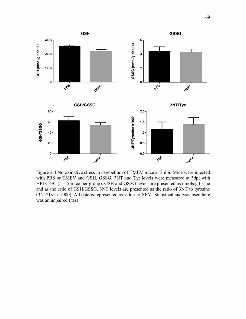
Figure 2.4 No oxidative stress in cerebellum of TMEV mice at 3 dpi. Mice were injected with PBS or TMEV and GSH, GSSG, 3NT and Tyr levels were measured at 3dpi with HPLC-EC (n = 5 mice per group). GSH and GSSG levels are presented as nmols/g tissue and as the ratio of GSH/GSSG. 3NT levels are presented as the ratio of 3NT to tyrosine (3NT/Tyr x 1000). All data is represented as values ± SEM. Statistical analysis used here was an unpaired t test

CHAPTER 3

HIPPOCAMPAL TNFα SIGNALING CONTRIBUTES TO

HYPEREXCITABILITY IN AN INFECTION-

INDUCED MOUSE MODEL OF

LIMBIC EPILEPSY

Introduction

Encephalitis as a consequence of viral infections of the CNS is often associated

with the occurrence of acute seizures and a dramatically increased probability of the

subsequent development of acquired epilepsy (Vezzani et al., 2016). Indeed, a

retrospective epidemiological study found an overall 22-fold increase in the risk for

developing epilepsy following viral encephalitis if patients present with acute seizures

(Misra et al., 2008). CNS infection can cause intense inflammatory responses and

parenchymal damage in the brain contributing to acute seizures which, in turn, can

exacerbate inflammatory conditions, ensuring further CNS damage and the development

of recurring chronic seizures (Vezzani et al., 2011; Vezzani et al., 2016). Therefore,

investigating the role of inflammation as a consequence of CNS infection could provide

valuable insight for the development of next generation disease-modifying therapies for

the prevention of epilepsy.

Theiler’s murine encephalomyelitis virus (TMEV)-infected C57BL/6J mice have

71

acute seizures between 3 and 8 days postinfection (dpi), exhibit pathological and

physiological changes such as astrogliosis, microgliosis, neuronal loss in CA1, and

increased excitatory synaptic transmission in CA3 pyramidal neurons in the

hippocampus. Importantly, the mice survive the infection, clear the virus from the brain,

present with cognitive impairment and anxiety-like symptoms and develop chronic

spontaneous seizures after a latent period (Broer et al., 2016; Kirkman et al., 2010;

Libbey et al., 2008; Loewen et al., 2016; Smeal et al., 2012; Stewart et al., 2010a;

Umpierre et al., 2014). Thus, TMEV-infected mice recapitulate many clinical

observations from patients suffering from infection-induced temporal lobe epilepsy

(TLE) and offer a unique opportunity to study the molecular mechanism(s) underlying

the process of infection-induced epileptogenesis.

TMEV infection results in a dramatic increase in the expression of cytokines,

chemokines, oxidative stress markers, and infiltration of macrophages in the first week of

infection which may contribute to the initiation and/or propagation of seizures during the

acute infection period in this model (Bhuyan et al., 2015; Cusick et al., 2013; Kirkman et

al., 2010). The level of tumor necrosis factor-α (TNFα) mRNA in the whole brain of

TMEV-infected mice exhibiting acute seizures was increased by 128-fold at 6 dpi

compared to control mice (Kirkman et al., 2010). Further, only 10% of TNFα receptor 1

knock-out (TNFR1 KO) mice developed acute behavioral seizures compared to 52% of

wild-type (WT) control mice, which implies a significant role of TNFR1-mediated effects

of TNFα in seizure development in this model (Kirkman et al., 2010).

TNFα signaling through the TNFR1 pathway has been shown to contribute to the

regulation of homeostatic synaptic scaling by modulating the postsynaptic expression of

72

α-amino-3-hydroxy-5-methyl-4-isoxazolepropionic acid receptors (AMPARs) under both

physiological and pathogenic conditions (Beattie et al., 2010). TNFα increases the

expression of GluR1-containing and GluR2-lacking AMPARs on the surface of cultured

hippocampal neurons and in pyramidal cells in acute rat hippocampal slices (Beattie et

al., 2002; Stellwagen et al., 2005; Stellwagen and Malenka, 2006). Whole cell patch-

clamp recordings from CA1 pyramidal neurons in acute hippocampal slices pretreated

with TNFα show significant increases in average amplitudes of miniature excitatory

postsynaptic currents (mEPSCs) (Stellwagen et al., 2005). This suggests that TNFα may

contribute to hyperexcitability by augmenting excitatory synaptic strength by increased

trafficking of AMPARs subunits to postsynaptic membranes (Stellwagen et al., 2005;

Stellwagen and Malenka, 2006). The effects of TNFα on regulating excitatory synaptic

strength have also been described in rodent models of spinal injury (Ferguson et al.,

2008), pain (Choi et al., 2010), and glaucoma (Cueva Vargas et al., 2015).

The present experiments were performed to evaluate the role of hippocampal

TNFα signaling in seizure generation during the acute period following TMEV infection.

We found a significant increase in expression of both hippocampal mRNA and protein

levels of TNFα following TMEV infection that was coincident with focal seizure activity.

In addition, a significant increase in the TNFR1:TNFR2 ratio in hippocampus suggests

that signaling through the TNFR1 pathway predominates during the acute infection

period. Consistent with increased TNFR1 signaling, increases in hippocampal cell surface

AMPA receptor expression was also observed during the acute period. While treatment

with XPro1595, a mutant form of human soluble TNFα (sTNFα) that acts as a dominant-

negative selective inhibitor of sTNFα, had no effect on seizures during the acute TMEV-

73

infection period, several lines of transgenic animals deficient in either TNFα or its

receptors were found to have robust changes in seizure incidence and severity following

TMEV infection. Taken together the present results suggest that increases in TNFα

signaling, likely through the TNFR1 pathway, contributes to hyperexcitability and the

increased probability of seizures in the hippocampus following TMEV infection.

Therefore, this pathway may provide a novel target for antiseizure and disease modifying

treatments following CNS infection.

Methods

Animals

C57BL/6J mice (WT (#006460), TNFR2-/- (#002620), TNFR1-/-TNFR2-/-

(#003243), and TNFα-/- (#005540)) aged between 4-6 weeks were purchased from

Jackson Laboratory. TNFR2-/-, TNFR1-/-TNFR2-/-, and TNFα-/- mice were bred at our

vivarium. All the KO mice were on C57BL/6J background and the deletion of the target

protein was confirmed by PCR. All the experiments were carried out in male mice unless

otherwise specified. After arrival, mice were allowed to acclimatize for at least 3 days

prior to the experiment. Mice were provided food and water ad libitum and kept in a

facility providing 12 h of light and lark cycle starting at 6:00 AM. All the procedures

performed were in accordance with the guidelines provided and approved by the

Institutional Animal Care and Use Committee of the University of Utah.

74

Method of TMEV infection and seizure monitoring

Mice are briefly anesthetized with 3% isoflurane and injected with 20 µl of either

phosphate-buffered saline (PBS) or DA-TMEV solution intracortically in the right

hemisphere by inserting the needle at a 90° angle to the skull. The injection region is

located slightly medial to the equidistant point on the imaginary line connecting the eye

and the ear. A sterilized syringe containing a plastic jacket on the needle exposing 2.5

mm of needle is used for infection to restrict the injection site to the somatosensory

cortex without damaging the hippocampus. TMEV titer injected per mouse ranged from

2x10⁴ to 3x10⁵ PFU (plaque forming units) depending on the experiment.

Mice were briefly agitated by shaking their cages and monitored for behavioral

seizures twice daily in the morning and afternoon separated by a minimum of 2 h until 10

dpi. Seizure intensity was graded using modified Racine scale as follows: stage 1, mouth

and facial movements; stage 2, head nodding; stage 3, forelimb clonus; stage 4, forelimb

clonus, rearing; and stage 5, forelimb clonus, rearing, and falling; stage 6, intense

running, jumping, repeated falling, and severe clonus. Seizure frequency was reported as

an average number of seizures during the entire acute seizure period, whereas seizure

severity/intensity was represented as an average cumulative seizure burden at each dpi

during acute seizure period. Cumulative seizure burden at each dpi for a mouse was

calculated by summing all of its seizure scores up to that dpi.

Tissue collection for mRNA and protein analysis

TMEV-infected mice that had acute behavioral seizures were used for all the

biochemical studies. TMEV- and PBS-treated mice were sacrificed at 1, 4, 5, and 14 dpi

75

depending on the experiment. The ipsilateral and contralateral hippocampi were rapidly

isolated and collected separately. All the tissue samples were flash frozen using 2-

methylbutane chilled on dry ice and stored at -80 ºC until further processing.

Multiplex cytokine array

The protein levels of an array of cytokines were measured in mouse ipsilateral

hippocampal lysates using a multiplex kit (V-PLEX™, K15048D) from Mesoscale

Discovery according to manufacturer’s instructions. Briefly, ipsilateral hippocampi were

homogenized in MSD Tris lysis buffer in a 1:10 weight by volume ratio, centrifuged and

the supernatant collected. Protein concentration was determined using the Bradford assay

and 250 µg of protein was loaded per well to measure cytokines. The levels of analytes

were determined by measuring the intensity of emitted light at 620 nm using a Sector

Imager 2400.

Gel electrophoresis and western blot

Protein expressions of TNFRs were quantified by western blot analysis. The

ipsilateral hippocampi were homogenized in 10 µl of lysis buffer (25 mM Tris-HCl, 150

mM NaCl, 1 mM EDTA, 1% Igepal CA-630, 5% glycerol, protease inhibitors (cocktail

tablet, Roche), and 1 mM sodium orthovanadate) per mg of tissue and the supernatant

was collected after centrifugation. Total protein concentration was measured by BCA

protein assay (Pierce) and 10 µg of total protein was electrophoresed using

polyacrylamide gel (4-12% Bis-Tris gel, NuPAGE™, Invitrogen) under denaturing

conditions. The proteins were transferred to a PVDF membrane and detected by

76

chemiluminescence (NEL105001EA, PerkinElmer) using rabbit polyclonal antibodies

against TNFR1 (#ab64006, 1:25000, Abcam), TNFR2 (#3727, 1:1000, Cell Signaling

Technologies) and actin (#A2103, 1:250000, Sigma) followed by HRP-conjugated

secondary antibody (#65-6120, 1:3000, Invitrogen). Densitometric analysis of protein

levels was performed using ImageJ software (National Institute of Health, NIH).

Quantitative reverse transcription polymerase

chain reaction (RT-qPCR)

Total RNA was isolated from the hippocampi samples by Trizol/chloroform

extraction and purified to remove genomic DNA contamination by DNase treatment

followed by a spin column-based method (RNeasy Mini Kit, Qiagen) according to

manufacturer’s instructions. Quality and quantity of RNA were validated by

spectrophotometry and acrylamide gel electrophoresis which showed intense discrete

ribosomal RNA bands devoid of genomic DNA. cDNA was synthesized from RNA using

random primers (SuperScript® VILO Master Mix, Invitrogen) and amplified by qPCR

(LightCycler®480, Roche) using 1 µg cDNA with Luminaris Color HiGreen qPCR

Master Mix (Thermo Fisher Scientific) and 0.3 µM primers for TNFR1 (Forward (F), 5’-

AGAGAAAGTGAGTGCGTCCC-3’; Reverse (R), 5’-AGCCTTCTCCTCTTTGAC-

AGG-3’), TNFR2 (F, 5’-AGCTGCAGTTCTTCCTGTACC-3’; R, 5’-GATGCTACA-

GATGCGGTGGG-3’), TNFα (F, 5’-CTGAACTTCGGGGTGATCGG-3’, R, 5’-GGC-

TTGTCACTCGAATTTTGAGA-3’, β-actin (F, 5’-AGATCAAGATCATTGCTCC-

TCC-3’, R, 5’-ACGCAGCTCAGTAACAGTCC-3’) and GAPDH (F, 5’-AGCTAC-

TCGCGGCTTTACG-3’, R, 5’-GGCCAAATCCGTTCACACC-3’). LC480 software

77

used a second derivative formula to calculate threshold cycle (CT). Primer efficiency for

each analyte was determined using four dilutions of cDNA, and the primer efficiency was

used to calculate ΔCT and ΔΔCT relative to the reference gene as well as PBS-treated

control group. Primer specificity was confirmed by melt curves followed by agarose gel

electrophoresis for the expected size product.

Treatment of TMEV-infected mice with XPro1595

TMEV-infected mice were treated with 10 or 100 mg/kg of XPro1595

subcutaneously either every third day or daily. Vehicle contained 150 mM NaCl, 10 mM

L-histidine, and 0.01% w/v Tween-20 in deionized water (pH 6.51). For

intracerebroventricular (i.c.v.) administration of XPro1595, the guide cannula was

surgically implanted into the left lateral ventricle using stereotaxic coordinates of -1.1

mm lateral, -0.5 mm posterior, and -3.0 mm ventral from the bregma as described in

detail previously (DeVos and Miller, 2013). Briefly, mice were anesthetized by 10 ml/kg

i.p. injection of a mixture of ketamine and xylazine solution in sterile PBS (final

concentration: ketamine – 10.4 mg/ml, Xylazine – 1.6 mg/ml) and the skull was exposed

in a sterile surgical environment. Guide cannulas (C315GS-5/SP, 3 mm below pedestal,

PlasticsOne) were inserted into the left lateral ventricle using a sharp beveled end of the

cannula and glued to the skull. A dummy cannula (C315DCS-5/SPC, PlasticsOne) was

placed in the guide cannula to prevent the exposure of the ventricle to the outside

environment. Mice were allowed to recover from surgery for 12-15 days before initiating

the experiment. Mice were immobilized during drug infusion by anesthetizing them using

isoflurane (VetEquip® isoflurane vaporizer) and the dummy cannula was removed just

78

before the drug infusion in a laminar flow hood under aseptic conditions. An internal

cannula (C315IS-5/SPC, PlasticsOne) connected to a 10 µl Hamilton® syringe via tubing

was inserted into the guide cannula to infuse either XPro1585 or the vehicle solution. An

infusion pump (PHD 2000 programmable, Harvard Apparatus) was used to infuse either

5 µl of 40 mg/ml XPro1595 at 1 µl/min (0 and 2 dpi) or 2.5 µl of 80 mg/ml XPro1595 at

0.5 µl/min (4 and 6 dpi). The internal cannula was kept in place for about 1 min after

infusion and then slowly removed to avoid leakage. The dummy cannula was secured

into the guide cannula immediately.

Cell surface biotinylation assay

Horizontal brain slices (350 µm) from control and TMEV-infected mice with

seizures were prepared by vibratome at 5 dpi in ice-cold sucrose solution (concentrations

in mM: 200 sucrose, 3 KCl, 26 NaHCO3, 1.4 NaH2PO4, 10 glucose, and 3 MgSO4 and 1

CaCl2 added before use; 4°C) and collected in artificial cerebrospinal fluid (aCSF,

concentrations in mM: 126 NaCl, 3 KCl, 26 NaHCO3, 1.4 NaH2PO4, 10 glucose, and 2

MgSO4 and 2 CaCl2 added before use; pH, 7.33-7.35; osmolality, 297-303 mOsm/kg) at

room temperature. Only brain slices ipsilateral to the injection were used for further

processing. The slices were incubated in aCSF (31°C) for 45-50 min to recover from the

surface damage inflicted during slicing. All the remaining steps were conducted at 4°C.

The slices were washed with aCSF to remove dead surface cells and debris and incubated

in 1 mg/ml solution of Sulfo-NHS-SS-biotin (Pierce) for 30 min to biotinylate cell

surface proteins. The excess biotin solution was washed off using aCSF, quenched by

incubating the slices in 100 mM glycine solution, and again washed with aCSF. The

79

hippocampal regions were dissected out quickly, collected in 250 µl lysis buffer (recipe

same as described in western blot procedure), homogenized, centrifuged (14,000 x g, 15

min, 4°C) and the supernatant was collected. All the solutions used for processing brains

and brain slices were continuously oxygenated with the mixture of 95% oxygen and 5%

carbon dioxide.

Total protein (biotinylated surface proteins and nonbiotinylated intracellular

proteins) concentrations in the supernatant were measured by a BCA protein assay

(Pierce). To isolate biotinylated surface proteins (SP) from the total proteins (TP), 50 µg

of TP was incubated with 25 µl of streptavidin beads (NeutrAvidin™, Thermo scientific)

overnight at 4°C on a rotator. The appropriate ratio of beads to TP for each protein of

interest was empirically measured by incubating a constant volume of beads with a range

of TP in order to isolate the corresponding SP, as described in detail (Gabriel et al.,

2014). We chose a ratio of beads to TP of 1:2, which was found to isolate SP in a linear

range. The mixture of beads and TP was centrifuged and the beads were washed with

lysis buffer. The SP were eluted by incubating the beads in 20 µl of 2X SDS-PAGE

reducing sample buffer containing 50 mM DTT in the final mixture (4X SDS-PAGE

sample buffer: 106 mM Tris-HCl, 141 mM Tris-base, 0.51 mM EDTA, 2% SDS, 10%

glycerol) for 30 min with continuous gentle mixing at room temperature. The supernatant

containing SP was collected after centrifugation (17,000 x g, 2 min). The expression of

GluA1 and GluA2 subunits of AMPARs were measured in both SP and TP fractions by

SDS-PAGE and western blot (anti-GluA1 mAb #MAB2263 and anti-GluA2 mAb

#MAB397, Millipore) as described above. The entire volume of supernatant containing

SP (isolated from 50 µg of TP) and 10 µg of TP were electrophoresed in the same gel.

80

Patch-clamp electrophysiology

Mice were sacrificed and their brains were removed between 3-7 dpi, and 350 µM

horizontal brain slices were cut in ice-cold sucrose solution and incubated in aCSF

solution for 1 h at room temperature. Miniature EPSCs were recorded from the dentate

granule cells (DGCs) by whole-cell patch-clamp in aCSF solution containing 1 µM

tetrodotoxin as described previously (Smeal et al., 2012). The internal recording solution

contained (in mM): 129 potassium gluconate, 6 CsCl, 10 HEPES, 1 EGTA, 0.5 CaCl2, 10

glucose, 2 ATP, 0.5 GTP, 5 QX314, 1 NaCl, and 5 tetraethylammonium chloride. The

equilibrium potential for ionotropic glutamate receptors (iGluR ENa+/K+) was 4.66 mV at

room temperature and mEPSCs were recorded by clamping the cell at -70 mV. Properties

of the miniature currents analyzed included amplitude, interevent interval, frequency, rise

time and decay time (Smeal et al., 2012).

Statistics

Datasets involving continuous variables are represented by the average and the

standard error of the mean (SEM), and the datasets with ordinal variables by frequency

distribution. Experimental design involving two groups with one continuous dependent

variable was analyzed by unpaired two-tailed t test, whereas design involving more than

two groups with two categorical independent variables and one continuous dependent

variable was analyzed by two-way ANOVA. Multiple comparisons were performed by

Bonferroni posttest. Average cumulative seizure burden, which was calculated from a

ranked dataset, was analyzed by Scheirer-Ray-Hare test which is an extension of the

Kruskal-Wallis test for two randomized factorial designs (Scheirer et al., 1976). Two

81

groups with binomial outcome were analyzed by Fisher’s exact test. “Survival” (%

seizure free) curves and cumulative distributions were analyzed by log-rank test and

Kolmogorov-Smirnov test, respectively. Densitometry of immunoblot images and the

analysis of mEPSCs were conducted by Image J (NIH) and Mini Analysis Program

(Version 6.0.7, Synaptosoft), respectively. Statistical calculations were conducted using

GraphPad Prism® 5 and Microsoft Excel.

Results

Increased expression of TNFα and other cytokines in the

hippocampus following TMEV infection

Previous studies have demonstrated that TNFα mRNA levels increase

dramatically by 128-fold in whole brain homogenates of TMEV-infected mice with acute

behavioral seizures at 6 dpi compared to noninfected control mice (Kirkman et al., 2010).

TMEV infection results in significant cell loss in the CA1 region, increased c-fos

immunoreactivity in the hippocampus following TMEV-induced acute seizures, and

increased excitability in the CA3 region of the hippocampus, a brain region often

associated with seizure initiation in TLE (Smeal et al., 2012). Therefore, it is important to

determine the role of hippocampal TNFα in TMEV-induced seizures. We measured the

expression levels of TNFα mRNA and protein in the hippocampus of TMEV-infected

mice and PBS-injected control mice. TMEV-infected mice exhibit acute behavioral and

electrographic seizures between 3-8 dpi. Therefore, we measured the levels of TNFα at 1,

5, and 14 dpi to compare the levels before, during, and after the acute seizure period.

Only handling-induced behavioral seizures were evaluated in these and the following

82

studies, as the presence of depth electrodes could influence the cytokine response. The

mRNA level of TNFα in TMEV-infected mice was not significantly different at 1 dpi

compared to control mice (n = 4) (Figure 3.1a). However, it dramatically increased by

161-fold (n = 4, p<0.001) at 5 dpi during the peak of the acute seizure activity and it was

still 88-fold higher at 14 dpi (n = 4, p<0.001) (Figure 3.1a). Similarly, immunoassay

studies found no significant increase in the protein expression of TNFα in the

hippocampus of TMEV-infected mice compared to control mice at 1 dpi, but TNFα was

elevated 206-fold (p<0.001) and 35-fold (p<0.05) at 5 and 14 dpi, respectively (Figure

3.1b, Table 3.1) (Control: n = 5 (all time points); TMEV: n = 8 (1 dpi), 6 (5 dpi), and 5

(14 dpi)). The absolute average (± SEM) levels of TNFα were measured as follows: 4.89

± 1.19 pg/ml (control) and 33.80 ± 3.09 pg/ml (TMEV) at 1 dpi; 0.54 ± 0.22 pg/ml

(control) and 110.50 ± 7.96 pg/ml (TMEV) at 5 dpi; and 1.41 ± 0.65 pg/ml (control) and

48.92 ± 10.02 pg/ml (TMEV) at 14 dpi. Additionally, protein levels of many other

cytokines were also increased in the hippocampus in TMEV-infected mice with seizures

at 5 dpi (Table 3.1), notably interferon-γ (IFNγ) which was 21,734-fold elevated

compared to control mice. In addition, the antiinflammatory cytokine, interleukin-10 (IL-

10), was also increased by 47-fold, which suggests that negative immune feedback also

occurs during acute TMEV infection to control excessive inflammation.

Increased protein expression ratio of TNFR1:TNFR2

in hippocampus during acute seizures

The TNFα receptors, TNFR1 and TNFR2, mediate contrasting effects of TNFα in

various disease models (Fischer et al., 2015) and differential changes in the protein levels

83

of TNFR1 and TNFR2 have been shown in the hippocampus in a rat model of limbic

epilepsy (Weinberg et al., 2013). Therefore, we measured the expression of TNFR1 and

TNFR2 in hippocampus following TMEV infection by western blot. Figure 3.1c shows a

representative western blot image for TNFR1, TNFR2, and actin (gel loading control)

from TMEV-infected mice at 1, 4, and 14 dpi along with PBS-treated control mice.

Optical density (OD) analysis revealed a slight but significant reduction in the expression

of TNFR1 in TMEV-infected mice at 4 dpi (OD: 0.97 ± 0.012 vs. 0.83 ± 0.01, n = 5-6,

p<0.05) and 14 dpi (OD: 0.95 ± 0.03 vs. 0.75 ± 0.064, n = 5-6, p<0.01) after normalizing

the data with actin levels (Figure 3.1d). However, the expression of TNFR2 protein levels

were dramatically reduced at 4 dpi (OD: 0.90 ± 0.029 vs. 0.51 ± 0.038, n = 5-6, p<0.001)

and 14 dpi (OD: 0.99 ± 0.049 vs. 0.42 ± 0.087, n = 5-6, p<0.001) (Figure 3.1d). The ratio

of TNFR1:TNFR2, indicating the relative expressions of both TNFRs in the hippocampus

during the acute infection period, was significantly elevated (1.08 ± 0.033 vs. 1.67 ±

0139, p<0.05) at 4 dpi and was further increased at 14 dpi (0.97 ± 0.057 vs. 2.04 ± 0.255,

p<0.001) (Figure 3.1d) which suggests that TNFα may mediate its downstream effects

predominantly through TNFR1 in the hippocampus during the acute TMEV infection

period.

We also measured the mRNA levels of both TNFRs using qPCR. In contrast to

TNFα, we found opposite changes in the levels of both TNFR1 and TNFR2 for mRNA

and protein expression. The expression of TNFR1 mRNA was significantly elevated by

2.59, 5.5, and 4.65-fold at 1, 5, and 14 dpi, respectively, (n = 4); whereas the protein

levels were slightly reduced (Figure 3.1e). Similarly, mRNA expression of TNFR2 was

increased by 12.94- and 16.8-fold at 5 and 14 dpi (n = 4); whereas the protein levels were

84

reduced by 0.57- and 0.42-fold at 4 and 14 dpi (Figure 3.1f). These changes result in a

progressive decrease in the relative mRNA expression ratios of TNFR1:TNFR2 and an

increase in the relative protein expression ratios of TNFR1:TNFR2 in the hippocampus

during acute TMEV infection (Figure 3.1g). This suggests that TNFR expression

undergoes posttranslational regulation.

Lack of seizure control following peripheral administration

of XPro1595 on TMEV-induced acute seizures

There is a significant increase in whole brain and hippocampal TNFα mRNA, an

increase in protein expression of TNFα, and an increase in the ratio of TNFR1:TNFR2

expression in the hippocampus as a consequence of TMEV infection. In addition, prior

work has demonstrated that seizure incidence is dramatically reduced in TNFR1-/- mice

(Kirkman et al., 2010). Therefore, we hypothesized that pharmacological inhibition of

TNFR1 could decrease the incidence and/or severity of TMEV-induced acute seizures.

TNFα is expressed as a homotrimeric transmembrane protein (tmTNFα) and the

extracellular portion of tmTNFα can be cleaved by TNFα converting enzyme (TACE) to

form a soluble TNFα (sTNFα). Both tmTNFα and sTNFα are active in their trimeric

composition and mediate a variety of cellular activities via TNFRs (McCoy and Tansey,

2008). sTNFα predominantly mediates its effects via TNFR1, while TNFR2 is fully

activated only by tmTNFα (Grell et al., 1995; Grell et al., 1998). While anti-TNFα

antibodies are approved for the treatment of peripheral inflammatory conditions, they do

not cross blood brain barrier (BBB) (Tracey et al., 2008). Therefore, we selected the

investigational BBB-permeant compound, XPro1595, to test the hypothesis that TNFα

85

signaling through TNFR1 contributes to seizure generation. XPro1595 is a mutant form

of human sTNFα and acts as a dominant-negative selective inhibitor of sTNFα (Steed et

al., 2003). It dose-dependently exchanges with endogenous monomers of sTNFα to form

an inactive heterotrimer which does not bind to TNFRs and lacks intrinsic bioactivity

(Steed et al., 2003). Since sTNFα predominantly mediates its effects via TNFR1,

XPro1595 indirectly inhibits TNFR1 functions. XPro1595 has been shown to provide

beneficial effects in animal models of several peripheral as well as CNS inflammatory

conditions including experimental autoimmune encephalomyelitis (EAE), Parkinson’s

disease, spinal cord injury, and focal cerebral ischemia by selectively inhibiting sTNFα-

TNFR1 signaling and sparing beneficial functions of TNFR2 (Brambilla et al., 2011;

Clausen et al., 2014; McCoy et al., 2006; Novrup et al., 2014; Zalevsky et al., 2007).

Since the dosing regimen of 10 mg/kg of subcutaneous (s.c.) XPro1595 administered

every third day was effective in models of other CNS diseases (Barnum et al., 2014;

Brambilla et al., 2011; Clausen et al., 2014), we tested whether peripheral treatment of

TMEV-infected mice with XPro1595 could prevent the development of handling-induced

acute seizures. Handling sessions were captured by video monitoring and experimenters

scoring seizures were blinded to the treatment groups as described in the methods.

Administration of 10 mg/kg of XPro1595 (s.c.) at 1, 4, and 7 dpi did not decrease average

number of seizures (Vehicle: 5.27 ± 0.37, XPro1595: 5.63 ± 0.45, n = 30, p=0.5313) or

have any effect on average cumulative seizure burden at any time point during the acute

seizure period when compared to vehicle-treated mice. To determine if the lack of

efficacy on seizure incidence and severity was due to an inappropriate dosing regimen,

we increased the dosing frequency of XPro1595 (10 mg/kg) to every day, starting from 2

86

h postinfection through 9 dpi. This dosing regime was also ineffective in decreasing

either seizure frequency (Vehicle: 5.13 ± 0.71, XPro1595: 5.13 ± 0.77, n = 15, p=1.00) or

severity. Finally, we increased the dose to 100 mg/kg and started treatment 2 days prior

to infection through 7 dpi. Despite a 10-fold increase in the dose and initiating the

treatment before infection, both XPro1595 and vehicle-treated groups had similar average

numbers of seizures (Vehicle: 6.63 ± 1.24, XPro1595: 6.25 ± 0.77, n = 8, p=0.801) and

average cumulative seizure burdens during the acute seizure period. Thus, systemic

administration of high doses of XPro1595 was not effective in controlling seizures

induced by TMEV infection.

Lack of seizure control following CNS administration of

XPro1595 on TMEV-induced acute seizures

Despite numerous studies demonstrating central activity of XPro1595 in animal

models of neurological disorders, a recent study found that systemic injection of

XPro1595 was insufficient to provide therapeutic efficacy in a mouse model of spinal

cord injury (SCI), whereas central administration of XPro1595 provided neuroprotection

and ameliorated motor dysfunction in this model (Novrup et al., 2014). The concentration

of XPro1595 in the CSF (1-6 ng/ml) has been found to be 1000-fold reduced compared to

that in the plasma (1-8 µg/ml) from rats after 2-3 days of treatment with XPro1595 (10

mg/kg, s.c.) (Barnum et al., 2014). Furthermore, as in the model of SCI, where acute

inflammatory response occurs rapidly in the CNS, the protein levels of TNFα and other

inflammatory cytokines increase rapidly in the hippocampus after TMEV infection. Thus,

we hypothesized that XPro1595 did not achieve a sufficient concentration in the brain to

87

prevent signaling through the TNFR1 system following subcutaneous treatment in

TMEV-infected mice Therefore, we tested the effectiveness of CNS administration of

XPro1595 by free-hand bolus injection of 5 µl of XPro1595, equivalent to about 10

mg/kg of mouse, at 2 dpi into the left lateral ventricle. Surprisingly, the average number

of seizures was significantly reduced by treatment with XPro1595 (Vehicle: 5.6 ± 0.40,

XPro1595: 3.1 ± 0.77, n = 10, p=0.0097), as well as average cumulative seizure burden at

each day during 6-8 dpi (Figure 3.2a). However, the results could not be replicated when

we repeated the free-hand i.c.v bolus experiment (average number of seizures, Vehicle:

5.2 ± 0.63, XPro1595: 5.3 ± 0.79, n = 10, p=0.9222) (Figure 3.2b). Single bolus

administration of XPro1595 might not have been sufficient to suppress endogenous

TNFα during the entire acute seizure activity period. Therefore, mice were implanted

with guide cannulas into the left lateral ventricle for multiple drug infusion in the CNS.

Infusions of XPro1595 (10 mg/kg starting at 5-6 h after infection) or vehicle were

repeated every other day for a total 4 infusions. As was observed following systemic

injections, central administration of XPro1595 had no effect on either average number of

seizures (Vehicle: 4.18 ± 1.32, XPro1595: 2.75 ± 1.02, n = 11-12, p=0.3952) or average

cumulative seizure burden at any day during 3-8 dpi (Figure 3.2c, 3.2d) compared to

TMEV-treated mice receiving vehicle infusions. The placement of the guide cannula into

the ventricle was confirmed in all the mice enrolled for this study by infusing 0.1%

Evan’s blue dye at 9 dpi (Figure 3.2e). Therefore, regardless of administration route,

dosing regimen, or dose, XPro1595 was ineffective in preventing seizures following

TMEV infections.

88

Differential susceptibility of TNFRs KO mice to develop

TMEV-induced acute seizures

Previous work has demonstrated that only 10% of TNFR1-/- mice (n = 2/20)

infected with 2x10⁴ PFU of TMEV developed acute behavioral seizures, suggesting that

TNFR1-mediated effects could be involved in seizure generation in this model (Kirkman

et al., 2010). As XPro1595 did not block acute seizures, we used transgenic animals to

further test the hypothesis that TNFα signaling contributes to hyperexcitability following

infection. Seizure incidence and severity in TNFα-/-, TNFR2-/-, and TNFR1-/-TNFR2-/-

mice during the acute infection period was therefore evaluated (Figure 3.3). All the mice

were infected with 2x10⁴ PFU of TMEV to compare the data with the published findings

in TNFR1-/- mice. Although the numbers of infected mice that developed acute

behavioral seizures were similar for WT and TNFα-/- (67% seized mice, n = 18), seizure

frequency and severity, as measured by average number of seizures and average

cumulative seizure burden, were significantly reduced in TNFα-/- mice (Figure 3.3a).

Interestingly, TNFR2-/- mice experienced severe seizures compared to WT as evidenced

by an increase in the average (± SEM) number of seizures (WT: 2.8 ± 0.42, TNF2-/-: 4.6

± 0.43, p=0.0042, n = 27 (WT), n = 30 (TNFR2-/-)) and an average cumulative seizure

burden at 7 and 8 dpi (Figure 3.3b). The latency to develop the first seizure was also

significantly reduced in TNFR2-/-, as 11 of 30 TNFR2-/- mice experienced seizures at 3

dpi compared to just 1 of 27 WT mice. In addition, the overall percentage of seizure free-

mice for TNFR2-/- and WT mice over the entire acute seizure period was significantly

different (p=0.0046) (Figure 3.3c). Since previous work demonstrated that TNFR1-/- mice

were less susceptible to developing acute seizures (Kirkman et al., 2010) and TNFR2-/-

89

developed severe TMEV-induced seizures, we reasoned that TNFR1-/-TNFR2-/- mice

might have seizure patterns similar to WT mice. However, TNFR1-/-TNFR2-/- exhibited a

significantly reduced average number of seizures (WT: 3.1 ± 0.50, TNFR1-/-TNF2-/-: 1.0

± 0.34, p=0.001, n = 28 (WT), n = 30 (TNFR1-/-TNFR2-/-)) as well as average cumulative

seizure burden at 6, 7, and 8 dpi (Figure 3.3d) compared to WT mice. The percentages of

total numbers of infected mice that developed acute behavioral seizures in TNFR1-/-,

TNFR1-/-TNFR2-/-, TNFα-/-, WT, and TNFR2-/- were 10% (n = 2/20) (Kirkman et al.,

2010), 33% (n = 10/30), 67% (n = 12/18), 73% (n = 40/55), and 93% (n = 28/30),

respectively (Figure 3.3e). Both male and female mice had similar seizure responses in

all the strains tested, and thus, the data from both genders are pooled. In contrast to the

lack of seizure control observed with XPro1595 treatment, results in the transgenic

mouse strains suggest that TNFα signaling through the TNFR1 pathway could indeed be

a prominent mechanism through which hyperexcitability and seizure activity occur

following TMEV infection. In addition, TNFα signaling through the TNFR2 pathway, as

is the case in other neurological disorders, may be involved in dampening excitability,

since mice lacking TNFR2 have a higher incidence and greater severity of seizures.

Increase in the surface levels of AMPAR subunits

during acute seizures in WT B6 mice

AMPARs are the primary glutamate receptors that mediate fast excitatory

neurotransmission in the brain (Traynelis et al., 2010). TNFα has been shown to increase

AMPAR trafficking into postsynaptic neuronal membranes via neuronal TNFR1 (Beattie

et al., 2002; Stellwagen et al., 2005). In addition, we have previously demonstrated that

90

the amplitudes of mEPSCs are increased during the acute infection period in CA3

pyramidal neurons, suggesting an increase in AMPA receptor surface expression (Smeal

et al., 2012). Given the hypothesized role of TNFα in AMPAR trafficking and the

observed increase in mEPSC amplitudes following TMEV infection, we evaluated

hippocampal AMPA receptor expression using a cell surface biotinylation assay in mice

treated with TMEV. Biotinylated surface proteins were separated from nonbiotinylated

intracellular proteins using avidin beads in tissue prepared from acute hippocampal slices

obtained from TMEV- and PBS-treated mice at 5 dpi as described here (Gabriel et al.,

2014). The GluA1 and GluA2 subunits of AMPARs were probed in the surface protein

(SP) as well as in the total protein (TP) fractions by western blot (Figure 3.4a). The ratios

of surface/total levels of GluA1 that indicate the relative levels of GluA1 present on the

cell surface were significantly elevated in the TMEV-infected mice with seizures

compared to control (OD: 0.31 ± 0.011 vs. 0.45 ± 0.018, p<0.0001, n = 6). We also

probed for phosphate-activated glutaminase (PAG), a mitochondrial protein, to control

for the biotinylation of intracellular proteins. The levels of PAG in the SP fraction were

less than 5% compared to those in the TP fraction (OD: 0.015 ± 0.0026 in PBS, 0.03 ±

0.0015 in TMEV) indicating that the biotinylation was largely restricted to cell surface

proteins. The surface/total ratio for the levels of GluA2 was also significantly increased

in TMEV-infected mice compared to control (OD: 0.53 ± 0.018 vs. 0.7 ± 0.025,

p=0.0007, n = 6) (Figure 3.4b). The cell surface levels of GluA1 and GluA2 subunits

were increased by 48% (p<0.0001) and 33% (p=0.0002), respectively, in TMEV-infected

mice compared to PBS-injected mice (Figure 3.4c). The total protein levels of GluA1 and

GluA2 in the TMEV-infected mice were significantly reduced by 46% and 55%,

91

respectively, compared to control mice (Figure 3.4d). Since TMEV-infected mice have a

pronounced neuronal loss in the CA1 region, it is not surprising that the total protein

levels of GluA1 and GluA2 are decreased in TMEV-infected mice.

Since TNFR2-/- mice had severe acute seizures compared to WT mice, we also

conducted cell surface biotinylation assay in TNFR2-/- mice to test if TNFR2-/- mice had

higher cell surface levels of GluA1 and GluA2 at 5 dpi. Similar to TMEV-infected WT

C57BL/6J mice, the ratios of surface/total levels of GluA1 and GluA2 were significantly

elevated in the TMEV-infected TNFR2-/- mice with seizures compared to PBS-injected

TNFR2-/- mice (For GluA1, OD: 0.28 ± 0.012 vs. 0.40 ± 0.021, p=0.0011, n = 6; and for

GluA2, OD: 0.45 ± 0.016 vs. 0.61 ± 0.036, p=0.0049, n = 6) (Figure 3.5a, 3.5b). The cell

surface levels of GluA1 and GluA2 subunits were increased by 44% (p=0.0011) and 34%

(p=0.0049), respectively, in the TMEV group compared to control mice (Figure 3.5c).

The total protein levels of GluA1 and GluA2 in the TMEV-infected TNFR2-/- mice were

significantly reduced by 42% and 48%, respectively, compared to control mice (Figure

3.5d), likely due to the widespread neurodegeneration observed in CA1 of the

hippocampus.

mEPSCs recorded in DGCs are not affected by TMEV infection

TMEV exhibits a strong tropism for limbic brain regions and viral particles can be

detected in CA1 and CA2 regions of hippocampus in the first week of infection (Buenz et

al., 2009; Kirkman et al., 2010; Stewart et al., 2010b). Abundant c-Fos immunoreactivity,

an indirect marker for neuronal activation, was found in the CA3 region and the dentate

gyrus (DG) of hippocampus within 2 h after TMEV-induced acute seizures (Smeal et al.,

92

2012). Since the CA1 region is severely damaged by TMEV within 4-5 days of infection,

initial studies investigating hyperexcitability in the hippocampus were focused on the

CA3 region. Patch-clamp studies in acute brain slices found an increase in the amplitude

and frequency of spontaneous and miniature EPSCs in CA3 pyramidal neurons during the

period of 3-7 days post-TMEV infection (Smeal et al., 2012). The DG is another key

region of the hippocampus that is frequently involved in seizure generation in limbic

epilepsy models (Sloviter et al., 2012). Further, TNFα and TNFRs regulate homeostatic

synaptic plasticity by enhancing excitatory synaptic strength in the DGCs in response to

denervation-induced injury (Becker et al., 2015; Becker et al., 2013). Given our finding

that there is an increased ratio of cell surface to total AMPA receptors in the biotinylation

studies, we performed whole cell patch clamp recordings in DGCs to determine if there

was an increase, as observed in CA3, in excitatory synaptic transmission during the acute

infection period when seizures are observed. Miniature EPSCs in DGCs in hippocampal

brain slices were obtained the during acute seizure period. Representative traces of

mEPSCs from control and TMEV-infected mice are shown in Figure 3.6a. A total of 11

DGCs from 8 mice in the control group and 15 DGCs from 10 TMEV-infected mice with

acute seizures were included in the analysis. The cumulative fraction analysis of mEPSC

amplitude as well as interevent interval found no significant differences between

treatment groups (p>0.05, KS test) (Figure 3.6b, 3.6c). The average (± SEM) mEPSC

amplitudes were 14.3 ± 0.66 pA and 14.9 ± 0.59 pA in the control and TMEV mice,

respectively (p=0.5), and the average (± SEM) frequencies were 2.30 ± 0.36 Hz and 2.35

± 0.23 Hz for the control and TMEV mice, respectively (p=0.9). These data suggest that,

unlike CA3 pyramidal neurons, DGCs do not have an increase in excitatory synaptic

93

transmission during the acute infection period.

Discussion

The present series of experiments evaluated the role of TNFα signaling in the

hippocampus, a brain region involved in seizure generation, following TMEV infection.

TNFα and its effects mediated via the activation of TNFR1, and/or reduced signaling

through TNFR2, could be one of the major inflammatory pathways contributing to acute

seizures. We demonstrate here, for the first time, that there is a substantial increase in the

protein level of TNFα that is coincident with an increase in the protein expression ratios

of TNFR1:TNFR2 in the hippocampus. The role of TNFα in contributing to seizure

generation following TMEV infection was also supported by our findings in a number of

transgenic mice. We found that there is a decrease in the seizure incidence, frequency,

and severity of acute seizures in TNFR1-/- and TNFR1-/-TNFR2-/- mice, whereas there is

an increase in frequency and severity of acute seizures in TNFR2-/- mice. In addition,

consistent with the hypothesized role of TNFα in hippocampal AMPA receptor

trafficking, we demonstrated that there is a significant increase in the cell surface to total

AMPA receptor ratio in TMEV transfected mice. This increase in cell surface expression

likely underlies the increased amplitudes of mEPSCs that we have previously observed in

recordings obtained in CA3 pyramidal cells in brain slices obtained from TMEV-infected

mice (Smeal et al., 2012). While peripheral as well as central administration of

XPro1595, an inhibitor of sTNFα, failed to inhibit TMEV-induced acute seizures, the

data acquired here from WT and transgenic animals suggests that signaling through the

TNFα system may play an important role in seizure generation during the acute infection

94

period and may serve as an important therapeutic target in conditions in which

inflammation contributes to seizure generation.

TNFα signaling contributes to the regulation of homeostatic synaptic plasticity by

modulating the postsynaptic surface levels of AMPARs under physiological conditions

(Beattie et al., 2010). TNFα has been shown to induce synaptic scaling via neuronal

TNFR1 by increasing the cell surface expression of GluA1-containing and GluA2-

lacking AMPARs in cultured hippocampal neurons (Beattie et al., 2002; Stellwagen et

al., 2005; Stellwagen and Malenka, 2006), in the rat hippocampal slice preparation

(Stellwagen et al., 2005; Stellwagen and Malenka, 2006), in an animal model of SCI

(Ferguson et al., 2008), and also in the dorsal spinal cord neurons in an intraplantar

carrageenan model of inflammatory pain (Choi et al., 2010). Elevated levels of AMPARs

in postsynaptic membranes can increase the strength of excitatory synaptic transmission.

Furthermore, GluA2-lacking AMPARs are Ca2+-permeable and increased intracellular

concentration of Ca2+ can result into excitotoxicity (Liu and Zukin, 2007). We report that

the levels of GluA1 and GluA2 subunits of AMPARs on the cell surface are elevated in

the hippocampus of mice with acute seizures compared to noninfected mice. Differences

in the trafficking of GluA2 subunits between SCI, pain models, and the present study

may reflect the differences in underlying pathological, anatomical, and experimental

conditions. Indeed, TNFα has been shown to exert varying effects on the regulation of

excitatory synaptic strength contingent upon factors such as brain region. TNFα drives

the internalization of GluA1 and GluA2 subunits of AMPARs in the medium spiny

neurons in striatum, which is in contrast to its effects in hippocampal neurons (Lewitus et

al., 2014). The data acquired here suggests that the increased cell surface expression of

95

AMPAR subunits most likely occurs in CA3 pyramidal neurons because 1) AMPA

receptors are expressed in principal cells of the hippocampus (Osten et al., 2006), 2) CA1

pyramidal neurons start to degenerate by 3 dpi and exhibit extensive loss by 4 dpi

(Loewen et al., 2016), 3) significant increases in amplitude and frequency of mEPSCs

recorded from CA3 pyramidal neurons in TMEV-infected mice with seizures between 3-

7 dpi (Smeal et al., 2012), and 4) no change in mEPSCs in DGCs in TMEV-infected mice

with seizures between 3-7 dpi. AMPARs are also present in glial cells although with

much less density than in neurons (Traynelis et al., 2010), and hippocampal interneurons

express mainly GluA1 and GluA4, but lack GluA2 (Osten et al., 2006); therefore, we do

not rule out the possibility of some postsynaptic AMPAR dynamics in these cells. A

novel approach for real-time in vivo monitoring of AMPAR subunits has recently been

described (Zhang et al., 2015), which could be utilized to address the question of spatial

and cellular subunit targeting in the hippocampus where AMPAR trafficking occurs

during TMEV-induced seizures.

TNFR1-/- and TNFR1-/-TNFR2-/- mice were found to be highly resistant to

developing TMEV-induced acute seizures, and those with seizures had much less severe

seizures compared to WT mice. In addition, although the percentage of TMEV-infected

TNFα-/- mice developing acute seizures was similar to WT mice, frequency and severity

were significantly reduced in TNFα-/- mice. Taken together with the AMPAR trafficking

findings, we conclude that TNFα is likely implicated in synaptic scaling via TNFR1

resulting in hippocampal hyperexcitability and seizures. However, additional studies that

utilize pharmacological approaches to modulate the level of TNFα and the functions of

TNFRs in the brain will be necessary to determine the mechanism through which

96

increased levels of TNFα impact excitatory strength and seizure generation.

Inhibition of sTNFα-TNFR1-mediated effects and sparing tmTNFα-TNFR2-

mediated effects of TNFα has been a well-rationalized strategy to treat many peripheral

and CNS inflammatory conditions which also minimizes adverse effects associated with

absolute inhibition of TNFα (Fischer et al., 2015). In previous studies, the concentration

of XPro1595 in CSF and plasma were found to be 1-6 ng/ml and 1-8 µg/ml, respectively,

following subcutaneous treatment of rats with 10 mg/kg XPro1595 (Barnum et al., 2014).

A 10-fold higher level of XPro1595, such as used in the present experiments, should

therefore exchange 99% of endogenous sTNFα (Steed et al., 2003). In the present studies,

the average concentration of TNFα in the hippocampus was 110.5 pg/ml at 5 days post-

TMEV infection in mice with acute seizures. Thus, if the CSF concentration of XPro1595

in rats after 10 mg/kg s.c. dosing could be extrapolated to TMEV-infected mice, 10

mg/kg s.c. dosing should have been sufficient to remove the endogenously active form of

sTNF. The present finding that XPro1595 treatment was unable to influence TMEV-

induced seizures could be due to an insufficient concentration of XPro1595 in the

hippocampus. This could also have been the limitation in a recent study where only CNS,

but not peripheral, administration of XPro1595 had beneficial effects in the mouse model

of SCI (Novrup et al., 2014). However, multiple i.c.v. infusion of XPro1595 also failed to

reduce TMEV-induced seizures in the present study. Further data on the

pharmacokinetics of XPro1595 in TMEV-infected mice could facilitate a better design of

dosing regimen for treatment with this compound. However, even if XPro1595

sufficiently removed endogenous sTNFα, TNFR1 may still be activated by tmTNFα.

Although sTNFα is a major ligand for TNFR1, tmTNFα has been shown to induce

97

inflammation and cytotoxicity through TNFR1 (Horiuchi et al., 2010). For example, a

recent study using XPro1595 to prevent peripheral inflammation in a model of gout

determined that the inflammatory response observed in that model was independent of

sTNFα and was likely due to tmTNFα (Amaral et al., 2016). Thus, XPro1595 was unable

to prevent the inflammation observed in that model. In addition, lymphotoxin (LTα3),

formerly known as TNFβ, is structurally similar to sTNFα and binds to both TNFRs

(Tracey et al., 2008). LTα3-TNFR1 signaling can cause inflammation similar to sTNFα-

TNFR1 effects which could partly explain the lack of effects of XPro1595 on TMEV-

induced seizures. Several TNFR1-specific inhibitors are under development (Fischer et

al., 2015), which should be leveraged in the future to test the hypothesis of a pathogenic

role of TNFR1-mediated signaling in TMEV-induced seizures. Finally, it has been found

in previous studies that IL-6 signaling can also contribute to seizure generation during the

TMEV acute infection period (Kirkman et al., 2010; Libbey et al., 2011). Animals

deficient in IL-6, or treated with wogonin to reduce infiltrating macrophages, which are

the major source of IL-6 (Cusick et al., 2013), also have a decrease in acute seizure

activity in this model (Kirkman et al., 2010). Therefore a polytherapy approach involving

both TNF and IL-6 modulators may prove the most efficacious in preventing seizure

activity following TMEV infection.

In addition to increasing AMPAR trafficking to the synaptic membrane, TNFα

has been proposed to increase excitation by modulating the functions of astrocytes.

Pathogenic levels of TNFα in hippocampus may induce the release of glutamate from

astrocytes into the synapse where glutamate can bind to presynaptic NMDA receptors to

further increase the synaptic release of glutamate (Habbas et al., 2015; Santello et al.,

98

2011). TNFα has also been shown to decrease expression of the astrocyte glutamate

transporter (GLT1) (Carmen et al., 2009). Recently, TNFα has been demonstrated to

downregulate connexin-43 and GLT1 on spinal astrocytes, causing increased

glutamatergic neurotransmission and neuropathic pain in mice (Morioka et al., 2015).

Astrogliosis occurs throughout the hippocampus within 3 days of TMEV infection and

persists up to 4-6 months postinfection (Loewen et al., 2016; Stewart et al., 2010a). It is a

matter of future interest to determine if increased levels of TNFα affect the expression of

glutamate transporters and gap junction proteins on astrocytes to tip the balance of

synaptic function in favor of hyperexcitatory conditions.

Our results that TNFα contributes to neuronal hyperexcitability in the

hippocampus through TNFR1-mediated signaling, whereas TNFR2-mediated signaling

imparts antiictogenic effects, are not specific to the TMEV model. Similar diametric roles

of TNFRs in modulating seizure activity have been reported in kainic acid and kindling

models of limbic seizures (Weinberg et al., 2013). Our results also corroborate clinical

findings in which the levels of TNFR1 and TNFR1-signaling proteins are increased in the

resected hippocampal tissues from patients with refractory TLE (Yamamoto et al., 2006).

Although animal studies that investigate the role of the TNFα system in various models

of limbic seizures suggest that TNFR1-mediated signaling contributes to ictogenic

effects, the consequences of pathogenic levels of TNFα on seizures are highly context-

dependent. For example, overexpression of murine TNFα in astrocytes decreased kainate-

induced seizures (Balosso et al., 2005), whereas overexpression of murine TNFα in

neurons either caused seizures (Probert et al., 1995) or had no effects on kainate-induced

seizure activity (Weinberg et al., 2013). Multiple factors in the CNS including the source

99

and the type of TNFα, the cell types expressing TNFRs, levels of TNFα and relative

density of TNFRs in the tissue can influence the seizure outcome (Probert, 2015). These

factors must be investigated in detail by designing animal studies that reflect clinical

findings from the patients with TLE.

In conclusion, we have demonstrated that pathogenic level of TNFα and an

increase in the expression ratio of TNFR1:TNFR2 in the hippocampus are associated

with acute behavioral as well as focal hippocampal electrographic seizures in TMEV-

infected mice. TNFα might cause hyperexcitation in TMEV-infected mice by

strengthening excitatory synapses via a TNFR1-mediated mechanism, whereas TNFR2

may provide antiictogenic effects during acute infection. Inflammation occurs in other

models of epilepsy and many patients with epilepsy present with CNS inflammatory

conditions. Antiinflammatory therapies could be useful as stand-alone therapy or in

combination with antiseizure medications. Anti-TNFα antibodies are widely prescribed

for peripheral inflammatory conditions, however, they also cause serious adverse effects

by inhibiting desirable functions of TNFα (Traynelis et al., 2010). Therefore, TNFR1-

specific inhibitors and/or TNFR2-specific agonists might be a better strategy for reducing

seizures.

References Amaral, F.A., Bastos, L.F., Oliveira, T.H., Dias, A.C., Oliveira, V.L., Tavares, L.D.,

Costa, V.V., Galvao, I., Soriani, F.M., Szymkowski, D.E., Ryffel, B., Souza, D.G., Teixeira, M.M., 2016. Transmembrane TNF-alpha is sufficient for articular inflammation and hypernociception in a mouse model of gout. Eur. J. Immunol. 46, 204-211.

Balosso, S., Ravizza, T., Perego, C., Peschon, J., Campbell, I.L., De Simoni, M.G.,

Vezzani, A., 2005. Tumor necrosis factor-alpha inhibits seizures in mice via p75

100

receptors. Ann. Neurol. 57, 804-812. Barnum, C.J., Chen, X., Chung, J., Chang, J., Williams, M., Grigoryan, N., Tesi, R.J.,

Tansey, M.G., 2014. Peripheral administration of the selective inhibitor of soluble tumor necrosis factor (TNF) XPro(R)1595 attenuates nigral cell loss and glial activation in 6-OHDA hemiparkinsonian rats. J. Parkinsons Dis. 4, 349-360.

Beattie, E.C., Stellwagen, D., Morishita, W., Bresnahan, J.C., Ha, B.K., Von Zastrow,

M., Beattie, M.S., Malenka, R.C., 2002. Control of synaptic strength by glial TNFalpha. Science 295, 2282-2285.

Beattie, M.S., Ferguson, A.R., Bresnahan, J.C., 2010. AMPA-receptor trafficking and

injury-induced cell death. Eur. J. Neurosci. 32, 290-297. Becker, D., Deller, T., Vlachos, A., 2015. Tumor necrosis factor (TNF)-receptor 1 and 2

mediate homeostatic synaptic plasticity of denervated mouse dentate granule cells. Sci. Rep. 5, 12726.

Becker, D., Zahn, N., Deller, T., Vlachos, A., 2013. Tumor necrosis factor alpha

maintains denervation-induced homeostatic synaptic plasticity of mouse dentate granule cells. Front. Cell. Neurosci. 7, 257.

Bhuyan, P., Patel, D.C., Wilcox, K.S., Patel, M., 2015. Oxidative stress in murine

Theiler's virus-induced temporal lobe epilepsy. Exp. Neurol. 271, 329-334. Brambilla, R., Ashbaugh, J.J., Magliozzi, R., Dellarole, A., Karmally, S., Szymkowski,

D.E., Bethea, J.R., 2011. Inhibition of soluble tumour necrosis factor is therapeutic in experimental autoimmune encephalomyelitis and promotes axon preservation and remyelination. Brain 134, 2736-2754.

Broer, S., Kaufer, C., Haist, V., Li, L., Gerhauser, I., Anjum, M., Bankstahl, M.,

Baumgartner, W., Loscher, W., 2016. Brain inflammation, neurodegeneration and seizure development following picornavirus infection markedly differ among virus and mouse strains and substrains. Exp. Neurol. 279, 57-74.

Buenz, E.J., Sauer, B.M., Lafrance-Corey, R.G., Deb, C., Denic, A., German, C.L.,

Howe, C.L., 2009. Apoptosis of hippocampal pyramidal neurons is virus independent in a mouse model of acute neurovirulent picornavirus infection. Am. J. Pathol. 175, 668-684.

Carmen, J., Rothstein, J.D., Kerr, D.A., 2009. Tumor necrosis factor-alpha modulates

glutamate transport in the CNS and is a critical determinant of outcome from viral encephalomyelitis. Brain Res. 1263, 143-154.

Choi, J.I., Svensson, C.I., Koehrn, F.J., Bhuskute, A., Sorkin, L.S., 2010. Peripheral

inflammation induces tumor necrosis factor dependent AMPA receptor trafficking

101

and Akt phosphorylation in spinal cord in addition to pain behavior. Pain 149, 243-253.

Clausen, B.H., Degn, M., Martin, N.A., Couch, Y., Karimi, L., Ormhoj, M., Mortensen,

M.L., Gredal, H.B., Gardiner, C., Sargent, II, Szymkowski, D.E., Petit, G.H., Deierborg, T., Finsen, B., Anthony, D.C., Lambertsen, K.L., 2014. Systemically administered anti-TNF therapy ameliorates functional outcomes after focal cerebral ischemia. J. Neuroinflammation 11, 203.

Cueva Vargas, J.L., Osswald, I.K., Unsain, N., Aurousseau, M.R., Barker, P.A., Bowie,

D., Di Polo, A., 2015. Soluble Tumor Necrosis Factor Alpha Promotes Retinal Ganglion Cell Death in Glaucoma via Calcium-Permeable AMPA Receptor Activation. J. Neurosci. 35, 12088-12102.

Cusick, M.F., Libbey, J.E., Patel, D.C., Doty, D.J., Fujinami, R.S., 2013. Infiltrating

macrophages are key to the development of seizures following virus infection. J. Virol. 87, 1849-1860.

DeVos, S.L., Miller, T.M., 2013. Direct intraventricular delivery of drugs to the rodent

central nervous system. J Vis Exp, e50326. Ferguson, A.R., Christensen, R.N., Gensel, J.C., Miller, B.A., Sun, F., Beattie, E.C.,

Bresnahan, J.C., Beattie, M.S., 2008. Cell death after spinal cord injury is exacerbated by rapid TNF alpha-induced trafficking of GluR2-lacking AMPARs to the plasma membrane. J. Neurosci. 28, 11391-11400.

Fischer, R., Kontermann, R., Maier, O., 2015. Targeting sTNF/TNFR1 Signaling as a

New Therapeutic Strategy. Antibodies 4, 48. Gabriel, L.R., Wu, S., Melikian, H.E., 2014. Brain slice biotinylation: an ex vivo

approach to measure region-specific plasma membrane protein trafficking in adult neurons. J Vis Exp.

Grell, M., Douni, E., Wajant, H., Lohden, M., Clauss, M., Maxeiner, B., Georgopoulos,

S., Lesslauer, W., Kollias, G., Pfizenmaier, K., Scheurich, P., 1995. The transmembrane form of tumor necrosis factor is the prime activating ligand of the 80 kDa tumor necrosis factor receptor. Cell 83, 793-802.

Grell, M., Wajant, H., Zimmermann, G., Scheurich, P., 1998. The type 1 receptor

(CD120a) is the high-affinity receptor for soluble tumor necrosis factor. Proc. Natl. Acad. Sci. U. S. A. 95, 570-575.

Habbas, S., Santello, M., Becker, D., Stubbe, H., Zappia, G., Liaudet, N., Klaus, F.R.,

Kollias, G., Fontana, A., Pryce, C.R., Suter, T., Volterra, A., 2015. Neuroinflammatory TNFalpha Impairs Memory via Astrocyte Signaling. Cell 163, 1730-1741.

102

Horiuchi, T., Mitoma, H., Harashima, S., Tsukamoto, H., Shimoda, T., 2010. Transmembrane TNF-alpha: structure, function and interaction with anti-TNF agents. Rheumatology (Oxford) 49, 1215-1228.

Kirkman, N.J., Libbey, J.E., Wilcox, K.S., White, H.S., Fujinami, R.S., 2010. Innate but

not adaptive immune responses contribute to behavioral seizures following viral infection. Epilepsia 51, 454-464.

Lewitus, G.M., Pribiag, H., Duseja, R., St-Hilaire, M., Stellwagen, D., 2014. An adaptive

role of TNFalpha in the regulation of striatal synapses. J. Neurosci. 34, 6146-6155.

Libbey, J.E., Kennett, N.J., Wilcox, K.S., White, H.S., Fujinami, R.S., 2011. Interleukin-

6, produced by resident cells of the central nervous system and infiltrating cells, contributes to the development of seizures following viral infection. J. Virol. 85, 6913-6922.

Libbey, J.E., Kirkman, N.J., Smith, M.C., Tanaka, T., Wilcox, K.S., White, H.S.,

Fujinami, R.S., 2008. Seizures following picornavirus infection. Epilepsia 49, 1066-1074.

Liu, S.J., Zukin, R.S., 2007. Ca2+-permeable AMPA receptors in synaptic plasticity and

neuronal death. Trends Neurosci. 30, 126-134. Loewen, J.L., Barker-Haliski, M.L., Dahle, E.J., White, H.S., Wilcox, K.S., 2016.

Neuronal injury, gliosis, and glial proliferation in two models of temporal lobe epilepsy. J. Neuropathol. Exp. Neurol. 75, 366-378.

McCoy, M.K., Martinez, T.N., Ruhn, K.A., Szymkowski, D.E., Smith, C.G., Botterman,

B.R., Tansey, K.E., Tansey, M.G., 2006. Blocking soluble tumor necrosis factor signaling with dominant-negative tumor necrosis factor inhibitor attenuates loss of dopaminergic neurons in models of Parkinson's disease. J. Neurosci. 26, 9365-9375.

McCoy, M.K., Tansey, M.G., 2008. TNF signaling inhibition in the CNS: implications

for normal brain function and neurodegenerative disease. J. Neuroinflammation 5, 45.

Misra, U.K., Tan, C.T., Kalita, J., 2008. Viral encephalitis and epilepsy. Epilepsia 49

Suppl 6, 13-18. Morioka, N., Zhang, F.F., Nakamura, Y., Kitamura, T., Hisaoka-Nakashima, K., Nakata,

Y., 2015. Tumor necrosis factor-mediated downregulation of spinal astrocytic connexin43 leads to increased glutamatergic neurotransmission and neuropathic pain in mice. Brain. Behav. Immun. 49, 293-310.

103

Novrup, H.G., Bracchi-Ricard, V., Ellman, D.G., Ricard, J., Jain, A., Runko, E., Lyck, L., Yli-Karjanmaa, M., Szymkowski, D.E., Pearse, D.D., Lambertsen, K.L., Bethea, J.R., 2014. Central but not systemic administration of XPro1595 is therapeutic following moderate spinal cord injury in mice. J. Neuroinflammation 11, 159.

Osten, P., Wisden, W., Sprengel, R., 2006. Molecular mechanisms of synaptic function in

the hippocampus: neurotransmitter exocytosis and glutamatergic, GABAergic, and cholinergic transmission, in: Andersen, P., Morris, R., Amaral, D., Bliss, T., O'Keefe, J. (Eds.), Hippocampus Book. Oxford University Press, USA, Cary, US.

Probert, L., 2015. TNF and its receptors in the CNS: The essential, the desirable and the

deleterious effects. Neuroscience 302, 2-22. Probert, L., Akassoglou, K., Pasparakis, M., Kontogeorgos, G., Kollias, G., 1995.

Spontaneous inflammatory demyelinating disease in transgenic mice showing central nervous system-specific expression of tumor necrosis factor alpha. Proc. Natl. Acad. Sci. U. S. A. 92, 11294-11298.

Santello, M., Bezzi, P., Volterra, A., 2011. TNFalpha controls glutamatergic

gliotransmission in the hippocampal dentate gyrus. Neuron 69, 988-1001. Scheirer, C.J., Ray, W.S., Hare, N., 1976. The analysis of ranked data derived from

completely randomized factorial designs. Biometrics 32, 429-434. Sloviter, R.S., Bumanglag, A.V., Schwarcz, R., Frotscher, M., 2012. Abnormal dentate

gyrus network circuitry in temporal lobe epilepsy, in: Noebels, J.L., Avoli, M., Rogawski, M.A., Olsen, R.W., Delgado-Escueta, A.V. (Eds.), Jasper's Basic Mechanisms of the Epilepsies, 4th ed, Bethesda (MD).

Smeal, R.M., Stewart, K.A., Iacob, E., Fujinami, R.S., White, H.S., Wilcox, K.S., 2012.

The activity within the CA3 excitatory network during Theiler's virus encephalitis is distinct from that observed during chronic epilepsy. J. Neurovirol. 18, 30-44.

Steed, P.M., Tansey, M.G., Zalevsky, J., Zhukovsky, E.A., Desjarlais, J.R., Szymkowski,

D.E., Abbott, C., Carmichael, D., Chan, C., Cherry, L., Cheung, P., Chirino, A.J., Chung, H.H., Doberstein, S.K., Eivazi, A., Filikov, A.V., Gao, S.X., Hubert, R.S., Hwang, M., Hyun, L., Kashi, S., Kim, A., Kim, E., Kung, J., Martinez, S.P., Muchhal, U.S., Nguyen, D.H., O'Brien, C., O'Keefe, D., Singer, K., Vafa, O., Vielmetter, J., Yoder, S.C., Dahiyat, B.I., 2003. Inactivation of TNF signaling by rationally designed dominant-negative TNF variants. Science 301, 1895-1898.

Stellwagen, D., Beattie, E.C., Seo, J.Y., Malenka, R.C., 2005. Differential regulation of

AMPA receptor and GABA receptor trafficking by tumor necrosis factor-alpha. J. Neurosci. 25, 3219-3228.

Stellwagen, D., Malenka, R.C., 2006. Synaptic scaling mediated by glial TNF-alpha.

104

Nature 440, 1054-1059. Stewart, K.A., Wilcox, K.S., Fujinami, R.S., White, H.S., 2010a. Development of

postinfection epilepsy after Theiler's virus infection of C57BL/6 mice. J. Neuropathol. Exp. Neurol. 69, 1210-1219.

Stewart, K.A., Wilcox, K.S., Fujinami, R.S., White, H.S., 2010b. Theiler's virus infection

chronically alters seizure susceptibility. Epilepsia 51, 1418-1428. Tracey, D., Klareskog, L., Sasso, E.H., Salfeld, J.G., Tak, P.P., 2008. Tumor necrosis

factor antagonist mechanisms of action: a comprehensive review. Pharmacol. Ther. 117, 244-279.

Traynelis, S.F., Wollmuth, L.P., McBain, C.J., Menniti, F.S., Vance, K.M., Ogden, K.K.,

Hansen, K.B., Yuan, H., Myers, S.J., Dingledine, R., 2010. Glutamate receptor ion channels: structure, regulation, and function. Pharmacol. Rev. 62, 405-496.

Umpierre, A.D., Remigio, G.J., Dahle, E.J., Bradford, K., Alex, A.B., Smith, M.D., West,

P.J., White, H.S., Wilcox, K.S., 2014. Impaired cognitive ability and anxiety-like behavior following acute seizures in the Theiler's virus model of temporal lobe epilepsy. Neurobiol. Dis. 64, 98-106.

Vezzani, A., French, J., Bartfai, T., Baram, T.Z., 2011. The role of inflammation in

epilepsy. Nat. Rev. Neurol. 7, 31-40. Vezzani, A., Fujinami, R.S., White, H.S., Preux, P.M., Blumcke, I., Sander, J.W.,

Loscher, W., 2016. Infections, inflammation and epilepsy. Acta Neuropathol. 131, 211-234.

Weinberg, M.S., Blake, B.L., McCown, T.J., 2013. Opposing actions of hippocampus

TNFalpha receptors on limbic seizure susceptibility. Exp. Neurol. 247, 429-437. Yamamoto, A., Schindler, C.K., Murphy, B.M., Bellver-Estelles, C., So, N.K., Taki, W.,

Meller, R., Simon, R.P., Henshall, D.C., 2006. Evidence of tumor necrosis factor receptor 1 signaling in human temporal lobe epilepsy. Exp. Neurol. 202, 410-420.

Zalevsky, J., Secher, T., Ezhevsky, S.A., Janot, L., Steed, P.M., O'Brien, C., Eivazi, A.,

Kung, J., Nguyen, D.H., Doberstein, S.K., Erard, F., Ryffel, B., Szymkowski, D.E., 2007. Dominant-negative inhibitors of soluble TNF attenuate experimental arthritis without suppressing innate immunity to infection. J. Immunol. 179, 1872-1883.

Zhang, Y., Cudmore, R.H., Lin, D.T., Linden, D.J., Huganir, R.L., 2015. Visualization of

NMDA receptor-dependent AMPA receptor synaptic plasticity in vivo. Nat. Neurosci. 18, 402-407.

105

Figure 3.1 Increase in the levels of TNFα and in a ratio of the protein expression of TNFR1:TNFR2 in the hippocampus of TMEV-infected mice during acute seizure activity period. (a) mRNA levels of TNFα, as measured by RT-qPCR, are significantly increased in TMEV-infected mice at 5 and 14 days postinfection (dpi) by 161- and 88-fold, respectively, compared to PBS-infected control mice (n = 4 for TMEV and control). (b) 206- and 35-fold increase in the protein expression levels of TNFα in TMEV-infected mice at 5 and 14 dpi compared to the PBS-injected control mice (Control: n = 5; TMEV: n = 8 (1 dpi), 6 (5 dpi), and 5 (14 dpi)). (c) Representative immunoblot shows the protein expression of TNFR1, TNFR2, and actin in the hippocampus from PBS- and TMEV-infected mice (n = 3). (d) Densitometric analysis of the immunoblots shows the expressions of TNFR1 and TNFR2 normalized to the expression levels of actin (Control: n = 5; TMEV: n = 5 (1 dpi), 6 (4 and 14 dpi); O.D., Optical density). The relative expression levels of TNFR1 and TNFR2 (TNFR1:TNFR2) are significantly increased by 1.54- and 2.1-fold at 4 and 14 dpi, respectively, in the TMEV-infected mice compared to control mice. (e-g) Comparison of mRNA and protein levels of TNFR1 and TNFR2 in the hippocampus of TMEV-infected mice before (1 dpi), during (5 dpi), and after (14 dpi) acute seizures. The ratio of TNFR1 to TNFR2 for mRNA is significantly reduced during the acute infection period (g, upper panel), whereas the ratio for the protein expression is significantly increased over the acute infection period (g, lower panel). The data are shown as mean ± SEM. Statistics: Two-way ANOVA followed by Bonferroni posttest; *p<0.05, **p<0.01, and ***p<0.001.

106

107

Figure 3.2 CNS administration of XPro1595 does not affect TMEV-induced acute seizure frequency and intensity. (a) Single i.c.v. bolus administration of XPro1595 (10 mg/kg) at 2 dpi in TMEV-infected mice (n = 10) significantly reduces an average number of generalized tonic-clonic seizures and an average cumulative seizure burden starting from 6 dpi compared to vehicle-treated TMEV-infected mice. (b) The repetition of single i.c.v. bolus administration study does not replicate the findings shown in (a). (c) Slow infusion of XPro1595 (10 mg/kg) at 0, 2, 4, and 6 dpi into the left lateral ventricle using a surgically implanted guide cannula does not reduce average seizure frequency and severity (n = 12, XPro1595; n = 11, vehicle). Each circle represents an individual mouse and the horizontal line shows average number of seizures per group. (d) Average seizure burden corresponding to each stage of modified Racine scale for generalized tonic-clonic seizures shows no difference between vehicle- and XPro1595-treated TMEV-infected mice. Only those mice which had acute seizures are included in this analysis. (e) The surgical placement of the guide cannula into the left lateral ventricle was confirmed by i.c.v. infusion of 0.1% Evans blue dye at 9 dpi in all TMEV-infected mice treated with either XPro1595 or vehicle. The panel shows an example of a diffusion of the dye into the ventricular system.

108

Figure 3.3 TMEV-induced acute behavioral seizure susceptibility in WT, TNFα-/-, TNFR2-/-, and TNFR1-/-TNFR2-/- mice. (a) TNFα-/- mice have a significant reduction in the seizure frequency (upper panel), plotted as a total number of seizures per mouse, and in the seizure severity (lower panel) as measured by an average cumulative seizure burden during acute seizure period (3-8 dpi) compared to WT mice. Each circle represents individual mouse and the horizontal line shows an average number of seizures per group. (b) TNFR2-/- mice show an increase in the average seizure frequency as well as severity. (c) Reduced latency to develop a first TMEV-induced acute seizure in TNFR2-/- mice compared to WT mice. (d) Reduced average seizure frequency and severity in TNFR1-/-TNFR2-/- mice. (e) Percentage of total infected mice that show acute behavioral seizures during 3-8 dpi. Statistics: unpaired t test (frequency), Scheirer-Ray-Hare test (severity), Fisher’s exact test (% seized mice); and long-rank test (% seizure free); **p<0.01, ***p<0.001.

109

Figure 3.4 Increase in the cell surface levels of GluA1 and GluA2 subunits of AMPARs in TMEV-infected WT C57BL/6J mice during acute seizures. (a) Representative immunoblots from two mice show the levels of GluA1 in the total as well as the cell surface fractions of proteins isolated from ipsilateral hippocampus at 5 days postinjection of either PBS (control) or TMEV. Data in the first (total) and the third (surface) lanes from the left are from the same mouse, and the second (total) and the fourth (surface) lanes correspond to the other mouse. The surface proteins were isolated from the intracellular proteins by cell surface biotinylation procedure in acute hippocampal slices. The levels of GluA1 were quantified by densitometry and the data are shown as a ratio of surface to total protein which is significantly increased in TMEV-infected mice (n = 6). Phosphate-activated glutaminase (PAG) is a mitochondrial protein and serves as an intracellular control protein. (b) Similar to GluA1, the ratio of surface/total level for GluA2 is also increased in TMEV-infected mice (n = 6). (c) The ratios of surface/total protein expression for GluA1 and GluA2 are increased by 48% and 33%, respectively (data normalized to control). (d) About 50% decrease in the total expressions of GluA1 and GluA2 in TMEV-infected mice compared to the control group. Statistics: Unpaired two-tailed t test.

110

Figure 3.5 Increase in the cell surface levels of GluA1 and GluA2 subunits of AMPARs in TMEV-infected TNFR2-/- mice during acute seizures. (a) Representative immunoblots from two mice show the levels of GluA1 in the total as well as the cell surface fractions of proteins isolated from ipsilateral hippocampus at 5 days postinjection of either PBS (control) or TMEV. The surface proteins were isolated by cell surface biotinylation procedure in acute hippocampal slices. The levels of GluA1 were quantified by densitometry and the data are shown as a ratio of surface to total protein that is significantly increased in TMEV-infected mice (n = 6). Phosphate-activated glutaminase (PAG) is a mitochondrial protein and serves as an intracellular control protein. (b) Similar to GluA1, the ratio of surface/total level for GluA2 is also increased in TMEV-infected mice (n = 6). (c) The ratios of surface/total protein expression for GluA1 and GluA2 are increased by 44% and 34%, respectively (data normalized to control). (d) About 50% decrease in the total expressions of GluA1 and GluA2 in TMEV-infected mice compared to the control group. Statistics: Unpaired two-tailed t test.

111

Figure 3.6 No difference in the properties of miniature excitatory postsynaptic currents (mEPSCs) of dentate granule cells (DGCs) between PBS-injected (control) and TMEV-infected mice during acute seizure activity period. (a) Representative traces of mEPSCs measured in the DGCs from the control group and TMEV-infected mice during 3-7 dpi. (b) Cumulative fraction distribution of the amplitude of mEPSCs shows no difference between the control and the TMEV groups. Average amplitudes of mEPSCs are plotted in the lower panel (Control: n = 8, TMEV: n = 10). (c) Cumulative fraction distribution of the interevent interval (IEI) of mEPSCs shows no difference between both the treatment groups. The lower panel shows the average frequency of mEPSCs. Statistics: Kolmogorov-Smirnov test (cumulative fraction), unpaired t test (average amplitude and frequency).

112

Table 3.1 Significant increase in the protein levels of various inflammatory mediators in the hippocampus of TMEV-infected mice during acute seizure activity period. Statistics: Two-way ANOVA, Bonferroni posttest, #p<0.05, *p<0.01, †p<0.001; SEM, standard error of the mean. (TNFα, tumor necrosis factor-α; IFNγ, interferon-γ; IL, interleukin; CXCL1, C-X-C motif chemokine ligand 1)

Cytokine Fold change (relative to PBS-injected control mice)

Average SEM Average SEM Average SEM 1 dpi (n = 8) 5 dpi (n = 6) 14 dpi (n = 5)

TNFα 6.9 0.6 206.2† 14.9 34.8# 7.1 IFNγ 4.5 0.8 21734.4† 3123.2 99.1 21.6 IL-1β 6.9 1.1 58.2† 5.4 46.6† 13.5 IL-10 2.3 0.3 47.2† 6.0 9.6 1.9 IL-12p70 2.5 0.3 3.0# 0.5 6.0† 1.0 IL-4 1.0 0.2 0.8 0.1 1.3 0.3 IL-2 2.4 0.3 14.4† 4.2 4.9 0.7 IL-5 2.0 0.1 10.8* 2.5 2.6 1.2 CXCL1 4.6 0.3 30.0† 4.3 2.4 0.3

CHAPTER 4

CANNABIDIOL TREATMENT PREVENTS SEIZURES

FOLLOWING CNS INFECTION WITH THEILER’S

MURINE ENCEPHALOMYELITIS VIRUS

Introduction

Theiler’s Murine Encephalomyelitis Virus (TMEV) infection in the CNS of

C57BL/6J mice causes a robust inflammatory response and oxidative stress in the

hippocampus that is coincident with an acute seizure activity period. This acute seizure

period is followed by a latent period and the majority of mice exhibiting acute seizures go

on to develop epilepsy (Stewart et al., 2010). Protein levels of several cytokines and

chemokines, including TNFα, IFNγ, IL-1β, IL-6, IL-2, IL-5, IL-12, and CXCL1, are

significantly increased in the hippocampus of TMEV-infected mice during the acute

behavioral seizure period (Table 3.1) (Cusick et al., 2013; Kirkman et al., 2010). In

addition, mice deficient in TNFR1, TNFα, or IL-6 are much less susceptible to

developing TMEV-induced acute seizures compared to WT mice (Figure 3.3a) (Kirkman

et al., 2010). Oxidative and nitrative stress markers, such as elevated levels of 3-

nitrotyrosine and impaired glutathione redox status, as evidenced by a decrease in the

ratio of GSH:GSSG, have also been observed in the hippocampus of TMEV-infected

mice with seizures as early as 3 dpi (Bhuyan et al., 2015). Thus, proinflammatory and

114

redox changes in the brain following TMEV infection may contribute to the development

of acute seizures. Previous studies investigated the cellular sources of inflammatory

mediators in the brain during acute TMEV infection period. In the CNS, glial cells,

especially microglia, are the major source of cytokines and chemokines. In addition,

neurons, endothelial cells, and infiltrating peripheral immune cells can also release

inflammatory molecules under pathogenic conditions in the brain (Vezzani et al., 2011).

Microgliosis, astrogliosis, and infiltration of macrophages have all been shown to occur

in the hippocampus of TMEV-infected mice during acute seizures and thus likely

contribute to the observed inflammatory response (Cusick et al., 2013; Loewen et al.,

2016). Microglia and infiltrating macrophages are the major sources of TNFα and IL-6,

respectively, in TMEV-infected mice with acute seizures (Cusick et al., 2013). Inhibiting

infiltration of macrophages and microgliosis by minocycline or wogonin treatment has

been shown to decrease the numbers of TMEV-infected mice that developed acute

seizures (Cusick et al., 2013; Libbey et al., 2011). This further supports that inflammation

likely plays a significant role in driving seizures in TMEV-infected mice, thus,

antiinflammatory therapies, either as monotherapy and/or in combination with antiseizure

drugs (ASDs), could be effective in reducing acute seizures and perhaps even prevent the

development of epilepsy in TMEV-treated mice.

Cannabidiol (CBD), a nonpsychogenic phytoconstituent of the Cannabis sativa

plant, has recently attracted clinical interest for the treatment of drug refractory epilepsy

(Friedman and Devinsky, 2015). CBD imparts antiseizure effects in several animal

models by altering neuronal excitability through a variety of mechanisms at doses devoid

of adverse motor or psychoactive effects (Friedman and Devinsky, 2015; Rosenberg et

115

al., 2015). CBD may also modulate neuronal excitability by decreasing presynaptic

release of glutamate by inhibiting G-protein-coupled receptor 55 (GRP55)-mediated

release of intracellular Ca2+ (Sylantyev et al., 2013). CBD can also exert antioxidant and

antiinflammatory effects (Burstein, 2015; Rosenberg et al., 2015). CBD decreases the

release of TNFα in lipopolysaccharide-treated rats (Liou et al., 2008). CBD is also known

to decrease the levels of other proinflammatory cytokines including IL-2, IFNγ, IL-6, IL-

12p40, and IL-17 in mice (Hegde et al., 2011). Furthermore, CBD can enhance adenosine

signaling by inhibiting adenosine uptake mechanisms (Carrier et al., 2006; Liou et al.,

2008). Adenosine is known to function as an endogenous immunosuppressant (Hasko and

Cronstein, 2004) and can also directly inhibit seizures and confer neuroprotective effects

(Boison, 2012); therefore, CBD may also suppress inflammation and seizure activity

through these mechanisms. In addition, CBD can ameliorate behavioral symptoms in an

experimental autoimmune encephalomyelitis mouse model of multiple sclerosis

potentially by suppressing T cell proliferation and microglial activity in the spinal cord

(Kozela et al., 2011). CBD treatment also provides long-term improvement in motor

deficits in TMEV-induced demyelinating disease, a model of multiple sclerosis in SJL/J

mice, by decreasing infiltration of leukocytes, microglial activation, and proinflammatory

cytokines IL-1β and TNFα (Mecha et al., 2013). Finally, isobolographic studies

conducted by Dr. Misty D. Smith (University of Utah) found synergistic effects with 1:1

combination of CBD and levetiracetam in the 6 Hertz (6 Hz) psychomotor seizure test

using CF1 mice (Smith et al., 2015). Based on these data, we hypothesized that CBD

could be effective in suppressing acute behavioral seizures following TMEV infection.

The present studies were aimed at testing the efficacy of CBD in preventing acute

116

behavioral seizures in the TMEV model in C57BL/6J mice. While low doses of CBD had

no effect on acute seizures that were induced by TMEV infection, the highest dose of

CBD (180 mg/kg) significantly reduced the frequency and severity of acute seizures. The

time to peak effect (TPE) of CBD in the 6 Hz psychomotor seizure test was observed at 2

h post-CBD treatment. This suggests that the lack of observed effects at the lower doses

of CBD may have been due to the time at which animals were tested. Nevertheless, the

results reported herein suggest that CBD may prove to be efficacious in this model of

infection-induced seizures.

Methods

Animals

Male C57BL/6J mice (#006460) aged 5-6 weeks were purchased from Jackson

Laboratory. After arrival, mice were allowed to acclimatize for at least 3 days prior to the

experiment. Mice were provided food and water ad libitum and kept in a facility

providing a 12 h light/dark cycle starting at 6:00 AM. All the procedures performed were

in accordance with the guidelines provided and approved by the Institutional Animal

Care and Use Committee of the University of Utah.

Method of TMEV infection and seizure monitoring

Mice are briefly anesthetized with 3% isoflurane and injected with 20 µl of either

phosphate-buffered saline (PBS) or 2x10⁵ PFU (plaque forming units) DA-TMEV

solution intracortically in the right hemisphere by inserting the needle at a 90° angle to

the skull. The injection region is located slightly medial to the equidistant point on the

117

imaginary line connecting the eye and the ear. A sterilized syringe containing a plastic

jacket on the needle exposing 2.5 mm of needle is used for infection to restrict the

injection site to the somatosensory cortex.

Acute behavioral seizures were induced by briefly agitating the mice by shaking

their cages and the seizures were video-monitored twice daily at 11:00 AM and at 4:00

PM between 2 to 8 days postinfection (dpi). Seizure intensity was graded using modified

Racine scale as follows: stage 1, mouth and facial movements; stage 2, head nodding;

stage 3, forelimb clonus; stage 4, forelimb clonus, rearing; stage 5, forelimb clonus,

rearing, and falling; and stage 6, intense running, jumping, repeated falling, and severe

clonus. Seizure scores were assigned after verifying seizure video recordings by an

investigator blinded to the treatment groups. Seizure frequency was reported as the

average number of seizures during the entire acute seizure period, whereas seizure

severity/intensity was represented as the average cumulative seizure burden at each dpi

during the acute seizure period. Cumulative seizure burden at each dpi for a mouse was

calculated by summing all of the seizure score numbers up to that dpi.

CBD treatment

CBD was received from the National Institute on Drug Abuse (NIDA). Since

CBD does not dissolve in aqueous solvents, it was formulated as an oil-in-water emulsion

using a mixture of 100% ethanol, Kolliphor®, and 0.9% saline (1:1:18). CBD was first

dissolved in 100% ethanol and equal amount of Kolliphor® (polyoxyl castor oil, BASF)

was added. The mixture was vortexed and a small amount of 0.9% saline was added

gradually while vortexing the mixture vigorously after every addition to make an

118

emulsion. The mixture of CBD in 100% ethanol and Kolliphor® appears transparent and

after addition of a small amount of 0.9% saline, the emulsion appears milky. The

emulsion was kept on ice and vortexed immediately before injecting each mouse i.p. at

10 ml/kg. Since the CBD emulsion destabilizes after about 1 h of preparation, it was

made fresh and used within 20 min of preparation.

Statistics

The dataset involving continuous variables is represented by the mean and

standard error of the mean (SEM), and the dataset with ordinal variables is presented as a

frequency distribution. Experimental design involving two groups with one continuous

dependent variable was analyzed by unpaired two-tailed t test, whereas design involving

two categorical independent variables and one continuous dependent variable was

analyzed by two-way ANOVA. Multiple comparisons were performed by Bonferroni

posttest. Cumulative seizure burden, which was calculated from a ranked dataset, was

analyzed by the Scheirer-Ray-Hare test, which is an extension of Kruskal-Wallis test for

two randomized factorial designs (Scheirer et al., 1976). The statistical calculations were

conducted using GraphPad Prism® 5 and Microsoft Excel.

Results

High dose CBD (180 mg/kg) effectively reduces TMEV-induced

acute seizure frequency and intensity

The ED50 of CBD had been calculated previously in the 6 Hz seizure model of

pharmacoresistant partial seizures (Smith et al., 2015). Male CF1 mice were treated with

119

different doses of CBD, ranging from 25-300 mg/kg i.p., and subjected to the 6 Hz

psychomotor seizure test, and the numbers of mice protected from seizures, that is, an

absence of jaw chomping, twitching of vibrissae, and forelimb clonus, were determined

(Barton et al., 2001). From this dose-response study, the ED50 of CBD was calculated to

be 180 mg/kg by probit analysis. Therefore, we used this dose to determine the

antiseizure potential of CBD in the TMEV-induced infection model.

The first dosing regimen chosen for treatment with CBD was a “prophylactic”

approach. CBD administration was begun two days prior to TMEV infection and

continued for 8 days after infection. Therefore, as shown in Figure 4.1a, mice were

treated with CBD (180 mg/kg; i.p. twice daily every 12 h) for a total 10 days. Acute

seizures were video-monitored and assessed using a modified Racine seizure scale from

stage 3 to 6 as described in the Methods and represented as a heat map (Figure 4.1b). All

TMEV-infected mice in the vehicle treatment group developed acute seizures, whereas 3

mice in the CBD group did not have seizures. CBD-treated mice had a dramatic reduction

in the average number of seizures that occurred over the acute seizure period (CBD: 1.5 ±

0.40, Vehicle: 8.3 ± 0.73, n = 10, p<0.0001. Data are presented as average seizures ±

SEM.) (Figure 4.1c) and seizure severity between 4-8 dpi (Figure 4.1d) compared to

vehicle-treated mice. Although 70% of CBD-treated mice had at least one behavioral

seizure, CBD treatment delayed the development of seizures and significantly prolonged

the seizure freedom period (p<0.0001, log-rank test) (Figure 4.1e).

After observing beneficial effects of CBD in the prophylactic regimen, we treated

TMEV-infected mice with a more relevant therapeutic treatment regimen. Instead of

beginning treatment before the infection, CBD treatment was started on day 3 after

120

infection. A significant portion of TMEV-infected mice begin to have acute seizures on 3

dpi, so the mice were treated with CBD between 3-7 dpi (Figure 4.2a). As in the first

study, CBD was administered at 7:00 AM and 7:00 PM and seizures were evaluated at 4

and 9 h after the morning injection. The seizure scores for all the mice are presented in

the heat map (Figure 4.2b). In both treatment groups, mice had similar numbers of

seizures on 3 dpi (the first day of treatment). However, the seizure frequency and severity

were dramatically reduced in the CBD group at 4 dpi and these effects were continued

during the remaining period of CBD treatment. Average number of seizures was reduced

by 67% in CBD-treated mice compared to vehicle-treated mice (CBD: 2.4 ± 0.60,

Vehicle: 6.6 ± 0.75, n = 10, p=0.0004. Data are presented as average seizures ± SEM.)

(Figure 4.2c). In addition, seizure severity was also significantly decreased in the CBD-

treated mice between 5-8 dpi (Figure 4.2d). In contrast to the prophylactic treatment

regimen, analysis of seizure freedom over time postinfection found no difference between

the treatment groups in this treatment regimen (p=0.3751, log-rank test) since the

majority of the mice in both the groups developed seizures within 3-4 dpi (Figure 4.2e).

Lower CBD doses have no effect on

TMEV-induced acute seizures

Next we conducted a dose-response study to determine the ED50 of CBD in this

model. Mice were divided into 6 groups (n = 10), with 5 of the groups being treated with

different doses of CBD (22.5, 45, 90, and 180 mg/kg) or vehicle starting at 3 dpi through

7 dpi every 12 h. One group served as noninfected control and only injected with PBS

intracortically. Surprisingly, all the doses of CBD, except 180 mg/kg, were ineffective in

121

reducing the average number of seizures (Vehicle: 10.2 ± 0.68, 22.5 mg/kg CBD: 8.3 ±

0.70, 45 mg/kg CBD: 9.6 ± 1.02, 90 mg/kg CBD: 10.1 ± 0.94, and 180 mg/kg CBD: 4.4

± 0.85. Data are presented as average seizures ± SEM.) and average cumulative seizure

burden compared to TMEV-infected vehicle-treated mice. However, as shown

previously, the high dose of CBD (180 mg/kg) consistently reduced seizure frequency

(p<0.0001 compared to vehicle, 45 and 90 mg/kg groups, p<0.05 compared to 22.5

mg/kg group) (Figure 4.3a) and the average cumulative seizure burden from 4-7 dpi was

also significantly reduced compared to the vehicle treated group (Figure 4.3b).

Although CBD (180 mg/kg) was efficacious in reducing TMEV-induced seizures,

mice treated with this dose of CBD lost weight starting from 4 dpi through 9 dpi

compared to mice in the other treatment groups (Figure 4.3c). Weight loss was coincident

with the CBD treatment period as mice started to recover after 8 dpi. CBD (180 mg/kg)-

treated mice were also observed to have ruffled fur, which may suggest a hypothermic

effect of treatment, and hypothermia can contribute to a reduction in seizures (Guilliams

et al., 2013; Wang et al., 2011). Therefore, we measured body temperature of CBD (180

mg/kg)-treated mice up to 4 h post-CBD treatment from 3-7 dpi by a rectal temperature

probe. As shown in Figure 4.3d, average body temperature measured immediately before

CBD treatment (baseline) ranged from 37.5 to 38.3 °C. On 3 dpi, the average body

temperature was reduced by 3.4 °C from the baseline at 2 h post-CBD treatment and that

could be considered mildly hypothermic, however, the temperature quickly recovered by

4 h post-CBD treatment. While a reduction in average body temperature of up to 2.1 °C

occurred at 2 h post-CBD treatment everyday from 4-7 dpi, that extent of a temperature

reduction is not generally considered to be hypothermic (Wong et al., 1997). Further,

122

seizure frequency and severity were similar between CBD (180 mg/kg)-treated mice and

vehicle-treated mice on 3 dpi, but significantly reduced during 4-7 dpi (Figure 4.2b).

Therefore, these data suggest that hypothermia likely did not contribute to the observed

decrease in acute seizures. Mechanism(s) for the antiseizure effects of the high dose of

CBD (180 mg/kg) are currently unknown and should be investigated in future studies.

In an additional set of experiments, we tested the efficacy of 135, 150, and 165

mg/kg CBD in reducing TMEV-induced acute seizures. None of these additional three

doses had any effect on either the average number of seizures or the average cumulative

seizure burden when compared to the TMEV-infected vehicle treatment group (Figure

4.4a, 4.4b).

CBD (150 mg/kg) reduces TMEV-induced acute seizures

at 4 h, but not at 9 h, posttreatment

Acute seizure monitoring was conducted at 4 and 9 h post-CBD treatment in all

the experiments described above. If the time to peak effect (TPE) of CBD in this model is

less than 4 h posttreatment, we might not have detected any potential antiseizure effects

of CBD with doses less than 180 mg/kg. Therefore, the TPE of CBD was investigated in

male C57BL/6J mice using the 6 Hz psychomotor seizure test as previously described

(Barton et al., 2001). The TPE for CBD was found to be 2 h post-CBD treatment, with a

diminished efficacy at 4 h (unpublished data, Dr. Misty D. Smith, University of Utah).

Therefore, to detect any beneficial effect of CBD treatment on seizures at the 4 h

posttreatment observation time point, we analyzed the data for the two daily monitoring

time points separately. Seizure frequency and severity were not different at 4 h post-CBD

123

treatment in the groups treated with 22.5, 45, 90, 135, or 165 mg/kg of CBD compared to

vehicle-treated control mice. However, mice treated with 150 mg/kg CBD had a

significant reduction in the average number of seizures during the acute seizure activity

period monitored at 4 h posttreatment (CBD: 0.5 ± 0.15, Vehicle: 1.45 ± 0.31, n = 20,

p=0.0095; Data are presented as average seizures ± SEM.), but not at 9 h (CBD: 1.15 ±

0.25, Vehicle: 1.55 ± 0.26, n = 20, p=0.2747. Data are presented as average seizures ±

SEM.) (Figure 4.5). Likewise, the average cumulative seizure burden, calculated by

analyzing seizures only at the 4 h post-CBD time point was significantly reduced in 150

mg/kg CBD group at 6 and 7 dpi (Figure 4.5b). These data suggest that the doses of CBD

investigated (22.5-165 mg/kg) might be effective in reducing TMEV-induced acute

seizures if seizures were monitored at the TPE of CBD.

Discussion

The experiments conducted here demonstrate that CBD (180 mg/kg) consistently

reduced both the frequency and severity of acute behavioral seizures following TMEV

infection, but the lower doses of CBD (ranging from 22.5 to 165 mg/kg) did not improve

seizure outcome during the acute infection period. However, we found a significant

reduction in frequency and severity of acute seizures monitored at 4 h post-CBD (150

mg/kg) treatment, whereas these beneficial effects were not observed when the seizures

were monitored at the 9 h post-CBD treatment. Based on this finding, and data suggesting

that the TPE for C57BL/6J is 2 h in acute seizure models, future studies should evaluate

seizure incidence in the TMEV model at or before the TPE. Other pharmacokinetic

parameters such as half-life and bioavailability of CBD in TMEV-infected mice are

124

unknown and the detailed understanding of these parameters will be helpful in

determining an optimum dosing paradigm of CBD for the treatment of TMEV-induced

acute seizures.

Acute seizures due to CNS infection greatly increase the probability for the

development of chronic spontaneous seizures (Berg, 2008; Hesdorffer et al., 1998). It will

be of utmost importance to investigate whether suppression of TMEV-induced acute

behavioral seizures by CBD treatment (180 mg/kg) provides disease-modifying effects

and can prevent the development of epilepsy in this model. Acute TMEV infection causes

a robust increase in the levels of proinflammatory cytokines and chemokines as well as

oxidative stress markers (Bhuyan et al., 2015; Kirkman et al., 2010), and CBD exerts

antiinflammatory and antioxidant actions (Rosenberg et al., 2015). Therefore, the changes

in the levels of the proinflammatory cytokines and oxidative stress markers, coincident

with the changes in acute seizures due to CBD treatment, should be measured in future

studies to assess the contribution of inflammation and oxidative stress in driving seizures

in this model and also to assess the mechanism of action of CBD.

The present study highlights the importance of the TMEV model of limbic

epilepsy as a drug screening tool for the development of novel therapeutic approaches for

the treatment of acute seizures as well as for the prevention of epileptogenesis. The

efficacy of carbamazepine and valproate has been assessed in this model (Barker-Haliski

et al., 2015). Valproate (200 mg/kg) and carbamazepine (20 mg/kg) given twice daily

during the first week of TMEV infection did not decrease numbers of TMEV-infected

mice developing acute seizures; however, valproate reduced the seizure severity (Barker-

Haliski et al., 2015). In contrast, carbamazepine treatment increased the numbers of mice

125

developing seizures and the concomitant seizure burden, and decreased the latency to

first seizure (Barker-Haliski et al., 2015). Thus, this model is refractory to at least one

commonly used ASD. Given intense scientific and public interest in cannabinoid-based

therapies for intractable epilepsies, thorough animal studies evaluating the safety and

efficacy of CBD either alone or in combination with available ASDs in a battery of

epilepsy models are highly desirable. In line with that goal, Dr. Misty D. Smith recently

conducted an isobolographic analysis of CBD in combination with five mechanistically

different ASDs (carbamazepine, clonazepam, levetiracetam, valproate, and lacosamide)

in their ED50 combinations of 3:1, 1:1 and 1:3 to investigate their efficacy in preventing

seizures in the 6 Hz model in CF1 mice. Only the 1:1 combination of CBD:levetiracetam

showed synergistic effects in suppressing seizures among all the other combinations

tested. Therefore, polytherapy using CBD and levetiracetam may provide superior control

of seizures with minimal adverse effects because 1) there are diverse mechanisms of

antiseizure actions of CBD and levetiracetam and 2) their complementary actions in

reducing distinct sets of cytokines as levetiracetam reduces the levels of IL-1β (Kim et

al., 2010) and CBD decreases expression of TNFα, IL-6, IL-2, IFNγ, IL-12p40, and IL-

17 (Burstein, 2015). Consistency of these findings should be evaluated using other

models of epilepsy including the TMEV model.

In conclusion, CBD (180 mg/kg) given twice daily intraperitoneally consistently

reduces acute behavioral seizures induced by TMEV infection. The pharmacology of

CBD in the TMEV model will be studied in detail by determining the pharmacokinetic

parameters which will better inform future study design and by investigating

mechanism(s) of antiseizure actions.

126

References Barker-Haliski, M.L., Dahle, E.J., Heck, T.D., Pruess, T.H., Vanegas, F., Wilcox, K.S.,

White, H.S., 2015. Evaluating an etiologically relevant platform for therapy development for temporal lobe epilepsy: effects of carbamazepine and valproic acid on acute seizures and chronic behavioral comorbidities in the Theiler's murine encephalomyelitis virus mouse model. J. Pharmacol. Exp. Ther. 353, 318-329.

Barton, M.E., Klein, B.D., Wolf, H.H., White, H.S., 2001. Pharmacological

characterization of the 6 Hz psychomotor seizure model of partial epilepsy. Epilepsy Res. 47, 217-227.

Berg, A.T., 2008. Risk of recurrence after a first unprovoked seizure. Epilepsia 49 Suppl

1, 13-18. Bhuyan, P., Patel, D.C., Wilcox, K.S., Patel, M., 2015. Oxidative stress in murine

Theiler's virus-induced temporal lobe epilepsy. Exp. Neurol. 271, 329-334. Boison, D., 2012. Adenosine dysfunction in epilepsy. Glia 60, 1234-1243. Burstein, S., 2015. Cannabidiol (CBD) and its analogs: a review of their effects on

inflammation. Bioorg. Med. Chem. 23, 1377-1385. Carrier, E.J., Auchampach, J.A., Hillard, C.J., 2006. Inhibition of an equilibrative

nucleoside transporter by cannabidiol: a mechanism of cannabinoid immunosuppression. Proc. Natl. Acad. Sci. U. S. A. 103, 7895-7900.

Cusick, M.F., Libbey, J.E., Patel, D.C., Doty, D.J., Fujinami, R.S., 2013. Infiltrating

macrophages are key to the development of seizures following virus infection. J. Virol. 87, 1849-1860.

Friedman, D., Devinsky, O., 2015. Cannabinoids in the Treatment of Epilepsy. N. Engl.

J. Med. 373, 1048-1058. Guilliams, K., Rosen, M., Buttram, S., Zempel, J., Pineda, J., Miller, B., Shoykhet, M.,

2013. Hypothermia for pediatric refractory status epilepticus. Epilepsia 54, 1586-1594.

Hasko, G., Cronstein, B.N., 2004. Adenosine: an endogenous regulator of innate

immunity. Trends Immunol. 25, 33-39. Hegde, V.L., Nagarkatti, P.S., Nagarkatti, M., 2011. Role of myeloid-derived suppressor

cells in amelioration of experimental autoimmune hepatitis following activation of TRPV1 receptors by cannabidiol. PLoS One 6, e18281.

127

Hesdorffer, D.C., Logroscino, G., Cascino, G., Annegers, J.F., Hauser, W.A., 1998. Risk of unprovoked seizure after acute symptomatic seizure: effect of status epilepticus. Ann. Neurol. 44, 908-912.

Kim, J.E., Choi, H.C., Song, H.K., Jo, S.M., Kim, D.S., Choi, S.Y., Kim, Y.I., Kang,

T.C., 2010. Levetiracetam inhibits interleukin-1 beta inflammatory responses in the hippocampus and piriform cortex of epileptic rats. Neurosci. Lett. 471, 94-99.

Kirkman, N.J., Libbey, J.E., Wilcox, K.S., White, H.S., Fujinami, R.S., 2010. Innate but

not adaptive immune responses contribute to behavioral seizures following viral infection. Epilepsia 51, 454-464.

Kozela, E., Lev, N., Kaushansky, N., Eilam, R., Rimmerman, N., Levy, R., Ben-Nun, A.,

Juknat, A., Vogel, Z., 2011. Cannabidiol inhibits pathogenic T cells, decreases spinal microglial activation and ameliorates multiple sclerosis-like disease in C57BL/6 mice. Br. J. Pharmacol. 163, 1507-1519.

Libbey, J.E., Kennett, N.J., Wilcox, K.S., White, H.S., Fujinami, R.S., 2011. Interleukin-

6, produced by resident cells of the central nervous system and infiltrating cells, contributes to the development of seizures following viral infection. J. Virol. 85, 6913-6922.

Liou, G.I., Auchampach, J.A., Hillard, C.J., Zhu, G., Yousufzai, B., Mian, S., Khan, S.,

Khalifa, Y., 2008. Mediation of cannabidiol anti-inflammation in the retina by equilibrative nucleoside transporter and A2A adenosine receptor. Invest. Ophthalmol. Vis. Sci. 49, 5526-5531.

Loewen, J.L., Barker-Haliski, M.L., Dahle, E.J., White, H.S., Wilcox, K.S., 2016.

Neuronal injury, gliosis, and glial proliferation in two models of temporal lobe epilepsy. J. Neuropathol. Exp. Neurol. 75, 366-378.

Mecha, M., Feliu, A., Inigo, P.M., Mestre, L., Carrillo-Salinas, F.J., Guaza, C., 2013.

Cannabidiol provides long-lasting protection against the deleterious effects of inflammation in a viral model of multiple sclerosis: a role for A2A receptors. Neurobiol. Dis. 59, 141-150.

Rosenberg, E.C., Tsien, R.W., Whalley, B.J., Devinsky, O., 2015. Cannabinoids and

Epilepsy. Neurotherapeutics 12, 747-768. Scheirer, C.J., Ray, W.S., Hare, N., 1976. The analysis of ranked data derived from

completely randomized factorial designs. Biometrics 32, 429-434. Smith, M.D., Wilcox, K.S., White, H.S., 2015. Abstract 1.215: Analysis of cannabidiol

interactions with antiseizure drugs, 69th Annual Meeting of American Epilespy Society, Philadelphia.

128

Stewart, K.A., Wilcox, K.S., Fujinami, R.S., White, H.S., 2010. Development of postinfection epilepsy after Theiler's virus infection of C57BL/6 mice. J. Neuropathol. Exp. Neurol. 69, 1210-1219.

Sylantyev, S., Jensen, T.P., Ross, R.A., Rusakov, D.A., 2013. Cannabinoid- and

lysophosphatidylinositol-sensitive receptor GPR55 boosts neurotransmitter release at central synapses. Proc. Natl. Acad. Sci. U. S. A. 110, 5193-5198.

Vezzani, A., French, J., Bartfai, T., Baram, T.Z., 2011. The role of inflammation in

epilepsy. Nat. Rev. Neurol. 7, 31-40. Wang, Y., Liu, P.P., Li, L.Y., Zhang, H.M., Li, T., 2011. Hypothermia reduces brain

edema, spontaneous recurrent seizure attack, and learning memory deficits in the kainic acid treated rats. CNS Neurosci. Ther. 17, 271-280.

Wong, J.P., Saravolac, E.G., Clement, J.G., Nagata, L.P., 1997. Development of a murine

hypothermia model for study of respiratory tract influenza virus infection. Lab. Anim. Sci. 47, 143-147.

129

Figure 4.1 Prophylactic treatment with 180 mg/kg CBD reduces average frequency and severity of TMEV-induced acute seizures. (a) Outline of prophylactic CBD treatment regimen. (b) Acute seizures were induced by handling the mice four times a day – during CBD/vehicle injections (7:00 AM, 7:00 PM) and during seizure monitoring (11:00 AM, 4:00 PM). Seizure severity was video-monitored with the experimenter blinded to the treatment groups, scored based on a modified Racine scale ranging from stage 3 to 6, and depicted as a heat map using green, yellow, orange, and red colors corresponding to seizure scores in increasing order (DI, during injection; dpi, days postinfection). (c, d) Prophylactic CBD treatment significantly reduces seizure frequency (c) plotted as total numbers of seizures per mouse and seizure severity (d) as measured by average cumulative seizure burden during acute seizure period (3-8 dpi) in TMEV-infected mice. Each circle represents an individual mouse and the horizontal line indicates the average number of seizures per group. (e) Development of the first acute seizure is significantly delayed in CBD-treated mice compared to vehicle-treated mice. Statistics: unpaired t-test (frequency), Scheirer-Ray-Hare test (severity), **p<0.01, ***p<0.001.

130

131

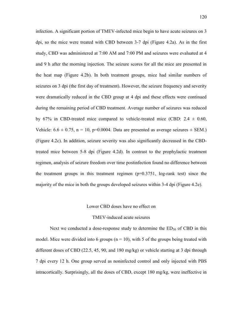
Figure 4.2 Therapeutic treatment with CBD (180 mg/kg) reduces average frequency and severity of TMEV-induced acute seizures. (a) Outline of therapeutic CBD treatment regimen. (b) Acute seizures were induced by handling the mice four times a day – during CBD/vehicle injections (7:00 AM, 7:00 PM) and during seizure monitoring (11:00 AM, 4:00 PM). Seizure severity was video-monitored with the experimenter blinded to the treatment groups, categorized based on modified Racine scale ranging from stage 3 to 6, and data depicted as a heat map using green, yellow, orange, and red colors corresponding to seizure scores in increasing order (DI, during injection; dpi, days postinfection). (c, d) Therapeutic CBD treatment significantly reduces seizure frequency (c) plotted as total numbers of seizures per mouse and seizure severity (d) as measured by average cumulative seizure burden during acute seizure period (3-8 dpi) compared to vehicle-treated TMEV-infected mice. Each circle represents an individual mouse and the horizontal line indicates the average number of seizures per group. (e) Development of the first seizure is not significantly different between treatment groups. Statistics: unpaired t-test (frequency), Scheirer-Ray-Hare test (severity), **p<0.01, ***p<0.001.

132

133

Figure 4.3 Only one dose of CBD (180 mg/kg; range of 22.5 to 180 mg/kg) reduces frequency and severity of TMEV-induced acute seizures. Five groups of mice (n = 10 per group) were infected with TMEV (2x10⁵ PFU/mouse) and treated with either vehicle or different doses of CBD (22.5, 45, 90, and 180 mg/kg) i.p. every 12 h for 5 days starting at 3 dpi. (a) CBD (180 mg/kg) treatment, but not the other doses, significantly reduces seizure frequency plotted as total numbers of seizures per mouse compared to vehicle as well as 22.5, 45, and 90 mg/kg CBD groups. Each circle indicates individual mouse and the horizontal line indicates average number of seizures per group. (b) Similarly, only 180 mg/kg CBD reduces seizure severity as measured by average cumulative seizure burden compared to all the other treatment groups, but the treatment with the lower doses of CBD does not affect seizure severity. (c) Changes in the average weight of mice in each treatment group over acute infection period shows a significant reduction in 180 mg/kg CBD treated mice compared to other treatment groups between 5-9 dpi. (d) Core body temperature of 180 mg/kg CBD-treated mice as measured immediately before CBD treatment (baseline) and at 15 min, 30 min, 1, 2, and 4 h posttreatment each day during 3-7 dpi shows a significant reduction on 3 dpi compared to other days. Statistics: unpaired t-test (frequency), Scheirer-Ray-Hare test (severity), two-way ANOVA, (weight and body temperature) *p<0.05, **p<0.01, ***p<0.001, †p<0.05, ‡p<0.01.

134

Figure 4.4 Low doses of CBD have no effect on TMEV-induced acute seizures. Mice were infected with TMEV (2x10⁵ PFU/mouse) and treated with either vehicle or CBD (135, 150, and 165 mg/kg) i.p. every 12 h for 5 days starting at 3 dpi. 135 mg/kg (a) as well as 150 and 165 mg/kg (b) CBD treatment does not reduce average frequency (upper panels) and intensity (lower panels) of TMEV-induced seizures. Data from two separate experiments testing 150 and 165 mg/kg CBD (n = 10/group) were pooled for seizure analysis.

135

Figure 4.5 CBD (150 mg/kg) administration decreases TMEV-induced seizures monitored at 4 h post-CBD treatment but not at 9 h post-CBD treatment. (a) Analysis of seizures monitored at 4 h post-CBD treatment shows a significant reduction in the average number of seizures (upper panel) and the average cumulative seizure burden at 6 and 7 dpi (lower panel) compared to the vehicle group. (b) CBD (150 mg/kg) treatment does not affect seizure endpoints measured at 9 h posttreatment. Statistics: unpaired t-test (frequency), Scheirer-Ray-Hare test (severity), *p<0.05, ***p<0.001.

CHAPTER 5

SUMMARY, FUTURE DIRECTIONS, AND PERSPECTIVES

The Theiler’s Murine Encephalitis Virus (TMEV) model is a recently developed

CNS infection-induced animal model of temporal lobe epilepsy (TLE) crucial to our

ability to investigate the cellular, molecular, and network level mechanisms implicated in

seizure generation, propagation and transforming homeostatic neural circuits into

epileptic circuits. The TMEV model also provides an important platform to test next-

generation therapeutics aimed at symptomatic seizure control (antiseizure) as well as for

disease modification (antiepileptogenic). Previous studies have discovered that innate

immune responses, mainly driven by the cytokines TNFα, IL-6 and complement protein

C3, cause inflammatory reactions in the brains of TMEV-infected mice concurrently with

acute behavioral seizures (Kirkman et al., 2010). Inflammation is known to contribute to

oxidative stress and mitochondrial dysfunction (Morris and Berk, 2015), whereas the

latter can cause injury by inducing inflammation, contributing to neurodegeneration, and

inducing hyperexcitability (Naik and Dixit, 2011). Both inflammation and oxidative

stress could therefore be both a cause and/or a consequence of brain damage and seizure

activity. Further, CNS infections and mitochondrial disorders are associated with

increased incidence of seizures and epilepsy in patients (Vezzani et al., 2016; Waldbaum

and Patel, 2010). Thus, a cross-talk can occur between inflammation, oxidative stress,

137

neuronal injury and seizures; however, the details of the relationship between these

factors have not been studied during TMEV infection-induced seizures and epilepsy.

Therefore, the present studies investigated the occurrence of oxidative stress and

inflammation in the hippocampus, a brain region known to generate limbic seizures,

during the TMEV-induced acute seizure period and how inflammation could modulate

acute seizures.

Summary and implications of findings

Our data show a strong correlation between impairment of redox status, a robust

increase in proinflammatory markers, and acute behavioral seizures following TMEV

infection. Among the numbers of cytokines elevated in the hippocampus during acute

seizures, we studied TNFα and its receptors in depth for their contribution to

hyperexcitability and seizures. TNFR1-mediated signaling of TNFα predominantly drives

hyperexcitatory synaptic changes and contributes to seizure occurrence, since TNFR1-/-

and TNFR1-/-TNFR2-/- mice are markedly resistant to developing TMEV-induced acute

seizures. In contrast, TNFR2-mediated signaling of TNFα could be a part of an

endogenous antiseizure mechanism, as the numbers and severity of seizures are worse in

TNFR2-/- mice than in WT. Given the changes in other cytokines and oxidative stress

markers, the mechanisms of seizure generation in this model are most likely

multifactorial and interactions between additional underlying factors should be

investigated further. Also, due to the observed increases in the levels of multiple

proinflammatory cytokines, we also tested the efficacy of cannabidiol (CBD) which

exhibits broad spectrum antiinflammatory and antiseizure activities (Burstein, 2015;

138

Rosenberg et al., 2015). CBD (180 mg/kg) effectively suppressed TMEV-induced acute

seizures, however, the pharmacokinetic and pharmacodynamic properties of CBD in this

model require further investigation.

The present research has strong translational implications by targeting TNFRs in

the brain for the treatment of acute seizures during infections and possibly for the

prevention of epilepsies caused by infection and CNS inflammation. The mechanism by

which TNFα causes hippocampal hyperexcitability through TNFR1 is not just limited to

the TMEV model of epilepsy. Indeed, a proictogenic role of TNFR1-mediated signaling

and an antiictogenic role of TNFR2-mediated signaling have also been reported in the

kainic acid-induced model of epilepsy (Balosso et al., 2005; Weinberg et al., 2013).

TNFRs have been proven to be successful targets for treating many peripheral

inflammatory diseases, and several biologics targeting the TNF system in the CNS are

under development (Fischer et al., 2015; McCoy and Tansey, 2008) which might be

helpful to treat inflammation-induced TLE. Inhibition of other inflammatory pathways

mediated by IL-1 receptor and Toll-like receptors also provides antiseizure effects in

various seizure models, and therefore, suggests that targeting inflammation could be an

important strategy to treat pharmacoresistant epilepsies (Vezzani et al., 2011b). In line

with previous findings, our results further substantiate the role of inflammation in causing

seizures. The present research also demonstrates the utilization of an animal model of

infection-induced epilepsy in testing novel compounds that have antiinflammatory as

well as antiseizure activities. CBD-based drugs are currently under clinical trials for the

treatment of Dravet syndrome, Lennox-Gastaut syndrome and other intractable forms of

epilepsies (Friedman and Devinsky, 2015). Further CBD studies should build upon the

139

results presented here by evaluating safety and antiseizure mechanisms of CBD that will

be beneficial in future clinical trial design.

Future directions

Future experiments will be directed toward site-specific and conditional deletion

of TNFR1 in excitatory neurons and other cell types using a TNFR1fl/fl mouse to

investigate a causal link between enhanced TNFR1 signaling and network

hyperexcitability. TNFR1fl/fl mice were obtained from Dr. George Kollias’ group (Van

Hauwermeiren et al., 2013). The effects of TNFα on neural circuit functions depend on

various factors, including the relative expression levels of TNFRs (TNFR1 and 2) in the

tissue and the cell types expressing them. For example, astrocyte dysfunction occurs in

many acquired epilepsies (Robel and Sontheimer, 2016; Steinhauser and Seifert, 2012)

and TNFα signaling through activation of TNFR1 has been shown to decrease expression

of the astrocyte glutamate transporter (GLT-1) (Carmen et al., 2009) and connexin-43

(Morioka et al., 2015). Pathogenic levels of TNFα also induce lasting synaptic alterations

and cognitive impairment through astrocytic TNFR1-mediated signaling (Habbas et al.,

2015). Thus, the role of TNFR1 signaling in astrocytes should be investigated and the use

of the TNFR1fl/fl mouse will be instrumental for such studies. TMEV-infected mice have

widespread astrogliosis in the hippocampus during the acute infection period (Loewen et

al., 2016). In addition, TMEV-infected mice exhibit anxiety-like behavior and cognitive

impairment (Umpierre et al., 2014). Whether signaling through the TNFR1 pathway in

astrocytes contributes to such tripartite synaptic alterations should be explored. On the

other hand, activation of TNFR2 may provide protection against seizures and neuronal

140

damage. Although the studies on TNFR2 agonists are limited, activation of signaling

through TNFR2 has been shown to ameliorate oxidative stress-induced cell death in

cultured dopaminergic neurons (Fischer et al., 2011). Thus, a combination of a TNFR1

antagonist and TNFR2 agonist might be an effective strategy for preventing TMEV-

induced seizures. In addition, since the protein expression of TNFR2 decreases in the

hippocampus during acute seizures following TMEV infection, the molecular

mechanisms that regulate synthesis and cell surface expression of TNFR2 could be

exploited to increase the level of TNFR2.

Considering the intracortical method of TMEV infection used and the observed

infiltration of macrophages into the brain (Cusick et al., 2013), the blood-brain barrier

(BBB) of TMEV-infected mice could be compromised. BBB injury is a contributing

factor to the development of epilepsy (van Vliet et al., 2007). Extravasation of albumin

from plasma into the brain occurs following BBB damage and albumin activation of

TGFβ receptors on astrocytes can cause astrocyte dysfunction by impairing the functions

of glutamate transporters, gap junction proteins, and inwardly-rectifying potassium

channel currents, thus leading to hyperexcitation (Ivens et al., 2007). Interestingly, TGFβ

expression in the hippocampus correlates with TMEV-associated seizure activity (Libbey

et al., 2008). Future studies should evaluate BBB damage and TGFβ signaling in

regulating astrocyte-neuronal functions following TMEV infection. In addition,

numerous other cytokines such as IL-6, IFNγ, and IL-1β are also upregulated in the

hippocampus during TMEV-induced acute seizures. Given the involvement of IL-6,

IFNγ, and IL-1β in neuronal excitability in other models of epilepsy (Getts et al., 2007;

Samland et al., 2003; Vezzani et al., 2011b), the contributions of these cytokines and

141

their interactions with TNFα following TMEV infection should be characterized. Such

studies would better inform antiinflammatory therapy for the treatment of seizures.

Perspectives

A large body of clinical evidence suggests that inflammation could be one of the

driving factors in precipitating seizures and mediating epileptogenesis (Vezzani et al.,

2011a). For example: 1) autoimmune diseases such as lupus and celiac disease increase

the risk for seizures (Devinsky et al., 2013), 2) febrile seizures, which can lead to

epilepsy, are correlated with immunological changes and inflammation (Choy et al.,

2014), 3) epileptic conditions with a strong inflammatory component, such as

Rasmussen’s encephalitis and paraneoplastic autoimmune encephalitis, are associated

with seizures that are often resistant to antiseizure drugs (Davis and Dalmau, 2013), 4)

CNS developmental conditions such as cortical dysplasia and tuberous sclerosis are also

associated with brain inflammation and increased seizure susceptibility (Aronica and

Crino, 2014), and 5) antiinflammatory therapies, for example, steroids, can prevent

seizures in several intractable epilepsies (Vincent et al., 2010). Confirmative clinical

studies linking specific inflammatory components with elevated seizure probability are

required. Thus, animal models mimicking clinical aspects of inflammation-associated

epilepsies, such as the TMEV model of limbic epilepsy, must be leveraged to identify

molecular mechanisms that could be targeted for future pharmacotherapies.

Several caveats must be considered before applying antiinflammatory treatment

for the treatment of seizures and for intervening in the process of epileptogenesis. First,

the diagnosis of infectious and/or the inflammatory origin of seizures could be very

142

challenging, particularly under latent CNS infections. Chronic mild to moderate

neuroinflammatory conditions may not reveal clinical symptoms for several years, as

observed in neurodegenerative diseases (Frank-Cannon et al., 2009). Therefore, studies

directed toward the development of biomarkers for CNS inflammation should be

prioritized, as such biomarkers will be highly useful for a timely pharmacological

intervention to prevent seizures and the development of subsequent epilepsy. Second,

timing for the initiation of antiinflammatory therapies will require a detailed

understanding of the purpose of inflammation under CNS pathologies associated with

seizures. Inflammation can be a double-edged sword, as it is necessary for the resolution

of the CNS insult and the clearance of infectious agents from the brain. It is not advisable

to resort to the use of broad-spectrum antiinflammatory drugs for a prolonged period as

that may also interfere with the beneficial functions of inflammatory mechanisms and

may cause collateral CNS damage. The ideal strategy should be to reestablish

homeostatic equilibrium between endogenous inflammatory and antiinflammatory

mechanisms and future research should help to determine the factors crucial in

maintaining the equilibrium between inflammatory processes. Finally, many antiseizure

drugs can affect innate and adaptive immune functions (Marchi et al., 2014). Therefore,

thorough animal studies must be conducted to evaluate interactions between antiseizure

and antiinflammatory drugs before initiating a combination therapy in the clinic for the

treatment of acute seizures and the prevention of epilepsy following infection. No doubt

the TMEV model of TLE, which has greatly contributed to our understanding of

infection-induced epilepsy, will be useful in the preclinical development of such novel

therapies for those patients at risk for developing epilepsy following a CNS infection.

143

References Aronica, E., Crino, P.B., 2014. Epilepsy related to developmental tumors and

malformations of cortical development. Neurotherapeutics 11, 251-268. Balosso, S., Ravizza, T., Perego, C., Peschon, J., Campbell, I.L., De Simoni, M.G.,

Vezzani, A., 2005. Tumor necrosis factor-alpha inhibits seizures in mice via p75 receptors. Ann. Neurol. 57, 804-812.

Burstein, S., 2015. Cannabidiol (CBD) and its analogs: a review of their effects on

inflammation. Bioorg. Med. Chem. 23, 1377-1385. Carmen, J., Rothstein, J.D., Kerr, D.A., 2009. Tumor necrosis factor-alpha modulates

glutamate transport in the CNS and is a critical determinant of outcome from viral encephalomyelitis. Brain Res. 1263, 143-154.

Choy, M., Dube, C.M., Ehrengruber, M., Baram, T.Z., 2014. Inflammatory processes,

febrile seizures, and subsequent epileptogenesis. Epilepsy Curr. 14, 15-22. Cusick, M.F., Libbey, J.E., Patel, D.C., Doty, D.J., Fujinami, R.S., 2013. Infiltrating

macrophages are key to the development of seizures following virus infection. J. Virol. 87, 1849-1860.

Davis, R., Dalmau, J., 2013. Autoimmunity, seizures, and status epilepticus. Epilepsia 54

Suppl 6, 46-49. Devinsky, O., Schein, A., Najjar, S., 2013. Epilepsy associated with systemic

autoimmune disorders. Epilepsy Curr. 13, 62-68. Fischer, R., Kontermann, R., Maier, O., 2015. Targeting sTNF/TNFR1 signaling as a new

therapeutic strategy. Antibodies 4, 48-70. Fischer, R., Maier, O., Siegemund, M., Wajant, H., Scheurich, P., Pfizenmaier, K., 2011.

A TNF receptor 2 selective agonist rescues human neurons from oxidative stress-induced cell death. PLoS One 6, e27621.

Frank-Cannon, T.C., Alto, L.T., McAlpine, F.E., Tansey, M.G., 2009. Does

neuroinflammation fan the flame in neurodegenerative diseases? Mol. Neurodegener. 4, 47.

Friedman, D., Devinsky, O., 2015. Cannabinoids in the Treatment of Epilepsy. N. Engl.

J. Med. 373, 1048-1058. Getts, D.R., Matsumoto, I., Muller, M., Getts, M.T., Radford, J., Shrestha, B., Campbell,

I.L., King, N.J., 2007. Role of IFN-gamma in an experimental murine model of West Nile virus-induced seizures. J. Neurochem. 103, 1019-1030.

144

Habbas, S., Santello, M., Becker, D., Stubbe, H., Zappia, G., Liaudet, N., Klaus, F.R., Kollias, G., Fontana, A., Pryce, C.R., Suter, T., Volterra, A., 2015. Neuroinflammatory TNFalpha Impairs Memory via Astrocyte Signaling. Cell 163, 1730-1741.

Ivens, S., Kaufer, D., Flores, L.P., Bechmann, I., Zumsteg, D., Tomkins, O., Seiffert, E.,

Heinemann, U., Friedman, A., 2007. TGF-beta receptor-mediated albumin uptake into astrocytes is involved in neocortical epileptogenesis. Brain 130, 535-547.

Kirkman, N.J., Libbey, J.E., Wilcox, K.S., White, H.S., Fujinami, R.S., 2010. Innate but

not adaptive immune responses contribute to behavioral seizures following viral infection. Epilepsia 51, 454-464.

Libbey, J.E., Kirkman, N.J., Smith, M.C., Tanaka, T., Wilcox, K.S., White, H.S.,

Fujinami, R.S., 2008. Seizures following picornavirus infection. Epilepsia 49, 1066-1074.

Loewen, J.L., Barker-Haliski, M.L., Dahle, E.J., White, H.S., Wilcox, K.S., 2016.

Neuronal injury, gliosis, and glial proliferation in two models of temporal lobe epilepsy. J. Neuropathol. Exp. Neurol. 75, 366-378.

Marchi, N., Granata, T., Janigro, D., 2014. Inflammatory pathways of seizure disorders.

Trends Neurosci. 37, 55-65. McCoy, M.K., Tansey, M.G., 2008. TNF signaling inhibition in the CNS: implications

for normal brain function and neurodegenerative disease. J. Neuroinflammation 5, 45.

Morioka, N., Zhang, F.F., Nakamura, Y., Kitamura, T., Hisaoka-Nakashima, K., Nakata,

Y., 2015. Tumor necrosis factor-mediated downregulation of spinal astrocytic connexin43 leads to increased glutamatergic neurotransmission and neuropathic pain in mice. Brain. Behav. Immun. 49, 293-310.

Morris, G., Berk, M., 2015. The many roads to mitochondrial dysfunction in

neuroimmune and neuropsychiatric disorders. BMC Med. 13, 68. Naik, E., Dixit, V.M., 2011. Mitochondrial reactive oxygen species drive

proinflammatory cytokine production. J. Exp. Med. 208, 417-420. Robel, S., Sontheimer, H., 2016. Glia as drivers of abnormal neuronal activity. Nat.

Neurosci. 19, 28-33. Rosenberg, E.C., Tsien, R.W., Whalley, B.J., Devinsky, O., 2015. Cannabinoids and

Epilepsy. Neurotherapeutics 12, 747-768. Samland, H., Huitron-Resendiz, S., Masliah, E., Criado, J., Henriksen, S.J., Campbell,

145

I.L., 2003. Profound increase in sensitivity to glutamatergic- but not cholinergic agonist-induced seizures in transgenic mice with astrocyte production of IL-6. J. Neurosci. Res. 73, 176-187.

Steinhauser, C., Seifert, G., 2012. Astrocyte dysfunction in epilepsy, in: Noebels, J.L.,

Avoli, M., Rogawski, M.A., Olsen, R.W., Delgado-Escueta, A.V. (Eds.), Jasper's Basic Mechanisms of the Epilepsies, 4th ed, Bethesda (MD).

Umpierre, A.D., Remigio, G.J., Dahle, E.J., Bradford, K., Alex, A.B., Smith, M.D., West,

P.J., White, H.S., Wilcox, K.S., 2014. Impaired cognitive ability and anxiety-like behavior following acute seizures in the Theiler's virus model of temporal lobe epilepsy. Neurobiol. Dis. 64, 98-106.

Van Hauwermeiren, F., Armaka, M., Karagianni, N., Kranidioti, K., Vandenbroucke,

R.E., Loges, S., Van Roy, M., Staelens, J., Puimege, L., Palagani, A., Berghe, W.V., Victoratos, P., Carmeliet, P., Libert, C., Kollias, G., 2013. Safe TNF-based antitumor therapy following p55TNFR reduction in intestinal epithelium. J. Clin. Invest. 123, 2590-2603.

van Vliet, E.A., da Costa Araujo, S., Redeker, S., van Schaik, R., Aronica, E., Gorter,

J.A., 2007. Blood-brain barrier leakage may lead to progression of temporal lobe epilepsy. Brain 130, 521-534.

Vezzani, A., French, J., Bartfai, T., Baram, T.Z., 2011a. The role of inflammation in

epilepsy. Nat. Rev. Neurol. 7, 31-40. Vezzani, A., Fujinami, R.S., White, H.S., Preux, P.M., Blumcke, I., Sander, J.W.,

Loscher, W., 2016. Infections, inflammation and epilepsy. Acta Neuropathol. 131, 211-234.

Vezzani, A., Maroso, M., Balosso, S., Sanchez, M.A., Bartfai, T., 2011b. IL-1

receptor/Toll-like receptor signaling in infection, inflammation, stress and neurodegeneration couples hyperexcitability and seizures. Brain. Behav. Immun. 25, 1281-1289.

Vincent, A., Irani, S.R., Lang, B., 2010. The growing recognition of immunotherapy-

responsive seizure disorders with autoantibodies to specific neuronal proteins. Curr. Opin. Neurol. 23, 144-150.

Waldbaum, S., Patel, M., 2010. Mitochondria, oxidative stress, and temporal lobe

epilepsy. Epilepsy Res. 88, 23-45. Weinberg, M.S., Blake, B.L., McCown, T.J., 2013. Opposing actions of hippocampus

TNFalpha receptors on limbic seizure susceptibility. Exp. Neurol. 247, 429-437.


Recommended