Influence of Physical and Chemical Treatments on Cell Survival and Acquisition of Pluripotency Ricardo Jorge Carvalho Correia Thesis to obtain the Master of Science Degree in Biological Engineering Supervisors: MD, PhD Petra de Sutter PhD Cláudia Alexandra Martins Lobato da Silva Examination Committee Chairperson: Professor Arsénio do Carmo Sales Mendes Fialho Supervisor: Professor Cláudia Alexandra Martins Lobato da Silva Member of the Committee: Professor Maria Margarida Fonseca Rodrigues Diogo November 2014
Transcript
Influence of Physical and Chemical Treatments on Cell Survival and
Acquisition of Pluripotency
Ricardo Jorge Carvalho Correia
Thesis to obtain the Master of Science Degree in
Biological Engineering
Supervisors: MD, PhD Petra de Sutter
PhD Cláudia Alexandra Martins Lobato da Silva
Examination Committee
Chairperson: Professor Arsénio do Carmo Sales Mendes Fialho
Supervisor: Professor Cláudia Alexandra Martins Lobato da Silva
Member of the Committee: Professor Maria Margarida Fonseca Rodrigues Diogo
November 2014
I
Acknowledgments
First of all, I would like to thank my external promoters, Professor Petra de Sutter and Björn Heindryckx, for
believing in me and accept and treat me as one of their own research group members, allowing me to develop
my knowledge and interest on this research field, and providing me the resources to conduct an organized and
scientifically useful project.
I would also like to thank Sharat Warrier for guiding me through all the past months, helping me during both the
practical experiences and writing process, and for teaching me almost everything. Also, his wife, Galbha Duggal,
for all her help. To both, I want to show my special gratitude for receiving me as more than a colleague, as a
friend. Without you, this work could never be possible.
To my internal promoter, Professor Cláudia Lobato Silva, for the constant availability to answer my questions and
for all the advices, helping me to finish my work.
To all the research team at the Ghent Hospital, for all the support, resources and time dedicated to my project.
To all my friends in Ghent, thank you for being my family during the last months and for making me feel home in
a city and country of which I didn’t know anything until then. Thank you for all the good moments we spent
together. I’m truly thankful for all the friends for life I made in the last months.
To all my family and friends in Portugal, thank you for all the daily messages that made me feel closer to my
country, and for all the support to overcome the distance between us.
I would also like to express my thankfulness to my parents for allowing me to have resources to live in another
country, for supporting me during the whole time, for believing in me, and for considering my graduation and
success as some of their biggest dreams. This thesis and all my commitment to my academic and work careers
are dedicated to you.
And last but not the least, I want to express my gratefulness to my girlfriend, Maria, who was the first to convince
me to face this adventure. For all the love, understanding and dedication during the whole time we were far
from each other. For making me feel close to her no matter the distance, and for giving me the strength when I
thought I didn’t have it. She was and will always be my biggest support.
To all, a heartfelt thank you.
II
Abstract
Pluripotent human embryonic stem cells represent a promising source to develop medical therapies and unveil
the secrets behind degenerative diseases. The necessity of large quantities of viable pluripotent cells is urgent.
Several ethical problems regarding the use of embryos for research result in the necessity of high throughput
reprogramming techniques. Recently, new data reported the possibility of reprogramming murine somatic cells
into a state of pluripotency through the induction of external strong stimuli, such as the exposure to an acidic
solution and/or constant physical stress. Intrigued by this, it was decided not only to try to reproduce the
accomplished results, but also to adapt the protocol to accomplish a reprogramming event using human cells.
Several different types of murine somatic cells, as well as embryoid bodies generated in vitro from both human
and mouse embryonic stem cells, were exposed to three consecutive physical trituration steps with increasingly
smaller lumen pipettes, followed by 30 minutes exposure to an acidic solution. Cell survival and resistance was
monitored during the experimental steps. Together, the results obtained showed incapacity of the described
technique to reprogram differentiated cells into a pluripotent state. The challenging protocol resulted in constant
cell death during the experiments. The possible reprogramming of somatic cells into pluripotency through this
simple and fast method would represent a remarkable improvement in the use of stem cells-based therapies.
I. Figures Index ................................................................................................................................... V
II. Tables Index .................................................................................................................................... XI
III. Symbols and Abbreviations Index ............................................................................................. XII
1. State of Art ...................................................................................................................................... 1
1.1. Introduction to Embryonic Stem Cells..................................................................................... 1
1.1.1. Establishment of the first stable Mouse Embryonic Stem Cell line in culture ................ 2
1.1.2. Establishment of Human Embryonic Stem Cell lines ....................................................... 3
4.4. Complementary work and Controls ...................................................................................... 69
4.4.1. Mouse Embryonic Stem cells cultured in sphere media ............................................... 69
4.4.2. Protocol performed directly on Mouse Embryonic Stem cells ..................................... 70
4.4.3. Negative Control – Embryoid Bodies are fully differentiated ....................................... 72
4.4.4. Positive Control – Mouse Embryonic Stem cells express Oct4 and Nanog ................... 73
4.4.5. Negative Control – Mouse Embryonic Fibroblast cells do not express pluripotency ... 74
4.5. Further Discussion ................................................................................................................. 75
5. Conclusion and Perspectives ......................................................................................................... 78
6. References ........................................................................................................................................ i
V
I. Figures Index
Figure 1: Three main reprogramming techniques converting differentiated somatic cells into a state of
pluripotency. Cell fusion of embryonic stem cells with a differentiated somatic cell results in the epigenetic
dominance of the undifferentiated cell, resulting in an undifferentiated pluripotent cell population. Through
somatic cell nuclear transfer, previously enucleated oocytes reprogram the nucleus of the differentiated cells
into a pluripotent state, thus resulting an embryo from which embryonic stem cells can be derived. Alternatively,
embryo can develop into a fully functional organism. Induced pluripotent stem cells are based on the
reprogramming event of differentiated somatic cells into a pluripotent state after being exogenously exposed to
Figure 2: Early embryogenesis in mouse and human follow different developmental stages. In the mouse, the
ICM segregates into a layer of cells representing the primitive endoderm surrounding the naïve epiblast, after
which an epithelium-like structure called egg cylinder arises. Contrastingly, in humans, a primed epiblast arises,
and an embryonic bilaminar disc is formed rather than an egg cylinder. The different pluripotent states of the
derived mouse and human embryonic stem cells are directly related with their in vivo counterparts, namely the
naïve and primed epiblast, respectively. .............................................................................................................. 13
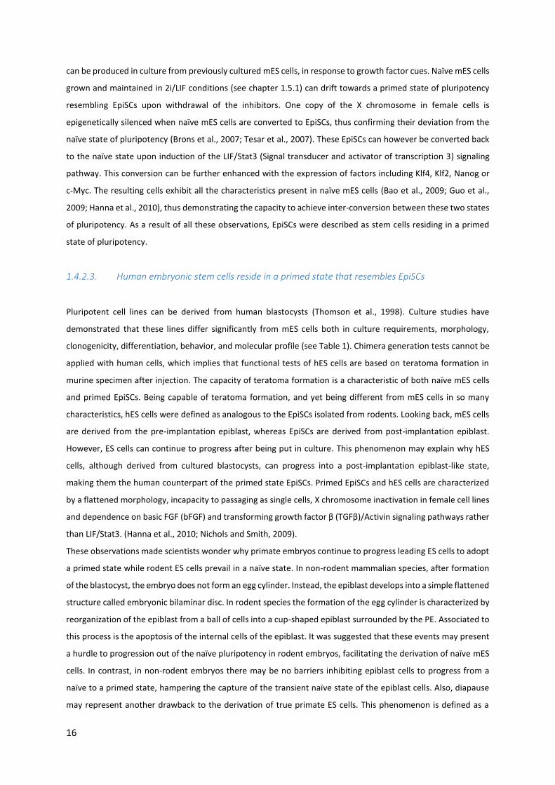
Figure 3: Signaling pathways leading to naïve pluripotency or differentiation. The maintenance of the
pluripotency state is facilitated by the use of LIF and BMP4. This two compounds lead to the activation of STAT3
and Smad1,5,8 respectively, which will posteriorly activate several genes encoding to transcription factors that
characterize the maintenance of pluripotency. The suppression of both GSK3β and Erk is fundamental, and
therefore two inhibitors (PD0325901 and CHIR99021) are essential to maintain a naïve pluripotent state. The
combination of these two inhibitors in the culture cocktail is sufficient to maintain naïve pluripotency, being
often used along with LIF to increase the efficiency. Also, Wnt leads to the suppression of GSK3β, thus
contributing to pluripotency. ................................................................................................................................ 20
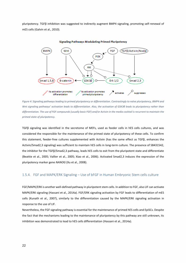
Figure 4: Signaling pathways leading to primed pluripotency or differentiation. Contrastingly to naïve
pluripotency, BMP4 and Wnt signaling pathways’ activation leads to differentiation. Also, the activation of GSK3β
leads to pluripotency rather than differentiation. The use of FGF compounds (usually basic FGF) and/or Activin
in the media cocktail is recurrent to maintain the primed state of pluripotency. ................................................ 22
Figure 5: Stimulus- triggered acquisition of pluripotency can be obtained by exposing somatic cells to a strong
external environment such as an acid solution. Also, extra cellular stress defined by physical trituration of the
cells may be helpful to successfully accomplish reprogramming event. .............................................................. 25
Figure 6: Main steps describing the adaptation of the protocol to use with embryoid bodies cultured in the lab.
Embryonic stem cells were expanded and differentiated into embryoid bodies during 14 days, after which they
were dissociated into single cells. Those isolated cells faced then three pipetting steps with increasingly smaller
lumen tips. Physical stress was followed by chemical stress described as 30 minutes of acid exposure. Cells were
then cultured during 7 days in sphere media, after which pluripotency was analyzed. ...................................... 38
Figure 7: Experimental procedure performed in murine somatic cells directly isolated from mice or cultured in
the lab. The procedure started directly with the exposure to the three physical stress steps, after which the
VI
exposure to an acidic solution during 30 minutes was imposed. After being cultured for 7 days in sphere media,
pluripotency of the cells was analyzed. ................................................................................................................ 39
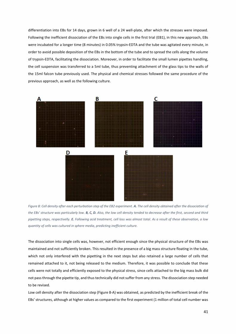
Figure 8: Cell density after each perturbation step of the EB2 experiment. A. The cell density obtained after the
dissociation of the EBs' structure was particularly low. B, C, D. Also, the low cell density tended to decrease after
the first, second and third pipetting steps, respectivelly. E. Following acid treatment, cell loss was almost total.
As a result of these observation, a low quantity of cells was cultured in sphere media, predicting inefficient
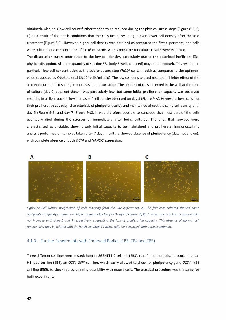
Figure 9: Cell culture progression of cells resulting from the EB2 experiment. A. The few cells cultured showed
some proliferation capacity resulting in a higher amount of cells after 3 days of culture. B, C. However, the cell
density observed did not increase until days 5 and 7 respectively, suggesting the loss of proliferation capacity.
This absence of normal cell functionality may be related with the harsh condition to which cells were exposed
during the experiment. ......................................................................................................................................... 42
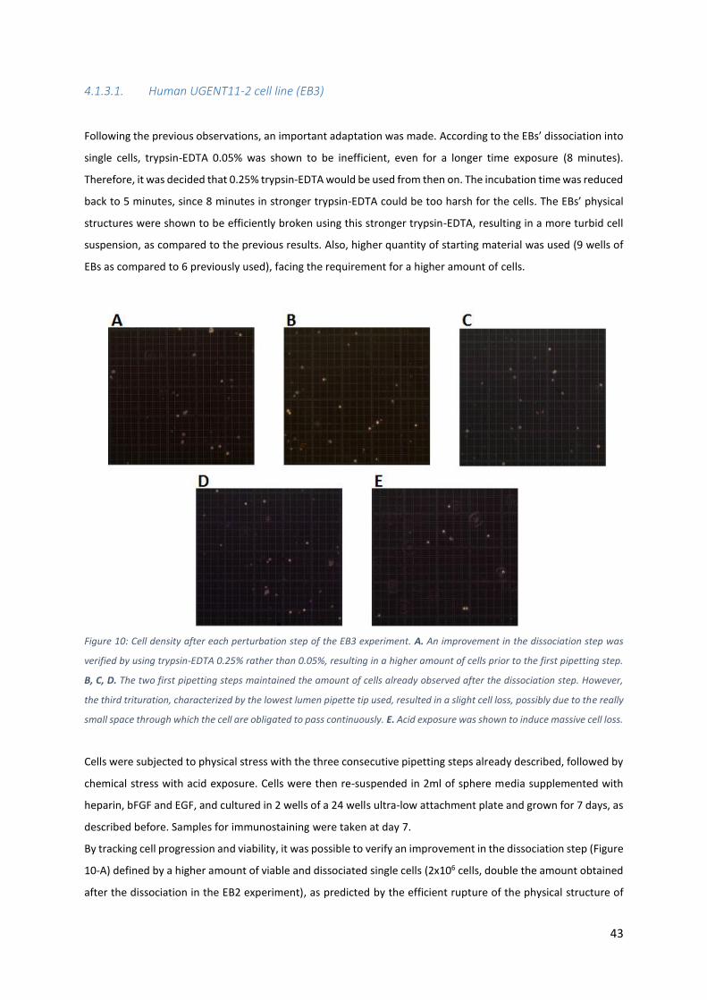
Figure 10: Cell density after each perturbation step of the EB3 experiment. A. An improvement in the dissociation
step was verified by using trypsin-EDTA 0.25% rather than 0.05%, resulting in a higher amount of cells prior to
the first pipetting step. B, C, D. The two first pipetting steps maintained the amount of cells already observed
after the dissociation step. However, the third trituration, characterized by the lowest lumen pipette tip used,
resulted in a slight cell loss, possibly due to the really small space through which the cell are obligated to pass
continuously. E. Acid exposure was shown to induce massive cell loss. .............................................................. 43
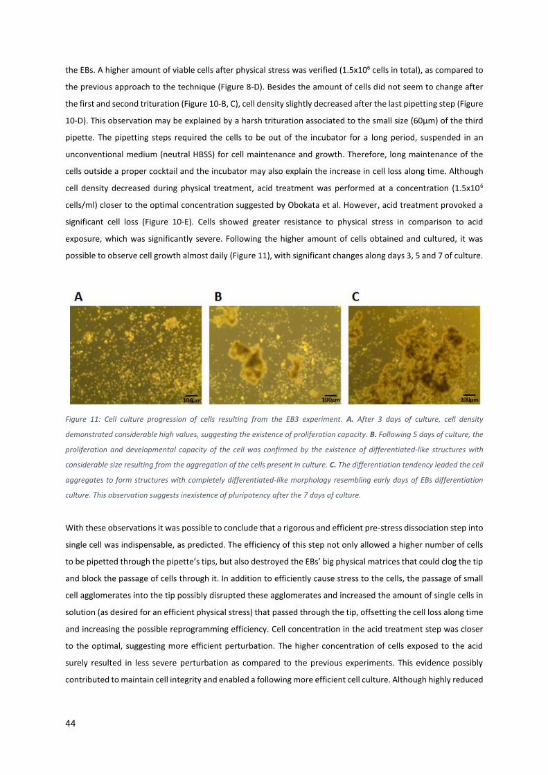
Figure 11: Cell culture progression of cells resulting from the EB3 experiment. A. After 3 days of culture, cell
density demonstrated considerable high values, suggesting the existence of proliferation capacity. B. Following
5 days of culture, the proliferation and developmental capacity of the cell was confirmed by the existence of
differentiated-like structures with considerable size resulting from the aggregation of the cells present in culture.
C. The differentiation tendency leaded the cell aggregates to form structures with completely differentiated-like
morphology resembling early days of EBs differentiation culture. This observation suggests inexistence of
pluripotency after the 7 days of culture. .............................................................................................................. 44
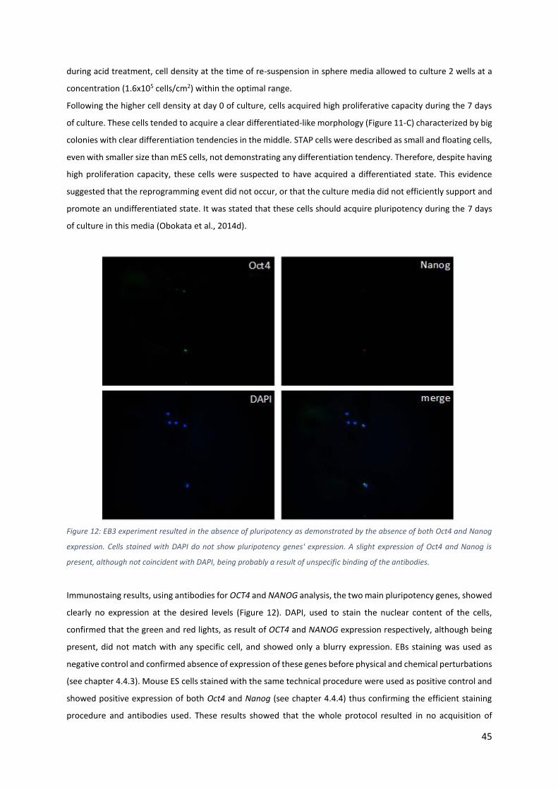
Figure 12: EB3 experiment resulted in the absence of pluripotency as demonstrated by the absence of both Oct4
and Nanog expression. Cells stained with DAPI do not show pluripotency genes' expression. A slight expression
of Oct4 and Nanog is present, although not coincident with DAPI, being probably a result of unspecific binding of
the antibodies. ...................................................................................................................................................... 45
Figure 13: Cell density after each perturbation step of the EB4 experiment. A. Cell density following dissociation
of the embryoid bodies' structures presented reasonable values. B, C, D. Cell density was maintained after the
first pipetting step (B) but reduced following the second one (C), being maintained again after the third trituration
(D). E. Cell density faced an intriguing increase following acid treatment. .......................................................... 46
Figure 14: Cell culture progression of cells resulting from the EB4 experiment. A. Even only after 3 days of culture,
cells showed differentiation tendencies characterized by several dark colored agglomerates. B, C. The
differentiation tendencies were still present after 5 (B) and 7 (C) days of culture. The colonies acquired an EB-like
Figure 15: Immunostaining results of the EB4 experiment from samples taken after 5 days of culture showed
absence of pluripotency factors Oct4 and Nanog expression, demonstrating that these cells still resided in a
differentiated state at this point. .......................................................................................................................... 48
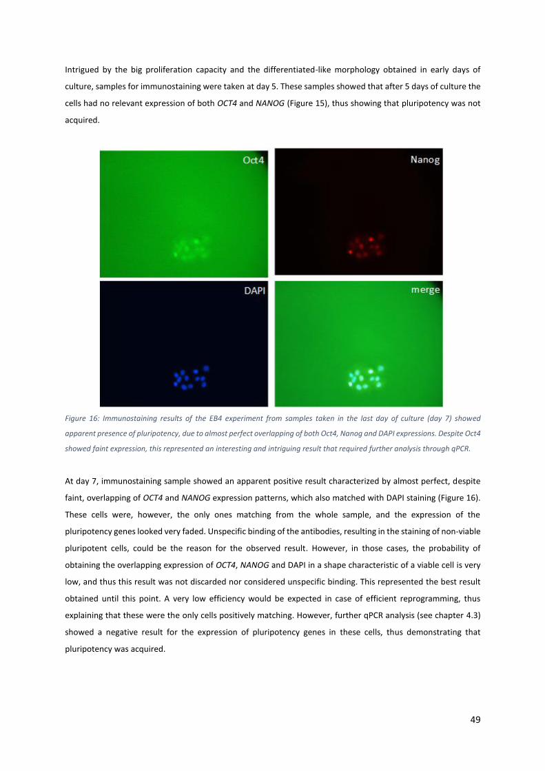
Figure 16: Immunostaining results of the EB4 experiment from samples taken in the last day of culture (day 7)
showed apparent presence of pluripotency, due to almost perfect overlapping of both Oct4, Nanog and DAPI
expressions. Despite Oct4 showed faint expression, this represented an interesting and intriguing result that
required further analysis through qPCR................................................................................................................ 49
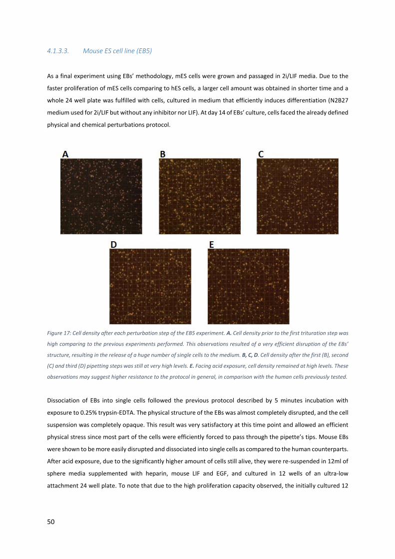
Figure 17: Cell density after each perturbation step of the EB5 experiment. A. Cell density prior to the first
trituration step was high comparing to the previous experiments performed. This observations resulted of a very
efficient disruption of the EBs’ structure, resulting in the release of a huge number of single cells to the medium.
B, C, D. Cell density after the first (B), second (C) and third (D) pipetting steps was still at very high levels. E. Facing
acid exposure, cell density remained at high levels. These observations may suggest higher resistance to the
protocol in general, in comparison with the human cells previously tested. ....................................................... 50
Figure 18: Cell culture progression of cells resulting from the EB5 experiment. A. Uncontrolled cell density was
observed in the first day of culture. B. Following that observation, excess cells were removed during medium
refreshing, resulting in a lower cell density. C. Cell density was verified to reach uncontrolled values again in the
next day and in the following days. ...................................................................................................................... 51
Figure 19: Immunostaining results obtained from a sample taken after the 7 days of culture showed apparent
positive results (bottom right especially) that clearly differ from the clearly negative results observed in the
sample of cells from the EB5 experiment. However, cells negatively expressing Oct4 and Nanog highly expressed
DAPI, whereas the apparent pluripotency-expressing cells showed very faint expression of DAPI. .................... 52
Figure 20: Cell density after each perturbation step of the GCs1 experiment. A. Granulosa cells isolated from
B6D2/F1 mice showed sufficient quantities prior to the first titration step. B. C, D, E. Cell density tended to
decrease after each one of the following trituration (B, C and D) and acid exposure (E) steps. .......................... 53
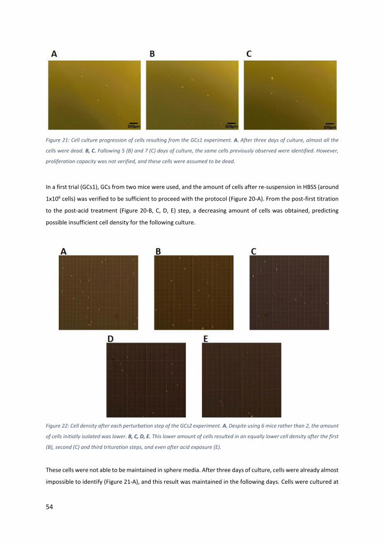
Figure 21: Cell culture progression of cells resulting from the GCs1 experiment. A. After three days of culture,
almost all the cells were dead. B, C. Following 5 (B) and 7 (C) days of culture, the same cells previously observed
were identified. However, proliferation capacity was not verified, and these cells were assumed to be dead. . 54
Figure 22: Cell density after each perturbation step of the GCs2 experiment. A. Despite using 6 mice rather than
2, the amount of cells initially isolated was lower. B, C, D, E. This lower amount of cells resulted in an equally
lower cell density after the first (B), second (C) and third trituration steps, and even after acid exposure (E). .. 54
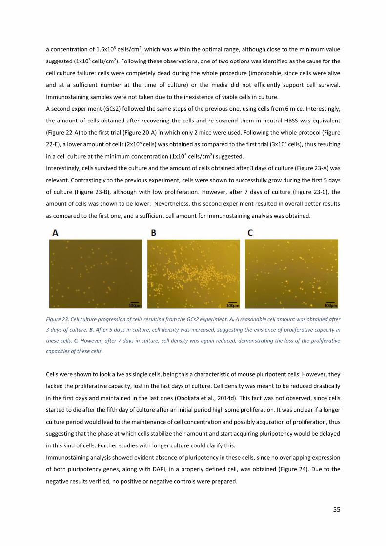
Figure 23: Cell culture progression of cells resulting from the GCs2 experiment. A. A reasonable cell amount was
obtained after 3 days of culture. B. After 5 days in culture, cell density was increased, suggesting the existence
of proliferative capacity in these cells. C. However, after 7 days in culture, cell density was again reduced,
demonstrating the loss of the proliferative capacities of these cells. .................................................................. 55
Figure 24: Immunostaining results of 7 days culture cells show clear absence of pluripotency in cells resulting
from the GCs2 experiment, due to impossible observation of overlapping expression of the pluripotency genes
Oct4 and Nanog. ................................................................................................................................................... 56
VIII
Figure 25: Cell density after each perturbation step of the TTs1 experiment. A. Following the trituration steps, a
high amount of cells was obtained. B. Acid exposure led the most part of the cells to die, thus resulting in
insufficient cell density for culture........................................................................................................................ 57
Figure 26: Cell culture progression of cells resulting from the TTs1 experiment. A. Following 3 days of culture,
low cell density was observed. B. Cell density increased after 5 days in culture in comparison with the third day.
C. The cell density stabilized. It is difficult to conclude about the viability of the present cells, since a lot of
contamination was present in the culture. ........................................................................................................... 57
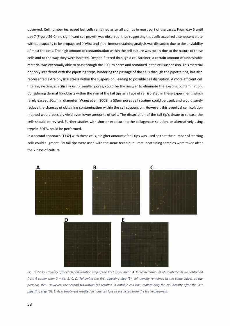
Figure 27: Cell density after each perturbation step of the TTs2 experiment. A. Increased amount of isolated cells
was obtained from 6 rather than 2 mice. B, C, D. Following the first pipetting step (B), cell density remained at
the same values as the previous step. However, the second trituration (C) resulted in notable cell loss,
maintaining the cell density after the last pipetting step (D). E. Acid treatment resulted in huge cell loss as
predicted from the first experiment. .................................................................................................................... 58
Figure 28: Cell culture progression of cells resulting from the TTs2 experiment. A. Higher cell density at the time
of culture was obtained, resulting in higher cell amounts after 3 days of culture. B, C. However, proliferation
capacity was not observed, leading to almost complete cell loss after 5 (B) and 7 (C) days in culture. ............... 59
Figure 29: Immunostaining analysis after 7 days of culture shows clear absence of pluripotency in cells from TTs2
experiment, defined by a completely blurred Oct4 and Nanog expressions, not identifying any specific cell stained
with DAPI............................................................................................................................................................... 60
Figure 30: Cell density after each perturbation step of the MEFs1 experiment. A. Sufficient cell amount was
obtained after isolation and prior to the first trituration. B, C, D. The first pipetting step (B) resulted in particularly
high cell loss, being the cell density maintained after the second (C) and third (D) pipetting steps. E. Acid
Figure 31: Cell culture progression of cells resulting from the MEFs1 experiment. A. The amount of cells cultured
was sufficient to proliferate resulting in a particularly high amount of cell after 3 days of culture. B, C. Proliferation
capacity was lost in the following days, and cell density decreased. Cell clusters with differentiated-like
morphology were observed both after 5 (B) and 7 (C) days of culture. ............................................................... 62
Figure 32: Immunostaining analyses showed some results that could apparently mean acquisition of pluripotency
in cells resulting from the MEFs1 experiment, defined by an almost perfect overlapping expression of both
pluripotency genes Oct4 and Nanog. However, DAPI expression, used to stain the nuclear content of the cells,
did not match perfectly with the pluripotency genes expression, thus suggesting a fake positive result. .......... 62
Figure 33: Cell density after each perturbation step of the MEFs2 experiment. A. The amount of isolated cells
was considerably higher. B, C, D. Cell density decreased following the first physical stress imposed (B), similarly
to the result observed in the first experiment. Cell density was maintained through the next pipetting steps (C
and D). E. Cell density slightly decreased again following acid exposure. Globally, the amount of cells obtained in
this second experiment was considerably higher. ................................................................................................ 63
Figure 34: Cell culture progression of cells resulting from the MEFs2 experiment. A. Cells showed high
proliferation capacity in the first days in culture, thus resulting in the formation of morphologically differentiated-
like structures at day 3. B, C. Cell proliferation was not observed in the next culture days. Instead, all the cells
IX
surrounding the apparently differentiated clumps started to die after 5 days, thus resulting in apparently
differentiated aggregates at day 7. These clumps had a morphology resembling MEFs defined by the formation
of elongations structures rather than round borders. .......................................................................................... 64
Figure 35: Immunostaining analysis confirms the absence of pluripotency in cells from the MEFs2 experiment.
DAPI perfectly identified cells, whereas Nanog and Oct4 expressions observed were dispersed and blurry. ..... 64
Figure 36: Cell density after each perturbation step on MEFs with physical stress only. A. Cell density after
isolation had considerable values, as verified in the previous experiment using MEFs. B. Similarly to the previous
results, the whole physical stress procedure, including the three steps, led to high cell loss. However, a sufficient
number of cells for posterior cell culture was obtained. ...................................................................................... 65
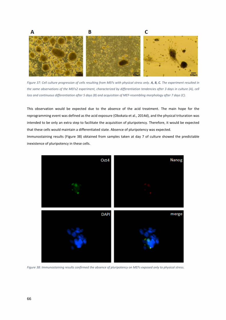
Figure 37: Cell culture progression of cells resulting from MEFs with physical stress only. A, B, C. The experiment
resulted in the same observations of the MEFs2 experiment, characterized by differentiation tendencies after 3
days in culture (A), cell loss and continuous differentiation after 5 days (B) and acquisition of MEF-resembling
morphology after 7 days (C).................................................................................................................................. 66
Figure 38: Immunostaining results confirmed the absence of pluripotency on MEFs exposed only to physical
Figure 43: Mouse ES cells were directly exposed to the protocol and cultured in sphere media. A. After 3 days in
culture, high cell density was observed, along with differentiation tendencies. B, C. After 5 (B) and 7 (C) days in
culture, cells tended to aggregate into completely differentiated clumps. These observations suggested that
undifferentiated pluripotent cell can survive the whole experiment, maintaining their capabilities to proliferate
and differentiate in vitro. ...................................................................................................................................... 72
Figure 44: EBs' immunostaining results showed absence of pluripotency-expressing cells in the structure of the
EB after 14 days of differentiation culture. ........................................................................................................... 72
Figure 45: Immunostaining analysis performed of mouse ES cells showed correct expression of pluripotency
genes Oct4 and Nanog. This analysis confirmed the efficiency of the staining method being used. ................... 73
X
Figure 46: Immunostaining results show absence of pluripotency on MEF cells prior to the protocol. Oct4 and
Nanog expression was completely absence.......................................................................................................... 74
XI
II. Tables Index
Table 1: Naïve and Primed pluripotency states show different potentials and morphology. Naive stem cells are
the in vitro counterpart of the cells present in the mouse early epiblast, whereas primed stem cells, human
embryonic stem cells and mouse epiblast stem cells, correspond to the in vivo human embryonic bilaminar disc
and mouse egg cylinder cells, respectively. Both are capable of teratoma formation, although this test, when
performed with human cells, relies in an inter-species teratoma formation, following injection into mice.
However, only naïve pluripotent cells are capable of chimera formation and single cell passaging. Epigenetic
changes are visible, specially characterized by the activation of the two X chromosomes in naïve female cells,
whereas one of the X chromosomes is inactivated in the primed ones. Moreover, morphology analysis identifies
naïve colonies with a domed shape, while primed colonies maintain a flat appearance. ................................... 17
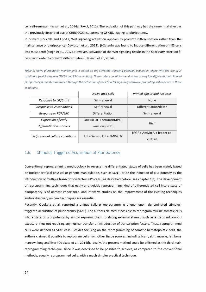
Table 2: Naïve pluripotency maintenance is based on the LIF/Stat3 signaling pathway activation, along with the
use of 2i conditions (which suppress GSK3β and ERK activation). These culture conditions lead to low or very low
differentiation. Primed pluripotency is mainly maintained through the activation of the FGF/ERK signaling
pathway, promoting self-renewal in these conditions. ........................................................................................ 24
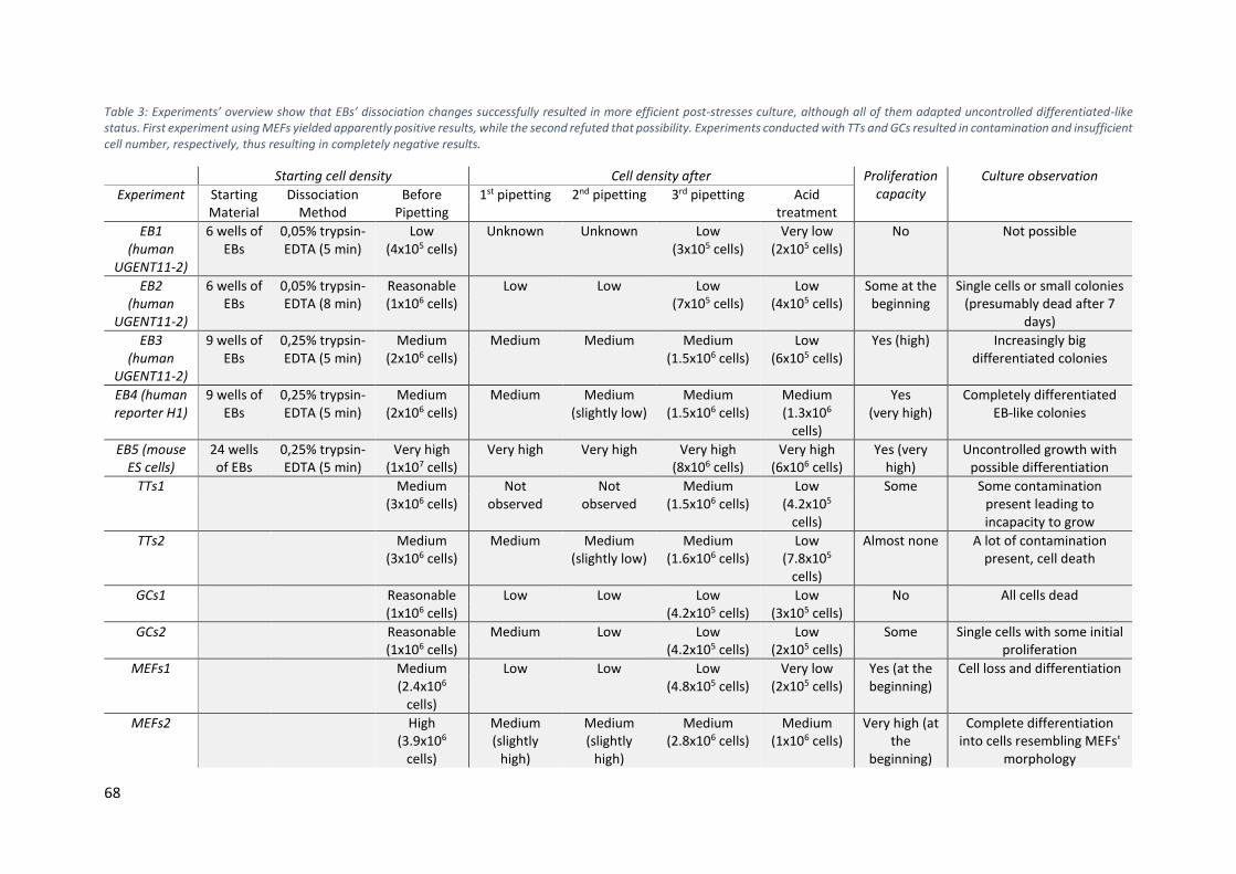
Table 3: Experiments’ overview show that EBs’ dissociation changes successfully resulted in more efficient post-
stresses culture, although all of them adapted uncontrolled differentiated-like status. First experiment using
MEFs yielded apparently positive results, while the second refuted that possibility. Experiments conducted with
TTs and GCs resulted in contamination and insufficient cell number, respectively, thus resulting in completely
Some samples (as mentioned) were collected to be analyzed by qPCR. This analysis included a comparison of
those samples with two control pluripotent lines (mES cell line for the mouse samples and human H1 ES cell line
for the human sample), observing expression fold change of pluripotency genes, to elucidate about the possible
acquisition of a pluripotent state following the experiments.
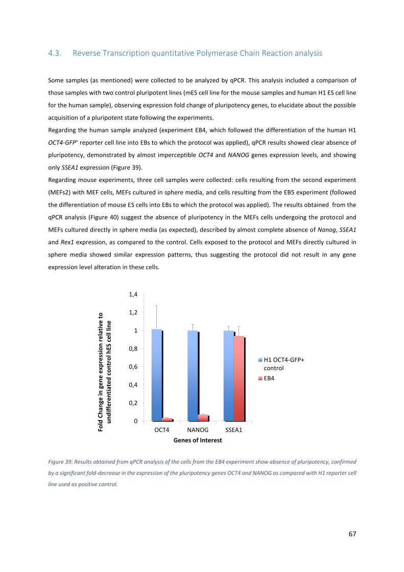
Regarding the human sample analyzed (experiment EB4, which followed the differentiation of the human H1
OCT4-GFP+ reporter cell line into EBs to which the protocol was applied), qPCR results showed clear absence of
pluripotency, demonstrated by almost imperceptible OCT4 and NANOG genes expression levels, and showing
only SSEA1 expression (Figure 39).
Regarding mouse experiments, three cell samples were collected: cells resulting from the second experiment
(MEFs2) with MEF cells, MEFs cultured in sphere media, and cells resulting from the EB5 experiment (followed
the differentiation of mouse ES cells into EBs to which the protocol was applied). The results obtained from the
qPCR analysis (Figure 40) suggest the absence of pluripotency in the MEFs cells undergoing the protocol and
MEFs cultured directly in sphere media (as expected), described by almost complete absence of Nanog, SSEA1
and Rex1 expression, as compared to the control. Cells exposed to the protocol and MEFs directly cultured in
sphere media showed similar expression patterns, thus suggesting the protocol did not result in any gene
expression level alteration in these cells.
Figure 39: Results obtained from qPCR analysis of the cells from the EB4 experiment show absence of pluripotency, confirmed
by a significant fold-decrease in the expression of the pluripotency genes OCT4 and NANOG as compared with H1 reporter cell
line used as positive control.
0
0,2
0,4
0,6
0,8
1
1,2
1,4
OCT4 NANOG SSEA1Fold
Ch
ange
in g
en
e e
xpre
ssio
n r
ela
tive
to
u
nd
iffe
ren
tiat
ed
co
ntr
ol h
ES c
ell
line
Genes of Interest
H1 OCT4-GFP+control
EB4
68
Table 3: Experiments’ overview show that EBs’ dissociation changes successfully resulted in more efficient post-stresses culture, although all of them adapted uncontrolled differentiated-like status. First experiment using MEFs yielded apparently positive results, while the second refuted that possibility. Experiments conducted with TTs and GCs resulted in contamination and insufficient cell number, respectively, thus resulting in completely negative results.
Starting cell density Cell density after Proliferation capacity
Single cells or small colonies (presumably dead after 7
days)
EB3 (human
UGENT11-2)
9 wells of EBs
0,25% trypsin-EDTA (5 min)
Medium (2x10⁶ cells)
Medium Medium Medium (1.5x10⁶ cells)
Low (6x10⁵ cells)
Yes (high) Increasingly big differentiated colonies
EB4 (human reporter H1)
9 wells of EBs
0,25% trypsin-EDTA (5 min)
Medium (2x10⁶ cells)
Medium Medium (slightly low)
Medium (1.5x10⁶ cells)
Medium (1.3x10⁶
cells)
Yes (very high)
Completely differentiated EB-like colonies
EB5 (mouse ES cells)
24 wells of EBs
0,25% trypsin-EDTA (5 min)
Very high (1x10⁷ cells)
Very high Very high Very high (8x10⁶ cells)
Very high (6x10⁶ cells)
Yes (very high)
Uncontrolled growth with possible differentiation
TTs1 Medium (3x10⁶ cells)
Not observed
Not observed
Medium (1.5x10⁶ cells)
Low (4.2x10⁵
cells)
Some Some contamination present leading to incapacity to grow
TTs2 Medium (3x10⁶ cells)
Medium Medium (slightly low)
Medium (1.6x10⁶ cells)
Low (7.8x10⁵
cells)
Almost none A lot of contamination present, cell death
GCs1 Reasonable (1x10⁶ cells)
Low Low Low (4.2x10⁵ cells)
Low (3x10⁵ cells)
No All cells dead
GCs2 Reasonable (1x10⁶ cells)
Medium Low Low (4.2x10⁵ cells)
Low (2x10⁵ cells)
Some Single cells with some initial proliferation
MEFs1 Medium (2.4x10⁶
cells)
Low Low Low (4.8x10⁵ cells)
Very low (2x10⁵ cells)
Yes (at the beginning)
Cell loss and differentiation
MEFs2
High (3.9x10⁶
cells)
Medium (slightly
high)
Medium (slightly
high)
Medium (2.8x10⁶ cells)
Medium (1x10⁶ cells)
Very high (at the
beginning)
Complete differentiation into cells resembling MEFs'
morphology
69
Figure 40: Results obtained by qPCR showed absence of pluripotency in the MEF-related experiments. EB5 experiment
presented some interesting expression of Rex1 and Gbx2 comparing to the pluripotent control. However, low expression of
both Nanog and SSEA1 suggests differentiation.
Contrastingly, cells from EB5 experiment showed some interesting results. Gbx2, the mouse homeobox gene,
known for being expressed in mES cells, having its expression decreased in differentiated cells (Chapman et al.,
1997), showed similar expression levels in the EB5 sample as compared to the control. Also, Rex1, a zinc finger
protein expressed mainly in undifferentiated cells (Scotland et al., 2009) showed an expression level that even
comparable to the undifferentiated ES control cells. Despite showing evidences of pluripotency, even evaluating
the slight Nanog expression observed, these cells did not acquire pluripotency. The surface marker gene SSEA1
expression level was particularly low. Since the expression level of this gene is known to decrease upon
differentiation (Zhao et al., 2012), this consisted in an evidence of differentiation of these cells.
4.4. Complementary work and Controls
4.4.1. Mouse Embryonic Stem cells cultured in sphere media
Somatic cells, following acid treatment, were stated to be reprogrammed into a pluripotent state during a 7 days
culture (Obokata et al., 2014d), in the termed sphere media, being supposed not to attach and to form small
round shaped cells. Only mouse cell data is available, and therefore it is unclear at this point if human cells,
0
0,2
0,4
0,6
0,8
1
1,2
1,4
1,6
mES Cell control MEF (cultured insphere media)
MEFs2 EB5
Fold
Ch
ange
in G
en
e E
xpre
ssio
n r
ela
tive
to
u
nd
iffe
ren
tiat
ed
mES
C c
on
tro
l
Different media for STAP induction
NANOG
SSEA1
REX1
GBX2
70
following these experiment, should adopt a morphology characteristic of hES cells or instead a status
characteristic of naïve hES cells, since they were cultured in ultra-low attachment plates (and hES cells are usually
grown in feeder-dependent conditions, usually with MEFs, to which they attach). However, since cells following
the protocol should acquire pluripotency through the 7 days of culture, it was admitted that sphere media was
(despite not having any compound to promote naïve pluripotency except LIF) capable of sustaining/induce both
proliferative capacity and pluripotent state of the cells cultured in it. Otherwise, the supposition that pluripotent
cells can arise in culture in this medium would not make sense. Intrigued by this supposition, it was decided to
culture mouse ES cells directly in sphere media (supplemented with heparin, EGF and mouse LIF) without being
exposed to any stress, and check cell progression through several days of culture.
Figure 41: Naïve mouse ES cells directly cultured in sphere media could not survive the culture conditions during 7 days. A.
After 3 days in culture, cell density was particularly high. B. By 5 days in culture, cell density decreased comparing to the
previous days. C. Cell density continued to decrease until day 7. Sphere media was demonstrated to be incapable of
maintaining naïve mES cells in culture.
Interestingly, proliferative capacity was not demonstrated on mouse ES cells directly cultured in sphere media,
fact demonstrated by a cell loss from day 3 to days 5 and 7 (Figure 41). Since both inhibitors that characterize 2i
conditions were not present, it was expected that these cells could not maintain a naïve state of pluripotency.
However, the medium should at least be capable of supporting cell survival, and it would be expected that these
cells could acquire a random proliferative capacity and differentiation tendency (due to the absence of 2i
conditions). Both of them were not verified. It is intriguing why a culture medium that is in fact incapable of
maintaining both naïve state of pluripotency and cell survival was suggested as capable of maintaining the
cultured cells for 7 days and induce them to acquire pluripotency. Therefore, this result represented an evidence
arguing against the possible acquisition of pluripotency by the cells cultured in this medium.
4.4.2. Protocol performed directly on Mouse Embryonic Stem cells
Intrigued by the response of pluripotent cells after being exposed to the stresses included in the protocol,
pluripotent mouse ES cells (the same used to grow embryoid bodies, EB5) were collected and directly exposed
both to the physical trituration steps and to acidic HBSS.
71
Figure 42: Naive mouse embryonic stem cells faced the whole protocol and showed high resistance to every step. A. High cell
density was used as starting material. B, C, D. High resistance to the trituration steps resulted in high cell density after the
first (B), second (C) and third (D) pipetting steps. E. Cell quantity was maintained even after acid exposure.
The protocol followed the exact same steps used for the EB5 experience, after the dissociation of those EBs into
single cells. High cell density was verified as starting material (Figure 42-A), after both the trituration steps (Figure
42-B, C, D), and even after acid exposure (Figure 42-E). Mouse ES cells showed, therefore, high resistance to the
described protocol, maintaining their integrity along the experiment and showing a particularly good cell density
before being cultured.
Following culture in sphere media (supplemented with the proper mouse compounds, EGF, heparin and mouse
LIF), cell growth was monitored during the 7 days of culture. As demonstrated, these cells showed high
proliferation capacity during the culture period, increasing the number of cells, and forming aggregates with
differentiation tendencies (Figure 43).
It was therefore possible to conclude that pluripotent mouse ES cells successfully survived and maintained their
capacities after being exposed to the defined stresses. Since pluripotent cells (possibly present in the initial
material) survived the whole protocol, they could be present and contribute to the differentiated-like
morphology observed in most of the experiments performed. Although at low probability, this would also mean
that a single pluripotent cell, which could still remain in that state in the 14 days differentiated EBs and maintain
its epigenetic characteristics during the experiment, would be sufficient to result in a false positive result. It
would be uncertain if the pluripotency observed would be a result of a previously pluripotent cell rather than
reprogramming. A negative control showed absence of pluripotency in the 14 days EBs (see chapter 4.4.3).
72
Figure 43: Mouse ES cells were directly exposed to the protocol and cultured in sphere media. A. After 3 days in culture, high
cell density was observed, along with differentiation tendencies. B, C. After 5 (B) and 7 (C) days in culture, cells tended to
aggregate into completely differentiated clumps. These observations suggested that undifferentiated pluripotent cell can
survive the whole experiment, maintaining their capabilities to proliferate and differentiate in vitro.
4.4.3. Negative Control – Embryoid Bodies are fully differentiated
A negative control for Oct4 and Nanog expression was performed by staining mouse EBs grown for 14 days. To
avoid discrepancy between the experiments and this biological replicate, the exact same culture conditions were
used, including the exact same culture medium (N2B27 without adding mouse LIF nor any inhibitor).
Figure 44: EBs' immunostaining results showed absence of pluripotency-expressing cells in the structure of the EB after 14
days of differentiation culture.
73
This control mainly aimed to prove the absence of the pluripotency genes expression, Oct4 and Nanog, after 14
days in culture. This analysis was useful to exclude the possibility of having pluripotent cells, after the 7 days of
culture in sphere media, which could be already present before the protocol, rather than being reprogrammed.
It was assumed that experiments using both mouse and human cells, despite using cells with different
characteristics and different differentiation culture media, would result in a similar differentiation level after 14
days culture. Therefore, this negative control performed with mES cells should be sufficient to elucidate about
the differentiation level after 14 days in all the experiments and the absence of pluripotency at this stage.
Immunostaining analysis showed clear absence of consistent expression of pluripotency genes Oct4 and Nanog
(Figure 44). Although there was some overlap between Oct4 expression and DAPI staining, thus suggesting that
some intact cells expressed Oct4 at the time of fixation, the absence of a Nanog expression overlapping with
Oct4 clarified about the absence of pluripotency. The observed expression of these genes was therefore due to
unspecific binding or eventually due to a possible existence of non-pluripotent Oct4 or Nanog expressing cells
(Ambady et al., 2010; Zangrossi et al., 2007), as a result of the spontaneous differentiation characteristic of EBs’
culture. It was therefore possible to conclude about the absence of pluripotency in EBs’ cells following 14 days
culture in the defined conditions.
4.4.4. Positive Control – Mouse Embryonic Stem cells express Oct4 and Nanog
Figure 45: Immunostaining analysis performed of mouse ES cells showed correct expression of pluripotency genes Oct4 and
Nanog. This analysis confirmed the efficiency of the staining method being used.
74
A positive control was performed, to test both the efficiency of the staining method applied and to prove the
successful expression of Oct4 and Nanog in pluripotent cells (naïve mES cells in this case).
For that, a sample of mouse ES cells, expanded to be differentiated into EBs (EB5 experiment), was collected,
fixed and stained using antibodies for Oct4 and Nanog genes (see chapter 3). These mouse ES cells reside in a
naïve state of pluripotency and, similarly to primed pluripotent cells, must perfectly express pluripotency-
associated genes Oct4 and Nanog (Loh et al., 2006; Nichols et al., 1998).
As demonstrated (Figure 45), Oct4 and Nanog expression patterns almost perfectly overlapped. Both genes also
overlapped with most part of the cells identified by DAPI, thus confirming those were intact pluripotent cells
(positive control for pluripotency expression) and that both the staining methods and the antibodies used for
the analysis were efficient.
4.4.5. Negative Control – Mouse Embryonic Fibroblast cells do not express pluripotency
Facing the apparently positive expression of pluripotency genes Oct4 and Nanog on the first experiment
conducted with MEF cells (MEFs1), a negative control proving the absence of pluripotency in those MEF cells
prior to be perturbed by any stress was necessary. Therefore, MEFs at passage 3 (the same used as starting
material for the experiment) were stained.
Figure 46: Immunostaining results show absence of pluripotency on MEF cells prior to the protocol. Oct4 and Nanog expression was completely absence.
75
Immunostaining results confirmed that completely functional and normal MEF cells, not being exposed to any
stress, showed clear absence of pluripotency, without any Oct4 or Nanog expression. This result demonstrated
that prior to the experiment, these cells did not show any pluripotency and thus the possible pluripotency
expression after the protocol would be due to reprogramming rather than being inherited.
4.5. Further Discussion
The reprogramming of somatic cells into pluripotency is generally associated with low efficiency, and the
requirement of a large amount of cells as starting material to obtain a proper quantity of pluripotent cells, no
matter the method used (Obokata et al., 2014d; Tachibana et al., 2013; Takahashi and Yamanaka, 2006), is a
well-known drawback of those techniques. In addition to the low efficiency predicted, several factors could have
contributed to the failure of the present work.
Experiments conducted with human cells during this work, through EBs’ growth, showed sufficient but not
particularly high amount of starting cells. Associated with the particularly low efficiency, in case of possible
reprogramming event, high amounts of reprogramed pluripotent cells could never be expected. Also, facing the
challenging protocol, which resulted in a lot of difficulties to maintain the cells alive during the experiments, it
was necessary to analyze cell survival and response to the perturbations as deeply as the pluripotency analysis
itself. Further studies using a higher amount of starting material (ideally two complete 24 well plates of EBs)
should be performed. A higher amount of starting cells would increase the probability of obtaining a pluripotent
cell population. Even so, a considerably high amount of material, following differentiation of mouse ES cells into
EBs (EB5), was exposed to the perturbations defined in the protocol. However, truly pluripotency was not
acquired. Further optimizations are necessary for this approach. The idea of starting with ES cells, posteriorly
differentiated into EBs, which were subjected to the reprogramming technique, should be related with a higher
facility to acquire reprogramming event, since EBs could reside in a less differentiated state than a functional
somatic cells, as suggested before. However, this methodology faced some drawbacks. In addition to the
necessity of large EBs’ cultures to have a larger amount of starting cells, the massive cell loss and inefficiency of
the dissociation step, which was indispensable before the cells were exposed to the physical perturbations,
represented issues that need to be revisited. Theoretically, the experiment EB5, following differentiation of mES
cells into EBs, should be the one from which the best results would be expected. Since the protocol was stated
to work with murine cells (Obokata et al., 2014d), and EBs should represent the possibly less differentiated
mouse cells used during this study, the reprogramming efficiency should be the highest possible to obtain. The
combination of the limited data available about this technique with the incapacity to reprogram mouse EBs into
a state of pluripotency suggested low expectations to the experiments using other kind of cells.
The necessity for a proper starting material was not only applied for the first methodology of the present work.
Isolation of somatic cells also requires efficient and high throughput methods. The method through which murine
somatic cells were isolated can be definitely related with the inefficiency of the procedure. Importantly, the
contamination observed during experiences with MEF cells was surely critical to the failure of the experiment.
More than perturbing cell culture, posterior to the whole treatment protocol, these impurities may have
76
interfered with the trituration procedures by blocking the passage of the cells through the pipette’s tip.
Moreover, the presence of these contaminations may have led to cells’ disruption following physical contact with
them, largely increasing cell death along the procedures. Again, the amount of cells needed for these
experiments was shown to be high, since high cell loss was verified even after the first pipetting step.
In all the experiments performed, the long procedure may have influenced negative results. A long maintenance
of the cells out of the incubator and in a buffer medium directly increased the amount of cells that were lost
during the experiment. The requirement of successive trituration steps followed by acid exposure, along with
several centrifugation rounds, left the cells in external extreme survival conditions. More than interfering with
the cell’s capacity to be maintained alive, this harsh conditions at which cells had to survive may have interfered
with the protocol itself, modifying the response of the cells to the external stimuli and possibly leaving them too
susceptible. During the present protocol these facts were unavoidable, since the whole protocol assumed the
necessity of a time-consuming physical treatment for the cells, which came out as a response for the failure and
incapacity to replicate the pre-defined protocol with acid treatment only. These experiments using chemical
treatment only, alleged not to work, were discarded and experiments including both physical and chemical
treatments were a priority. This evidence turned it impossible to decrease the experiment duration to the original
30 minutes only protocol. However, further experiments using the originally claimed protocol, with a single acid
exposure to induce pluripotency, should be performed, to analyze the results and compare expression levels of
pluripotency genes and morphology with the ones obtained with the present study.
Acid treatment on cells can be definitely considered as a severe method that leaves few chances of acquiring a
proper functional cell. By disrupting membrane proteins structure due to pH alterations, breaking their
supportive ionic bonds, the presence of an acidic environment leads the cells both to open potentially dangerous
channels in their membrane and lose their physical integrity (Campos et al., 2009; Lowes and Simmons, 2001;
Niero and Machado-Santelli, 2013). It is therefore more than expected that an acidic solution potentially kills the
cells that are exposed. Such evidence can explain both the cell loss verified after some of the acidic treatments
realized and the incompetence of the surviving cells to be maintained and proliferate after being cultured again,
associated with the intrinsic damage caused by that exposure to the acid. Different kinds of cells may have
different resistance to the exposure to equally different acidic solutions and/or compounds (Lampe et al., 2009).
Interestingly, acid can kill cancer cells (Mei et al., 2014). However, not all the cells have this response. Gastric
mucosa, the cells that line the stomach, are daily exposed to pH around 2 and still maintain their integrity. It is
therefore difficult at this point to predict the response of different cell types to the acidic solution, as performed
in the experiments. Further studies to evaluate the resistance of the different studied cell types to different acidic
solutions and pH levels could result in an optimal acid exposure, unveiling a relatively more optimized protocol
to be applied to each cell type.
The technical tips suggesting physical stress as essential to generate STAP cells came along with the idea that the
reprogramming event could be facilitated by any kind of stress induced on the cells. Therefore, following this
idea and as it was stated (Obokata et al., 2014d), other experimental procedures that equally expose the cells to
stressful conditions would or could have influence on the acquisition of pluripotency. The answer to the question
of whether strong external stimuli, of any kind, have any influence on a differentiated cell leading to a pluripotent
77
reprogrammed state, remains unclear. Cells’ trituration surely provoked several damage compromising their
integrity. It is hard to understand which effect this physical treatment could have on cells to influence them to
acquire pluripotency. Successive trituration may leave cells more susceptible and weak, and thus further
perturbations (such as the acid exposure) can be more efficient. This could eventually be the reason why this
physical treatment would facilitate reprogramming, since a direct relation between physical perturbation and
epigenetic change cannot be found. This way, the actual effect of the physical trituration is supposed to have on
the cells, which could scientifically explain an epigenetic change leading to a pluripotent state and justify the use
of such physical pipetting steps, remains undefined.
78
5. Conclusion and Perspectives
Together, the results obtained showed the incapacity of differentiated cells to acquire pluripotency following
acid exposure, contradicting what was previously claimed (Obokata et al., 2014d), even inducing various physical
stresses that were said to help the reprogramming event.
The ultimate goal to be achieved in the present work would be the efficient reprogramming using human cells,
assuming that the same reprogramming with mouse cells would be effectively possible and reproducible.
However, the results obtained showed clear divergence from a possible pluripotency acquisition. Moreover, it
was shown to be very difficult to conduct the whole protocol and reach the culture phase during which cells
would acquire pluripotency. All these difficulties faced during the study obligated the analysis to be equally
focused on the monitoring of the cells’ survival during the experiments and then on the possible acquisition of
pluripotency (if the cells efficiently reached the culture phase), rather than being only focused on the efficient
reprogramming event.
A completely adapted protocol, using embryoid bodies as starting material to be subjected to the perturbations,
was studied. In general, even starting from embryoid bodies, presumably in a less differentiated state than a fully
differentiated and functional somatic cells integrated in a leaving being, physical and chemical stresses together
were not shown to have the capacity to induce a reprogramming event. However, additional studies would be
necessary to evaluate these observations.
Experiments performed in functional and directly isolated, or cultured in vitro, murine somatic cells confirmed
the incapacity of this technique to induce pluripotency, contrastingly to what was stated. Mouse embryonic
fibroblasts represented an easily obtainable source of somatic cells for this study, since these cells are regularly
used as feeder layer for human embryonic stem cells culture studies. Although apparent positive results were
obtained in a first trial, a second experiment using a higher amount of cells from the same source, which could
possibly lead to more expressive positive results, refuted the first results, resulting in a fully differentiated cell
population. Cells from mouse tail tips showed no expression of pluripotency posteriorly to be exposed to these
perturbations. Moreover, experiments with these cells led to the presence of a large amount of impurities within
the cell culture. These contamination was directly related with the method by which mouse tail tip cells were
isolated, and thus this method should be improved. The amount of cells from both two and six tail tips was shown
to be insufficient due to the high cell loss verified during the protocol. Therefore, experiments involving these
cells can be hindered by ethical issues regarding the amount of animals that need to be sacrificed or damaged to
make ultimate experiments and achieve trustable results, with this and other different types of cells from the
organism. Also, following the facility in reprogramming granulosa cells into induced pluripotent stem cells, two
experiments were performed with this cell type, hoping that some pluripotency expression would be observed.
However, no pluripotency was acquired in neither the experiments. Moreover, the amount of cells obtained as
starting material for the experiments was shown to be particularly random, since cells extracted from six mice
yielded the same density as compared to only two mice. Therefore, the same issue already described for cells
from the tail tips (and for cells from any other tissue from the organism), related with the amount of animals
79
necessary to have a proper amount of cells to ultimately test this technique, is present with granulosa cells.
Accordingly to the amount of cells obtained as starting material from six sacrificed mice, it is expected to be quite
hard to obtain a proper amount of cells regarding both ethical and economical costs involved.
Although several optimizations were made during the experiences, regarding practical technique and handling,
as well as certain compounds used in certain steps, such as the use of trypsin-EDTA 0.25% that more effitiently
dissociated embryoid bodies into single cells, several improvements can be made in the whole protocol.
Generally, the volume of neutral HBSS in which cells were re-suspended before starting the first pipetting, the
volume of acid used to treat the cells, and the volume of sphere media in which cells were re-suspended and
posteriorly cultured, should be optimized. Although optimal values were suggested in Obokata’s papers, the
amount of cells obtained in every experiment varied drastically, and thus the optimal concentrations were not
obtained in all the experiments. The volume of neutral HBSS in which the starting material is suspended has a
crucial influence in the following physical stress steps, since a variation in the concentration of cells influences
the amount of cells that pass through the tips of the pipettes and suffer trituration by them. A too small cell
density means that a few cells are perturbed in each pipetting, whereas a too high cell density could mean
constriction of the tip, leading to an excessively severe trituration. An optimal volume of 2-3ml of HBSS was
suggested, at a concentration of 1x10⁶ cells/ml, and thus ideally a maximum of 3 million cells would be subjected
to the initial physical stress. Associated to the high cell loss observed during the protocol, this can be considered
a relatively slow amount of cells to be used as starting material, and surely reduces the chances to obtain a
proper amount of viable cells in culture. Relatively to the acidic HBSS treatment, the cell density is crucial. The
perfect cell concentration in this acidic suspension should be achieved in order to have a treatment that both
does not kill the cells and helps to reprogram them into a pluripotent state. A too small quantity of acid, relatively
to the amount of cells, may not have sufficient effect on the cells. On the other hand, a higher amount of acid
per cell can be too harsh for them, leading to death. Although an optimal concentration of 2x10⁶ cells/ml of acid
was suggested by Obokata et al, the issues faced during the present experiments regarding the limited quantity
of starting cells and the massive cell loss during the experiments rarely allowed to reach that concentration value.
Therefore, the lower concentration of cells in the acidic solution used in most part of the experiments surely
contributed for a particularly severe chemical perturbation, and may explain the extensive cell loss verified.
Despite all these optimizations that are necessary, the results obtained with this technique do not seem to create
any expectation regarding the possible acquisition of pluripotency following the exposure of cells to an external
strong environment, as previously stated, thus suggesting the incapacity of this technique to actually work.
The merely interesting results obtained would surely face several implications for the use of these cells for
therapeutics. Showing reprogramming capacity, this technique would be the third main reprogramming
possibility after somatic cell nuclear transfer and induced pluripotent stem cells. Interestingly, this
reprogramming method would have several advantages. Generally, as a reprogramming event, this technique
would allow the production of pluripotent stem cells from pre-existing differentiated somatic cells, thus
retrieving the usually necessary use of embryos as stem cells source. This would not only amplify the availability
of stem cells, but also overcome possible ethical issues regarding the use of embryos for research and
therapeutics. As a matter of fact, this technique would surpass somatic cell nuclear transfer, since it would not
80
be dependent of viable and good quality unfertilized donated oocytes. Also, somatic cell nuclear transfer
technically generates an embryo, thus generating a possible source of life. The reprogramming of somatic cells
rather than use of oocytes overcomes this ethical issue. Due to its simplicity, through the simple exposure of
somatic cells to an acidic solution for half an hour (and possibly to some extra physical treatment), it would be a
promising method to acquire the so much desired high amounts of pluripotent stem cells, necessary for
therapeutic purposes. Induced pluripotent stem cells’ technique can be considered the standard reprogramming
method, since this technique potentially overcomes all the issues enumerated above. However, problems related
with possible viral activation, since transgenes are transfected into the somatic cells through retroviruses to
acquire pluripotency, are a reality. Also, other direct reprogramming methods using plasmids or small molecules
can modify the genome of the cells, leading to mutations, despite overcoming the problems associated with the
use of virus. Interestingly, if this external stimuli-based reprogramming method would work, this issue related
with the virus-dependency would also be surpassed. However, the completely negative results obtained during
this work turn unclear if the process itself would result in several genome modifications, deviating the possibly
reprogrammed cells from an ES-like state, and thus not being suitable for proper research or therapeutics. Since
no viable pluripotent cells were obtained, is it not possibly to evaluate the capacity of the eventually generated
cells to be differentiated into different cells types with an efficiency comparable to ES cells. The unpredictability
of this stimulus-based reprogramming technique immediately argues against the possible use of the generated
pluripotent cells for research or therapeutics, since it would face a lot of safety-related issues.
The existence of STAP stem cells would be remarkable. The exceptionally easy method through which pluripotent
cells are supposed to be created is just unbeatable in the stem cells field. In the case of efficient reprogramming,
it would surely be the easiest and most accepted method to reprogram differentiated cells into pluripotency,
completely changing the way scientists would approach stem cells. However, if cells could be reprogrammed and
lose their functionality through external stresses, this would mean that a common cell from a common living
being could spontaneously lose its functionality anytime. The conditions involved in the reprogramming
technique are far away from the daily environment at which the organisms are exposed. However, the possibility
of a living being to acquire a tumor after reprogramming of its somatic cells due to being exposed to possibly
strong environmental condition (and all the living being are daily exposed to several severe conditions) cannot
be discarded. Following common sense, this fact surely argues against the capacity of this reprogramming
technique to reprogram somatic cells.
It is intriguing why a physical and/or chemical stress would lead to the acquisition of pluripotency the way it was
stated. Following the proof of mix or switch of cells and inclusion of fraudulent data by Obokata in her
publication, the possible acquisition of a pluripotent state triggered by an external stimulus remains a mystery.
However, at this points, the evidences of the obtained results suggest that this reprogramming method, in the
way it was suggested (Obokata et al., 2014d) and in the way it was adapted to the present work, is incapable of
inducing a differentiated cell into a state of truly pluripotency.
i
6. References
Aasen, T., Raya, A., Barrero, M.J., Garreta, E., Consiglio, A., Gonzalez, F., Vassena, R., Bilić, J., Pekarik, V., Tiscornia, G., et al. (2008). Efficient and rapid generation of induced pluripotent stem cells from human keratinocytes. Nat Biotechnol 26, 1276-1284. Aebi, U., Cohn, J., Buhle, L., and Gerace, L. (1986). The nuclear lamina is a meshwork of intermediate-type filaments. Nature 323, 560-564. Akhurst, R.J., Lehnert, S.A., Faissner, A., and Duffie, E. (1990). TGF beta in murine morphogenetic processes: the early embryo and cardiogenesis. Development 108, 645-656. Amabile, G., and Meissner, A. (2009). Induced pluripotent stem cells: current progress and potential for regenerative medicine. Trends Mol Med 15, 59-68. Ambady, S., Malcuit, C., Kashpur, O., Kole, D., Holmes, W.F., Hedblom, E., Page, R.L., and Dominko, T. (2010). Expression of NANOG and NANOGP8 in a variety of undifferentiated and differentiated human cells. Int J Dev Biol 54, 1743-1754. Avilion, A.A., Nicolis, S.K., Pevny, L.H., Perez, L., Vivian, N., and Lovell-Badge, R. (2003). Multipotent cell lineages in early mouse development depend on SOX2 function. Genes Dev 17, 126-140. Bao, S., Tang, F., Li, X., Hayashi, K., Gillich, A., Lao, K., and Surani, M.A. (2009). Epigenetic reversion of post-implantation epiblast to pluripotent embryonic stem cells. Nature 461, 1292-1295. Beattie, G.M., Lopez, A.D., Bucay, N., Hinton, A., Firpo, M.T., King, C.C., and Hayek, A. (2005). Activin A maintains pluripotency of human embryonic stem cells in the absence of feeder layers. Stem Cells 23, 489-495. Bechard, M., and Dalton, S. (2009). Subcellular localization of glycogen synthase kinase 3beta controls embryonic stem cell self-renewal. Mol Cell Biol 29, 2092-2104. Bernardo, A.S., Faial, T., Gardner, L., Niakan, K.K., Ortmann, D., Senner, C.E., Callery, E.M., Trotter, M.W., Hemberger, M., Smith, J.C., et al. (2011). BRACHYURY and CDX2 mediate BMP-induced differentiation of human and mouse pluripotent stem cells into embryonic and extraembryonic lineages. Cell Stem Cell 9, 144-155. Bourillot, P.Y., Aksoy, I., Schreiber, V., Wianny, F., Schulz, H., Hummel, O., Hubner, N., and Savatier, P. (2009). Novel STAT3 target genes exert distinct roles in the inhibition of mesoderm and endoderm differentiation in cooperation with Nanog. Stem Cells 27, 1760-1771. Briggs, R., and King, T.J. (1952). Transplantation of Living Nuclei From Blastula Cells into Enucleated Frogs' Eggs. Proc Natl Acad Sci U S A 38, 455-463. Brons, I.G., Smithers, L.E., Trotter, M.W., Rugg-Gunn, P., Sun, B., Chuva de Sousa Lopes, S.M., Howlett, S.K., Clarkson, A., Ahrlund-Richter, L., Pedersen, R.A., et al. (2007). Derivation of pluripotent epiblast stem cells from mammalian embryos. Nature 448, 191-195. Buehr, M., Meek, S., Blair, K., Yang, J., Ure, J., Silva, J., McLay, R., Hall, J., Ying, Q.L., and Smith, A. (2008). Capture of authentic embryonic stem cells from rat blastocysts. Cell 135, 1287-1298. Burridge, P.W., and Zambidis, E.T. (2013). Highly efficient directed differentiation of human induced pluripotent stem cells into cardiomyocytes. Methods Mol Biol 997, 149-161. Campbell, K.H., McWhir, J., Ritchie, W.A., and Wilmut, I. (1996). Sheep cloned by nuclear transfer from a cultured cell line. Nature 380, 64-66. Campos, F.M., Couto, J.A., Figueiredo, A.R., Tóth, I.V., Rangel, A.O., and Hogg, T.A. (2009). Cell membrane damage induced by phenolic acids on wine lactic acid bacteria. Int J Food Microbiol 135, 144-151. Carpenter, M.K., Rosler, E., and Rao, M.S. (2003). Characterization and differentiation of human embryonic stem cells. Cloning Stem Cells 5, 79-88. Cartwright, P., McLean, C., Sheppard, A., Rivett, D., Jones, K., and Dalton, S. (2005). LIF/STAT3 controls ES cell self-renewal and pluripotency by a Myc-dependent mechanism. Development 132, 885-896. Chambers, I., Colby, D., Robertson, M., Nichols, J., Lee, S., Tweedie, S., and Smith, A. (2003). Functional expression cloning of Nanog, a pluripotency sustaining factor in embryonic stem cells. Cell 113, 643-655. Chapman, G., Remiszewski, J.L., Webb, G.C., Schulz, T.C., Bottema, C.D., and Rathjen, P.D. (1997). The mouse homeobox gene, Gbx2: genomic organization and expression in pluripotent cells in vitro and in vivo. Genomics 46, 223-233. Chen, K.G., Mallon, B.S., McKay, R.D., and Robey, P.G. (2014). Human pluripotent stem cell culture: considerations for maintenance, expansion, and therapeutics. Cell Stem Cell 14, 13-26. Chung, Y., Klimanskaya, I., Becker, S., Li, T., Maserati, M., Lu, S.J., Zdravkovic, T., Ilic, D., Genbacev, O., Fisher, S., et al. (2008). Human embryonic stem cell lines generated without embryo destruction. Cell Stem Cell 2, 113-117.
ii
Chung, Y., Klimanskaya, I., Becker, S., Marh, J., Lu, S.J., Johnson, J., Meisner, L., and Lanza, R. (2006). Embryonic and extraembryonic stem cell lines derived from single mouse blastomeres. Nature 439, 216-219. Constantinescu, D., Gray, H.L., Sammak, P.J., Schatten, G.P., and Csoka, A.B. (2006). Lamin A/C expression is a marker of mouse and human embryonic stem cell differentiation. Stem Cells 24, 177-185. Cowan, C.A., Atienza, J., Melton, D.A., and Eggan, K. (2005). Nuclear reprogramming of somatic cells after fusion with human embryonic stem cells. Science 309, 1369-1373. Davidson, K.C., Adams, A.M., Goodson, J.M., McDonald, C.E., Potter, J.C., Berndt, J.D., Biechele, T.L., Taylor, R.J., and Moon, R.T. (2012). Wnt/β-catenin signaling promotes differentiation, not self-renewal, of human embryonic stem cells and is repressed by Oct4. Proc Natl Acad Sci U S A 109, 4485-4490. Dimos, J.T., Rodolfa, K.T., Niakan, K.K., Weisenthal, L.M., Mitsumoto, H., Chung, W., Croft, G.F., Saphier, G., Leibel, R., Goland, R., et al. (2008). Induced pluripotent stem cells generated from patients with ALS can be differentiated into motor neurons. Science 321, 1218-1221. Eckersley-Maslin, M.A., Bergmann, J.H., Lazar, Z., and Spector, D.L. (2013). Lamin A/C is expressed in pluripotent mouse embryonic stem cells. Nucleus 4, 53-60. Eminli, S., Foudi, A., Stadtfeld, M., Maherali, N., Ahfeldt, T., Mostoslavsky, G., Hock, H., and Hochedlinger, K. (2009). Differentiation stage determines potential of hematopoietic cells for reprogramming into induced pluripotent stem cells. Nat Genet 41, 968-976. Eminli, S., Utikal, J., Arnold, K., Jaenisch, R., and Hochedlinger, K. (2008). Reprogramming of neural progenitor cells into induced pluripotent stem cells in the absence of exogenous Sox2 expression. Stem Cells 26, 2467-2474. Evans, M. (2011). Discovering pluripotency: 30 years of mouse embryonic stem cells. Nat Rev Mol Cell Biol 12, 680-686. Evans, M.J. (1972). The isolation and properties of a clonal tissue culture strain of pluripotent mouse teratoma cells. J Embryol Exp Morphol 28, 163-176. Evans, M.J., and Kaufman, M.H. (1981). Establishment in culture of pluripotential cells from mouse embryos. Nature 292, 154-156. Fox, N., Damjanov, I., Martinez-Hernandez, A., Knowles, B.B., and Solter, D. (1981). Immunohistochemical localization of the early embryonic antigen (SSEA-1) in postimplantation mouse embryos and fetal and adult tissues. Dev Biol 83, 391-398. Gafni, O., Weinberger, L., Mansour, A.A., Manor, Y.S., Chomsky, E., Ben-Yosef, D., Kalma, Y., Viukov, S., Maza, I., Zviran, A., et al. (2013). Derivation of novel human ground state naive pluripotent stem cells. Nature 504, 282-286. Galvin, K.E., Travis, E.D., Yee, D., Magnuson, T., and Vivian, J.L. (2010). Nodal signaling regulates the bone morphogenic protein pluripotency pathway in mouse embryonic stem cells. J Biol Chem 285, 19747-19756. Gardner, R.L. (1983). Origin and differentiation of extraembryonic tissues in the mouse. Int Rev Exp Pathol 24, 63-133. Guo, G., Yang, J., Nichols, J., Hall, J.S., Eyres, I., Mansfield, W., and Smith, A. (2009). Klf4 reverts developmentally programmed restriction of ground state pluripotency. Development 136, 1063-1069. Gurdon, J.B. (1962). The developmental capacity of nuclei taken from intestinal epithelium cells of feeding tadpoles. J Embryol Exp Morphol 10, 622-640. Hanna, J., Cheng, A.W., Saha, K., Kim, J., Lengner, C.J., Soldner, F., Cassady, J.P., Muffat, J., Carey, B.W., and Jaenisch, R. (2010). Human embryonic stem cells with biological and epigenetic characteristics similar to those of mouse ESCs. Proc Natl Acad Sci U S A 107, 9222-9227. Hanna, J., Markoulaki, S., Mitalipova, M., Cheng, A.W., Cassady, J.P., Staerk, J., Carey, B.W., Lengner, C.J., Foreman, R., Love, J., et al. (2009). Metastable pluripotent states in NOD-mouse-derived ESCs. Cell Stem Cell 4, 513-524. Hanna, J., Markoulaki, S., Schorderet, P., Carey, B.W., Beard, C., Wernig, M., Creyghton, M.P., Steine, E.J., Cassady, J.P., Foreman, R., et al. (2008). Direct reprogramming of terminally differentiated mature B lymphocytes to pluripotency. Cell 133, 250-264. Hanna, J., Wernig, M., Markoulaki, S., Sun, C.W., Meissner, A., Cassady, J.P., Beard, C., Brambrink, T., Wu, L.C., Townes, T.M., et al. (2007). Treatment of sickle cell anemia mouse model with iPS cells generated from autologous skin. Science 318, 1920-1923. Hassani, S.N., Totonchi, M., Farrokhi, A., Taei, A., Larijani, M.R., Gourabi, H., and Baharvand, H. (2012). Simultaneous suppression of TGF-β and ERK signaling contributes to the highly efficient and reproducible generation of mouse embryonic stem cells from previously considered refractory and non-permissive strains. Stem Cell Rev 8, 472-481. Hassani, S.N., Totonchi, M., Gourabi, H., Schöler, H.R., and Baharvand, H. (2014a). Signaling roadmap modulating naive and primed pluripotency. Stem Cells Dev 23, 193-208.
iii
Hassani, S.N., Totonchi, M., Sharifi-Zarchi, A., Mollamohammadi, S., Pakzad, M., Moradi, S., Samadian, A., Masoudi, N., Mirshahvaladi, S., Farrokhi, A., et al. (2014b). Inhibition of TGFβ signaling promotes ground state pluripotency. Stem Cell Rev 10, 16-30. Hayashi, K., and Surani, M.A. (2009). Self-renewing epiblast stem cells exhibit continual delineation of germ cells with epigenetic reprogramming in vitro. Development 136, 3549-3556. Henderson, J.K., Draper, J.S., Baillie, H.S., Fishel, S., Thomson, J.A., Moore, H., and Andrews, P.W. (2002). Preimplantation human embryos and embryonic stem cells show comparable expression of stage-specific embryonic antigens. Stem Cells 20, 329-337. Hirai, H., Karian, P., and Kikyo, N. (2011). Regulation of embryonic stem cell self-renewal and pluripotency by leukaemia inhibitory factor. Biochem J 438, 11-23. Hochedlinger, K., and Jaenisch, R. (2006). Nuclear reprogramming and pluripotency. Nature 441, 1061-1067. Kim, J.B., Sebastiano, V., Wu, G., Araúzo-Bravo, M.J., Sasse, P., Gentile, L., Ko, K., Ruau, D., Ehrich, M., van den Boom, D., et al. (2009). Oct4-induced pluripotency in adult neural stem cells. Cell 136, 411-419. Kim, J.B., Zaehres, H., Wu, G., Gentile, L., Ko, K., Sebastiano, V., Araúzo-Bravo, M.J., Ruau, D., Han, D.W., Zenke, M., et al. (2008). Pluripotent stem cells induced from adult neural stem cells by reprogramming with two factors. Nature 454, 646-650. Kleinsmith, L.J., and Pierce, G.B. (1964). Multipotentiality of single embryonal carcinoma cells. Cancer Res 24, 1544-1551. Klimanskaya, I., Chung, Y., Becker, S., Lu, S.J., and Lanza, R. (2006). Human embryonic stem cell lines derived from single blastomeres. Nature 444, 481-485. Kunath, T., Saba-El-Leil, M.K., Almousailleakh, M., Wray, J., Meloche, S., and Smith, A. (2007). FGF stimulation of the Erk1/2 signalling cascade triggers transition of pluripotent embryonic stem cells from self-renewal to lineage commitment. Development 134, 2895-2902. Kurosawa, H. (2007). Methods for inducing embryoid body formation: in vitro differentiation system of embryonic stem cells. J Biosci Bioeng 103, 389-398. Lampe, K.J., Namba, R.M., Silverman, T.R., Bjugstad, K.B., and Mahoney, M.J. (2009). Impact of lactic acid on cell proliferation and free radical-induced cell death in monolayer cultures of neural precursor cells. Biotechnol Bioeng 103, 1214-1223. Li, Y., McClintick, J., Zhong, L., Edenberg, H.J., Yoder, M.C., and Chan, R.J. (2005). Murine embryonic stem cell differentiation is promoted by SOCS-3 and inhibited by the zinc finger transcription factor Klf4. Blood 105, 635-637. Li, Z., Fei, T., Zhang, J., Zhu, G., Wang, L., Lu, D., Chi, X., Teng, Y., Hou, N., Yang, X., et al. (2012). BMP4 Signaling Acts via dual-specificity phosphatase 9 to control ERK activity in mouse embryonic stem cells. Cell Stem Cell 10, 171-182. Liao, J., Cui, C., Chen, S., Ren, J., Chen, J., Gao, Y., Li, H., Jia, N., Cheng, L., Xiao, H., et al. (2009). Generation of induced pluripotent stem cell lines from adult rat cells. Cell Stem Cell 4, 11-15. Liu, H., Zhu, F., Yong, J., Zhang, P., Hou, P., Li, H., Jiang, W., Cai, J., Liu, M., Cui, K., et al. (2008). Generation of induced pluripotent stem cells from adult rhesus monkey fibroblasts. Cell Stem Cell 3, 587-590. Lo, B., and Parham, L. (2009). Ethical issues in stem cell research. Endocr Rev 30, 204-213. Loh, Y.H., Agarwal, S., Park, I.H., Urbach, A., Huo, H., Heffner, G.C., Kim, K., Miller, J.D., Ng, K., and Daley, G.Q. (2009). Generation of induced pluripotent stem cells from human blood. Blood 113, 5476-5479. Loh, Y.H., Wu, Q., Chew, J.L., Vega, V.B., Zhang, W., Chen, X., Bourque, G., George, J., Leong, B., Liu, J., et al. (2006). The Oct4 and Nanog transcription network regulates pluripotency in mouse embryonic stem cells. Nat Genet 38, 431-440. Lowes, S., and Simmons, N.L. (2001). Human intestinal cell monolayers are preferentially sensitive to disruption of barrier function from basolateral exposure to cholic acid: correlation with membrane transport and transepithelial secretion. Pflugers Arch 443, 265-273. Mao, J., Zhang, Q., Ye, X., Liu, K., and Liu, L. (2014). Efficient induction of pluripotent stem cells from granulosa cells by Oct4 and Sox2. Stem Cells Dev 23, 779-789. Martin, G.R. (1980). Teratocarcinomas and mammalian embryogenesis. Science 209, 768-776. Martin, G.R. (1981). Isolation of a pluripotent cell line from early mouse embryos cultured in medium conditioned by teratocarcinoma stem cells. Proc Natl Acad Sci U S A 78, 7634-7638. Martin, G.R., and Evans, M.J. (1974). The morphology and growth of a pluripotent teratocarcinoma cell line and its derivatives in tissue culture. Cell 2, 163-172. Matsuda, T., Nakamura, T., Nakao, K., Arai, T., Katsuki, M., Heike, T., and Yokota, T. (1999). STAT3 activation is sufficient to maintain an undifferentiated state of mouse embryonic stem cells. EMBO J 18, 4261-4269.
iv
Mei, W., Dong, C., Hui, C., Bin, L., Fenggen, Y., Jingjing, S., Cheng, P., Meiling, S., Yawen, H., Xiaoshan, W., et al. (2014). Gambogenic acid kills lung cancer cells through aberrant autophagy. PLoS One 9, e83604. Meissner, A., and Jaenisch, R. (2006). Mammalian nuclear transfer. Dev Dyn 235, 2460-2469. Mitsui, K., Tokuzawa, Y., Itoh, H., Segawa, K., Murakami, M., Takahashi, K., Maruyama, M., Maeda, M., and Yamanaka, S. (2003). The homeoprotein Nanog is required for maintenance of pluripotency in mouse epiblast and ES cells. Cell 113, 631-642. Munsie, M.J., Michalska, A.E., O'Brien, C.M., Trounson, A.O., Pera, M.F., and Mountford, P.S. (2000). Isolation of pluripotent embryonic stem cells from reprogrammed adult mouse somatic cell nuclei. Curr Biol 10, 989-992. Niakan, K.K., and Eggan, K. (2013). Analysis of human embryos from zygote to blastocyst reveals distinct gene expression patterns relative to the mouse. Dev Biol 375, 54-64. Nichols, J., Silva, J., Roode, M., and Smith, A. (2009). Suppression of Erk signalling promotes ground state pluripotency in the mouse embryo. Development 136, 3215-3222. Nichols, J., and Smith, A. (2009). Naive and primed pluripotent states. Cell Stem Cell 4, 487-492. Nichols, J., Zevnik, B., Anastassiadis, K., Niwa, H., Klewe-Nebenius, D., Chambers, I., Schöler, H., and Smith, A. (1998). Formation of pluripotent stem cells in the mammalian embryo depends on the POU transcription factor Oct4. Cell 95, 379-391. Niero, E.L., and Machado-Santelli, G.M. (2013). Cinnamic acid induces apoptotic cell death and cytoskeleton disruption in human melanoma cells. J Exp Clin Cancer Res 32, 31. Niwa, H., Ogawa, K., Shimosato, D., and Adachi, K. (2009). A parallel circuit of LIF signalling pathways maintains pluripotency of mouse ES cells. Nature 460, 118-122. Noggle, S., Fung, H.L., Gore, A., Martinez, H., Satriani, K.C., Prosser, R., Oum, K., Paull, D., Druckenmiller, S., Freeby, M., et al. (2011). Human oocytes reprogram somatic cells to a pluripotent state. Nature 478, 70-75. Obokata, H., Sasai, Y., and Niwa, H. Essential technical tips for STAP convertion culture from somatic cells. URL: E http://www.nature.com/protocolexchange/protocols/3013 (accessed 03.06.14) Obokata, H., Sasai, Y., Niwa, H., Kadota, M., Andrabi, M., Takata, N., Tokoro, M., Terashita, Y., Yonemura, S., Vacanti, C.A., et al. (2014a). Bidirectional developmental potential in reprogrammed cells with acquired pluripotency. Nature 505, 676-680. Obokata, H., Sasai, Y., Niwa, H., Kadota, M., Andrabi, M., Takata, N., Tokoro, M., Terashita, Y., Yonemura, S., Vacanti, C.A., et al. (2014b). Retraction: Bidirectional developmental potential in reprogrammed cells with acquired pluripotency. Nature 511, 112. Obokata, H., Wakayama, T., Sasai, Y., Kojima, K., Vacanti, M.P., Niwa, H., Yamato, M., and Vacanti, C.A. (2014c). Retraction: Stimulus-triggered fate conversion of somatic cells into pluripotency. Nature 511, 112. Obokata, H., Wakayama, T., Sasai, Y., Kojima, K., Vacanti, M.P., Niwa, H., Yamato, M., and Vacanti, C.A. (2014d). Stimulus-triggered fate conversion of somatic cells into pluripotency. Nature 505, 641-647. Okita, K., Ichisaka, T., and Yamanaka, S. (2007). Generation of germline-competent induced pluripotent stem cells. Nature 448, 313-317. Okita, K., Nakagawa, M., Hyenjong, H., Ichisaka, T., and Yamanaka, S. (2008). Generation of mouse induced pluripotent stem cells without viral vectors. Science 322, 949-953. Oldershaw, R.A., Baxter, M.A., Lowe, E.T., Bates, N., Grady, L.M., Soncin, F., Brison, D.R., Hardingham, T.E., and Kimber, S.J. (2010). Directed differentiation of human embryonic stem cells toward chondrocytes. Nat Biotechnol 28, 1187-1194. Parisi, S., Passaro, F., Aloia, L., Manabe, I., Nagai, R., Pastore, L., and Russo, T. (2008). Klf5 is involved in self-renewal of mouse embryonic stem cells. J Cell Sci 121, 2629-2634. Rais, Y., Zviran, A., Geula, S., Gafni, O., Chomsky, E., Viukov, S., Mansour, A.A., Caspi, I., Krupalnik, V., Zerbib, M., et al. (2013). Deterministic direct reprogramming of somatic cells to pluripotency. Nature 502, 65-70. Reubinoff, B.E., Itsykson, P., Turetsky, T., Pera, M.F., Reinhartz, E., Itzik, A., and Ben-Hur, T. (2001). Neural progenitors from human embryonic stem cells. Nat Biotechnol 19, 1134-1140. Rideout, W.M., Hochedlinger, K., Kyba, M., Daley, G.Q., and Jaenisch, R. (2002). Correction of a genetic defect by nuclear transplantation and combined cell and gene therapy. Cell 109, 17-27. Robertson, J.A. (2001). Human embryonic stem cell research: ethical and legal issues. Nat Rev Genet 2, 74-78. Röber, R.A., Weber, K., and Osborn, M. (1989). Differential timing of nuclear lamin A/C expression in the various organs of the mouse embryo and the young animal: a developmental study. Development 105, 365-378. Schöler, H.R., Hatzopoulos, A.K., Balling, R., Suzuki, N., and Gruss, P. (1989). A family of octamer-specific proteins present during mouse embryogenesis: evidence for germline-specific expression of an Oct factor. EMBO J 8, 2543-2550. Scotland, K.B., Chen, S., Sylvester, R., and Gudas, L.J. (2009). Analysis of Rex1 (zfp42) function in embryonic stem cell differentiation. Dev Dyn 238, 1863-1877.
v
Selwood, L., and Johnson, M.H. (2006). Trophoblast and hypoblast in the monotreme, marsupial and eutherian mammal: evolution and origins. Bioessays 28, 128-145. Sherman, M.I. (1975). Long term culture of cells derived from mouse blastocysts. Differentiation 3, 51-67. Shevinsky, L.H., Knowles, B.B., Damjanov, I., and Solter, D. (1982). Monoclonal antibody to murine embryos defines a stage-specific embryonic antigen expressed on mouse embryos and human teratocarcinoma cells. Cell 30, 697-705. Shi, Y., Desponts, C., Do, J.T., Hahm, H.S., Schöler, H.R., and Ding, S. (2008). Induction of pluripotent stem cells from mouse embryonic fibroblasts by Oct4 and Klf4 with small-molecule compounds. Cell Stem Cell 3, 568-574. Singh, A.M., Reynolds, D., Cliff, T., Ohtsuka, S., Mattheyses, A.L., Sun, Y., Menendez, L., Kulik, M., and Dalton, S. (2012). Signaling network crosstalk in human pluripotent cells: a Smad2/3-regulated switch that controls the balance between self-renewal and differentiation. Cell Stem Cell 10, 312-326. Smith, A.G., Heath, J.K., Donaldson, D.D., Wong, G.G., Moreau, J., Stahl, M., and Rogers, D. (1988). Inhibition of pluripotential embryonic stem cell differentiation by purified polypeptides. Nature 336, 688-690. Sokol, S.Y. (2011). Maintaining embryonic stem cell pluripotency with Wnt signaling. Development 138, 4341-4350. Tachibana, M., Amato, P., Sparman, M., Gutierrez, N.M., Tippner-Hedges, R., Ma, H., Kang, E., Fulati, A., Lee, H.S., Sritanaudomchai, H., et al. (2013). Human embryonic stem cells derived by somatic cell nuclear transfer. Cell 153, 1228-1238. Takahashi, K., Tanabe, K., Ohnuki, M., Narita, M., Ichisaka, T., Tomoda, K., and Yamanaka, S. (2007). Induction of pluripotent stem cells from adult human fibroblasts by defined factors. Cell 131, 861-872. Takahashi, K., and Yamanaka, S. (2006). Induction of pluripotent stem cells from mouse embryonic and adult fibroblast cultures by defined factors. Cell 126, 663-676. Takeuchi, H., Nakatsuji, N., and Suemori, H. (2014). Endodermal differentiation of human pluripotent stem cells to insulin-producing cells in 3D culture. Sci Rep 4, 4488. Tan, H.K., Toh, C.X., Ma, D., Yang, B., Liu, T.M., Lu, J., Wong, C.W., Tan, T.K., Li, H., Syn, C., et al. (2014). Human finger-prick induced pluripotent stem cells facilitate the development of stem cell banking. Stem Cells Transl Med 3, 586-598. Tesar, P.J., Chenoweth, J.G., Brook, F.A., Davies, T.J., Evans, E.P., Mack, D.L., Gardner, R.L., and McKay, R.D. (2007). New cell lines from mouse epiblast share defining features with human embryonic stem cells. Nature 448, 196-199. Thomson, J.A., Itskovitz-Eldor, J., Shapiro, S.S., Waknitz, M.A., Swiergiel, J.J., Marshall, V.S., and Jones, J.M. (1998). Embryonic stem cell lines derived from human blastocysts. Science 282, 1145-1147. Thomson, J.A., Kalishman, J., Golos, T.G., Durning, M., Harris, C.P., Becker, R.A., and Hearn, J.P. (1995). Isolation of a primate embryonic stem cell line. Proc Natl Acad Sci U S A 92, 7844-7848. Trounson, A. (2006). The production and directed differentiation of human embryonic stem cells. Endocr Rev 27, 208-219. Vacanti, C. Refined protocol for generating STAP cells from mature somatic cells. URL: https://research.bwhanesthesia.org/site_assets/51520d191eea6679ce000001/cterm/Refined_STAP_protocol-9a685fc86fec5ca857ad58ae75462d07.pdf (accessed 03.25.14) Vallier, L., Alexander, M., and Pedersen, R.A. (2005). Activin/Nodal and FGF pathways cooperate to maintain pluripotency of human embryonic stem cells. J Cell Sci 118, 4495-4509. Vallier, L., Touboul, T., Chng, Z., Brimpari, M., Hannan, N., Millan, E., Smithers, L.E., Trotter, M., Rugg-Gunn, P., Weber, A., et al. (2009). Early cell fate decisions of human embryonic stem cells and mouse epiblast stem cells are controlled by the same signalling pathways. PLoS One 4, e6082. Vazin, T., and Freed, W.J. (2010). Human embryonic stem cells: derivation, culture, and differentiation: a review. Restor Neurol Neurosci 28, 589-603. Wang, J., Dodd, C., Shankowsky, H.A., Scott, P.G., Tredget, E.E., and Group, W.H.R. (2008). Deep dermal fibroblasts contribute to hypertrophic scarring. Lab Invest 88, 1278-1290. Wang, Y.Z., Plane, J.M., Jiang, P., Zhou, C.J., and Deng, W. (2011). Concise review: Quiescent and active states of endogenous adult neural stem cells: identification and characterization. Stem Cells 29, 907-912. Watanabe, S., Umehara, H., Murayama, K., Okabe, M., Kimura, T., and Nakano, T. (2006). Activation of Akt signaling is sufficient to maintain pluripotency in mouse and primate embryonic stem cells. Oncogene 25, 2697-2707. Wernig, M., Meissner, A., Foreman, R., Brambrink, T., Ku, M., Hochedlinger, K., Bernstein, B.E., and Jaenisch, R. (2007). In vitro reprogramming of fibroblasts into a pluripotent ES-cell-like state. Nature 448, 318-324.
vi
Wernig, M., Zhao, J.P., Pruszak, J., Hedlund, E., Fu, D., Soldner, F., Broccoli, V., Constantine-Paton, M., Isacson, O., and Jaenisch, R. (2008). Neurons derived from reprogrammed fibroblasts functionally integrate into the fetal brain and improve symptoms of rats with Parkinson's disease. Proc Natl Acad Sci U S A 105, 5856-5861. Williams, R.L., Hilton, D.J., Pease, S., Willson, T.A., Stewart, C.L., Gearing, D.P., Wagner, E.F., Metcalf, D., Nicola, N.A., and Gough, N.M. (1988). Myeloid leukaemia inhibitory factor maintains the developmental potential of embryonic stem cells. Nature 336, 684-687. Xiao, L., Yuan, X., and Sharkis, S.J. (2006). Activin A maintains self-renewal and regulates fibroblast growth factor, Wnt, and bone morphogenic protein pathways in human embryonic stem cells. Stem Cells 24, 1476-1486. Xu, R.H., Sampsell-Barron, T.L., Gu, F., Root, S., Peck, R.M., Pan, G., Yu, J., Antosiewicz-Bourget, J., Tian, S., Stewart, R., et al. (2008). NANOG is a direct target of TGFbeta/activin-mediated SMAD signaling in human ESCs. Cell Stem Cell 3, 196-206. Yamanaka, S. (2009). A fresh look at iPS cells. Cell 137, 13-17. Yamanaka, S. (2012). Induced pluripotent stem cells: past, present, and future. Cell Stem Cell 10, 678-684. Ying, Q.L., Nichols, J., Chambers, I., and Smith, A. (2003). BMP induction of Id proteins suppresses differentiation and sustains embryonic stem cell self-renewal in collaboration with STAT3. Cell 115, 281-292. Ying, Q.L., Wray, J., Nichols, J., Batlle-Morera, L., Doble, B., Woodgett, J., Cohen, P., and Smith, A. (2008). The ground state of embryonic stem cell self-renewal. Nature 453, 519-523. Yuan, H., Corbi, N., Basilico, C., and Dailey, L. (1995). Developmental-specific activity of the FGF-4 enhancer requires the synergistic action of Sox2 and Oct-3. Genes Dev 9, 2635-2645. Zangrossi, S., Marabese, M., Broggini, M., Giordano, R., D'Erasmo, M., Montelatici, E., Intini, D., Neri, A., Pesce, M., Rebulla, P., et al. (2007). Oct-4 expression in adult human differentiated cells challenges its role as a pure stem cell marker. Stem Cells 25, 1675-1680. Zhang, K., Li, L., Huang, C., Shen, C., Tan, F., Xia, C., Liu, P., Rossant, J., and Jing, N. (2010). Distinct functions of BMP4 during different stages of mouse ES cell neural commitment. Development 137, 2095-2105. Zhang, P., Li, J., Tan, Z., Wang, C., Liu, T., Chen, L., Yong, J., Jiang, W., Sun, X., Du, L., et al. (2008). Short-term BMP-4 treatment initiates mesoderm induction in human embryonic stem cells. Blood 111, 1933-1941. Zhao, W., Ji, X., Zhang, F., Li, L., and Ma, L. (2012). Embryonic stem cell markers. Molecules 17, 6196-6236. Zhou, J., Su, P., Wang, L., Chen, J., Zimmermann, M., Genbacev, O., Afonja, O., Horne, M.C., Tanaka, T., Duan, E., et al. (2009). mTOR supports long-term self-renewal and suppresses mesoderm and endoderm activities of human embryonic stem cells. Proc Natl Acad Sci U S A 106, 7840-7845. Zhou, X., Sasaki, H., Lowe, L., Hogan, B.L., and Kuehn, M.R. (1993). Nodal is a novel TGF-beta-like gene expressed in the mouse node during gastrulation. Nature 361, 543-547. Zuo, B., Yang, J., Wang, F., Wang, L., Yin, Y., Dan, J., Liu, N., and Liu, L. (2012). Influences of lamin A levels on induction of pluripotent stem cells. Biol Open 1, 1118-1127.